Note: Descriptions are shown in the official language in which they were submitted.
CA 02421406 2008-09-08
A device and a method for separating undissolved
constituents out of biological fluids
The invention relates to a device and a method for
separating undissolved constituents out of biological
fluids, in particular, for the separation of blood plasma
out of whole blood. The separation of cellular
constituents out of cell cultures can be implemented to
obtain cytoplasm containing dissolved constituents.
Further example of suitable biological fluids are blood
serum, urine and liquor or other body fluids. Pure fluids
relieved of undissolved constituents can be provided with
the invention for e.g., analyzing purposes.
The invention is particularly suitable for laboratory
medicine diagnostics. In this situation, relatively low
quantities of biological fluid, e.g. blood plasma, which
are largely relieved of interfering components are
required for analysis purposes. Such interfering
components are cellular constituents, in particular, such
as leucocytes and erythrocytes.
An adequately pure blood plasma can be employed with
different known diagnosis methods such as for example the
so-called immuno assays.
Usually, the separation of blood plasma from whole blood
is carried out by centrifuging which is particularly
expensive and cost intensive.
With immuno chromatographic quick tests, separation
membranes are used as a standard when, e.g. whole blood is
utilized as a sample fluid. In this case, the separated
blood plasma generally remains within the membrane
material and will not be present as a pure fluid without
CA 02421406 2008-09-08
2
any substrate. This makes any quantitative analysis
impossible in most cases.
From EP 0 336 483 B1 it is known to employ a two part
assembly of a hydrophilic micropore type separating
membrane and a hydrophilic micropore type collecting
membrane. With such a separating membrane the haemacrotit
and blood plasma will be separated first, and the
separated blood plasma will be collected in the collecting
membrane. The collecting membrane containing blood plasma
will be subsequently separated from the separating
membrane, and the analysis of components of blood plasma
will be carried out with the collecting membrane.
Problems are associated during such handling and
determined analysis methods, in particular quantitative
analysis methods, where a measurement is carried out on a
pure fluid volume and is not carried out within a
membrane, cannot be readily used without any further
treatment.
From EP 0 785 012 Al it is known to perform a separation
by means of filtration. With this, one glass fibre
membrane and one microporous membrane are used which the
blood plasma is passed through, and the interfering cell
components are extracted by filtering. With such
filtration, however, the micropores of the membrane clog
very quickly due to the erythrocytes. The time required
for the separation is relatively long since it is only
allowed to be worked, with small (if any) pressure
gradients between both sides of the filter membranes in
order to avoid a haemolysis of the blood cells and a
pollution of the separated blood plasma, respectively.
CA 02421406 2008-09-08
3
It is a feature of the invention to propose a simple and
cost-effective way where undissolved constituents can be
separated from biological fluids, in particular blood
plasma out of whole blood, and where after separation the
biological fluid is present as a pure fluid volume without
any substrate.
In accordance with one embodiment of the present invention
there is provided a device for separating undissolved
constituents out of biological fluids, comprising a feed
chamber for the fluid and a cavity having a small height
which is connected to a flow channel or an opening; the
feed chamber and the cavity being separated by means of a
two-dimensional membrane for separating the undissolved
constituents from biological fluid wherein the biological
fluid is passed through the membrane into the cavity in an
orthogonal direction.
In accordance with another embodiment of the present
invention there is provided a device for separating
undissolved constituents out of biological fluids,
comprising a cavity having a small height located between
a feed chamber for the fluid and a flow channel or an
opening, and a transport membrane located with the cavity
for separating and carrying the undissolved constituents
toward the flow channel or the opening.
Yet another embodiment of the present invention provides a
method for separating undissolved constituents out of bio-
logical fluids, the method comprising the steps of placing
the biological fluid into a feed chamber; passing the
biological fluid, in an orthogonal direction, through a
membrane separating the biological fluid from undissolved
constituents; passing the fluid from the membrane into a
CA 02421406 2008-09-08
4
cavity having a small height; and transferring a pure
fluid therefrom into a volume, wherein a force selected
from the group consisting of suction, pressure, capillary
and hydrostatic pressure is utilized.
A further embodiment of the present invention provides a
method for separating undissolved constituents out of
biological fluids, the method comprising the steps of:
placing the biological fluid into a feed chamber; passing
the fluid in an orthogonal direction through a membrane
for separating the biological fluid from undissolved
constituents; passing the fluid from the membrane into a
transport membrane located within a cavity having a small
height wherein the effect of capillary force in the cavity
is greater than that of the membrane; and transferring
from the transport membrane, as a pure fluid, into a
volume; wherein the steps are carried out by at least one
of suction force, force of pressure, capillary forces and
hydrostatic pressure of a liquid column.
A still further embodiment of the present invention
provides a method for separating undissolved constituents
out of biological fluids, the method comprising the steps
of placing the biological fluid into a feed chamber;
passing the fluid from the feed chamber into a transport
membrane for separating undissolved constituents; and
transversely transferring the fluid, as a pure fluid, by
capillary forces of the transport membrane through a
cavity having a small height into a volume.
In the following, reference will be exclusively made to
whole blood as an example of a biological fluid, from
which blood plasma relieved of undissolved constituents is
to be separated. Although reference is made to whole
CA 02421406 2008-09-08
blood, it is understood that other biological fluids can
be utilized.
With the solution according to the invention, whole blood
5 is introduced into a feed chamber with the addition of
coagulation inhibiting means, if desired. The feed
chamber is separated from a per se closed cavity having a
small overall height and snugly fitting using one
membrane. The cavity is connected to a flow channel or an
opening from which the separated blood plasma can be
removed.
With the separating membrane the separation is taking
place completely or almost completely according to a
chromatographic principle wherein the constituents of the
fluid and the whole blood, respectively, are carried with
different velocities through the membrane, and where, for
example, the blood plasma is flowing more quickly than the
cellular constituents contained in the whole blood through
the membrane. The direction of motion is orthogonally to
the actual membrane plane of the membrane.
Since the blood plasma is passed more quickly through the
membrane, it is allowed to flow towards a successive flow
channel or an opening by means of an advantageously
tapering area of the cavity formed on the other membrane
side, and to be removed or collected therein, and to be
subsequently delivered as a pure fluid volume for an
analysis. The tapering area of the cavity is
advantageously located outside of the area covered by the
separating membrane.
Since the blood plasma is congregating within the membrane
on the side of the membrane facing toward the cavity
CA 02421406 2008-09-08
6
having a small height and is held therein by capillary
forces, equivalent forces have to act to permit the blood
plasma to be passed out of the membrane. Such forces may
be suction forces, forces of pressure and capillary forces
or the hydrostatic pressure acting through the introduced
sample of whole blood, wherein a combination of several of
these forces and pressures are also applicable. A
hydrostatic pressure is acting due to the liquid column
being above the separating membrane.
In this case, form and dimensioning of the cavity are
playing an advantageous role. In particular the small
height is preferably uniform across the whole surface and
is preferably smaller than 1 mm, more preferably in the
range of 0.01 to 0.5 mm, and most preferably at about 0.05
mm.
The wall and the bottom of the cavity can be provided with
textural elements in a contoured manner which supports or
enables the fluid to penetrate out of the exclusively
separating membrane by way of capillary forces. Thus,
profiles can be formed which are acting as capillaries and
which canalize the flow of fluid.
The individual channels of a cavity structured in this
manner should have free cross-sections for the fluid
transport under consideration of the surface energies,
which ensure an effect of capillary force being higher
than the actual separating membrane.
The surfaces of such channels can also be coated in order
to influence the surface tension and therefore the surface
energy as well under consideration of the desired higher
capillary forces.
CA 02421406 2008-09-08
7
The separation, transport and/or drawing off of the blood
plasma from the device can also take place with the
support of suction forces or forces of pressure such as
discussed with the alternative embodiment of the invention
which is described in the following.
However, it is also possible to employ a second further
membrane by means of which a lateral transport of the
blood plasma is achieved within this transport membrane to
the opening and the flow channel, respectively. This
transport membrane can be inserted into the cavity having
a small height and, should fill it up in an all-over
manner, if possible, and be in contact with the surface of
the bottom side of the exclusively separating membrane.
This transport membrane is selected such that it achieves
an effect of capillary force higher than the membrane
exclusively used for the separation such that the blood
plasma from the separating membrane is allowed to be
passed into the transport membrane by means of an increase
of capillary force, and will be carried within this
transport membrane laterally and thus orthogonally to the
direction of separation.
With the selection of an appropriate membrane material,
this transport membrane is not necessarily used for the
fluid transfer only, and, in addition it can also function
to separate other undesired components in a selective
manner.
However, the device according to the invention can also be
formed in an alternative manner such that merely one
transport membrane is located at least in the cavity
having a small height between a feed chamber for the fluid
CA 02421406 2008-09-08
8
from which the undissolved constituents are to be
separated and a flow channel or an opening by means of
which the appropriately separated fluid can be transferred
into a volume, where the transport membrane achieves the
transport function for the respective fluid as well as
separates the undissolved constituents out of the fluid.
With such a transport membrane the fluid, at least due to
its own effect of capillary force, is carried starting
from the feed chamber through the transport membrane
towards the flow channel and an opening, respectively.
The undissolved constituents will be chromatographically
separated by means of this transport membrane such that
fluid relieved of undissolved constituents can be removed
from the flow channel or opening. The time required for
the separation and the liquid volume are determined by the
characteristics of the material of the transport membrane,
the lateral length thereof, the thickness of the transport
membrane and the height of the cavity, respectively.
These parameters can be additionally influenced by applied
forces of pressure and/or suction forces.
A device according to the invention is applicable in
particular for the preparation of relatively small liquid
volumes, in the range of some few microliters (ul),
relieved of undissolved constituents.
The time and the achievable liquid volume per time unit
can also be influenced in that incisions, which are
limited in length and do not extend beyond the total
length of the transport membrane, can be formed at the end
of the transport membrane which faces towards the low
channel or opening in parallel to the flow direction of
the fluid (i.e., in the lateral direction).
CA 02421406 2008-09-08
9
Where such a transport membrane is to be used, the fluid
to be separated is passed from the feed chamber over the
end surface of the transport membrane facing towards the
feed chamber for the lateral transport and the separation
into the transport membrane.
It is also possible to contour and to dimension the
transport membrane such that it fills up in an all-over
manner both the cavity having a small height and the total
surface of the feed chamber. In this case, the fluid to
be separated is passed over the free surface of the
transport membrane, in the area of the feed chamber into
the transport membrane, and is carried therefrom in the
lateral direction toward the flow channel or opening
within the transport membrane through the cavity having a
small height. In this case, the velocity of the
undissolved constituents within the transport membrane is
smaller such that pure fluid is allowed to enter and
discharge, respectively, into the flow channel and at the
opening or can be transferred into a volume over a certain
time interval.
Appropriate membranes for the chromatographic separation
of blood plasma are multi-layer, e.g. three-layer
polyester membranes such as those available from the Prall
Company under the trade name of "Hemasep V".
For the transport membrane optionally located in the
cavity such membranes are allowed to be used which effect
the transfer of blood plasma by means of capillary forces.
For this, fibre membranes made of natural and synthetic
fibres can be used. A membrane which has been proven to
be particularly suitable is that available from the Prall
Company as well under the trade name "CytoSep 1660 or
CA 02421406 2008-09-08
1661", in particular in combination with the exclusively
separating membrane "Hemasep V". With this type and the
membrane types "CytoSep 1660, 1662, 1663 or Hemasep L"
separation continues during the lateral transport.
5
However, pure transport membranes such as, e.g., nylon
membranes (nylon 6,6), cellulose membranes, nitrocellulose
membranes, polyether sulfone membranes, borosilicate
membranes and glass fibre membranes can also be used,
10 although these achieve a reduced yield of blood plasma or
a less purity degree of the blood plasma.
The blood plasma separated by the first membrane isolating
the feed chamber and the cavity is situated at the bottom
of this membrane and can be transferred therefrom into a
volume by means of acting capillary forces due to the
shape and the height and, as the case may be with the
support of the further transport membrane located within
the transport membrane by means of hydrostatic forces.
Thus, as a rule, a quantity of blood plasma being
sufficient for analyses can be achieved within a time
interval of 10 minutes or more.
The separation time required can be significantly reduced
as suction forces and/or forces of pressure are
additionally used. In this case, the time interval for
the separation should not be greater than 10 min, if
possible, in order to ensure that pure blood plasma is
available within the volume.
A suction force can also be utilized by applying a
negative pressure. With this, a piston and cylinder unit,
such as a conventional syringe, can be joined at the
CA 02421406 2008-09-08
11
opening or the exit of a flow channel. By an adequate
motion of the piston within the cylinder a suction force
is applied both to the cavity and the bottom side of the
actual separating membrane by means of which the required
time can be reduced to a few minutes. The pure separated
blood plasma can be received immediately within the
cylinder and can be carried with the cylinder to a
location of analysis.
A force of pressure can also be exerted by itself or
additionally on the respective sample which has been
inserted into the feed chamber to temporarily reduce
separating. On that occasion, a plunger or piston can be
placed upon the surface of liquid and is allowed to press
against the sample liquid and membrane surface with the
gravitational force or with accessory forces, as the case
may be. The same effect can also be achieved with a
compressed gas, preferably an inert gas, however, which
will be pressed into the feed chamber closed after
charging. On that occasion, the total membrane surface
within the feed chamber should be covered with sample
fluid (whole blood).
The feed chamber being open per se on one side can also be
occluded after charging with the sample with a flexible
material, e.g., a foil, and the desired force of pressure
acting vertically upon the surface of the membrane can be
applied by simply pressing by hand due to the achieved
reduction of volume.
The cavity having small height which is located between
the actual separating membrane and the opening or the flow
channel represents an interface between these elements and
CA 02421406 2008-09-08
12
serves to carry the separated blood plasma into an
appropriate volume.
As a rule, on such a gap shaped cavity a taper towards an
opening and the flow channel, respectively, will be
formed. However, it is also conceivable to form two
diametrically opposing tapering areas or a plurality of
tapering areas being arranged such as in a star-like
manner on the cavity, which are running into flow channels
or openings and communicating with the cavity having a
small height. Thus the separation time can be reduced
and/or the quantity of blood plasma can be increased.
The cavity having a small height should be transferred
directly into a volume by the separated liquid up to the
area of the feed chamber and the opening, or should be
occluded in a fluid-tight manner in the area of the
opening communicating with a flow channel and an opening,
respectively, formed in a transport membrane. Separated
liquid is transferred into a volume through the flow
channel in order to avoid fluid from undesirably escaping,
and to selectively direct the flow of fluid toward the
openings.
In each case the relatively large available surface of the
feed chamber and cavity always has an advantageous effect.
With this invention the time required for the separation
can be shortened. An equivalent device is simply
constructed and fabricable in a low cost manner. It is
allowed to be used very simply. The separation is
carefully achieved, and the blood plasma is largely pure,
is available as a liquid phase without any interfering
CA 02421406 2008-09-08
13
membrane material, and thus being suitable for the most
different methods of analysis.
In the following, the invention will be explained in more
detail according to an example wherein:
Figure 1 shows an example of a device according to the
invention in a component drawing;
Figure 2 shows a sectional side view of the example
according to Figure 1;
Figure 3 shows a top view upon the example of a device
according to the invention;
Figure 4 shows a sectional side view of a device having an
auxiliary transport membrane; and
Figure 5 shows an example of a device having an
intermediate container.
The subsequently described example of a device according
to the invention is constructed in a relatively simple
manner and can be cost-effectively manufactured from a few
injection moulding parts of plastic.
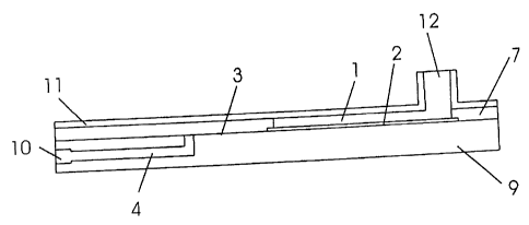
In Figure 1 the individual elements used in this example
are shown in a detail drawing.
Herein, the cover portion 7 is used with an opening
forming a feed chamber 1, wherein the thickness of the
cover portion 7 and the exposed cross-section surface of
the opening predetermine the volume in the feed chamber 1
provided for the sample fluid.
CA 02421406 2008-09-08
14
This example of a device according to the invention is
downwardly formed with a base portion 9. The cover
portion 7 and base portion 9 will be coupled with each
other before using. The two portions may be glued, welded
or connected with each other in a form-fit or friction-fit
manner by, for example, way of clips.
The based and cover portions can be manufactured from
plastic with an injection moulding method. However, they
can be composed of other materials as well.
The cavity 3 having a small height tapering in its width
can be formed by means of cooperating recesses formed in a
surface of the cover portion 7 or base portion 9 which are
facing each other.
However, with the example shown in the Figures 1 to 3 an
adhesive film 8 is used which will be coupled with the
cover portion 7 and base portion 9, and forms the one
sided wedge-shaped, tapering cavity 3 having a small
height by means of a stamped portion. The adhesive film 8
used herein has a thickness of 0.13 mm and predetermines
the height of the cavity.
The cavity 3 is dimensioned in a plane manner such that
the cross-section surface of the feed chamber 1 is
completely covered, and in addition a tapering portion
follows which is not covered by the membrane 2.
The membrane 2 is inserted into the feed chamber 1 for the
separation of the blood plasma such that a liquid sample
can be placed upon the surface of the membrane 2 into the
CA 02421406 2008-09-08
feed chamber 1 without unseparated sample fluid passing
into the cavity 3.
The membrane 2 used with this example is a "Hemasep V"
5 type membrane having a length of 30 mm, a width of 13 mm
and a thickness of 0.89 + 0.05 mm.
With this example, an auxiliary transport membrane 5 is
used which fills up the cavity 3 in an all-over manner.
10 In this example this transport membrane 5 has a length of
45 mm and a width of 13 mm as well. The smallest widths
of the transport membrane 5 and cavity 3 within the
tapered area are 5 mm with an angle of the taper of
approximately 15 .
In the base portion 9 a flow channel 4 can be formed
through which the separated blood plasma is guided toward
the opening 10. The blood plasma which at least is
carried laterally through the transport membrane 5 is
passed through an opening, which is located in the
tapering area of the cavity 3 having a small height, into
the flow channel 4 and can be drawn off therein. An
opening 6 which communicates with the inlet opening of the
flow channel 4 is formed in the transport membrane 5.
The separated blood plasma within the transport membrane 5
accumulates around this opening 6 and is allowed to be
drawn off into an appropriate volume by acting forces of
pressure or suction forces. Thus, a suction force is
allowed to act across the opening 10 in order to achieve
this. Because of the small dimensions of the opening
small forces are required. A suction force is acting upon
the relative small inner marginal surface of the opening 6
CA 02421406 2008-09-08
16
formed within the transport membrane 5 which is dominantly
determined by the thickness of the transport membrane S.
A hollow needle of a syringe formed correspondingly is
allowed to be fixed to the opening 10 of the flow channel
4, and the blood plasma separated thus in a suction force
supported manner can be drawn into the cylinder.
The transport membrane 5 can be formed from a material
mentioned in the general part of the description.
For the separation of blood plasma a whole blood sample of
approximately 500 microlitres (ul) to which an
anticoagulating substance can be added, is placed from
above into the open feed chamber 1, upon the surface of
the membrane 2.
The whole blood is vertically passed through the
horizontally oriented membrane 2, where the hydrostatic
forces for accelerating the blood plasma separation which
is achieved using chromatographic effects of the membrane
2, have a time-shortening effect. The blood plasma
passing quickly through the membrane with respect to the
erythrocytes and other cellular constituents contained in
the whole blood is received from the bottom side of the
membrane 2 by the transport membrane 5 which has greater
capillary forces and is laterally flowing with the support
of capillary forces in the direction of the tapering area,
and consequently toward the opening of the flow channel 4.
There, it is allowed to be removed with the mentioned
syringe using a suction force.
CA 02421406 2008-09-08
17
With the described arrangement approximately 50 ~i1 of
blood plasma is obtained from the whole blood sample of
500 ul in approximately 5 min.
The feed chamber 1 can be covered with a cover 11, and the
fluid can be placed through the opening 12 formed within
the cover 11 into the feed chamber 1. As a result,
spilling of sample fluid can be avoided.
With such a design a force of pressure can be exerted
across the opening 12. With this, e.g., as an example of
a piston and cylinder unit, a syringe drawn up with air
can be introduced into the opening 12 and positioned
therein. With moving the piston air is pressed into the
fed chamber 1 above the sample fluid, and a force of
pressure is exerted.
With the sectional view according to Figure 4, in
particular, the arrangement of a transport membrane 5
within the cavity 3 having a small height shall be
explained, wherein with the transport membrane 5 used here
an additional separating function can be achieved for
undissolved constituents in addition to the effect of its
inherent capillary force effect.
To support the separation, either a suction force at the
opening 10 or a force of pressure at the opening 12 can be
generated by positioning a piston and cylinder unit to at
least one of the openings 10 or 12. Then, with such a
piston and cylinder unit, a relative motion between the
piston and cylinder can be carried out in a continuous
manner, in an intermittent motion with at least two steps
or a motion restricted by an end stopper, and as a result
CA 02421406 2008-09-08
18
the suction force and the force of pressure can be
generated correspondingly.
With such an arrangement, the separation of blood plasma
out of whole blood is carried out with the support of a
suction force and/or force of pressure within a time
interval of maximum 10 minutes, wherein with a quantity of
whole blood of 550 ul, for example, which is heparinized
with Saarstedt type monovettes, a yield of plasma of up to
20% can be achieved.
With the example of a device according to the invention
shown in a sectional view of Figure 5 an auxiliary
intermediate container 14 for separated fluid is connected
to the cavity 3 having a small height, wherein with this
example a transport membrane can be used again in addition
to the separating membrane 2. The inlet opening for the
biological fluid relieved of undissolved constituents into
the intermediate container 14 is located at the opening 6
formed in the transport membrane 5.
The intermediate container 14 has an opening through which
the separated fluid can be removed from the fluid relieved
of undissolved constituents with a pipette or a
conventional syringe having a hollow needle such as for
carrying out subsequent analyses.
The intermediate container 14 should be temporarily
occluded outwardly with at least a fluid-tight cover 13.
Such a cover 13 may be a foil, for example, which is
circumferentially provided with a bonding agent in a
marginal area, and thus may be glued upon the cover and a
cover portion 7, respectively, for temporarily occluding
the opening of the intermediate container 14.
CA 02421406 2008-09-08
19
In the case, where the intermediate container 14 does not
comprise any further connection to the environment and the
separation is carried out with a support of force of
pressure, it is preferable to form this cover in a fluid-
tight, but gas permeable manner.
However, in the form shown in Figure 5 this is not
necessarily required since the intermediate container 14
is connected to the flow channel 4, and an opening 10 is
provided on the flow channel 4. With such a design, it
may additionally separate with the support of suction
force as already explained with the other examples and in
the general part of the description.
To avoid entering and discharging the fluid already
separated out of the intermediate container 14 through the
flow channel 4 and the opening 10, the inlet opening of
the flow channel 4 can be arranged on the intermediate
container 14 such that the level of the separated fluid
does not reach the inlet opening of the flow channel 4.
Another alternative to prevent this effect is to use a
membrane which is fluid-tight and permeable to gas which
can be located at the inlet opening or inside of the flow
channel 4.
However, in addition to the use of a foil as cover 13 for
the opening of the intermediate container 14 a cap can
also be used which is fixable in a friction-fit member or
a form-fit manner and made of plastic material, for
example, and which can be pressed simply into the opening.
Such a cap may be replaced in relatively simple manner for
removing separated fluid out of the intermediate container
CA 02421406 2008-09-08
14, or it is further possible for the cap as a cover 13 to
be pierced with a hollow needle of a conventional syringe
and thus to draw off the separated fluid out of the
intermediate container 14 which also applies logically to
5 the use of a foil as a cover 13.
15
25