Note: Descriptions are shown in the official language in which they were submitted.
CA 02421751 2003-03-07
WO 02/22685 PCT/US01/28548
MUC1 EXTRACELLULAR DOMAIN AND CANCER TREATMENT
COMPOSITIONS AND METHODS DERIVED THEREFROM
This application claims priority to Provisional Application Serial No.
60/231,841,
filed September 11, 2000. The United States government may own rights in the
present
invention pursuant to grant number R21-CA87421from the National Cancer
Institute,
National Institutes of Health, Department of Health and Human Services.
FIELD OF THE INVENTION
The present invention relates generally to the field of cancer therapy and
more
specifically to the use of modulators or agents that interact with MUC1 as a
point on
intervention in cancer therapy.
BACKGROUND OF THE INVENTION
The human MUC1 mucin glycoprotein is expressed on the apical borders of
secretory epithelial cells on the luminal surface of most glandular epithelia
(Kufe et al.,
1984). In carcinomas, MUC1 is highly overexpressed throughout the entire cell
membrane and cytoplasm (Kufe et al., 1984; Perey et al., 1992). As such, the
aberrant
pattern of MUC1 expression in carcinoma cells may confer a function for MUC1
normally found at the apical membrane to the entire cell membrane. The
hallmark of
MUC1 mucin is an ectodomain comprising a glycosylated 20 amino acid
extracellular
sequence that is tandemly repeated 25-100 times in each molecule (Strouss &
Decker,
1992). The mucin glycosylation level appears to be lower in cancer cells than
normal cells
of ductal epithelial tissue (Kufe, U.S. Pat. No. 5,506,343). This
hypoglycosylation results
in the exposure of tumor-specific epitopes that are hidden in the fully
glycosylated mucin.
Over ninety percent of breast cancers show an increased expression of MUC1
(also known as Mucin, Epithelial Membrane Antigen, Polymorphic Epithelial
Mucin,
Human Milk Fat Globule Membrane antigen, Episialin, DF-3, etc., see Barry &
Sharkey,
1
CA 02421751 2010-02-23
1985). Several clinical studies have suggested that mucinous tumor antigens
expressed
on the cell surface of tumor cells associate with poor prognosis of a variety
of cancer
types (Itzkowitz et al., 1990).
MUC1 is expressed as both a transmembrane form and a secreted form (Finn et
al., 1995). The repeating sialyl epitopes of MUC1 (the `ectodomain') are shed
into the
serum (Reddish et al., 1996). The N-terminal ectodomain (the extracellular
domain that
is cleaved) of MUC1 consists of a variable number of the 20-amino acid tandem
repeats
that are subject to 0-glycosylation. This mucin extends far above the cell
surface and
past the glycocalyx making it easily available for interactions with other
cells. The C-
terminal region of MUC1 includes a 37 amino acid transmembrane domain and a 72
amino acid cytoplasmic tail that contains sites for tyrosine phosphorylation.
A
approximately 45-amino acid extracellular domain remains following cleavage of
the
ectodomain. It is not known what enzyme is responsible for the cleavage of the
ectodomain at this time. The extracellular domain or "MUC1/ECD," remaining,
after
cleavage of the ectodomain, typically includes the amino acid sequence:
TINVHDVETQFNQYKTEAASRYNLTISDVSVSDVPFPFSAQSGAG. (SEQ ID
NO:1)
The cytoplasmic domain of MUC1 ("MUC 1 /CD") encompasses multiple sub-
domains that are important in intracellular signaling in cancer cells. p-
Catenin binds
directly to MUC1/CD at a SAGNGGSSL motif (Yamamoto et al., 1997). 13-Catenin,
a
component of the adherens junctions of mammalian epithelium, binds to
cadherins at the
intracellular surface of the plasma membrane and performs a signaling role in
the
cytoplasm as the penultimate downstream mediator of the wnt signaling pathway
(Takeichi, 1990; Novak & Dedhar, 1999). The ultimate mediator of the wnt
pathway is a
nuclear complex of P-catenin and lymphoid enhancer factor/T cell factor
(Lef/Tcf) which
stimulates the transcription of a variety of target genes (see e.g., Molenaar
et al., 1996;
Brunner et al., 1997). Defects in the P-catenin-Lef/Tcf pathway are involved
in the
development of several types of cancers (Novak & Dedhar, 1999).
Glycogen synthase kinase 313 (GSK3P) also binds directly to MUC1/CD and
phosphorylates serine in a DRSPY site adjacent to the P-catenin binding motif,
thereby
2
CA 02421751 2003-03-07
WO 02/22685 PCT/US01/28548
decreasing the association between MUC1 and f3-catenin (Li et al., 1998). In
addition,
the c-Src tyrosine kinase also binds to and phosphorylates a MUC1/CD SPYEKV
motif,
resulting in an increased interaction between MUC1/CD and P-catenin and a
decreased
interaction between MUC1/CD and GSK3P (Li et al., 2001).
MUC1 associates also constitutively with the epidennal growth factor receptor
(EGF-R, HER1) at the cell membrane and activated EGF-R induces phosphorylation
of
the MUC1/CD SPYKEV motif (Li et al., 2001(a)). EGF-R mediated phosphorylation
of
MUC1/CD appears to increase the interaction of MUC1 with c-Src and P-catenin
and
downregulate the interaction between MUC1 and GSK3P. These results support a
model
wherein MUC1 integrates the signaling among c-Src, P-catenin and GSK3P
pathways
and dysregulation of this integrated signaling by aberrant overexpression of
MUC1 in
cancer cells could promote the transformed phenotype (Li et al., 2001(a)).
The Armadillo protein p120en' also binds directly to MUC1/CD resulting in the
nuclear localization of p120 (Li & Kufe, 2001). P120 has been implicated in
cell
transformation and altered patterns of p120 expression have been observed in
carcinomas
(see e.g., Jawhari et al., 1999; Shimazui et al., 1996). P120 is a v-Src
tyrosine kinase
substrate, binds to E-cadherin, and is implicated as a transcriptional
coactivator (Reynolds
et al., 1989; Reynolds et al., 1994; Daniels & Reynolds, 1999). The
observations that
p120 localizes to both cell junctions and the nucleus have supported a role
for p120, like
P-catenin, in the regulation of both cell adhesion and gene transcription.
Decreased cell
adhesion resulting from association of MUC1 and p120 may be involved in
increased
metastatic potential of MUC1-expressing tumor cells.
Thus the available evidence indicates that MUC1/CD functions to transfer
signals
from the extracellular domain to the nucleus, and utilizes signaling
mechanisms that have
been implicated in adhesion receptor and growth factor signaling and cellular
transformation. It is desirable to identify compositions and methods related
to
modulation of the MUC1-mediated signaling and its putative role in cellular
transformation.
3
CA 02421751 2003-03-07
WO 02/22685 PCT/US01/28548
SUMMARY OF THE INVENTION
The present invention encompasses methods of use and pharmaceutical
compositions relating to the discovery that the extracellular domain of MUC1
provides
binding domains for endogenous ligands and that such binding is related to an
oncogenic
function of MUC1 and the proliferation of cancer cells.
Broadly the invention relates to cancer treatment compositions and methods
employing agents or treatment methodologies that comprise or include
antagonists of
MUC1/ECD modulated cell proliferation. Preferred are methods and compositions
that
comprise agents that bind to MUC1/ECD or that bind to 1MIUC1/ECD ligands that
activate
the oncogenic function of MUC1.
Thus, one aspect of the present invention provides for a method for inhibiting
the
proliferation of cancer cells, comprising administration of an effective
amount of a
MUC1/ECD antagonist. MUC1/ECD antagonists are agents that downregulate or
reduce
the quantity of MUC1/ECD presented on cell surfaces, or downregulate the level
of wild-
type MUC1/ECD ligands available for binding to MUC1/ECD, and/or MUC1/ECD
binding inhibitors. A "MUC1/ECD binding inhibitor" means a compound that
inhibits
the binding of MUC1 wild-type ligands, which may suitably include neuregulin 2
isoform
5 (SEQ ID NO: 2), neuregulin 2 isoform 6 (SEQ ID NO: 3), and appropriate
fragments
thereof, to MUC1/ECD or a compound that inhibits the binding of an antibody
that binds
to an epitope within SEQ ID NO. 4 to MUC1/ECD. Appropriate fragments of
neuregulin
2 isoform 5 (SEQ ID NO: 2) and neuregulin 2 isoform 6 (SEQ ID NO: 3) are those
that
bind to MUC1/ECD. MUC1/ECD binding inhibitors include antibodies, polypeptides
and small molecules that inhibit such binding. A "MUC1/ECD-P1 binding
inhibitor"
means a MUC1/ECD binding inhibitor identified by inhibition of the binding to
MUC1/ECD of an antibody the binds to an epitope within SEQ ID NO. 4.
In one embodiment of the invention, the MUC1/ECD inhibitor is the polypeptide
of SEQ ID NO: 1, or a fragment comprising at least four consecutive amino
acids of SEQ
ID. NO: 1 such as TINY, NVHD, VIHDV, DVET, VETQ, ETQF, TQFN, QFNQ, FNQY,
NQYK, QYKT, YKTE, KTEA, TEAA, EAAS, AASR, ASRY, SRYN, RYNL, YNLT,
4
CA 02421751 2010-02-23
NLTI, LTIS, TISD, ISDV, SDVS, DVSV, VSVS, SVSD, VSDV, SDVP, DVPF, VPFP,
PFPF, FPFS, PFSA, FSAQ, SAQS, AQSG, QSGA, and SGAG (SEQ ID NOs:19-58,
respectively). In other embodiments, the MUC1/ECD inhibitor is a conservative
variant
of the foregoing peptides. In another embodiment, the MUC1/ECD binding
inhibitor is
the polypeptide of SEQ ID NO: 4, SEQ ID NO: 5, or conservative variants
thereof.
In another embodiment of the present invention, the MUC1/ECD inhibitor is an
antibody that binds to one or more epitopes in the MUC1/ECD sequence SEQ ID
NO: 1.
In other embodiments of the invention, the MUC1/ECD inhibitor is an antibody
that
binds to an epitope within SEQ ID NO: 2 or SEQ ID NO: 3. The antibody may be a
polyclonal or a monoclonal antibody. Monoclonal antibodies may be humanized or
human monoclonal antibodies. It may also be a bispecific antibody or a
fragment which
comprises an antigen binding region. In some embodiments, the antibody is
conjugated
to a chemotherapeutic agent, radioisotope, toxin, or an effector that induces
a cytolytic or
cytotoxic immune response. Such conjugates may comprise a cytokine, an
antimetabolite, an anthracycline, a vinca alkaloid, an antibiotic, an
alkylating agent, a
naturally derived toxin, or an Fe region of a IgG1 immunoglobulin.
In another embodiment, the method further comprises the administration of a
chemotherapeutic agent or radiation in combination with a MUC1/ECD antagonist.
Chemotherapeutic agents typically include alkylating agents, topoisomerase
inhibitors,
antimetabolites, tubulin interactive agents, anti-hormonal agents, ornithine
decarboxylase inhibitors and tyrosine kinase inhibitors.
In various embodiments, the cancer cells are selected from the group
consisting
of skin cancer cells, prostate cancer cells, lung cancer cells, brain cancer
cells, breast
cancer cells, ovarian cancer cells, cervical cancer cells, liver cancer cells,
pancreatic
cancer cells, colon cancer cells, stomach cancer cells and leukemia cells.
Another aspect of the invention is a method for reducing tumor growth in a
mammal comprising administration of a therapeutic amount of a chemotherapeutic
agent
or radiation and an effective amount of a MUC1/ECD antagonist. In a preferred
embodiment, the mammal is human. In one embodiment the method is for treating
refractory tumors comprising administration of a therapeutic amount of a
5
CA 02421751 2003-03-07
WO 02/22685 PCT/US01/28548
chemotherapeutic agent or radiation and an effective amount of a M1IC1/ECD
antagonist
subsequent to treatment with one or more chemotherapeutic agents. In various
embodiments, the tumor is a tumor of the skin, prostate, lung, brain, breast,
ovary, cervix,
liver, pancreas, colon, stomach or heampoietic system.
Other aspects of the invention relate to pharmaceutical compositions
comprising
MUC1/ECD antagonists and a pharmaceutically acceptable carrier. Wherein the
antagonist is a MUC1/ECD binding inhibitor that may be the polypeptide of SEQ
ID NO:
1 or a fragment comprising at least four consecutive amino acids, or
conservative variants
thereof, and a pharmaceutically acceptable carrier. In some embodiments, the
MUC1/ECD inhibitor may be the polypeptide of SEQ ID NO: 4, SEQ ID NO: 5, or
conservative variants thereof. In other embodiments, the pharmaceutical
composition
comprises an antibody that is a MUC1/ECD binding inhibitor and binds to an
epitope
within sequences of the peptides selected from the group consisting of SEQ ID
NO: 1,
SEQ ID NO: 2 and SEQ ID NO: 3 and a pharmaceutically acceptable carrier.
The present invention also encompasses methods for screening MUC1/ECD
binding inhibitor activity. One embodiment comprises a method of identifying a
compound that inhibits the binding of ligands to MUC1/ECD, the method
comprising: (a)
providing a polypeptide comprising SEQ ID. NO: 1 or SEQ ID NO: 5; (b)
contacting said
polypeptide with a test compound and a ligand to the extracellular domain of
MUC1
selected from the group consisting of antibodies to MUC1/ECD that stimulate
MUC1
mediated cancer cell proliferation and wild type ligands that bind to MUC1/ECD
and
stimulate cancer cell proliferation; and (c) determining whether the binding
of said
antibody to MUC1/ECD or wild type ligand is decreased relative to an
appropriate
control. Appropriate controls include, but are not limited to, assays wherein
test
compounds are excluded. In one embodiment the M1JC1/ECD antibody that
stimulates
MUC1 mediated cancer cell proliferation is an antibody that binds to an
epitope within
SEQ ID NO. 4. In other embodiments, the wild type ligands may suitable include
neuregulin 2 isoform 5 (SEQ ID NO: 2) and appropriate fragments thereof and
neuregulin
2 isoform 6 (SEQ ID NO: 3) and appropriate fragments thereof, wherein
appropriate
6
CA 02421751 2003-03-07
WO 02/22685 PCT/US01/28548
fragments are those that bind to the SEQ ID NO: 1 and have suitable growth
stimulatory
activity.
Another embodiment is a method of identifying a compound that inhibits the
proliferation of MUC1-expressing cancer cells, the method comprising: (a)
providing a
population of MUC1-expressing cancer cells; (b) contacting said population of
MUC1-
expressing cancer cells with a test compound and a ligand to the extracellular
domain of
MUC1 selected from the group consisting of antibodies to MUC1/ECD that
stimulate
MUC1 mediated cancer cell proliferation and wild type ligands that bind to
MUC1/ECD
and stimulate cancer cell proliferation; and (c) determining whether the
proliferation of
the population of MUC1-expressing cancer cells is decreased by comparison to
an
appropriate control. Appropriate controls include, but are not limited to,
proliferation
assays wherein test compounds are excluded. In one embodiment the MUC1/ECD
antibody that stimulates MUC1 mediated cancer cell proliferation is an
antibody that
binds to an epitope within SEQ ID NO. 4. In other embodiments, the wild type
ligands
may suitable include neuregulin 2 isoform 5 (SEQ ID NO: 2) and appropriate
fragments
thereof and neuregulin 2 isoform 6 (SEQ ID NO: 3) and appropriate fragments
thereof,
wherein appropriate fragments are those that bind to the SEQ ID NO: 1 and have
suitable
growth stimulatory activity.
The present invention also provides methods for identifying compounds that
downregulate M1JC1/ECD expression. The method comprises: (a) providing a
population of MUC1-expressing cancer cells; (b) contacting said population of
MUC1-
expressing cancer cells with a test compound; (c) utilizing an anti-M1JC1/ECD
antibody
to identify polypeptides comprising MUC1/ECD in the MUC1-expressing cancer
cells;
and (d) determining whether the expression of polypeptides comprising MUC1/ECD
is
decreased in comparison to controls wherein the test compound was excluded.
The present invention also encompasses pharmaceutical compositions comprising
compounds identified by the foregoing methods and a pharmaceutically
acceptable
carrier.
7
CA 02421751 2010-02-23
In another aspect, the present invention provides a use of a MUC1
extracellular domain
(ECD) antagonist for the manufacture of a medicament to inhibit the
proliferation of MUC1-
expressing cancer cells, wherein said MUC I ECD antagonist is: (a) a
polypeptide comprising
at least 4 amino acids of SEQ ID NO: 1; or (b) an antibody or an antigen-
binding fragment
thereof that binds to an epitope within the MUC1 ECD.
In another aspect, the present invention provides a use of a MUC1
extracellular domain
(ECD) antagonist to inhibit the proliferation of MUC1-expressing cancer cells,
wherein said
MUC1 ECD antagonist is: (a) a polypeptide comprising at least 4 amino acids of
SEQ ID NO:
1; or (b) an antibody or an antigen-binding fragment thereof that binds to an
epitope within the
MUC1 ECD.
In another aspect, the present invention provides a pharmaceutical composition
for
inhibiting proliferation of MUC1-expressing cancer cells, said composition
comprising (a) a
MUC1 ECD antagonist, wherein said MUC1 ECD antagonist is: (i) an antibody or
an antigen-
binding fragment thereof that binds to an epitope within the MUC1 ECD; or (ii)
a polypeptide
comprising at least 4 amino acids of SEQ ID NO: 1; and (b) a pharmaceutically
acceptable
carrier.
In another aspect, the present invention provides a method of identifying a
compound
for inhibiting proliferation of MUC1-expressing cancer cells, the method
comprising: (a)
contacting a population of MUC1-expressing cancer cells with a test compound;
and (b)
determining whether MUC1 activity in said population of MUC1-expressing cancer
cells is
decreased in the presence of said test compound relative to the absence
thereof; wherein a
decrease in MUC1 activity in the presence of said test compound relative to
the absence
thereof is indicative that said test compound inhibits the proliferation of
MUC1-expressing
cancer cells.
In another aspect, the present invention provides a method of identifying a
compound
for inhibiting proliferation of MUC 1-expressing cancer cells, the method
comprising: (a)
providing an MUC1 extracellular domain (ECD) polypeptide; (b) contacting said
MUC1 ECD
polypeptide with a test compound; (c) determining whether said test compound
binds to said
MUC1 ECD polypeptide; and, if said test compound binds to said MUC1 ECD
polypeptide, (i)
contacting a population of MUC1-expressing cancer cells with said test
compound; and (ii)
determining whether MUC1 activity in said population of MUC1-expressing cancer
cells is
decreased in the presence of said test compound relative to the absence
thereof; wherein a
decrease in MUC1 activity in the presence of said test compound relative to
the absence
thereof is indicative that said test compound inhibits the proliferation of
MUC1-expressing
cancer cells.
7a
CA 02421751 2013-09-10
In another aspect, the present invention provides a use of a MUC1
extracellular
domain (ECD) antagonist for the manufacture of a medicament to inhibit the
proliferation of
MUC1-expressing cancer cells, wherein said MUC1 ECD antagonist is: (a) a
polypeptide
comprising at least 4 consecutive amino acids of SEQ ID NO: 1; or (b) an
antibody or an
antigen-binding fragment thereof that binds to an epitope within SEQ ID NO: 1,
SEQ ID NO:
4 or SEQ ID NO: 5.
In another aspect, the present invention provides a use of a polypeptide
comprising at
least 4 consecutive amino acids of SEQ ID NO: 1 for the manufacture of a
medicament to
inhibit the proliferation of MUC1-expressing cancer cells.
In another aspect, the present invention provides a use of a polypeptide
comprising at
least 4 consecutive amino acids of SEQ ID NO: 1 to inhibit the proliferation
of MUC1-
expressing cancer cells.
In another aspect, the present invention provides a use of a MUC1
extracellular
domain (ECD) antagonist to inhibit the proliferation of MUC1-expressing cancer
cells,
wherein said MUC1 ECD antagonist is: (a) a polypeptide comprising at least 4
consecutive
amino acids of SEQ ID NO: 1; or (b) an antibody or an antigen-binding fragment
thereof that
binds to an epitope within SEQ ID NO: 1, SEQ ID NO: 4 or SEQ ID NO: 5.
In another aspect, the present invention provides a pharmaceutical composition
for
inhibiting proliferation of MUC1-expressing cancer cells, said composition
comprising (a) a
MUCI ECD antagonist, wherein said MUC1 ECD antagonist is: (i) an antibody or
an antigen-
binding fragment thereof that binds to an epitope within SEQ ID NO: 1, SEQ ID
NO: 4 or
SEQ ID NO: 5; or (ii) a polypeptide comprising at least 4 consecutive amino
acids of SEQ ID
NO: 1; and (b) a pharmaceutically acceptable carrier.
In another aspect, the present invention provides a pharmaceutical composition
for
inhibiting proliferation of MUC1-expressing cancer cells, said composition
comprising (a) a
polypeptide comprising at least 4 consecutive amino acids of SEQ ID NO: 1; and
(b) a
pharmaceutically acceptable carrier.
In another aspect, the present invention provides a method of identifying a
compound
for inhibiting proliferation of MUC1-expressing cancer cells, the method
comprising: (a)
providing an MUC1 extracellular domain (ECD) polypeptide comprising SEQ ID NO:
1, SEQ
ID NO: 4 or SEQ ID NO: 5; (b) contacting said MUC1 ECD polypeptide with a test
compound; (c) determining whether said test compound binds to said MUC1 ECD
polypeptide; and, if said test compound binds to said MUC1 ECD polypeptide,
(i) contacting a
population of MUC 1-expressing cancer cells with said test compound; and (ii)
determining
7b
CA 02421751 2013-09-10
whether MUC1 activity in said population of MUC 1-expressing cancer cells is
decreased in
the presence of said test compound relative to the absence thereof; wherein a
decrease in
MUC1 activity in the presence of said test compound relative to the absence
thereof is
indicative that said test compound inhibits the proliferation of MUC1 -
expressing cancer cells.
7c
CA 02421751 2003-03-07
WO 02/22685 PCT/US01/28548
BRIEF DESCRIPTION OF THE DRAWINGS
The following drawings form part of the present specification and are included
to
further demonstrate certain aspects of the present invention. The invention
may be better
understood by reference to one or more of these drawings in combination with
the
detailed description of specific embodiments presented herein.
FIG. 1: Effect of anti-MUC1-P1 antibody on proliferation of ZR-75-1 breast
carcinoma cells.
FIG. 2: Effect of anti-MUC 1-P1 antibody on proliferation of SW480 cells
stably
expressing an empty vector (SW480/V) or MUC1 (SW480/MUC1).
FIG. 3: Effect of ZR-75-1 conditioned medium on proliferation of SW480 cells
stably expressing an empty vector (SW480/V) or MUC1 (SW480/MUC1).
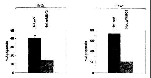
FIG. 4: Effect of MUC1 on H202 and taxol-induced apoptosis in HeLa cells
stably expressing an empty vector (HeLa/V) or MUC1 (HeLa/Muc1). The results
are
expressed as the percentage apoptosis (mean SE) of three separate experiments.
DETAILED DESCRIPTION OF THE INVENTION
I. Polyp eptides
The polypeptides of the present invention can be created by synthetic
techniques
or recombinant techniques which employ genomic or cDNA cloning methods.
Polypeptides can be routinely synthesized using solid phase or solution phase
peptide
synthesis. Methods of preparing relatively short polypeptides peptides, such
as PO (SEQ
ID NO: 9), PI (SEQ ID NO: 4), P2 (SEQ ID NO: 6) and P3 (SEQ BD NO: 7), by
chemical
synthesis are well known in the art. Such polypeptides could, for example be
produced
by solid-phase peptide synthesis techniques using commercially available
equipment and
reagents such as those available from Milligen (Bedford, Mass.) or Applied
Biosystems-
Perkin Elmer (Foster City, CA). Alternatively, segments of such polypeptides
could be
prepared by solid-phase synthesis and linked together using segment
condensation
methods such as those described by Dawson et al., (1994). During chemical
synthesis of
such polypeptides, substitution of any amino acid is achieved simply by
replacement of
the residue that is to be substituted with a different amino acid monomer.
8
CA 02421751 2003-03-07
WO 02/22685 PCT/US01/28548
Wild-type MUC1/ECD ligand polypeptides can be identified as exemplified in
Example 3 herein. Recombinant MUC1/ECD ligands can then be prepared by methods
known in the art.
The polypeptides of the present invention include variant polypeptides. By
"variant" polypeptide is intended a polypeptide sequence modified by deletion
or addition
of one or more amino acids at one or more sites in the sequence; or
substitution of one or
more amino acids at one or more sites within the sequence. Variant
polypeptides
encompassed by the present invention retain the desired biological activity of
the
polypeptide from which they are derived. Such variants will have at least 40%,
50%,
60%, 70%, generally at least 75%, 80%, 85%, preferably about 90% to 95% or
more, and
more preferably about 98% or more sequence identity to the amino acid sequence
of the
polypeptide from which they are derived. The percentage of sequence identity,
also
termed homology, between a polypeptide native and a variant sequence may be
determined by comparing the two sequences using the Gap program (Wisconsin
Sequence Analysis Package, Version 8 for Unix, Genetics Computer Group,
University
Research Park, Madison Wisconsin), which uses the algorithm of Smith and
Waterman,
(1981).
The polypeptides of the present invention also include variant polypeptides
with
one or more conservative substitutions. For the purposes of classifying amino
acid
substitutions as conservative, amino acids are grouped as follows: Group I
(hydrophobic
sidechains): norleucine, met, ala, val, leu, ile; Group II (neutral
hydrophilic side chains):
cys, ser, thr; Group DI (acidic side chains): asp, glu; Group IV (basic side
chains): asn,
gin, his, lys, arg; Group V (residues influencing chain orientation): gly,
pro; and Group VI
(aromatic side chains): trp, tyr, phe. Conservative substitutions involve
substitutions
between amino acids in the same class.
Also encompassed by the present invention are chemical derivatives of
polypeptides. "Chemical derivative" refers to a subject polypeptide having one
or more
residues chemically derivatized by reaction of a functional side group. Such
derivatized
residues include, for example, those molecules in which free amino groups have
been
derivatized to form amine hydrochlorides, p-toluene sulfonyl groups,
carbobenzoxy
9
CA 02421751 2003-03-07
WO 02/22685 PCT/US01/28548
groups, t-butyloxycarbonyl groups, chloroacetyl groups or formyl groups. Free
carboyl
groups may be derivatized to form salts, methyl and ethyl esters or other
types of esters or
hyrazides. Free hydroxyl groups may be derivatized to form 0-acyl or 0-alkyl
derivatives. The imadazole group of histidine may be derivatized to form N-
imbenzylhistidine.
The tem]. "polypeptide" as used herein indicates a molecular chain of amino
acids
and does not refer to a specific length of the product.
Antibodies
The teim "antibody" is used in the broadest sense and specifically covers
monoclonal antibodies (including full length monoclonal antibodies),
polyclonal
antibodies, multispecific antibodies (e.g., bispecific antibodies), and
antibody fragments,
so long as they exhibit the desired biological activity.
Methods for generating polyclonal antibodies are well known in the art.
Briefly, a
polyclonal antibody is prepared by immunizing an animal with an antigenic
composition
and collecting antisera from that immunized animal. A wide range of animal
species can
be used for the production of antisera including rabbit, mouse, rat, hamster,
guinea pig
and goat.
As is well known in the art, a given composition may vary in its
immunogenicity.
It is often necessary therefore to boost the host immune system, as may be
achieved by
coupling a polypeptide immunogen to a carrier. Exemplary and preferred
carriers are
keyhole limpet hemocyanin (KLH) and bovine serum albumin (BSA). Other albumins
such as ovalbumin, mouse serum albumin or rabbit serum albumin can also be
used as
carriers. Means for conjugating a polypeptide to a carrier protein are well
known in the art
and include glutaraldehyde, m-maleimidobenzoyl-N-hydroxysuccinimide ester,
carbodiimide and bis-biazotized benzidine. As is also well known in the art,
the
immunogenicity of a particular immunogen composition can be enhanced by the
use of
non-specific stimulators of the immune response, known as adjuvants. Exemplary
and
preferred adjuvants include complete Freund's adjuvant (a non-specific
stimulator of the
CA 02421751 2003-03-07
WO 02/22685 PCT/US01/28548
immune response containing killed Mycobacterium tuberculosis), incomplete
Freund's
adjuvants and aluminum hydroxide adjuvant.
The serum for an immunized animal may be used as is for various applications
or
the desired antibody fraction may be purified by well-known methods, such as
affinity
chromatography using another antibody or a peptide bound to a solid matrix.
Monoclonal antibodies (MAbs) may be readily prepared through use of well-
known techniques, such as those exemplified in U.S. Patent 4,196,265,
incorporated
herein by reference. Typically, this technique involves immunizing a suitable
animal with
a selected immunogen composition, e.g., a purified or partially purified
expressed
polypeptide. The immunizing composition is administered in a manner that
effectively
stimulates antibody producing cells.
The methods for generating monoclonal antibodies (MAbs) generally begin along
the same lines as those for preparing polyclonal antibodies. The use of rats
may provide
certain advantages (Goding, 1986), but mice are preferred, with the BALB/c
mouse being
the most routinely used and generally gives a higher percentage of stable
fusions.
Following immunization, somatic cells with the potential for producing
antibodies, specifically B lymphocytes (B cells), are selected for use in the
MAb
generating protocol. These cells may be obtained from biopsied spleens,
tonsils or lymph
nodes, or from a peripheral blood sample. Spleen cells and peripheral blood
cells are
preferred. Often, a panel of animals will have been immunized and the spleen
of animal
with the highest antibody titer will be removed and obtaining lymphocytes from
the
spleen.
The antibody-producing B lymphocytes from the immunized animal are then
fused with cells of an immortal myeloma cell, generally one of the same
species as the
animal that was immunized. Myeloma cell lines suited for use in hybridoma-
producing
fusion procedures preferably are non-antibody-producing, have high fusion
efficiency,
and have enzyme deficiencies that render them incapable of growing in certain
selective
media that support the growth of only the desired fused cells (hybridomas).
Selected
hybridomas are serially diluted and cloned into individual antibody-producing
cell lines,
which can then be propagated indefinitely to provide MAbs.
11
CA 02421751 20 03-03-0 7
WO 02/22685 PCT/US01/28548
In accordance with the present invention, fragments of the monoclonal antibody
of
the invention can be obtained from the monoclonal antibody produced as
described
above, by methods which include digestion with enzymes such as pepsin or
papain and/or
cleavage of disulfide bonds by chemical reduction. Alternatively, monoclonal
antibody
fragments encompassed by the present invention can be synthesized using an
automated
synthesizer, or by expression of full-length gene or of gene fragments in E.
coli or other
recombinant microorganisms and cell lines.
The present invention also encompasses various antibody conjugates. Conjugates
with fluorescein markers are prepared be methods known in the art, such as
conjugation
in the presence of coupling agents or by reaction with an isothiocyanate.
Conjugates with
metal chelates are similarly produced. Other moieties to which antibodies may
be
conjugated include radionuclides such as 131 I, 90 y, 105 Rh, 47 Sc, 67 Cu,
212Bi, 211 At, 188
Re, 109 pd, 47 se, 212
Pb, and 153 Sm and the like, as described in Gansow, 1991, which is
herein incorporated by reference.
Monoclonal antibodies of the invention can also be coupled to conventional
chemotherapeutic agents such as an antimetabolite, an anthracycline, a vinca
alkaloid, an
antibiotic or an alkylating agent. Drugs that may be coupled to the antibodies
for
targeting include compounds such as doxorubicin, cyclophosphamide, cisplatin,
adriamycin, estramustine, fluorouracil, ethinyl estradiol, mitoxantrone,
methotrexate,
finasteride, taxol, and megestrol. Methods of coupling may be direct via
covalent bonds,
or indirect via linking molecules, and will generally be known in the art for
the particular
drug selected and are made using a variety of bifunctional protein coupling
agents.
Examples of such reagents are SPDP, TT, bifunctional derivatives of
imidoesters such a
dimethyl adipimidate HC1, active esters such as disuccinimidyl suberate,
aldehydes such
as glutaraldehyde, bisazido compounds such as his (R-azidobenzoyl)
hexanediamine,
bisdiazonium derivatives such as bis-(R-diazoniumbenzoyDethylenediamine,
diisocyanates such as tolylene 2,6-diisocyanate, and bis-active fluorine
compounds such
as 1,5-difluoro-2,4-dinitrobenzene. (See, e.g., Thorpe et al., 1982, herein
incorporated by
reference).
12
CA 02421751 2003-03-07
WO 02/22685 PCT/US01/28548
The antibodies of the present invention may also be conjugated with various
toxin
molecules or an effector such as IgG1 immunoglobulin, which induces cytolytic
or
cytotoxic immune response. Thus, the two components may be chemically bonded
together by any of a variety of well-known chemical procedures. For example,
the linkage
may be by way of heterobifunctional cross-linkers, e.g. SPDP, carbodiimide,
glutaraldehyde, or the like. The toxin molecules may also be fused to the
antibody or
binding regions thereof by recombinant means, such as through the production
of single
chain antibodies. The genes encoding protein chains may be cloned in cDNA or
in
genomic form by any cloning procedure known to those skilled in the art (see
e.g.,
Sambrook et al., 1989). The recombinant production of various immunotoxins is
well-
known within the art and can be found, for example in Thorpe et al., 1982(a),
Waldmann,
1991, and Pastan et al., 1992, all herein incorporated by reference. A variety
of toxin
molecules are suitable for use as the cytotoxic domain in the antibody
conjugates or
fusion proteins described here. Any toxin known to be useful as the toxic
component of
an immunotoxin may be used, preferably a protein toxin that may be
recombinantly
expressed. Particularly useful as the cytotoxic domain are bacterial toxins
such as
Pseudomonas exotoxin A (PE), diphtheria toxin, shiga toxin and shiga-like
toxin, and
ribosome inactivating toxins derived from plants and fungi, including ricin, a-
sarcin,
restrictotocin, mitogellin, tricanthosin, saporin-G, saporin-1, momordin,
gelonin,
pokeweed antiviral protein, abrin, modeccin and others described in
Genetically
Engineered Toxins, ed. A. Frankel, Marcel Dekker, Inc., 1992, herein
incorporated by
reference, and any recombinant derivatives of those proteins (see Olsnes 1981;
U.S. Pat.
No. 4,675,382; and U.S. Pat. No. 4,894,443, herein incorporated by reference).
The antibody may also be a bispecific antibody which recognizes both the
MUC1/ECD and an antigen which promotes the release of a cytokine such as IL-1,
TNF
alpha and CD16, CD2, CD3 L.C. CD28, which in turn, will activate the release
of lFN
gamma or TNF alpha, respectively.
The MAb's of the present invention encompass chimeric Mobs, including,
"humanized" fomis of non-human (e.g., murine) Mabs. Humanized MAbs are
chimeric
antibodies which contain minimal sequence derived from non-human
immunoglobulin.
13
CA 02421751 2003-03-07
WO 02/22685 PCT/US01/28548
For the most part, humanized antibodies are human immunoglobulins (recipient
antibody)
in which residues from a hypervariable region of the recipient are replaced by
residues
from a hypervariable region of a non-human species (donor antibody) such as
mouse, rat,
rabbit or nonhuman primate having the desired specificity, affinity, and
capacity. In some
instances, framework residues of the human immunoglobulin are replaced by
corresponding non-human residues. Furthermore, humanized antibodies may
comprise
residues which are not found in the recipient antibody or in the donor
antibody. These
modifications are made to further refine antibody performance. In general, the
humanized
antibody will comprise substantially all of at least one, and typically two,
variable
domains, in which all or substantially all of the hypervariable regions
correspond to those
of a non-human immunoglobulin and all or substantially all of the framework
regions are
those of a human immunoglobulin sequence. The humanized antibody optionally
also
will comprise at least a portion of an immunoglobulin constant region (Pc),
typically that
of a human immunoglobulin. (see Jones et al., 1986; Riechmann et al., 1988;
and Presta,
1992). Fully human MAbs are preferred in the therapeutic methods of the
present
invention.
"Single-chain FV' or "sFv" antibody fragments of the present invention
comprise
the VH and VL domains of antibody, wherein these domains are present in a
single
polypeptide chain. Generally, the Fv polypeptide further comprises a
polypeptide linker
between the VH and VL domains which enables the sFy to form the desired
structure for
antigen binding (see Pluckthun, 1994).
III. Screening and Diagnostic Assays
The present invention provides for methods for identifying compounds that
inhibit
the binding of various ligands to MUC1/ECD. The binding ligands include
neuregulin 2
isoform 5 (SEQ ID NO: 2), neuregulin 2 isoform 6 (SEQ ID NO: 3) and fragments
of
either isoform that bind to MUC1/ECD and, in a preferred embodiment, an
antibody that
binds to an epitope within SEQ ID NO: 4.
In one embodiment, the screening method utilizes an in vitro competitive
binding
assay, wherein the capacity of a test compound to inhibit the binding of the
14
CA 02421751 2003-03-07
WO 02/22685 PCT/US01/28548
aforementioned ligands to a polypeptide comprising SEQ ID NO.1 or SEQ ID NO: 5
is
assessed. In such an assay, the polypeptide comprising MUC1/ECD derived
sequences
SEQ ID NO.1 or SEQ ID NO: 5 may be conjugated to another protein or produced
as a
fusion protein, e.g., the GST-MUC1/ECD fusion protein exemplified herein in
Example
3. Other suitable conjugates and fusion proteins may be made by one of skill
in the art
utilizing procedures know in the art. The polypeptides or MUC1/ECD ligands may
be
labeled with a radioisotope or fluorescent label (e.g., phycobiliproteins,
such as
phycoerythrin and allophycocyanins, fluorescein and Texas red). Alternatively
an
enzyme, such as peroxidase, may be used and conjugated either directly or
indirectly via a
biotin and avidin or streptavidin system. Decreased binding upon introduction
of a test
compound is indicative of competitive binding.
A compound that inhibits the binding of ligands to MUC1/ECD may be a
modulator, that is an antagonist or agonist of the biological activity
initiated by
M1JC1/ECD binding by neregulin 2 isoforms 5 or 6. E.g., the antibody raised to
polypeptide P1 (SEQ ID. NO 4) is expected to inhibit binding of the wild-type
ligands but
acts as an agonist for the MUC1/ECD binding site, i.e., it stimulates
proliferation of
carcinoma cells. In contrast, appropriate compounds, such as the MUC1/ECD
polypeptide SEQ ID NO. 1, will bind to the endogenous wild-type ligands
thereby
preventing binding to MUC1/ECD and consequently acting as an antagonist, i.e.,
preventing or decreasing the proliferation of carcinoma cells that would be
otherwise
observed upon binding of the MUC1/ECD ligands.
An alternative screening assay can discriminate between M1IC1/ECD binding
inhibitors that exhibit antagonist and agonist activity in regard to the
proliferation of
MUC1-expressing cancer cells. The method requires a population of MUC1-
positive
cancer cells, preferably human cancer cells. This could be a population of
cells that
constitutively expresses M1JC1, but the population is preferably of a cell
type engineered
to express MUCl. The latter are more versatile in regard to providing cells
for
appropriate controls, e.g. cells engineered with an empty vector, and also for
enabling the
construction of cells expressing MUC1 mutants. Examples of engineered MUC1
cancer
cells include, but are not limited to, SW480 and HCT116 colon cancer cells as
CA 02421751 2010-02-23
exemplified in Examples 2 and 4 herein. Inhibition of MUC1/ECD ligand-induced
cell
proliferation will indicate a test compound with antagonist activity. Controls
may
comprise incubation of cancer cells engineered with an empty vector (i.e. MUC1-
negative) or incubation of MUC1 -positive cells in the absence of either the
test
compound or the MUC1/ECD ligand. One of the latter controls will identify
agonists,
i.e., stimulation of cancer cell proliferation observed in incubations in
which the test
compound is present and the MUC1/ECD ligand is absent. Specificity of the
agonist
activity is established by use of engineered MUCl-negative cells.
Yet another screening assay monitors MUC1/ECD ligand induced
phosphorylation of the intracellular domain of MUCl. Alternate screening
methodologies employ monitoring of MUC1/ECD ligand induced association of MUC1
with EGF-R, s-Src, 13-catenin, GSK3f3 or p120. Methods for monitoring such
phosphorylation and protein associations are described in Li et al., (1998),
Li et al.,
(2001), Li et al., (2001(a)) and Li & Kufe, (2001).
The present invention also provides for methods for identifying compounds that
downregulate the expression of MUC1/ECD. In some embodiments of the invention,
labeled antibodies to MUC1/ECD are utilized to visualize the expression of
MUC/ECD
in appropriate cell lines by flow cytometry or by immunohistochemistry, using
methods
know in the art. Alternatively, the expression of MUC1 can be estimated by
immunoblotting or by probing total cellular RNA with labeled DNA probes, e.g.,
as
described in Example 7 herein.
Estimation of the expression of MUC1/ECD can also be used for diagnostic
methodologies, wherein antibodies to MUC1/ECD are utilized to investigated the
expression of MUC1/ECD on or in cells derived from a subject. Such antibodies
can
also be utilized for imaging of cancer cells within a subject. Imaging is
performed by
labeling the anti-MUC1/ECD antibody, e.g., with a radiolabel, and injecting
the antibody
to a subject and monitoring the location of the antibody within the body of
said subject.
16
CA 02421751 2003-03-07
WO 02/22685 PCT/US01/28548
IV. Combination with Chemotherapeutic Agents
The present invention encompasses the use of the MUC1/ECD antagonists in
combination with chemotherapeutic agents. While not being limited by any
particular
theory, MUC1 inhibits the apoptotic response to genotoxic stress induced by
certain
chemotherapeutic agents, and thereby induces resistance to such agents.
MUC1/ECD
antagonists may be used to mitigate this MUC1 mediated response to
chemotherapeutic
agents, thereby enhancing the effectiveness of such agents. In this regard,
MUC1/ECD
antagonists will be useful for the treatment cancer cells resistant to
chemotherapeutic
agents, including residual cancers remaining or reoccurring after cancer
chemotherapy.
The foregoing rational also pertains to the combination of MUC1/ECD
antagonists and
ionizing radiation.
The chemotherapeutic agents useful in the methods of the invention include the
full spectrum of compositions and compounds which are known to be active in
killing
and/or inhibiting the growth of cancer cells. The chemotherapeutic agents,
grouped by
mechanism of action include DNA-interactive agents, antimetabolites, tubulin
interactive
agents, anti-hormonals, anti-virals, ODC inhibitors and other cytotoxics such
as hydroxy-
urea. Any of these agents are suitable for use in the methods of the present
invention.
DNA-interactive agents include the alkylating agents, e.g., cisplatin,
cyclophosphamide, altretamine; the DNA strand-breakage agents, such as
bleomycin; the
intercalating topoisomerase If inhibitors, e.g., dactinomycin and
doxorubicin); the
nonintercalating topoisomerase II inhibitors such as, etoposide and
teniposide; and the
DNA minor groove binder plicamycin.
The alkylating agents form covalent chemical adducts with cellular DNA, RNA
and protein molecules and with smaller amino acids, glutathione and similar
chemicals.
Generally, these alkylating agents react with a nucleophilic atom in a
cellular constituent,
such as an amino, carboxyl, phosphate, sulfhydryl group in nucleic acids,
proteins, amino
acids, or glutathione. The mechanism and the role of these alkylating agents
in cancer
therapy is not well understood. Typical alkylating agents include: nitrogen
mustards,
such as chlorambucil, cyclophosphamide, ifosfamide, mechlorethamine,
melphalan,
uracil mustard; aziridine such as thiotepa; methanesulphonate esters such as
busulfan;
17
CA 02421751 2003-03-07
WO 02/22685 PCT/US01/28548
nitroso ureas, such as carmustine, lomustine, streptozocin; platinum complexes
such as
cisplatin, carboplatin; bioreductive alkylators, such as mitomycin and
procarbazine,
dacarbazine and altretemine; DNA strand-breaking agents including bleomycin.
Topoisomerases are ubiquitous cellular enzymes which initiate transient DNA
strand breaks during replication to allow for free rotation of the strands.
The functionality
of these enzymes is critical to the replication process of DNA. Without them,
the
torsional strain in the DNA helix prohibits free rotation, the DNA strands are
unable to
separate properly, and the cell eventually dies without dividing. Topo I links
to the 3'-
terminus of a DNA single strand break, while Topo II links to the 5'-terminus
of a double
strand DNA break. DNA topoisomerase II inhibitors include the following:
intercalators
such as amsacrine, dactinomycin, daunorubicin, doxorubicin, idarubicin and
mitoxantrone; nonintercalators such as etoposide and teniposide; camtothecins
including
irinotecan (CPT-II) and topotecan. A representative DNA minor groove binder is
plicamycin.
The antimetabolites generally exert cytotoxic activity by interfering with the
production of nucleic acids by one or the other of two major mechanisms. Some
of the
drugs inhibit production of the deoxyribonucleoside triphosphates that are the
immediate
precursours of DNA synthesis, thus inhibiting DNA replication. Some of the
compounds
are sufficiently like purines or pyrimidines to be able to substitute for them
in the
anabolic nucleotide pathways. These analogs can then be substituted into the
DNA and
RNA instead of their normal counterparts. The antimetabolites useful herein
include:
folate antagonists such as methotrexate and trimetrexate; pyrimidine
antagonists such as
fluorouracil, fluorodeoxyuridine, azacitidine, cytarabine, and floxuridine;
purine
antagonists include mercaptopurine, 6-thioguanine, fludarabine, pentostatin;
sugar
modified analogs include cytarabine, fludarabine; ribonucleotide reductase
inhibitors
include hydroxyurea.
Tubulin interactive agents interfere with cell division by binding to specific
sites
on Tubulin, a protein that polymerizes to form cellular microtubules.
Microtubules are
critical cell structure units. When the interactive agents bind on the
protein, the cell
18
CA 02421751 2003-03-07
WO 02/22685 PCT/US01/28548
cannot properly form microtubules. Tubulin interactive agents include
vincristine and
vinblastine, both alkaloids and the taxanes (paclitaxel and docetaxel).
Although their mechanisms of action are different, both taxanes and vinca
alkaloids exert their biological effects on the cell microtubles. Taxanes act
to promote
the polymerization of tubulin, a protein subunit of spindle microtubles. The
end result is
the inhibition of depolymerization of the microtubles, which causes the
formation of
stable and nonfunctional microtubles. This disrupts the dynamic equilibrium
within the
microtuble system, and arrests the cell cycle in the late G2 and M phases,
which inhibits
cell replication.
Like taxanes, vinca alkaloids also act to affect the microtuble system within
the
cells. In contrast to taxanes, vinca alkaloids bind to tubulin and inhibit or
prevent the
polymerization of tubulin subunits into microtubles. Vinca alkaloids also
induce the
depolymerization of microtubles, which inhibits microtuble assembly and
mediates
cellular metaphase arrest. Vinca alkaloids also exert effects on nucleic acid
and protein
synthesis; amino acid, cyclic AMP, and glutathione synthesis; cellular
respiration; and
exert immunosuppressive activity at higher concentrations.
Antihormonal agents exert cytotoxic activity by blocking hormone action at the
end-receptor organ. Several different types of neoplasm require hormonal
stimulation to
propagate cell reproduction. The antihormonal agents, by blocking hormone
action,
deprive the neoplastic cells of a necessary stimulus to reproduce. As the
cells reach the
end of their life cycle, they die normally, without dividing and producing
additional
malignant cells. Antihormonal agents are typically derived from natural
sources and
include: estrogens, conjugated estrogens and ethinyl estradiol and
diethylstibesterol,
chlortrianisen and idenestrol; progestins such as hydroxyprogesterone
caproate,
medroxyprogesterone, and megestrol; androgens such as testosterone,
testosterone
propionate; fluoxymesterone, methyltestosterone.
Adrenal corticosteroids are derived from natural adrenal cortisol or
hydrocortisone. They are used because of their anti-inflammatory benefits as
well as the
ability of some to inhibit mitotic divisions and to halt DNA synthesis. these
compounds
include prednisone, dexamethasone, methylprednisolone, and prednisolone.
19
CA 02421751 2003-03-07
WO 02/22685 PCT/US01/28548
Leutinizing-releasing hormone agents or gonadotropin-releasing hormone
antagonists are
used primarily in the treatment of prostate cancer. These include leuprolide
acetate and
goserelin acetate. They prevent the biosynthesis of steroids in the testes.
Anti-hormonal agents include antiestrogenic agents such as tamoxifen,
antiandrogen agents such as flutamide, and antiadrenal agents such as mitotane
and
aminoglutethimide.
ODC (or ornithine decarboxylase) inhibitors inhibit cancerous and pre-
cancerous
cell proliferation by depleting or otherwise interfering with the activity of
ODC, the rate
limiting enzyme of polyamine biosynthesis important to neoplastic cell growth.
In
particular, polyamine biosynthesis wherein ornithine is converted to the
polyamine,
putrescine, with putrescine being subsequently by converted to spermidine and
spermine
appears to be an essential biochemical event in the proliferation of
neoplastic growth in a
variety of cancers and cancer cell lines and the inhibition of ODC activity or
depletion of
ODC in such neoplastic cells has been shown to reduce polyamine levels in such
cells
leading to cell growth arrest; more differentiated cell morphology and even
cellular
senescence and death. In this regard, ODC or polyamine synthesis inhibitors
are
considered to be more cytotoxic agents functioning to prevent cancer
reoccurrence or the
conversion of pre-cancerous cells to cancerous cells than cytotoxic or cell
killing agents.
A suitable ODC inhibitor is eflornithine or a-difluoromethyl-ornithine, an
orally
available, irreversible ODC inhibitor, as well as a variety of polyamine
analogs which are
in various stages of pre-clinical and clinical research.
Other cytotoxics include agents which interfere or block various cellular
processes
essential for maintenance of cellular functions or cell mitosis as well as
agents which
promote apoptosis. In this regard, hydroxyurea appears to act via inhibitors
of the
enzyme ribonucleotide reductase whereas asparaginase enzymatically converts
asparagine
into non-functional aspartic acid thereby blocking protein synthesis in a
tumor.
Compositions of the MUC1/ECD antagonists of present invention can also be
used in combination with antibodies to HER-2, such as Trastuzumab (Herceptin
(H)). In
addition, the present invention also emcompasses the use of MUC1 domain
antoagonists
in combination with epidermal growth factor recpetor-interactive agents such
as tyrosine
CA 02421751 2003-03-07
WO 02/22685 PCT/US01/28548
kinase inhibitors. Tyrosine kinase inhibitors suitably include imatinib
(Norvartis), OSI-
774 (OSI Pharmaceuticals), ZD-1839 (AstraZeneca), SU-101 (Sugen) and CP-701
(Cephalon).
When used in the treatment methods of the present invention, it is
contemplated
that the chemotherapeutic agent of choice can be conveniently used in any
formulation
which is currently commercially available, and at dosages which fall below or
within the
approved label usage for single agent use.
V. Ionizing Radiation
In the present invention, the term "ionizing radiation" means radiation
comprising
particles or photons that have sufficient energy or can produce sufficient
energy via
nuclear interactions to produce ionization (gain or loss of electrons). An
exemplary and
preferred ionizing radiation is an x-radiation. Means for delivering x-
radiation to a target
tissue or cell are well known in the art. The amount of ionizing radiation
needed in a
given cell generally depends on the nature of that cell. Means for determining
an
effective amount of radiation are well known in the art. Used herein, the term
"an
effective dose" of ionizing radiation means a dose of ionizing radiation that
produces cell
damage or death when given in conjunction with the MUC1/ECD antagonists of the
present invention, optionally further combined with a chemotherapeutic agent.
Dosage ranges for x-rays range from daily doses of 50 to 200 roentgens for
prolonged periods of time (3 to 4 weeks), to single doses of 2000 to 6000
roentgens.
Dosage ranges for radioisotopes vary widely, and depend on the half-life of
the isotope,
the strength and type of radiation emitted, and the uptake by the neoplastic
cells.
Any suitable means for delivering radiation to a tissue may be employed in the
present invention, in addition to external means. For example, radiation may
be delivered
by first providing a radiolabeled antibody that immunoreacts with an antigen
of the
tumor, followed by delivering an effective amount of the radiolabeled antibody
to the
tumor. In addition, radioisotopes may be used to deliver ionizing radiation to
a tissue or
cell.
21
CA 02421751 2003-03-07
WO 02/22685 PCT/US01/28548
VI. Downregulation of MUC1/ECD Expression
The present invention also encompass compounds that downregulate MUC1/ECD
expression. One such compound is the isocoumarin NM-3 (2-(8-hydroxy-6-methoxy-
1-
oxo-1 H-2-benzopyran-3-y1) propionic acid). NM-3 and other isocoumarins
suitable to
downregulate the expression of MUC1/ECD are disclosed in U.S. patent No.
6,020,363,
the entirety of which is herein incorporated by reference. Other suitable
compounds
include 2-substituted estradiol compounds such as 2-methoxyestradiol and 2-
hydroxyrestradiol. These and other suitable estradiol derivatives are
disclosed in U.S.
Patent No. 6,239,123, the entirety of which is herein incorporated by
reference. Other
compounds suitable for downregulating MUC1/ECD expression include antisense
oilgonucleotides that target nucleic acid molecules encoding MUC1, as
described below.
VII. Antisense Oligonucleotides
The present invention also employs antisense compounds, particularly
oilgonucleotides, for use in modulating the function of nucleic acid molecules
encoding
MUC1 and MUC1/ECD wild-type ligands, such as neuregulin 2 isoforms 5 and 6.
Inhibition of MUC1 expression will decrease the levels of MUC1/ECD available
for
binding to MUC1/ECD ligands. Inhibition of the expression of the endogenous
ligands
of MUC1/ECD will prevent or decrease the proliferative effect on cancer cells
associated
with the binding of such ligands to MUC1/ECD. Antisense methodology takes
advantage
of the fact that nucleic acids tend to pair with "complementary sequences." By
complementary, it is meant that polynucleotides are those capable of base-
pairing
according to the standard Watson-Crick complementary rules. The
oligonucleotides of
the present invention may be targeted wholly or in part to informational
sequences, i.e.,
those coding for a protein, and other associated ribonucleotides such 5'-
untranslated
regions, 3'-untranslated regions, 5' cap regions and intron/exon junctions.
Thus, the
invention provides oilgonucleotides which specifically hybridize with nucleic
acids,
preferably mRNA, encoding MUC1 and/or MUC1/ECD wild-type ligands such as
neuregulin 2 isoforms 5 and 6. The overall effect of interference with mRNA is
modulation of expression of neuregulin isoforms 5 and/or 6. Such modulation
can be
22
CA 02421751 2003-03-07
WO 02/22685 PCT/US01/28548
measured in ways that are routine in the art. In addition, effects on cancer
cell
proliferation or tumor growth can be assessed.
It is understood that an oligonucleotide need not be 100% complementary to its
target nucleic acid sequence to be specifically hybridizable. An
oligonucleotide is
specifically hybriclizable when binding of the oligonucleotide to the target
interferes with
the normal function of the target molecule to cause a loss of utility, and
there is a
sufficient degree of complementarity to avoid non-specific binding of the
oligonucleotide
to non-target sequences under conditions in which specific binding is desired,
i.e., under
physiological conditions in the case of in vivo assays or therapeutic
treatment.
The antisense compounds in accordance with this invention preferably comprise
from about 4 to about 50 nucleobases. Particularly preferred are antisense
oligonucleotides comprising from about 8 to about 30 linked nucleobases. The
oligonucleotides used in accordance with this invention may be conveniently
and
routinely made through the well-known technique of solid phase synthesis. In
the context
of this invention, the term "oligonucleotide" refers to an oligomer or polymer
of
ribonucleic acid or deoxyribonucleic acid. This term includes oligonucleotides
composed
of naturally-occurring nucleobases, sugars and covalent intersugar (backbone)
linkages as
well as oligonucleotides having non-naturally-occurring portions which
function
similarly. Such modified or substituted oligonucleotides are often preferred
over native
forms because of desirable properties such as, for example, enhanced cellular
uptake,
enhanced binding to target and increased stability in the presence of
nucleases. Examples
of some preferred modified oligonucleotides include those containing
phosphorothioates,
phosphotriesters, methyl phosphonates, short chain alkyl or cycloalkyl
intersugar linkages
or short chain heteroatomic or heterocyclic intersugar linkages.
Another additional or alternative modification of the oligonucleotides of the
present invention involves chemically linking to the oligonucleotide one or
more
lipophilic moieties which enhance the cellular uptake of the oligonucleotide.
Such
lipophilic moieties may be linked to an oligonucleotide at several different
positions on
the oligonucleotide. Some preferred positions include the 3' position of the
sugar of the 3'
terminal nucleotide, the 5' position of the sugar of the 5' terminal
nucleotide, and the 2'
23
CA 02421751 2003-03-07
WO 02/22685 PCT/US01/28548
position of the sugar of any nucleotide. The antisense compounds of the
present
invention also include bioequivalent compounds, including pharmaceutically
acceptable
salts and prodrugs.
The terms "specifically hybridizable" and "complementary" are used to indicate
a
degree of complementarity sufficient to result in stable and specific binding
between the
antisense oligonucleotide and the target nucleic acid sequence.
An oligonucleotide need not be 100% complementary to its target nucleic acid
sequence
to be specifically hybridizable. An oligonucleotide considered "specifically
hybridizable"
when binding of the oligonucleotide to the target interferes with the normal
function of
the target molecule to cause a loss of utility and decrease in expression of
the product
protein, and there is a sufficient degree of complementarity to avoid non-
specific binding
of the oligonucleotide to non-target sequences.
The neuregulin 2 protein family comprises a number of alternatively spliced
isoforms (Ring et al., 1999). The coding sequences for neuregulin 2 isoforms 5
and 6
share the same nucleotide sequence from exons 1 through 6 of the neuregulin 2
gene that
code for the first 416 amino acids of each protein but differ in the sequence
coding for the
carboxy terminal 10 amino acids of isoform 5 and for the carboxy terminal 6
amino acids
of isoform 6. The coding DNA sequences of exons 1 through 6 are incorporated
in SEQ
ID NO: 10 through SED ID. NO: 15 respectively. The sequences coding for the
first 416
amino acids of isoforms 5 and 6 are nucleotides 313 through 1012 of SEQ ID. NO
10,
nucleotides 51-222 of SEQ ID. NO: 11, nucleotides 230-348 of SEQ ID NO: 12,
nucleotides 100 through 220 of SEQ ID. NO: 13, nucleotides 111 through 187 of
SEQ
ID. NO: 14 and nucleotides 123 through 181 of SEQ ID. NO: 15. The sequences
coding
for the carboxy terminals of isofatin 5 and isoform 6 are nucleotides 132
through 164 of
SEQ ID NO: 16 and nucleotides 30 through 50 of SEQ ID NO: 17 respectively.
As SEQ ID NO: 16 and SEQ ID NO: 17 are apparently not shared by other
neuregulin gene products, in a preferred embodiment, the antisense
oligonucleotide
comprises a sequence of at least 4 nucleotides that is complementary to a
region between
nucleotides 132 and 164 of SEQ ID. NO: 16 or nucleotides 30 through 50 of SEQ
ID NO:
17. In a more preferred embodiment, the antisense oilgonucleotides comprises a
24
CA 02421751 2003-03-07
WO 02/22685 PCT/US01/28548
sequence of at least 8 nucleotides that is complementary to a region between
nucleotides
132 and 164 of SEQ ID. NO: 16 or nucleotides 30 through 50 of SEQ ID NO: 17.
In other embodiments, the antisense oligonucleotide comprises a sequence of at
least 4 nucleotides that is complementary to a region between nucleotides 313
through
1012 of SEQ ID. NO 10, or a region between nucleotides 51-222 of SEQ lD. NO:
11, or a
region between nucleotides 230-348 of SEQ ID NO: 12, or a region between
nucleotides
100 through 220 of SEQ ID. NO: 13, or a region between nucleotides 111 through
187 of
SEQ ID. NO: 14, or a region between nucleotides 123 through 181 of SEQ ID. NO:
15.
In another embodiment the antisense oligonucleotide is at least 8 nucleotides
that is
complementary to a region of the one the foregoing nucleotides sequences.
In other embodiments, the antisense oligonucleotide comprises a sequence of at
least 4 nucleotides, and preferably a sequence of at least 8 nucleotides, that
is
complementary to a non-coding region of SED ID NOS: 10 through 17.
In other embodiments of the invention, MUC1 directed antisense
oligonucleotides
comprise a sequence of at least 4 nucleotides that is complementary to SEQ ID
NO: 18.
In preferred embodiments, the antisense oligonucleotides comprises a sequence
of at least
8 nucleotides that is complementary to SEQ ID NO: 18
The present invention also encompasses expression vectors comprising an
expression control system that directs production of a transcript of the
foregoing antisense
oilgonucleotides. In addition, the present invention provides for methods of
hybridization
comprising providing one of the forgoing antisense oilgonucleotides and
contacting such
oligonucleotide with a nucleic acid comprising the target sequence under
conditions that
permit hybridization of the oligonucleotide with the nucleic acid. Also
included are
methods of inhibiting translation of mRNA comprising providing one of the
forgoing
antisense oilgonucleotides and providing a cell comprising mRNA comprising the
target
sequence and introducing the oligonucleotide into the cell, wherein the
oligonucleotide
inhibits translation of the mRNA in the cell.
Another aspect of the present invention provides for pharmaceutical
compositions
comprising an antisense oligonucleotide of the invention and a
pharmaceutically
acceptable carrier.
CA 02421751 2003-03-07
WO 02/22685 PCT/US01/28548
VIII. Vaccines
The present invention also encompasses the use of MUC1/ECD peptides, e.g,.
SEQ ID NO: 1 or fragments thereof, wherein such fragments comprise four or
more
consecutive amino acids of SEQ ID NO. 1, in a vaccine wherein the host mammal
generates antibodies to the polypeptide which also act against the host's own
MUC1/ECD. Vaccine preparation techniques are generally known in the art as
described
by Duffy (1980), and references cited therein, all of which are incorporated
herein by
reference.
The MUC 1/ECD peptides may be conjugated to a carrier molecule such as a
protein or Ficoll. The carrier protein is preferably one with a molecular
weight of at least
about 40,000 dalton and more preferably at least about 60,000 dalton. The
vaccine
formulation may comprise a pharmaceutically acceptable carrier and may also
include
adjuvant systems for enhancing the immunogenicity of the formulation, such as
oil-in
water systems and other systems known in the art. Since the peptides or
conjugates may
be broken down in the stomach, the vaccine is preferably administered
parenterally (for
instance, subcutaneous, intramuscular, intravenous, or intradermal injection).
The dosage
will depend on the specific activity of the vaccine and can be readily
determined by
routine experimentation. The formulations may be presented in unit-dose or
multi-dose
containers, for example, sealed ampoules and vials and may be stored in a
freeze-dried
condition requiring only the addition of the sterile liquid carrier
immediately prior to use.
IX. Formulations
The MUC1/ECD antagonists including binding inhibitors or oligonucleotides
employed in the compositions and methods of the present invention can be
formulated in
a variety of conventional pharmaceutical formulations and administered to
cancer
patients, in need of treatment, by any one of the drug administration routes
conventionally
employed including oral, intravenous, intraarterial, parental or
intrapenitoneal.
For oral administration the compositions of the present invention may be
formulated, for example, with an inert dilutent or with an assimiable edible
carrier, or
enclosed in hard or soft shell gelatin capsules, or compressed into tablets,
or incorporated
26
CA 02421751 2003-03-07
WO 02/22685 PCT/US01/28548
directly with the food of the diet. For oral therapeutic administration, the
active
compound may be incorporated with excipients and used in the form of
ingestible tablets,
buccal tablets, troches, capsules, elixirs, suspensions, syrups, wafers, and
the like. Such
compositions and preparations may, of course, be varied and may conveniently
be
between about 2 to about 60% of the weight of the unit. The amount of active
compounds in such therapeutically useful compositions is such that a suitable
dosage will
be obtained.
The tablets, troches, pills, capsules and the like may also contain the
following: a
binder, a gum tragacanth, acacia, cornstarch, or gelatin; excipients, such as
dicalcium
phosphate; a disintegrating agent, such as corn starch, potato starch, alginic
acid and the
like; a lubricant, such as magnesium stearate; and a sweetening agent, such as
sucrose,
lactose or saccharin may be added or a flavoring agent, such as peppermint,
oil of
wintergreen, or cherry flavoring. When the dosage unit for is a capsule, it
may contain, in
addition to materials of the above type, a liquid carrier. Various other
materials may be
present as coatings or to otherwise modify the physical form of the dosage
unit. For
instance, tablets, pills, or capsules may be coated with shellac, sugar or
both. A syrup or
elixir may contain the active compounds sucrose as a sweetening agent methyl
and
propylparabens as preservatives, a dye and flavoring, such as cherry or orange
flavor. Of
course, any material used in preparing a dosage unit form should be
pharmaceutically
pure and substantially non-toxic in the amounts employed. In addition, other
chemotherapeutic compounds may be incorporated into sustained-release
preparation and
formulations.
In regard to formulations comprising oligonucleotides, colloidal dispersion
systems may be used as delivery vehicles to enhance the in vivo stability of
the
oligonucleotides and/or to target the oligonucleotides to a particular organ,
tissue or cell
type. Colloidal dispersion systems include, but are not limited to,
macromolecule
complexes, nanocapsules, microspheres, beads and lipid-based systems including
oil-in-
water emulsions, micelles, mixed micelles, liposomes and lipid:oligonucleotide
complexes of uncharacterized structure.
27
CA 02421751 2003-03-07
WO 02/22685 PCT/US01/28548
Pharmaceutical formulations of the compositions of the present invention which
are suitable for injectable use include sterile aqueous solutions or
dispersions and sterile
powders for the extemporaneous preparation of sterile injectable solutions or
dispersions.
In all cases the form must be sterile and must be fluid to the extent that
each syringability
exists. It must be stable under the conditions of manufacture and storage and
must be
preserved against the contaminating action of microorganisms, such as bacteria
and fungi.
The carrier can be a solvent or dispersion medium containing, for example, by
the use of
a coating, such as lecithin, by the maintenance of the required particle size
in the case of
dispersion and by the use of surfactants. The prevention of the action of
microorganisms
can be brought about by various antibacterial and antifungal agents, for
example,
parabens, chlorobutanol, phenol, sorbic acid, thimerosal, and the like. In
many cases, it
will be preferable to include isotonic agents, for example, sugars or sodium
chloride.
Prolonged absorption of the injectable compositions can be brought about by
the use in
the compositions of agents delaying absorption, for example, aluminum
monostearate and
gelatin.
Sterile injectable solutions are prepared by incorporating the compositions of
the
present invention in the required amount in the appropriate solvent with
various of the
other ingredients enumerated above, as required, followed by filtered
sterilization.
Generally, dispersions are prepared by incorporating the various sterilized
active
ingredients into a sterile vehicle which contains the basic dispersion medium
and the
required other ingredients from those enumerated above. In the case of sterile
powders
for the preparation of sterile injectable solutions, the preferred methods of
preparation are
vacuum-drying and freeze-drying techniques which yield a powder of the active
ingredient plus any additional desired ingredient from a previously sterile-
filtered solution
thereof.
As used herein, "pharmaceutically acceptable carrier" includes any and all
solvents, dispersion media, coatings, antibacterial and antifungal agents,
isotonic and
absorption delaying agents and the like. The use of such media and agents for
pharmaceutical active substances is well known in the art. Except insofar as
any
conventional media or agent is incompatible with the active ingredient, its
use in the
28
CA 02421751 2003-03-07
WO 02/22685 PCT/US01/28548
therapeutic compositions is contemplated. Supplementary active ingredients can
also be
incorporated into the composition.
X. Treatment Methods
Tumors that can be suitably treated with the methods of the present invention
include; but are not limited to, tumors of the brain (glioblastomas,
medulloblastoma,
astrocytoma, oligodendroglioma, ependymomas), lung, liver, spleen, kidney,
lymph node,
small intestine, pancreas, blood cells, colon, stomach, breast, endometrium,
prostate,
testicle, ovary, skin, head and neck, esophagus, bone marrow, blood and other
tissue. The
tumor may be distinguished as metastatic and non-metastatic. Pre-malignant
lesions may
also be suitably treated with the methods of the present invention.
The treatment with the MUC1/ECD antagonists of the present invention may
precede or follow irradiation and/or chemotherapy by intervals ranging from
seconds to
weeks and/or be administered concurrently with such treatments. In embodiments
where
the M1JC1/ECD antagonists and irradiation and/or chemotherapy are applied
separately to
the cell, steps should be taken to ensure that a significant period of time
does not expire
between the time of each delivery, such that the combination of the two or
three
treatments would still be able to exert an advantageously combined effect on
the cell. In
such instances, it is contemplated that one would contact the cell with the
treatment
agents or modalities within amount 0.1 to 25 h of each other and, more
preferably, within
about 1 to 4 h of each other, with a delay time of only about 1 h to about 2 h
being most
preferred. In some situations, it is desirable to extend the time period of
treatment
significantly, however, where several days (2, 3, 4, 5, 6 or 7) or several
weeks (1, 2, 3, 4,
5, 6, 7 or 8) lapse between the respective administrations. In any case, the
invention
contemplates that the MUC1/ECD antagonists may be given before, after or even
simultaneously with the ionizing radiation and/or chemotherapeutic agent.
Some chemotherapeutic agents transiently induce MUC1 expression on cancer
cells 0.5 to 12 hours after contact of the carcinoma cells with the
chemotherapeutic
agents. Thus, in some embodiments the administration of MUC1/ECD binding
inhibitors, especially antibodies to the MUC/ECD sequence SEQ ID NO. 1,
optionally
29
CA 02421751 2010-02-23
conjugated to a toxin or radionucleotide, is coordinated with the increased
expression of
MUC1 on the cancer cells. In other embodiments, agents other than
chemotherapeutic
agents may be used to increase MUC1 expression prior to treatment with a MUC1
binding inhibitor, especially antibodies to the MUC/ECD sequence SEQ ID NO. 1,
optionally conjugated to a toxin or radionucleotide.
In the methods of the present invention, the actual dosage of MUC1/ECD
antagonists employed will depend on a variety of factors including the type
and severity
of cancer being treated, and the additive or synergistic treatment effects of
the
MUC1/ECD antagonists and the other treatment modality or modalities selected.
EXAMPLES OF THE INVENTION
Example 1: Peptides
The MUC1/ECD polypeptide sequence, as typically found in MUC1-expressing
cells, is provided by SEQ ID NO: 1. A number of polypeptide sequences have
been
synthesized by standard techniques. These include peptides P1 (SEQ ID NO: 4),
P2
(SEQ ID NO: 5) and P3 (SEQ ID NO:6), which are polypeptide fragments of
MUC1/ECD. P1 (SEQ ID NO: 4) represents amino acids 5 through 20 of MUC1/ECD
(SEQ ID NO: 1) with a cysteine added at the carboxy terminal. P2 (SEQ ID NO:
5)
represents amino acids 13 through 28 of MUC1/ECD (SEQ ID NO: 1) with a
cysteine
added at the carboxy terminal. P3 (SEQ ID NO: 6) represents amino acids 27
through 44
of MUC1/ECD (SEQ ID NO: 1) with a cysteine at the carboxy terminal. In
addition, the
synthesized polypeptide sequence SEQ ID NO: 7 incorporates amino acids 6
through 24
of MUC1/ECD (SEQ ID NO: 1) with a cysteine added at the carboxy terminal. The
synthesized polypeptide PO (SEQ ID NO: 8) incorporates a 19 amino acid
sequence
occurring in the MUC1 protein occurring just prior to the amino terminus of
MUC1/ECD and represents the a potential cleavage site. A cysteine was again
added at
the carboxy terminal of the sequence at it occurs in the MUC 1 sequence.
CA 02421751 2003-03-07
WO 02/22685 PCT/US01/28548
Example 2: Anti-MUCI.-Pl. Antibody
A. Generation of Antibody
The polypeptide P1 (SEQ. ID NO: 4), contains the QYK motif and other
sequences homologous to ligand binding domains of cytokine receptors (Zrihan-
Licht, et
al., 1994). A polyclonal antibody was raised in rabbits against polypeptide P1
(SEQ ID
NO. 4) conjugated to KLH. Serum was prepared by standard methods.
Polyclonal antibodies were also raised against the polypeptide SEQ ID NO: 7,
whereby the immunogen was formed by conjugating the polypeptide SEQ ID NO: 7
to
KLH. Antibodies have been obtained from 2 rabbits, designated 3402-1 and 3402-
2.
Both serum and affinity purified antibody preparations have been prepared by
standard
methodologies.
B. Stimulation of Human Carcinoma Cells by Anti-MUCI,P1-Antibody
Human ZR-75-1 carcinoma cells were grown to 80% confluence in RPM[ 1640
medium containing 10% fetal bovine serum (1-13S) and then passed onto a 6-well
plate at
1x10⁴ cells per well. After overnight starvation in medium containing 0.1%
PBS, anti-
MUC1-P1 antibody was added to each well it the amounts indicated in FIG. 1 and
incubated for 48 hours. Cell numbers were quantified after another 3 days of
incubation
in the presence of 0.1 % FBS. As shown in FIG. 1, anti-MUCi-P1 stimulates the
growth
of ZR-75-1 cells in a dose dependent fashion.
To assess the specificity of anti-MUC1-P1 antibody stimulation, human MUC1-
negative SW480 colon cancer cells were stably transfected to express empty
vector
(5W480N) or MUC1 (SW480/MUC1). 5W80 colon cancer cells were transfected with
either pCMV-IE-akl-dhfr vector or pCMV-IE-akl-dhfr-MUC1 using lipofectamide
(Ligtenburger et al., 1992). Cells were grown in the presence of 800 g/ml
G418
(neomycin) and serially diluted to single-cell populations. Single cell clones
that express
MUC1 (SW480/MUC1) were selected. Both SW480 cells types were grown to 80 %
confluence in DMEM containing 10% PBS and plated onto 6-well plates at 5x10⁴
cells
per well. After overnight starvation in medium containing 0.1% PBS, anti-MUC1-
P1
antibody was added at the indicated concentrations and incubated for 48 hr.
Cell numbers
were quantified after a further day of incubation (3 days total). As shown in
FIG. 2, anti-
31
CA 02421751 2010-02-23
MUC1-P1 stimulates the growth of SW480/MUC1 cells but not SW48-ON cells. These
findings confirm that the anti-MUCl-P1 antibody stimulates the growth of human
carcinoma cells by specifically interacting with MUC1.
Example 3: Endogenous MUC1 ECD Ligands
The finding that anti-MUCl-P1 stimulates growth of human carcinoma cells is
suggestive of the potential existence of natural MUC1 ECD ligands. To
investigate this
potential, ZR-75-1 cells were screened as a possible source of MUC1 ligand(s).
Conditioned medium was prepared by culturing ZR-75-1 cells in RPMI 1640 medium
containing 0.1% FBS for 72 hr and then preparing a supernatant. The
conditioned
medium was added to SW480N and SW480/MUC1 cells that were growth arrested in
DME containing 0.1% FBS as previously described. Conditioned medium was added
at
the concentration indicated in Fig. 3 and the cells were maintained for 3 days
prior to
quantification of cell numbers. As shown in FIG. 3, ZR-75-1 cells express a
soluble
ligand that stimulates carcinoma cell growth by binding to MUC1.
To identify the soluble MUC1 ligand(s), a fusion protein comprising the
MUC1/ECD (SEQ ID. No: 1) and glutathione S-transferase (GST) was prepared. GST
was amplified by PCR using a set of primers as follows:
5'-ATTAGGCTAGCCTGGTTCCGCGTGGTTCTATGTCCCCTATACTAGGTTA-3'
(SEQ ID NO:59), and
5'-CAAGGGGATCCCTACGGAACCAGATCCGATTTTGG-3' (SEQ ID NO:60),
and inserted between the Nhe 1 and BamH1 sites of pET-11d (the "pET-11d-GST
vector"). MUC1/ECD was amplified by PCR using a set of primers as follows:
5'-TCTGGCCATGGGAGAAGGTACCATCAAT-3' (SEQ ID NO:61), and
5'-AGCGCGCTAGCCCAGCCTGGCACCCCAGC-3' (SEQ ID NO:62),
and inserted between the Ncol and Nhel sites of pET11d-GST vector (the "pET11d-
GST-MUC1/ECD" vector).
32
CA 02421751 2010-02-23
The GST fusion protein was prepared by incubating logarithmically growing E.
coli BL21(D3)pLysS cells transformed with pET11d-GST-MUC1/ECD or pET11d-GST
with 0.1 mM isopropyl-P-D-thiogalatopyranoside for 6 hr at 25 C. Cells were
pelleted
and resuspended in PBS containing 20 % sucrose, 5 mM MgC12, 0.5 % NP-40, then
sonicated. Debris was removed by centrifugation at 10,000 x g for 30 mM. at 4
C. The
32a
CA 02421751 2003-03-07
WO 02/22685 PCT/US01/28548
supernatant was applied to bulk glutathione sepharose 4B and incubated for 4
hr at 4 C
prior to washing and packing into a column. The fusion protein was eluted with
10 mM
glutathione in 50 mM Tris-HC1, pH 9.5. The purified fusion protein was
dialyzed with
PBS.
Cytosolic fractions of cultured cells were prepared by harvesting ZR75-1 cells
in
PBS containing 40mM EDTA. After washing with PBS, cells were resuspended with
ice-cold homogenized buffer (20 mM Hepes-KOH, pH 7.5, 10 mM KC1, 1.5 mM MgC12,
1 mM dithiothreitol, 1 mM EGTA, 1 mM EDTA and protease inhibitors cocktail
(Roche)
and put on ice for 15 min. The suspension was homogenized with a Dounce
homogenizer. The homogenate was centrifuged at 1,00 x g for 5 min at 4 C. The
supernatant was collected as a cytosol fraction and stored at 0 C.
The GST-MUC1/ECD fusion protein (1.8 mg) was immobilized on 400 iul of
glutathione-sepharose 4B, which was packed into a column and equilibrated with
buffer
A (30 mM Tris-HC1, pH 7.5, 5 mM MgC12, 1 mM EDTA, and 1 mM dithiothreitol).
The
cytosolic fraction was first precleared by passing it through a glutathione-
sepharose 4B
column and was then loaded onto the GST-MUC1/ECD affinity column which was
then
washed twice with 2 x 10 ml of buffer A. The protein bound to the column was
eluted by
the addition of 2 ml of buffer B (buffer A containing 0.15 M NaC1), and
fractions of 0.4
ml each were collected. The second and third fractions were mixed and loaded
on
sodium dodecyl sulfate-polyacyralmide gel electrophoresis (SDS-PAGE). As a
control,
the GST protein (1.5 mg) was immobilized on 400 IA of glutathione-sepharose 4B
and the
experiment performed as described above. The results demonstrated that
MUC1/ECD
binds to a 45 kDa protein. A similar experiment was performed with lysates of
human
MCF-7 breast carcinoma cells confirmed the binding of MUC1/ECD with a 45 kDa
protein.
The 45 kDa from the GST-M1JC1/ECD adsorbate was excised from the gel,
dehydrated with acetonitrile, and then redhydrated with 100 mM ammonium
bicarbonate.
The gel pieces were then suspended in 12.5 ng/ 1 trypsin/50 mM ammonium
bicarbonate.
Digestion was carried out at 37 C for 10-12 hr. The masses of the trypsin-
digested
33
CA 02421751 2003-03-07
WO 02/22685 PCT/US01/28548
peptides were analyzed by matrix assisted laser desorption/ionization-time of
flight-mass
spectroscopy (MALDI-TOF-MS) using a Voyager DE-PRO (Perceptive Biosystem Inc.,
Framingham, MA). Two related proteins, designated ML-1 and ML-2, were
identified by
mass fingerprinting. The sequences of ML-1 (SEQ ID NO: 3) and ML-2 (SEQ ID NO:
4)
are as those previously disclosed for two neuregulin isoforms, NRG2 splice
isofonn 5 and
NRG2 splice isofonn 6 respectively.
Example 4: MUC1 as an Oncogene
A. MUC1 supports growth in soft agar
The expression of MUC1 was shown to be functionally significant regarding the
exhibition of the malignant phenotype. Cell lines that stably express MUC1
were
generated. HCT116 colon cancer cells were transfected with plRES-puro2 vector
or
pIRES-puro2-MUC1 by lipofectamine and selected for puromycin-resistance.
Studies
were performed with human MUC1-negative HCT116 colon cancer cells with single
clones that stably express either the empty vector (VCT116/V) or MUC1
(HCT116/MUC1). The HCT 116/V and HCT116/MUC1 cells were assayed for
anchorage-independent growth in soft agar. Cells (1 x 10⁵ / 60 mm dish) were
suspended
in 0.33% agarose-containing D1VLEM medium supplemented with 10 % EBS and
layered
over an agarose plug (0.5 % agarose in DMEM supplemented with 10 % PBS). The
cells
were incubated to 4 weeks, during which time fresh medium was added to the
plates
every week. Colonies larger than 70 pm in diameter were counted after 4 weeks.
Expression of wild-type MUC1 was associated with a marked increase in the size
and
number of colonies compared to that obtained with HCT/116/V cells. These
findings
were confirmed by similar studies performed with SW480/V and SW480/MUC1 cells
wherein MUC1 expression was again shown to support anchorage-independent
growth of
SW480 cells.
B. MUC1 supports human tumor formation in nude mice
To assess the effects of MUC1 on human tumor growth in vivo. Five to six week
old athymic, Balbc/nu/nu mice (Taconic, Germantown, NY) were injected
subcutaneously in the right flank with 1 x 10⁶ HCT116/V or HCT 116/MUC1 cells.
34
CA 02421751 2003-03-07
WO 02/22685 PCT/US01/28548
Tumors (4 mice/group) were measured twice a week. Tumor volumes were
calculated by
the following formula: 1/2(length x width2). Experiments were terminated when
tumor
volume exceeded 2 cm3. Measurements of tumor volume over time demonstrated
little
HCT116/V cell growth. By comparison there was a marked increase in the growth
of
Example 5: MUC1 Expression is Induced by Oxidative and Genotoxic Stress
To determine whether MUC1 is induced in response to oxidative stress, MCF-7
cells were treated with hydrogen peroxide. Cell lysates were analyzed by
immunoblotting
with anti-DF3/MUC1 antibody. Lysates were prepared by suspending MCF-7 cells
in
Similar studies were performed with genotoxic agents. The results demonstrate
Stress
To determine whether MUC1 inhibits the apoptotic response to oxidative stress,
HeLa cells stably expressing the empty vector or MUC1 were treated with 1 mM
CA 02421751 2010-02-23
cells with sub-G1 DNA content were determined with a MODFIT LT program,
(Verity
Software House, Topsham, ME) (Yuan et al., 1997). The cells were analyzed for
induction of sub-G1 DNA by flow cytometry as a marker of apoptosis. As shown
in
FIG. 4, the results demonstrate that MUC1 inhibits the apoptotic response to
oxidative
stress.
The induction of apoptosis in cells treated with 0.01 mM taxol for 20 hr was
assessed by measuring sub-G1 DNA content of HeLa cells expressing empty vector
or
MUC1. As shown in FIG. 4, as with oxidative stress, taxol-induced apoptosis
was
inhibited by MUC1 expression.
Example 7: Effect of NM-3 on MUC1 Expression
MCF-7 cells were treated with NM-3 (2-(8-hydroxy-6-methoxy-1-oxo-1 H-2-
benzopyran-3-y1) propionic acid) at 100-400 g/ml for 48 hr. DF3 antigen
levels were
visualized by immunoblot analysis with DF3 MAb (Kufe, U.S. Patent 5,506,343).
A decrease in the intracellular levels of DF3 antigen of NM-3 treated cells
relative to non-treated cells was observed. Similar results were found in ZR-
75-1 and
BT-20 cell lines. The NM-3 mediated decrease of intracellular DF3 antigen was
shown
to be both dose- and time dependent. To determine whether NM-3 impaired the
extracellular localization of the MUC1 DF3 antigen, the antigen levels in cell
culture
medium supernatants and on MCF-7 cells after NM-3 treatment were investigated.
The
levels of DF3 antigen were reduced in both localizations. These findings
suggest that
NM-3 inhibits MUC1 protein expression.
Hybridization studies were performed to determine whether the effect of NM-3
on MUC1-expression was detectable at the transcriptional level. A 32P-
labaelled DF3
DNA probe hybridized to two transcripts of 4.5 and 7.0 kb in total cellular
RNA after
NM-3 treatment on MCF-7 cells for 48 hr. The levels of both mRNA were
decreased
relative to controls wherein MCF-7 cells were incubated in the absence of NM-
3. The
same results were observed using ZR-75-1 and BT-20 cell lines. These findings
suggest
that DF3 expression is regulated at the transcriptional level after NM-3
treatment on
those cell lines.
36
CA 02421751 2003-03-07
WO 02/22685 PCT/US01/28548
To determine whether NM-3 inhibits the expression of cellular surface proteins
as
well, the level of epidermal growth factor receptor (EGF-R) expression after
NM-3
treatment was tested on MCF-7, ZR-75-1 and BT-20 cell lines. Compared to
controls
wherein cells were incubated in the absence of NM-3, there were no detectable
changes in
EGF-R expression after NM-3 treatment. These results indicate a selective
effect of
NM-3 on MUC1 expression without inhibition of surface molecular expression.
The present invention has been shown by both description and examples. The
Examples are only examples and cannot be construed to limit the scope of the
invention.
One of ordinary skill in the art will envision equivalents to the inventive
process
described by the following claims that are within the scope and spirit of the
claimed
invention.
37
CA 02421751 2003-03-07
WO 02/22685 PCT/US01/28548
REFERENCES
The following references, to the extent that they provide exemplary procedural
or
other details supplementary to those set forth herein, are specifically
incorporated herein
by reference.
Barry & Sharkey, Hum. Pathol., 16:225-7, 1985.
Brunner et al., Nature, 385:829-833, 1997.
Daniels & Reynolds, Mol. Cell. Biol., 19:3614-23, 1999.
Dawson et al., Science 266:776, 1994.
Duffy, in Vaccine Preparation Techniques, Noyes Data Corporation of Park
Ridge, N.J.,
1980.
Finn et at., Immunol. Rev. 145:61-89, 1995.
Gansow, Int. J. Rad Appl. Instrum. [B], Nucl. Med. Biol. 18:369-381, 1991.
Goding, In: Monoclonal Antibodies: Principles and Practice, 2d ed., Orlando,
Fla.,Academic Press, pp. 60-61, 1986.
Itzkowitz et al., Cancer, 66:1960-6, 1990.
Jawhari et at., J. Pathol., 189:180-85, 1999.
Jones et at., Nature 321:522-525, 1986.
Kufe et at., Hybridoma, 3:223-232, 1984.
Li et al., Mol Cell Biol., 18:7216-24, 1998.
Li et al., J. Biol. Chem., 276:6061-64, 2001.
Li et at., J. Biol Chem, e-publication manuscript C100359200, August 1,
2001(a).
Li & Kufe, Biochem Biophys. Res. Commun., 281, 440-43, 2001.
Ligtenburger et al., Cancer Res., 15:2318-2324, 1992.
Molenaar et al., Cell, 86:391-399, 1996.
Novak & Dedhar, Cell Mol. Life Sci., 523-37, 1999.
Olsnes & Pihl, Pharmac. Ther. 25:355-381, 1981.
Pandey et al., Cancer Res., 55:4000-4003, 1995.
Pastan et al., Ann. Rev. Biochem. 61:331-354, 1992.
Perey et at., Cancer Res., 52:2563-68, 1992.
38
CA 02421751 2003-03-07
WO 02/22685 PCT/US01/28548
Pluckthun in The Pharmacology of Monoclonal Antibodies, vol. 113:269-315,
Rosenburg and Moore eds. Springer-Verlag, New York, 1994.
Presta, Curr. Op. Struct Biol. 2:593-596, 1992.
Reddish et al., Cancer Immunol. Immunother., 42:303-9, 1996.
Reynolds et al., Mol. Cell. Biol. 9:629-38, 1989.
Reynolds et al., Mol. Cell. Biol., 14:8333-41, 1994.
Riechmann et al., Nature 332:323-329, 1988.
Ring et al., Human Genet., 104:326-32, 1999.
Sambrook et al., Molecular Cloning: A Laboratory Manual, Cold Spring Harbor
Laboratory Press, Cold Spring Harbor, N.Y. 1989.
Shimazui et al., Cancer Res., 56:4154-58, 1996.
Smith & Waterman, Adv. Appl. Math., 2:482-489, 1981.
Strouss & Decker, Crit. Rev. Biochem. Mol. Biol., 27:57-92, 1992.
Takeichi, Annu. Rev. Biochem., 59:237-52, 1990.
Thorpe et al., Immunological Rev. 62:119-158, 1982.
Thorpe et al., Monoclonal Antibodies in Clinical Medicine, pp. 168-190
Academic Press,
NY 1982(a).
Waldmann, Science, 252:1657, 1991.
Yamamoto et al., J. Biol. Chem. 272:12492-94, 1997.
Yuan et al., PNAS, 94:1437-1440, 1997.
Zrihan-Licht, et al., FEBS Let., 356:130-36, 1994.
U.S. Pat. No. 4,675,382
U.S. Pat. No. 4,894,443,
U.S. Pat. No. 5,506,343
39