Note: Descriptions are shown in the official language in which they were submitted.
CA 02421797 2003-03-04
WO 02/22050 PCT/USO1/28850
SELECTIVELY ETCHED RADIOPAQUE
INTRALUMINAL DEVICE
FIELD OF THE INVENTION
The present invention relates to selectively etched gold plated intraluminal
devices, and in particular, to selectively etched gold plated stems. The
entire surface of a
preform in the form of a metal sheet or tubular shape is electroplated with
gold. A strut
pattern may be formed in the preform either before or after electroplating.
The gold is
then removed only in the desired areas using a predetermined strut pattern.
BACKGROUND OF THE INVENTION
Stenoses occur when the diameter of a vessel or an artery becomes
narrower most often due to a build-up of fatty deposits on the interior walls
of the vessel
or artery which ultimately restricts the flow of blood through the vessel.
Stents are useful in the treatment of artherosclerotic stenoses in blood
vessels and arteries. Stems function to "prop" open the vessel with the
stenosis or
blockage, and allow an adequate flow of blood through the vessel. They are
generally
tubular shaped devices that are open at both ends and are designed for facile
and accurate
insertion into body vessels. Stems, as opposed to angioplasty balloons, are
generally left
in the vessels permanently, and reduce the chance of restenosis, i.e.
reclosing or
reblockage of the vessel, occurring, although stents may be temporarily placed
in vessels
as well.
Stents are also commonly used in other medical procedures as well
including repair and support of injured tissue, in urological procedures (i.e.
prostate
surgery for holding open tracts of the urinary system), in reproductive
surgeries, and so
forth. Other commonly used devices include grafts and stmt-grafts which serve
similar
purposes.
Various techniques are used in order to ensure accurate placement of the
stmt at the desired bodily location, and also to identify the position of the
stent at some
later date. It is critical to the success of the procedure that the stmt does
not shift from
its position and consequently must be checked at various times after
placement. One
CA 02421797 2003-03-04
WO 02/22050 PCT/USO1/28850
very common technique is to use a radiopaque material with the stmt so that
the stmt
image may be viewed using fluoroscopy. The stmt may itself be produced from
such a
material, or various means may be utilized to attach a radiopaque material to
a stmt that
itself does not otherwise fluoresce. This technique allows the stent to be
rechecked at
later dates using fluoroscopy.
There are some limitations to using such a technique, however. For
instance, some types of materials such as tantalum, fluoresce so brightly that
they
obscure visibility of the lesion. Likewise, plating or coating the whole scent
with a
radiopaque material can result in the same effect.
A further problem with coating or plating the entire stmt with a
radiopaque material is that it is more difficult to ascertain the orientation
of the scent. For
instance, some stems are placed at branches in a blood vessel. These stents
are typically
referred to as bifurcated stems, and are used where blockages occur at blood
vessel
junctions. A bifurcated stent is typically designed such that a second stmt
may be
positioned through an opening in the bifurcated stmt at a later date such that
the stems
together function to keep the branched blood vessel open to blood flow. It is
particularly
important in this instance to be able to readily ascertain where the opening
of the
bifurcated stmt is to allow for accurate positioning of the second stmt which
is inserted
later. If the entire stmt is fluorescing, the opening of the stent as well as
the blood
vessel, may be obscured.
A further problem with the stmt fluorescing or illuminating too brightly is
that it makes it difficult for the person inserting the stent to accurately
assess the stmt
length and diameter.
Another problem that may occur is that the addition of a radiopaque
coating or layer to the stent can increase the rigidity of the stent and
affect its expansion
properties.
US 5725572 to Lam et al. describes a radiopaque marker that is affixed to
portions of the undeformed components at least at the distal end and the
proximal end of
an intravascular stmt so that the radiopaque material is visible under
fluoroscopy and the
distal stmt end and the proximal stent end can be easily located in the body
lumen where
the stent is implanted. The patent describes plating a portion of the
circumference of the
2
CA 02421797 2003-03-04
WO 02/22050 PCT/USO1/28850
stmt or the entire circumference of the stent thereby producing a stmt with a
band of
radiopaque material at its distal and proximal ends.
A need remains for a method of affixing radiopaque materials to only
certain portions of a stmt that overcomes the aforementioned problems.
SUMMARY Of THE INVENTION .
The present invention relates to an intraluminal device having radiopaque
material selectively affixed thereon, and to methods of making the same.
The present invention further relates to a method of forming a radiopaque
intraluminal device having a distal end, a proximal end, and a body having a
center
portion, the intraluminal device having a radiopaque material selectively
affixed thereon
comprising the steps of providing a preform in the form of a sheet or a tubes,
optionally
having a strut pattern formed therein, affixing at least one radiopaque
material to said
preform, and removing some of the radiopaque material from said preform in
preselected
areas by a process selected from laser cutting and chemical etching.
Where the preform is in the form of a sheet, the method further comprises
the step of forming the preform into a tubular shape. The tubular shape can be
retained
by joining the edges of the sheet together and using a method of securing the
edges
together such as welding.
A pattern, typically referred to as a sti ut pattern, may be formed in the
preform either before of after affixing the radiopaque material. The strut
pattern is
formed by the removal of portions of the stmt preform to form a patterned
preform
having radiopaque material selectively affixed to portions thereon. The
formation of the
strut pattern may be accomplished by laser cutting or etching, chemical
etching, metal
stamping, or by any other processes currently used to accomplish the removal.
The process of removing radiopaque material from the desired areas of the
stent may be accomplished by laser cutting, chemical etching, metal stamping,
and so
forth.
Mechanical removal processes useful for removal of the radiopaque
material from the desired areas also include microblasting and machine
grinding, for
instance.
CA 02421797 2003-03-04
WO 02/22050 PCT/USO1/28850
The radiopaque material can be completely removed from areas of the
intraluminal device, or the method of the present invention can be used to
vary the
thickness of the radiopaque material over the surface of the stmt. The result
is that the
stent is more illuminates more brightly in some areas than others. This can be
extremely
helpful for accurate placement of the device in an artery or vein.
The present invention also relates to an intraluminal device or stmt
formed by this process. Radiopaque material can be selectively and
advantageously
affixed to both bifurcated and nonbifurcated stems using the method of the
present
invention.
This summary is not intended to limit the scope of the present invention.
Various embodiments of the present invention are discussed in the Detailed
Description
below.
BRIEF DESCRIPTION OF THE FIGURES
Fig. 1 is a perspective view of an example of a bifurcated stent having a
radiopaque coating selectively affixed to the distal end, proximal end, and a
portion of
the body of the stmt.
Fig. 2 is a perspective view of a nonbifurcated stent in a tubular,
unexpended state having radiopaque coating selectively affixed to the proximal
and distal
ends of the stmt.
Fig. 3 is a flat view of the same nonbifurcated stmt as shown in fig. 2
having radiopaque material over the entire surface prior to selective removal
of the
radiopaque material from those areas where none is desired.
Fig. 4 is a perspective view of another non-bifurcated stmt having
radiopaque coating selectively affixed to the proximal and distal ends of the
stmt.
Fig. 5 is a flat view of the same stent shown in fig. 4 having radiopaque
coating over the entire surface of the stent prior to selective removal of the
radiopaque
material from those areas where none is desired.
DETAILED DESCRIPTIONS OF THE PREFERRED EMBODIMENTS
While this invention may be embodied in many different forms, there are
4
CA 02421797 2003-03-04
WO 02/22050 PCT/USO1/28850
described in detail herein specific preferred embodiments of the invention.
This
description is an exemplification of the principles of the invention and is
not intended to
limit the invention to the particular embodiments illustrated.
Generally, the intraluminal devices, or stems useful herein include
tubular, flexible, expandable vascular or endoluminal stems adapted for
deployment in a
vessel or tract of a patient to maintain an open lumen. The stems are
typically radially
expandable stems formed from either a hollow tube or a sheet which may be
polymeric,
biocompatible metal, or metal-like materials with metal or metal-like
materials being
preferred.
The stems have selectively affixed thereon, a radiopaque material to allow
an operator to easily view the stems using fluoroscopy for accurate placement
and
positioning of the stmt within an artezy or vein. These radiopaque coatings
are discussed
in detail below.
Some examples of radial expandable stems useful herein are described
generally in Application Serial Nos. 081511,076; 09/111,531; and 09/197,276
all now
pending, the entire contents of which are herein incorporated by reference.
Other radial
expandable stents are described generally in US 5807404, US 5836964 and US
5922005,
the entire contents of which are herein incorporated by reference.
The stents typically have a multitude of openings in the stmt wall, and are
open at both the proximal and the distal end. These openings in the stmt wall
are pattern
etched into the sheet or tube. This can be accomplished by laser etching or
cutting, by
chemical etching, by metal stamping, and so forth. This etching, cutting or
stamping
process therefore creates the stmt strut pattern.
The stems axe fabricated having a predetermined inner diameter in a
production state and are adapted for expansion to a larger diameter upon
deployment in a
vessel or tract.
The method of the present invention is suitable for providing bifurcated
stents with radiopaque coatings. A stmt designed for placement at a branch in
an artery
or vessel may be referred to as a bifurcated stmt. Sometimes the build-up of
fatty
substances or lesions that restrict the blood flow of an artery may form at
the intersection
between two arteries, that is, where the section where the two arteries form a
generally
5
CA 02421797 2003-03-04
WO 02/22050 PCT/USO1/28850
"Y" shaped configuration (e.g. bifurcate, trifurcate, and so on).
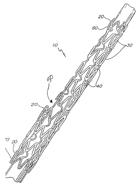
Fig. 1 illustrates generally at 10 a particular embodiment of this type of
stmt. The stmt has an opening 50 through which a second stmt may be maneuvered
and
subsequently positioned at the branch. The two stents will therefore
substantially form
the "Y" shape at the obstructed intersection so that one stmt may be placed in
the first
branch and the second stmt may be placed in the second branch. The second stmt
may
be placed in the vessel or artery during a subsequent procedure, or during the
same
procedure but following the placement and expansion of the first stmt.
The stmt therefore advantageously has a radiopaque coating 20, located at
the distal end 60, at the proximal end 70 and around the opening 50. The
distal end 60
has a smaller diameter than the proximal end 70, the distal end being inserted
into the
patient first. The stent can therefore not only be located, but the
orientation of the stmt
may also be determined accurately using fluoroscopy. This is important for
proper
placement of the second stmt through the opening of the first stent and into
the branch of
the vessel.
Fig. 2 illustrates a non-biftircated stmt of the type found in US
Application Serial No. 08/511,076 incorporated by reference herein. As shown
in fig. 2,
a radiopaque coating 20 is affixed 'to each of the distal end 22 and the
proximal end 24,
actually interchangeable in this figure.
Fig. 3 shows a flat view of the same stmt as found in fig. 2, hereinafter
I referred to as a stmt preform. The stmt may be formed of a flat preform
shown in fig. 2
that is formed into a tubular shape (fig. 1) by rolling the pattern so that it
brings edges 12
and 14 together. Alternatively, the preform may already be a one piece tubular
form that
requires no rolling or welding. The edges may then be joined by welding or the
like to
provide a configuration such as that shown in fig. 2. The stmt preform has
been cut into
a pattern of substantially parallel struts 16. Pairs of struts are
interconnected at
alternating end portions 19a and 19b. The radiopaque material 20 is shown over
the
entire surface of the patterned preform. As will be discussed in more detail
below, the
radiopaque material may be affixed to the preform either before or after strut
pattern
formation.
Fig. 4 shows generally at 30 a nonbifurcated stmt of the type found in US
6
CA 02421797 2003-03-04
WO 02/22050 PCT/USO1/28850
5807404, US 5836964 and US 5922005 incorporated by reference herein. As shown
in
fig. 4, a radiopaque coating 28, such as gold, is affixed to each of the
distal end 32 and
the proximal end 34, actually interchangeable in this figure. Struts 38 have
been cut,
etched, stamped, or so forth into the stmt structure.
Fig. 5 is a flat view of the same stem found in fig. 4 showing the general
pattern of the stmt which can be referred to as a stmt preform. The preform
may be
rolled into a tubular shape (fig. 1) to bring the edges 42 and 44 together.
Alternatively,
the preform may already be a one piece tubular form having the strut pattern
cut or
etched into it. The tubular form thus requires no rolling or welding. The
edges may then
be joined by welding or the like to provide a configuration such as that shown
in fig. 4.
The stmt preform has been cut into a pattern of substantially parallel struts
38. Pairs of
struts are interconnected at alternating end portions 39a and 39b. The
radiopaque
material 40 is shown over the entire surface of the patterned preform. As will
be
discussed in more detail below, the radiopaque material may be affixed to the
preform
either before or after strut pattern formation.
The method of forming stems with radiopaque materials, or of affixing
radiopaque coating to the stems of the present invention allows the radiopaque
material
to be accurately and conveniently placed in only those areas of the stmt where
the
radiopaque material is desired.
The radiopaque material may be affixed to a stmt preform in either a
sheet or tubular form. This may be done either before or after the strut
pattern has been
cut, etched or stamped in the preform. If a tubular preform is being used and
the strut
pattern has already been formed in the stmt, this preform is basically a
finished stent. All
of these forms will, however, hereinafter be referred to as stmt preforms. The
stent
preform may be in the form of a sheet or a tube, and may be made of various
materials
including polymeric materials, although in the present invention metals or
metal-like
materials are preferable for use. The stmt may be formed of at least one
metal, but may
be comprised of more than one metal such as in the case of an alloy. Stent
formation
from such materials is well known to those of skill in the art. Biocompatible
metals such
as stainless steel, Nitinol (nickel-titanium alloys), and so forth.
Nitinol is discussed in US 6059810 herein incorporated by reference in its
7
CA 02421797 2003-03-04
WO 02/22050 PCT/USO1/28850
entirety. This patent refers to an article by L. McDonald Schetky for a
discussion of such
alloys entitled "Shape-Memory Alloys" at pp 74-82 of Volume 241 (5) November
1979,
SCIENTIFIC AMERICAN, and to "A Source Manual For Information On Nitihol and
Ni Ti", first revision, by David Goldstein, Research and Technology
Department, Feb. l,
1980, Naval Surface Weapons Center, Dalgren, Va. 22448 (NSWC TR 80-59), both
of
which are incorporated by reference herein.
Other alloys useful for stmt formation, in addition to Nitinol, are
discussed in US 5725570 herein incorporated by reference. 'This discussion
includes
stainless steel, as well as other superelastic materials including, e.g.,
silver-cadmium
(Ag-Cd), gold-Cadmium (Au-Cd), gold-copper-zinc (Au-Cu-Zn), copper-aluminum-
nickel (Cu-Al-Ni), copper-gold-zinc (Cu-Au-Zn), copper-zinc/(Cu-Zn), copper-
zinc-
aluminum (Cu-Zn-Al), copper-zinc-tin (Cu-Zn-Sn), copper-zinc-xenon (Cu-Zn-Xe),
iron-beryllium (Fe3-Be), iron-platinum (Fe3-Pt), indium-thallium (In-Tl), iron-
manganese (Fe-Mn), nickel-titanium-vanadium (Ni-Ti-V), iron-nickel-titanium-
cobalt
(Fe-Ni-Ti-Co), and copper-tin (Cu-Sn). See also Schetsky, L. McDonald, "Shape
Memory Alloys", Encyclopedia of Chemical Technology (3rd ed.), John Wiley &
Sons,
1982, vol. 20. pp. 726-736 for a full discussion of superelastic alloys,
herein incorporated
by reference. ~ One method of affixing the radiopaque material to the stmt
preform is by electroplating the sheet or tubular preform with the radiopaque
material.
This may be accomplished by dipping the sheet or tubular member in the
electroplating
solution. At this point, the entire surface of the sheet or tube is coated
with the
radiopaque material. There are optional steps that may be undertaken during
the
electroplating process. This can improve the adhesion of the radiopaque
material to the
stmt in those situations where good adhesion may be difficult to achieve.
A first optional step which may be referred to in the industry as acid
etching involves the placement of the metal sheet or tube in an acid bath
prior to
electroplating to remove oxides from the surface of the metal. The use of this
technique
is known to one of skill in the art and is sometimes referred to as
"pickling." This may
be accomplished out of a sulfuric acid bath, for instance. The metal is
typically rinsed
both prior to and after the acid etching step.
A second optional step involves application to the metal tube or sheet of
8
CA 02421797 2003-03-04
WO 02/22050 PCT/USO1/28850
what is referred to as a "strike" layer. The strike layer is a very thin
electrochemically
deposited layer that prevents reformation of oxides on the surface of the
metal thereby
improving the adhesion of the subsequent coating.
During the strike, the metal is plated with a solution of a metal salt
S wherein a thin layer of metal, i.e. the "strike", is immediately deposited
on the surface of
the metal sheet or tube. This layer is controlled by the concentration of the
solution, the
time of exposure, and the amperage of the current, to a thickness of
preferably about 5 x
10'6 cm to about 1.5 x 10'5 cm (about 0.5 ,um to about l.S,um; about 500 to
about 1500
A). A typical strike layer is approximately 1.3 x 10'5 cm (about 1.3 ,um or
1270 A). This
thin layer has no affect on the rigidity or expansion properties of the stmt
thereby
allowing the entire surface to be coated with a simple immersion step. The
"strike" layer
may be optionally added to improve the adhesion of the subsequently applied
radiopaque
coating to the metal.
The very thin "strike" layer allows the entire metal sheet or tube to be
dipped in a plating solution of the metal because the thin coating will not
have an adverse
affect on the stmt properties, such as decreased flexibility or increased
radiopacity.
The strike layer may be comprised of any metal typically used for a
"strike" including any of the noble metals such as gold, silver, nickel, and
so forth. A
gold "strike" is desirable. Gold has been found to be a preferable choice
because it tends
to cause less thrombus, tissue irritation and/or allergic reaction and so
forth than other
metals such as nickel, for instance.
The sheet or tube may then be electroplated with the desired radiopaque
material. The present invention allows for a simplistic electroplating method
because the
entire metal form may be immersed in the electroplating solution.
Electroplating
methods are well known to those of ordinary skill in the art.
Any radiopaque material may be used in the present invention including
the noble metals such as gold, platinum, tantalum, rhenium and iridium, and
the non-
noble metal, silver. Radiopaque materials useful for providing stems with
radiopacity
are discussed in US 5725570 herein incorporated by reference in its entirety.
Some
metals, such as tantalum, irradiate more brightly than others and the metal
can therefore
be selected on such basis. Some particular embodiments of the present
invention utilize
9
CA 02421797 2003-03-04
WO 02/22050 PCT/USO1/28850
gold as the metal of choice. Gold is useful due to its nonallergenic
qualities, as well as
its radiopacity.
Again, as noted above, gold is a desirable choice because it is known to
produce less thrombus, tissue irritation and/or allergic reaction.
The thickness of the coating may be varied, but is preferably between
about 1 ,um to about 20 ,um (about 3.9 x 10'5 inches to about 7.9 x 10'4
inches),
preferably from about 2 ,um to about l2,um (about 7.9 x 10'5 inches to about
4.7 x 10'4
inches). The desired thickness may be achieved, for instance, through one or
more
electrochemical depositions.
The coating thickness can be varied over the surface of the stmt in order
to vary the radiopacity over the surface of the stmt. It may be desirable to
coat some
areas thicker than others to increase the irradiation in those areas where the
radiopaque
material is coated thicker. This variation in thickness can be controlled by
how much
material is plated on the surface, as well as by the removal process itself.
For instance,
etching can be used to remove more radiopaque coating from some areas than
others
therefore controlling the coating thickness in this manner.
Further, different radiopaque materials can be coated on different portions
of the stmt to vary the radiopacity of the stmt as well. For instance,
tantalum can be
coated on one portion and gold on another portion. The tantalum will irradiate
more
brightly than the gold. For example, in a bifurcated stmt, it may be desirable
to mark the
opening in the stent with tantalum so that it illuminates more brightly. Or,
it may be
desirable that only the ends of the stent are clearly visible, and
consequently, it may be
desirable to mark the ends so that they illuminate brightly, and to mark the
center portion
so that it is only slightly radiopaque. Using the method of the present
invention,
selective marking of the stems is easily accomplished.
The ability to control how much coating is located on various parts of the
stmt is extremely helpful for accurate.placement of the stmt in a body lumen.
For a
bifurcated stent, for instance, it may be desirable that the proximal and
distal ends of the
stmt are radiopaque, as well as the portion of the stmt surrounding the
opening, i.e. the
bifurcation, where the second stent is to be placed as is shown in Fig. 1.
This type of .
pattern allows for an easy determination as to stmt orientation and position,
as well as
CA 02421797 2003-03-04
WO 02/22050 PCT/USO1/28850
the length and diameter of the stmt. The position of the scent is particularly
critical for
bifurcated stents because it is necessary that the operator can accurately
determine if the
opening of the stmt is matched with the branch in the artery or vessel. For
this reason, it
may be desirable that the opening in the center illuminate more brightly so
that the
operator who is positioning the stmt can accurately place the opening at the
branch in the
artery. Furthermore, when placing the second stmt through the opening, the
operator
will be able to accurately ascertain where the opening easy for easier stent
placement.
In the case of nonbifurcated stents, it may be desirable that only the ends
illuminate, or that the ends illuminate more brightly so that the proper
position and
orientation of the stmt may be determined not only during insertion, but also
at later
dates to determine if the stmt position has changed as shown in Figs. 2 and 3.
A strut pattern may be formed in the stmt preform either before or after
the electroplating process. The strut pattern is formed by laser cutting or
etching,
chemical etching, metal stamping, and so forth. Following the formation of the
strut
pattern, the gold plating or coating may then be removed from those areas
where it is
desired that the stent be non-radiopaque, consequently leaving the gold
coating on those
areas of the stmt where radiopacity is desired. This may also be accomplished
using an
etching process, either laser or chemical etching processes may be used. This
process is
therefore used to remove the radiopaque material from certain areas of the
stent preform.
If it is a metal sheet that has been electroplated and etched, it will then be
necessary to form the sheet into a tube, and to join the tube together at the
seam.
There axe various advantages to using the method of the present invention.
One advantage is that it simplifies the production process. The selective
etching method
of the present invention eliminates the need for premasking of the stmt prior
to plating.
Furthermore, using this method of plating only portions of the stent
eliminates wash out or haloing. These phenomena can occur where the whole stmt
is
either itself formed of a radiopaque material, such as tantalum, or the entire
stmt is
coated with a relatively thick layer of a radiopaque material. This makes the
stmt
difficult to view under fluoroscopy because it illuminates too brightly,
making the lines
of the stmt indistinct, and not clearly discernible. Furthermore, it makes it
difficult to
accurately determine the location and orientation of the stmt. Also, if the
stmt is very
11
CA 02421797 2003-03-04
WO 02/22050 PCT/USO1/28850
luminous, the haloing can obscure visibility of the blood vessel lesion, and
can actually
mask it, making it difficult to repair the vessel.
Further to the present invention, selective plating allows the expansion
characteristics of the stmt to be controlled. For instance, application of the
coating layer
in certain areas can increase the rigidity of the stmt in those areas. This
can result in
either a "watermelon seed" effect wherein the stent shoots from the desired
location of
scent placement, or if the middle deploys first, the stmt may lodge in the
vessel. It has
been noted that the coating can increase the rigidity of the stmt in those
areas where it is
plated. This fact must be taken into account because it can change the method
by which
the stent opens. For instance, the balloon may open the center or body of the
stent first if
the end portions are plated but not the center. However, controlling the mode
of
deployment wherein the ends or center portion open first may be desirable in
some cases.
The thicker the coating, the more likely that the stmt may deploy first at
those portions
where the coating is not as thick. With the method of the present invention,
the stent can
be balanced easily by selectively plating certain areas, thereby preventing
undesirable
movement of the stent. For instance, if a portion of the middle of the stent
is plated, as
well as the distal and proximal ends, the center will have less of a tendency
to open first.
Furthermore, the thickness to which the areas are plated can be easily
controlled. For
instance, certain portions of the stmt may be plated with a thicker coating,
causing the
stmt to illuminate more brightly in certain areas. This would allow not only
the location
of the stmt to be easily determined, but the orientation of the stmt as well.
The current invention allows the use of a base stainless steel stmt which
maintains good flexibility. The application of the radiopaque material to only
the ends or
specified subsections does not increase stmt rigidity.
In addition to being directed to the embodiments described above and
claimed below, the present invention is further directed to embodiments having
different
combinations of the features described above and claimed below. As such, the
invention
is also directed to other embodiments having any other possible combination of
the
dependent features claimed below.
The above figures and disclosure are intended to be illustrative and not
exhaustive, and will suggest many variations and alternatives to one of
ordinary skill in
12
CA 02421797 2003-03-04
WO 02/22050 PCT/USO1/28850
this art. All these alternatives and variations are intended to be included
within the scope
of the attached claims. Those familiar with the art may recognize other
equivalents to the
specific embodiments described herein which equivalents are also intended to
be
encompassed by the claims attached hereto.
While the embodiments discussed above have focused to a large degree
on stems, other implantable medical devices may be treated using the method of
the
present invention without detouring from the spirit of the present invention.
The contents of parent application No.09/659,571, filed September12, 2000, is
incorporated herein by reference in its entirety.
13