Note: Descriptions are shown in the official language in which they were submitted.
CA 02421967 2003-03-10
WO 02/21994 PCT/USO1/26964
A METHOD AND IMPLANT FOR EXPANDING A SPINAL CANAL
Invented By:
D. Greg Anderson
Related Applications
The present application claims priority from copending U.S. Utility
Application Serial No. 09/659,180, filed September 11, 2000, entitled "A
Method and
Implant for Expanding a Spinal Canal" .
Field Of The Invention
The present invention relates generally to spinal surgery, and more
particularly to a method and apparatus for expanding a spinal canal to relieve
pressure on
spinal nerves.
Background Of The Invention
Spinal Stenosis, or narrowing of the spinal canal, inflicts millions of people
with back and leg pain due to compression of spinal nerves. Severe spinal
stenosis often
leads to surgery in an effort to relieve compressed nerves and lessen back and
leg pain.
Spinal laminectomy is the traditional operation performed to treat spinal
stenosis. In the
spinal laminectomy, posterior aspects of the spinal column are removed to "un-
roof" the
spinal canal to relieve the pressure on the nerves. Specifically, a spinous
process, lamina
and portions of various facet joints are the posterior aspects of the spinal
column surgically
excised.
Although the spinal laminectomy is often successful in relieving pressure on
the nerves of the spinal canal, several problems and disadvantages arise as a
result of the
laminectomy. First, the laminectomy removes important sites of back muscle
attachment
1
CA 02421967 2003-03-10
WO 02/21994 PCT/USO1/26964
leading to back muscle dysfunction and pain. Second, the laminectomy exposes
the nerve
sac causing scar tissue to form around the nerves. Scar tissue may prevent
normal motion
of the nerves, leading to recurrent pain. Third, the laminectomy can
destabilize the spine
resulting in a forward slippage of one vertebra on another. Vertebral slippage
can cause
recurrent pain and deformity. Fourth, the laminectomy requires a large
surgical exposure
and significant blood loss, making the laminectomy dangerous for older
patients. Finally,
spinal stenosis can recur following the laminectomy, requiring risky revision
surgery.
Laminectomy risks have Ied surgeons to seek an alternative for patients with
severe spinal stenosis. Some surgeons choose to treat spinal stenosis with
multiple
laminotomics. Laminotomies involve removing bone and soft tissue from the
posterior
aspect of the spine making "windows" into the spinal canal over areas of nerve
compression. Multiple laminotomies remove less tissue than the laminectomy,
resulting in
less scaring, vertebral instability and blood loss.
Multiple laminotomies, however, also suffer from problems and
disadvantages. Laminotomies may not adequately relieve nerve compression and
the pain
may continue. Laminotomies are more difficult to correctly perform than the
laminectomy.
Laminotomies expose the nerves and may cause nerve scaring. Patients receiving
multiple
laminotomies also often have recurrent spinal stenosis requiring risky
revision surgery.
Zucherman, et. al. , discloses another approach (differing from
laminectomies, laminotomies and the present invention) to spinal stenosis in
U. S. patent
5,836,948, where a device and method is described to distract (spread apart)
spinous
processes of adjacent vertebrae and prevent extension of the spine. While the
Zucherman
technique may help to relieve some spinal canal narrowing due to in folding of
posterior
soft tissues, the bony spinal canal remains unchanged in size. Without
expanding the spinal
2
CA 02421967 2003-03-10
WO 02/21994 PCT/USO1/26964
canal area, the Zucherman technique offers limited benefit for spinal stenosis
sufferers.
Furthermore, arthritic facet spurs, the main cause of degenerative spinal
stenosis, remain
unaffected by the Zucherman approach and continue to cause pain. Also, the
distraction of
the spinous processes as described by Zucherman creates a forward curvature of
the spine
called kyphosis. Lumbar kyphosis is a spinal deformity often associated with
back pain and
dysfunction.
Information relevant to a wide variety of spinal implants including screw and
rod constructs are described by Biedermann et. al. in U.S patent 5,725,527,
Tsou in U.S.
patent 5,176,678, Kambin in U.S. patent 5,480,440 and Mathews in U.S. patent
5,496,322. These implants are useful for stabilizing the spine and the
correcting spinal
deformities, however, these references are not capable of expanding the spinal
canal or
treating spinal stenosis. Other implants operating as intervertebral spacers
are described by
Errico et. al in U.S. patent 5,653,763 and Kuslich in U.S. patent 5,059,13.
These implants
are useful in expanding the intervertebral disc space and assisting with
spinal fusion,
however, these references are also not capable of expanding the spinal canal
or treating
spinal stenosis.
For the foregoing reasons, there is a strong need for a different and better
method for relieving the symptoms of spinal stenosis without the drawbacks of
currently
available techniques. A method is needed that expands the spinal canal,
relieving pressure
on the spinal nerves, while being simple, safe and permanent.
Summary of the Invention
The present invention provides a simple, safe and permanent method and
apparatus for treating spinal stenosis by expanding the spinal canal area to
provide
additional space for the spinal nerves, relieving pressure on the spinal
nerves.
3
CA 02421967 2003-03-10
WO 02/21994 PCT/USO1/26964
An object of the present invention is to maintain the integrity of the spinal
canal so that the function of normal tissues is not destroyed or significantly
altered as with a
laminectomy and laminotomy.
Another object of the present invention is to avoid scarring around the spinal
nerves by avoiding an open exposure of the nerves.
Another object of the present invention is to avoid causing spinal
instability,
where one vertebra slips forward on another vertebra causing recurrent pain
and deformity.
Yet another object of the present invention is to decompress the spinal nerves
with a quick, safe approach resulting in minimal blood loss.
Yet another object of the present invention is to provide a permanent solution
to spinal stenosis, where no tendency exists for recurrence.
In one aspect of the present invention, a method for correcting spinal
stenosis is introduced where a spinal canal is enlarged by cutting a vertebra,
separating the
vertebral cut and then stabilizing the cut, allowing the vertebra to heal with
the spinal canal
expanded, permanently creating more space for the spinal nerves thus relieving
compression on the nerves.
In another aspect of the present invention, the method of expanding the
spinal canal begins with cutting a vertebra (performing an osteotomy) at each
spinal pedicle
of the vertebra in an oblique fashion, beginning at a base of a transverse
process of the
vertebra and coursing medially and anteriorly at approximately a 45-degree
angle to
complete the cut through a medial wall of the pedicle to the spinal canal.
Each osteotomy
(bone cut) is then distracted (expanded) by inserting an implant into the
osteotomy,
increasing the diameter (and thus the area) of the spinal canal. The implant
is stabilized,
securing the osteotomies and the vertebra with the spinal canal in an expanded
state.
4
CA 02421967 2003-03-10
WO 02/21994 PCT/USO1/26964
In another aspect of the present invention, the implant comprises a stent and
a screw. The stent is inserted into the vertebral cut, expanding the spinal
canal. The screw
is inserted through the stent and threaded into the vertebra, securing the
stmt, the vertebral
cut and the expanded spinal canal.
In another aspect of the present invention, the implant comprises two stems
and two screws. Each stmt is inserted into one of two vertebral cuts, each
stmt insertion
expanding the spinal canal. Each screw is inserted through one stent and
threaded into the
vertebra, securing the stmt, the vertebral cut and the expanded spinal canal.
In another aspect of the present invention, the implant includes two stems,
two screws, two washers and a cable. Each stmt is inserted into one of two
vertebral cuts,
each stmt insertion expanding the spinal canal. Each screw is inserted through
one washer
and one stent. Each screw is then threaded into an anterior portion of the
vertebra,
securing the washer, the stmt, the vertebral cut and the expanded spinal
canal. Each
washer is connected to one of two ends of the cable, the cable being strapped
around or
through a posterior portion of the vertebra, securing the posterior portion of
the vertebra to
the anterior portion of the vertebra.
In another aspect of the present invention, the screw can include self
tapping, bone gripping threads. The screw can also include a rounded head.
In another aspect of the present invention, the stem is designed to
accommodate the insertion of bone graft material to facilitate healing of the
vertebra with
expanded spinal canal. The stmt can be U-shaped for this purpose. The stent
can include
a rounded, wedge-shaped end for ease in penetrating and separating the
vertebral cut. The
stent can include a flange designed to prevent the stmt from penetrating into
the spinal
s
CA 02421967 2003-03-10
WO 02/21994 PCT/USO1/26964
canal. The stent can also include a rounded depression adapted to rotatably
accept a
rounded washer and/or a rounded head on the screw for securing the stent to
the vertebra.
In another aspect of the present invention, the washer can include a rounded
shape substantially similar to and engagable with a rounded head on the screw
and a
S rounded depression in the stmt. The rounded head of the screw would reside
within the
rounded washer and both would be rotatably housed within the rounded
depression in the
stent.
The present invention has the following advantages over current, unrelated
techniques for treating spinal stenosis:
(1) Normal spine structures are not removed and thus normal muscle attachments
are maintained.
(2) There is less chance of spinal instability.
(3) There is less manipulation of the spinal nerves.
(4) There is less scaring around the spinal nerves.
1S (S) Spinal decompression is more complete.
(6) The operation is quicker and safer with less blood loss.
(7) The expanded spinal canal is permanent, preventing recurrent spinal
stenosis.
Brief Description Of The Drawings
For the purpose of illustrating the invention, there is shown in the drawings
a
form which is presently preferred; it being understood, however, that this
invention is not
limited to the precise arrangements and instrumentalities shown.
Fig. 1 illustrates an exploded view of an implant comprising a screw, a
rounded washer with a cable end-attachment and a stent, in accordance with the
present
invention;
6
CA 02421967 2003-03-10
WO 02/21994 PCT/USO1/26964
Fig. 2 illustrates a perspective view of the implant of Fig. 1 without the
cable end-attachment on the rounded washer, to show the relationship of the
screw, the
rounded washer and the stmt;
Fig. 3 illustrates a perspective view of the implant of Fig. 1 having an angle
between the stmt and the screw typical of the implant when inserted into a
vertebra;
Fig. 4a illustrates a perspective view of the rounded washer as shown in Fig.
1 with the cable end-attachment;
Fig. 4b illustrates an underside perspective view of the rounded washer of
Fig. 4a with the cable end-attachment;
Fig. 4c illustrates a top plan view of the rounded washer of Fig. 4a with the
cable end-attachment;
Fig. 4d illustrates a top plan view of a rounded washer with a cable side-
attachment, in accordance with the present invention;
Fig. 4e illustrates a perspective view of the rounded washer of Fig. 4d with
the cable side-attachment;
Fig. 5 illustrates a cross-section view of a vertebra, where dashed lines
represent locations of vertebral cuts (osteotomies), in accordance with the
present
invention;
Fig. 6 illustrates a cross-section view of a vertebra after expanding the
spinal
canal by inserting the stents in the osteotomies, in accordance with the
present invention;
Fig. 7 illustrates a cross-section view of the vertebra of Fig. 6, with screws
stabilizing the expanded spinal canal by insertion through the stents and into
the vertebra, in
accordance with the present invention;
CA 02421967 2003-03-10
WO 02/21994 PCT/USO1/26964
Fig. 8 illustrates a cross-section view of the vertebra of Fig. 7, with the
screws, rounded washers and a cable stabilizing the expanded spinal canal, in
accordance
with the present invention; .
Fig. 9a illustrates a perspective view of a vertebra with pedicle osteotomies'
in place and a stmt ready for impaction into one of the osteotomies, in
accordance with the
present invention;
Fig. 9b illustrates a perspective view of the vertebra of Fig. 9a, after
expanding the spinal canal by inserting stems in the osteotomies, in
accordance with the
present invention;
Fig. 9c illustrates a perspective view of the vertebra of Fig. 9b, with
screws,
rounded washers and a cable stabilizing the expanded spinal canal, in
accordance with the
present invention; and
Fig. 10 illustrates a perspective view of the vertebra of Fig. 9a, viewed from
the top right side, showing the relationship of the screw, a rounded washer
with a side-
attachment and the cable, in accordance with the present invention.
Detailed Description Of The Invention
Referring now to the drawings, wherein like numerals indicate like elements,
there is shown in Fig. 1 an exploded view of an implant 11 comprising a screw
26, a
rounded washer with end-attachment 32a, a cable 40 and a stent 24. The stent
24 is
designed for impaction into a vertebral osteotomy (bone cut), to expand the
sides of the
osteotomy apart. Once in place, the stem 24 is designed to contain bone graft
material to
assist the healing of the osteotomy.
The stmt 24 has a rounded, wedge shaped end 48, a U-shaped sidewall 58
and an expanded end 56. There is a flange 50 at the juncture of the expanded
end 56 and
s
CA 02421967 2003-03-10
WO 02/21994 PCT/USO1/26964
the U-shaped sidewall' S8. The expanded end 56 contains a rounded depression
54 to
accept the screw 26 and/or the rounded washer 32a. The rounded, wedge-shaped
end 48 is
designed to enter and wedge apart the sides of the osteotomy. The U-shaped
sidewall 58
surrounds an interior section 52 of the stent 24 which can be filled with bone
graft after the
impaction of the stmt 24 into the osteotomy.
The screw 26 includes a head 34 having a screwdriver socket 35 and a
rounded outer surface 33. The rounded outer surface 33 is adapted to engage an
inner
surface 43 of the rounded washer 32a. The screw 26 also includes a shank 36
with threads
38 designed to grip vertebral bone and can be the self tapping variety.
The rounded washer with end-attachment 32a includes a central passage 46
for the screw 26 and an inner surface 43 adapted to accept the outer surface
33 of the screw
head 34. The rounded washer 32a also includes a rounded outer surface 42,
adapted to
engage the rounded depression 54 of the stmt 24 allowing the rounded washer
32a to
rotatably reside within the rounded depression 54. The outer surface 42 of the
rounded
washer 32a has a connector 44 for attaching an end of a cable 40 to the
rounded washer
32a.
Figs. 4a through 4e illustrate two embodiments of the rounded washer 32a,
32b. The rounded washer with end-attachment 32a for the cable 40 is shown in
Figs. 4a
through 4c. A rounded washer with side-attachment 32b for the cable 40 is
shown in Figs.
4d and 4e. Fig. 4a illustrates a perspective view of the rounded washer with
end-
attachment 32a as shown in Fig. 1. Fig. 4b illustrates a second perspective
view of the
rounded washer 32a, showing the central passage 46 from below and the
connector 44 for
end-attachment of the cable 40. Fig. 4c illustrates a top plan view of the
rounded washer
32a, also showing the central passage 46, the connector 44 and the cable 40.
9
CA 02421967 2003-03-10
WO 02/21994 PCT/USO1/26964
Figs. 4d and 4e illustrate the rounded washer with side-attachment 32b. The
rounded washer with side-attachment 32b has a similarly shaped central passage
46,
rounded inner surface 43 and rounded outer surface 42 as the rounded washer
with end-
attachment 32a. As such, the rounded washer 32b is similarly designed to
accept the screw
S 26 and to rotatably reside within the rounded depression S4 of the stem 24.
The rounded washer with side-attachment 32b differs from the rounded
washer with end-attachment 32a only in its cable 40 attachment means, having a
side
passage 4S with a tunnel 47 slightly larger than the cable 40 so that the
cable 40 is slidably
movable within the tunnel 47. The slidable movement of the cable 40 through
the tunnel 47
allows tensioning of the cable 40 to remove cable slack. The cable 40 may then
be secured
to the side passage 4S by crushing or plastically deforming the side passage
4S around the
cable 40 to firmly hold the cable 40 in place. Alternatively, the side passage
4S could be
secured to the cable 40 by other means, such as by setscrew.
Fig. 2 illustrates the screw 26, the rounded washer 32a and the stent 24
1S engaged together. The connector 44 and the cable 40 (shown in Fig. 1) are
removed in
Fig. 2 to detail the engagement of the screw 26, the rounded washer 32a and
the stmt 24.
The head 34 of the screw 36 is seated within the rounded washer 32a. The
rounded washer
32a is seated within the rounded depression S4 of the expanded end S6 of the
stent 24. The
shape of the head 34 of the screw 26, the rounded washer 32a and the rounded
depression
S4, allow the screw 26 and the rounded washer 32a to move or rotate within the
rounded
depression S4 of the stmt 24 (illustrated by the arrow 29). Therefore, the
angle between
the screw 26 and the stent 24 is variable to assist with accurate placement of
the implant 11.
Fig. 3 illustrates the implant 11 with the screw 26, the rounded washer with
end-attachment 32a, the cable 40 and the stmt 24 engaged and positionally
angled (relative
to
CA 02421967 2003-03-10
WO 02/21994 PCT/USO1/26964
to one another) similar to implant 11 positioning within a vertebra in
accordance with the
present invention. The screw 26 is seated within the rounded washer 32a, the
rounded
washer 32a is seated within the rounded depression 54 of the stent 24. The
stmt 24 and
screw 26 form an acute angle 31, positioning the connector 44 and the cable 40
in an
upward direction relative to the stmt 24.
The implant 11 could be made of materials including but not limited to:
stainless steel, titanium alloy, cobalt chromium alloy, bone, polyethylene,
and polymers of
lactide, glycolide, caprolactone, polydioxanone, trimethylene carbonate,
polyorthoesters,
polyethelene oxide or blends of the above polymers.
Fig. 5 illustrates a cross-section of a vertebra 15 comprising a vertebral
body
12, a pedicle 21, a transverse process 14, a superior articular process 18, a
lamina 17, a
spinous process 16 and spinal canal (unexpanded) 20a. The dashed lines
represent the site
of the osteotomies (bone cuts) 22. Each osteotomy 22 begins at the base of the
transverse
process 14 and courses at approximately a 45-degree angle (relative to a
coronal plane)
towards a medial wall of the pedicle 21. Cutting the pedicle 21 as described
(as shown by
the dashed lines) separates a posterior portion of the vertebra (the spinous
process 16, the
superior articular process 18, and the lamina 17) from an anterior portion of
the vertebra
(the pedicle 21, the transverse process 14, and the vertebral body 12).
The osteotomy (bone cut) 22 is performed by any commonly known method
for cutting bone, such as using a wire saw, a oscillating power saw or an
osteotome. Of
course, care should be taken to protect the spinal nerves while performing the
osteotomy.
Fig. 6 illustrates a cross-section of the vertebra 15 showing an expanded
spinal canal 20b. Osteotomies 22 were completed through both pedicles 21 and
stents 24
were placed into each osteotomy 22. The stents 24 distract (separate) and hold
the edges of
11
CA 02421967 2003-03-10
WO 02/21994 PCT/USO1/26964
the osteotomy 22 apart. Distracting the osteotomies 22 by inserting stents 24
expands the
spinal canal 20b, increasing its diameter relative to that of the unexpanded
spinal canal 20a
shown in Fig. 5.
Fig. 7 illustrates a cross-section of the vertebra 15 showing the expanded
spinal canal 20b, with a screw 26 threaded into a pedicle 21 to secure the
stmt 24 inserted
into each osteotomy 22. The screws 26 are seated within the rounded depression
54 of the
stmt 24, preventing the stmt 24 from sliding out of the osteotomy 22. The
flange 50 on
the stent 24 prevents the stent 24 from penetrating the spinal canal 20b, by
preventing
further movement of the stmt 24 into the osteotomy 22.
Prior to placement of each screw 26 into the pedicle 21, bone graft material
(not shown) should be placed into the interior section 52 of each stmt 24 to
assist with
healing of the osteotomy 22. The screw 26, the U-shaped sidewall 58 of the
stmt 24 and
the sides of the osteotomy 22 completely contain the bone graft material
placed within the
interior section 52 of the stent 24, preventing a release of the bone graft
material prior to
vertebral healing.
The spinous process 16, the lamina 17 and the superior articular process 18
rest on the stmt 24 and are held in place (fixed against the stmt 24) by
surrounding soft
tissues. Concluding the procedure in this manner (relying on surrounding soft
tissue to
secure the posterior portion of the vertebra to the anterior portion) would be
appropriate in
stiff, arthritic spines where there is no tendency for the vertebrae to slip
on one another.
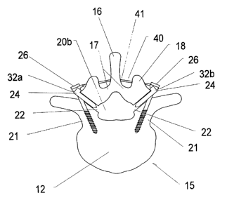
Fig. 8 illustrates a cross-section of the vertebra 15 showing the expanded
spinal canal 20b. In addition to the screws 26 threaded into the pedicles 21
to secure the
stents 24 in the osteotomies 22, rounded washers 32a, 32b and a cable 40 are
added to
12
CA 02421967 2003-03-10
WO 02/21994 PCT/USO1/26964
secure the spinous process 16, the lamina 17 and the superior articular
process 18 against
the scents 24 for healing of the vertebra 15.
The screws 26 pass through the rounded washers 32a, 32b and are threaded
into the bone of the pedicle 21. The cable 40 is secured at each end by
attachment to the
rounded washers 32a, 32b. First, the rounded washer with end-attachment 32a
for the
cable 40 (with the cable 40 attached) is mounted into the rounded depression
54 in the stmt
24 and the screw 26 is inserted through the rounded washer 32a and threaded
into the
pedicle 21. Next, the cable 40 is aligned across a top (dorsal) surface of the
lamina 17 and
inserted through a hole 41 drilled in a base of the spinous process 16. The
cable is then
aligned across a top (dorsal) surface of the other lamina 17 and is inserted
through the
tunnel 47 in the side passage 45 of the rounded washer with side-attachment
32b. The
cable 40 is tensioned to remove all slack (by pulling the cable 40 taut
through the tunnel 47)
and the side passage 45 is crushed, plastically deformed or bolted to fixedly
connect the
cable 40 to the rounded washer 32b, firmly securing the cable 40 between the
rounded
washers 32a, 32b and the stents 24. Alternatively, two rounded washers with
side-
attachment 32b could be used, depending on operational conditions and user
preference.
The cable 40 secures the spinous process 16, the lamina 17 and the superior
articular process 18 (the posterior portions of the vertebra 15) to the
pedicle 21 and the
vertebral body 12, yielding a mechanically stable spinal canal. The addition
of the cable 40
and the rounded washers 32a, 32b lends stability to the osteotomies 22,
preventing slippage
of the vertebra 15. Again, bone graft material should be placed within the
interior section
52 of the stent 24 prior to placement of the screws 26, the rounded washers
32a, 32b and
the cable 40.
13
CA 02421967 2003-03-10
WO 02/21994 PCT/USO1/26964
Fig. 9a illustrates a perspective view of a vertebra 15 after creation of an
osteotomy (bone cut) 22 through each pedicle 21. Each osteotomy 22 extends
obliquely
from the base of the transverse process 14 through the pedicle 21 to the
spinal canal
(unexpended) 20a. The stmt 24 is ready for impaction into one of the
osteotomies 22. The
rounded, wedge-shaped end 48 of the stmt 24 is placed at the edge of the
osteotomy 22.
An impactor 60 having a mallet end 62 and an impact end 64 is placed into
contact with the
expanded end 56 of the stmt 24. The impact end 64 of the impactor 60 is
adapted for flush
engagement with the expanded end 56 of the stmt 24. Mallet blows to the mallet
end 62 of
the impactor 60 drives the rounded, wedge-shaped end 48 of the stmt 24 into
the osteotomy
22, causing the osteotomy 22 to wedge open as the stent 24 enters the
osteotomy 22. The
stent 24 is driven into the osteotomy 22 until the flange 50 contacts the
osteotomy 22 edge
at the base of the transverse process 14, preventing further movement of the
stmt 24 into
the osteotomy 22. To expand the spinal canal 20a in a symmetric fashion, the
opposite
osteotomy 22 also receives a stent 24.
IS Fig. 9b illustrates the vertebra 15 of Fig. 9a with an expanded spinal
canal
20b after placement of the stems 24 into the osteotomies 22. The stents 24
hold the
osteotomies 22 open, expanding the spinal canal 20b. The stems 24 are slightly
shorter in
length than the osteotomies 22, preventing stmt 24 projection into the
expanded spinal
canal 20b. The flange 50 abuts the edge of the osteotomy 22 at the base of the
transverse
process 14, preventing the stmt 24 from sliding into the expanded spinal canal
20b. With
the stmt 24 in place, the interior section 52 of the stent 24 can be filled
with bone graft
material which assists in healing each osteotomy 22.
Fig. 9c illustrates the vertebra 15 of Figs. 9a and 9b with the expanded
spinal canal 20b after placement of the screws 26, the rounded washers 32a,
32b (the
14
CA 02421967 2003-03-10
WO 02/21994 PCT/USO1/26964
rounded washer 32b is not visible) and the cable 40. The screws 26 are placed
through the
rounded washers 32a, 32b and are threaded into the bone of the pedicle 21. The
rounded
washer 32a is seated in the rounded depression 54 of the stmt 24, preventing
the stent 24
from backing out of (or moving side to side in) the osteotomy 22. The rounded
washer 32a
is attached to the cable 40, the cable 40 is strapped around (lies on) each
lamina 17 and
passes through a hole 41 in the base of the spinous process 16. Alternatively,
the cable 40
can be strapped around (instead of through) the spinous process 16. The cable
40 is
tensioned and secured to the rounded washer 32b on the opposite side (not
shown). The
screws 26, the rounded washers 32a, 32b and the cable 40 secure the vertebra
15 during
healing of the osteotomy 22.
Fig. 10 illustrates a perspective view of the vertebra 15 of Fig. 9c from the
upper right side, showing the head 34 of the screw 26, the rounded washer with
side-
attachment 32b, the stent 24 and the cable 40. The cable 40 passes over the
lamina 17 after
passing and through the hole 41 in the base of the spinous process 16. The
rounded washer
32b includes a side passage 45 having a tunnel 47 slidably receiving the cable
40. The side
passage 45 of the rounded washer 32b allows the cable 40 to be tensioned
(remove slack)
prior to attachment of the cable 40 to the rounded washer 32b. After the cable
40 is pulled
taut through the tunnel 47, the side passage 45 is crushed or plastically
deformed around
the cable 40, fixedly securing the cable 40 to the rounded washer 32b.
In operation, one aspect of the method for expanding a spinal canal can be
summarized as follows (referring to Figs. 1-10): oblique osteotomies 22 are
made through
both pedicles 21 (Figs. 5, 9a); the stents 24 are impacted into the
osteotomies 22, opening
the osteotomies 22 and expanding the spinal canal 20b (Figs. 6, 9a, 9b); bone
graft material
is placed into the interior section 52 of the stmt 24 to assist with healing
(Figs. 1, 6, 9b);
is
CA 02421967 2003-03-10
WO 02/21994 PCT/USO1/26964
the screws 26 are placed through the rounded washers 32a, 32b and the stents
24, the
screws 26 are then threaded into the pedicles 21 to secure to stems 24 in
place; a rounded
washer with end-attachment 32a for the cable 40 may be used at one osteotomy
22, while a
rounded washer with side-attachment 32b for the cable 40 is used at the second
osteotomy
22 (Figs. 1, 2, 3, 4, 8, 9c, 10); the cable 40 is passed through a hole 41
drilled through a
base of the spinous process 16 (Figs. 8, 9c, 10); the cable 40 is passed
through the tunnel
47 in the side passage 45 of the rounded washer with side-attachment 32b, the
cable 40 is
then tensioned to remove all slack (Figs. 8, 9c, 10); and the side passage 45
of the rounded
washer 32b is crushed or plastically deformed to fixedly secure the cable 40
in place.
These and other advantages of the present invention will be apparent to those
skilled in the art from the foregoing specification. Accordingly, it will be
recognized by
those skilled in the art that changes or modifications may be made to the
above-described
embodiments without departing from the broad inventive concepts of the
invention. It
should therefore be understood that this invention is not limited to the
particular
embodiments described herein, but is intended to include all changes and
modifications that
are within the scope and spirit of the invention.
16