Note: Descriptions are shown in the official language in which they were submitted.
CA 02422167 2007-01-12
1
TITLE: Intraocular lens for implantation in an eye and instrument and methods
for
insertion of such a lens
FIELD AND BACKGROUND OF THE INVENTION
The invention relates to an intraocular lens comprising an optical portion of
a
transparent, deformable material, at least one haptic radially projecting from
the optical
portion for supporting the optical portion in a position parallel to and
against an anterior
iris surface plane, and at least one aperture bounded by said haptic, to an
instrument for
inserting such an intraocular lens into an eye, to a method for preparing such
an intraocular
lens for insertion into an eye and to a method for inserting an intraocular
lens into an eye,
including such a preparatory method. Such a lens, such an instrument and such
methods
are known from United States patent 4,573,998.
Implantation of an intraocular lens after surgical removal of the opaque lens,
a
structure having a thickness of about 5 millimeters and diameter of about 9
millimeters,
from the eye of a cataract patient is one of the most common forms of eye
surgery. The
lens is usually implanted in the anterior chamber of the eye (in front of the
iris) or in the
posterior chamber of the eye (behind the iris) in the capsular bag or in the
sulcus.
Another indication for the prescription of intraocular lenses is optical
correction of
the natural lens. For that purpose the lens is implanted in the anterior
chamber of the eye,
in front of the natural lens in its natural position. An example of such a
lens is disclosed in
United States patent 5,192,319. This lens has a rigid optical portion and,
disposed along
the circumference of the optical portion, haptics in the form of pairs of arms
which are
flexible but stiff enough to pinch a plea of iris material between free ends
thereof for
retaining the lens relative to the iris.
The implantation of an intraocular lens involves making a corneal or
corneoscleral
incision. The intraocular lens is inserted through this incision into the eye.
It has long been
recognized that it is advantageous if the lens to be implanted can be passed
through a small
incision, in particular if the natural lens is not removed or if the natural
lens is removed
after having been emulsified, so that the size of the incision does not have
to meet
requirements originating from the need to remove the natural lens through that
incision. A
CA 02422167 2007-01-12
2
disadvantage of the rigid intraocular lens is that insertion of the lens
requires a relatively
large incision in the ocular tissue.
For the purpose of reducing the required size of the incision through which
the lens
is inserted into the eye, it is described in the aforementioned United States
patent
4,573,998 to provide a lens with a deformable optical portion. A wide variety
of inserting
instruments, lenses and methods is disclosed in this document.
One method of deforming the lens disclosed in this document involves deforming
the intraocular lens by engaging a distal portion of the lens and urging the
lens through a
relatively small incision made in the ocular tissue. One of the lenses
disclosed in this
document has haptics in the form of appendages of the compressible-integral
support type,
which are uniplanar with the optical zone portion of the lens. An internal
support element
extends closely along a rim of the appendage.
A specially designed inserting instrument, which may generally be described as
a
single micro hook device comprising a very thin, relatively rigid shaft having
an
engagement berid in the forward portion, engages the distal rim or hole of the
intraocular
lens and effects insertion of the lens through the incision. During surgery,
the micro hook
device engaged with the lens is initially inserted through the incision and
the lens
undergoes defoimation to an appropriate diameter by compression of the lens
caused by
the pressure exerted by corneal tissue around the incision. The lens is
thereafter fully
inserted into the eye.
Another method for implantation of the lens in the eye disclosed in this
document
includes the use of a double micro hook type device to stretch the intraocular
lens in a
direction parallel to the direction of insertion, thereby deforming the lens
in the plane of
the incision sufficiently to allow insertion of the lens through a relatively
small incision.
Disadvantages of this method of implantation are that it is cumbersome to
engage
the lens with the instrument and that control of the position of the lens
relative to the
instrument is difficult. Moreover, the hook can easily dislocate the
positioned lens when
the instrument is withdrawn from the eye.
Another option described in this document is to insert the deformable lens via
a
channel with a circular cross-section. The lens is released from the channel
behind the
incision. Release of the lens and the position of the lens before insertion in
the tube and
after release from the tube are difficult to control.
CA 02422167 2007-01-12
3
In United States patent 5,047,051, it is proposed to mount the deformable
optical
portion of the lens to a semi-rigid haptic anchor plate surrounding the
deformable optical
portion to which anchor plate relatively short looped haptics are attached.
However, the
semi-rigid anchor plate reduces compressibility of the lens and unfolding of
the semi-rigid
plate in the anterior chamber of the eye entails a risk of damaging eye tissue
bounding the
anterior chamber and in particular the cornea.
In United States patent 5,147,395, it is proposed to provide a lens with a
fixation
member including a deformable element integral with the deformable optic and
at least
one resilient stiffening element within the deformable element and the optic.
This entails
that the stiffenirig element extends within the optic and accordingly reduces
the effective
optical area of the lens.
In United States patent 5,562,676, it is mentioned to push, pull or carry a
lens
through a lumeri projecting into an eye, for inserting the lens into an eye.
For pulling or
carrying the lens through the lumen, the use of a forceps is mentioned, which
forceps
enters the lumen proximally. This entails that the forceps, which needs to
extend in the
lumen along the lens, occupies a relatively large portion of the cross-section
of the lumen
in the section of the lumen where the lens is located. Moreover, reliable
engagement of the
forceps extending through a narrow lumen is difficult to ensure. The lens has
relatively
slender haptics,which can easily be damaged during passage through the lumen.
In international patent application publ. no. WO 95/21594, it is described to
suck a
lens having a deformable optic into a tube having an internal diameter of 4 mm
using a
loading funnel. After the distal end of the tube is inserted into the eye, the
lens is ejected
from the tube by applying pressure to fluid behind the lens. The emergence of
the lens
from the tube is difficult to control, in particular with respect to the
velocity with which
the lens regains its original shape and the orientation of the lens after
emergence from the
tube.
In European patent application 0,766,952 a lens is proposed of which the
haptics
and the optical part are of shape-recovery materials, the material of the
haptics recovering
shape more quickly than the material of the lens. Shape recovery is obtained
by hydration
or temperature. This requires stringent control of the humidity or temperature
of the lens
before insertion. Furthermore, preparation of the lenses requires hydration or
heating,
CA 02422167 2008-01-31
4
deformation, and drying or cooling in deformed condition, which is relatively
cumbersome.
In United States patent 5,843,187, it is described to reduce the transverse
dimensions of an intraocular lens during passage through an incision in the
eye by
stretching the lens in the direction of insertion. To achieve this, holes in
the haptics are
engaged by micro hooks. Disadvantages of this treatment are that engaging the
lens with
the micro hooks is cumbersome and that a further incision in the eye is made
for insertion
of the second micro hook instrument that pulls the lens into the eye.
Furthermore,
coordinated control of the two instruments inserted into the eye via different
incisions is
relatively difficult.
SUMMARY OF THE INVENTION
It is an aim of the present invention to facilitate control over a lens,
which, for
inserting the lens into an eye, is passed through a passage, such as an
incision or a channel
in which the lens is inserted in preparation of insertion into the eye.
According to one aspect of the invention, there is provided an
intraocular lens comprising an optical portion of a transparent, deformable
material, and at least one haptic projecting in a radial direction from the
optical portion for supporting the optical portion in a position parallel to
and
against an anterior iris surface plane, said at least one haptic comprising a
pair of flexible, pincer-like arms defining a nip between said arms for
pinching and fixating an anterior surface portion of iris tissue, at least one
aperture in the at least one haptic being bounded by and located between
said pincer-like arms of the at least one haptic, wherein said at least one
haptic is dimensioned andlor is of a material having a higher specific
stiffness
than the material of the optical portion, such that said at least one haptic
has
a higher stiffness against bending about an axis in said radial direction than
the optical portion, such that flexing of the optical portion in a central
zone
in-line with said at least one haptic is counteracted, and wherein said at
least
one haptic has a width measured parallel to said plane and perpendicular to
said radial direction, which is smaller than the size of the optical portion
in
the direction of said width, leaving flexibly deformable zones of said optical
portion laterally of said central zone.
CA 02422167 2008-01-31
According to another aspect of the invention, there is provided an
inserting instrument for inserting an intraocular lens into an eye, comprising
an elongate inserting member and, at a distal end of said inserting member,
a hook projecting transversely from said inserting member, for engaging a
5 haptic of an intraocular lens distally from an optical portion of the lens
wherein said distal end of said inserting member includes a wide portion
having a width for engaging said lens in at least laterally spaced apart
positions, and wherein said hook structure including a first section
projecting
transversely from said inserting member and a second section projecting
distally from said first section, and wherein said second section of said hook
structure includes at least a portion of said wide portion.
According to yet another aspect of the invention there is provided a
method for preparing an intraocular lens for insertion into an eye including
providing an intraocular lens having an optical portion of a transparent
material and at least one haptic radially projecting from the optical portion
for supporting the optical portion in a position parallel to a plane, at least
one
aperture being bounded by said haptic, providing an inserting instrument for
inserting an intraocular lens into an eye, comprising an elongate inserting
member and, at a distal end of said inserting member, a hook projecting
_ laterally from said inserting member, and engaging said hook to said haptic,
said haptic being positioned distally from said optical portion, wherein said
lens is engaged by a wide portion of said distal end portion engaging said
lens
in at least laterally spaced apart positions, and wherein said hook includes a
first section projecting transversely from said inserting member and a second
section projecting distally from said first section, wherein said lens is
positioned with said optical portion against said inserting member and a
portion of said haptic under said second section of said hook, such that said
inserting member supports said lens engaged by a said hook, and wherein
said second section of said hook structure, includes at least a portion of
said
wide portion.
'The improved control over the orientation of the lens facilitates handling of
the
lens and immediately after release from the deformed condition in the passage,
it reduces
the risk of the lens touching sensitive tissue within the eye when released
from the
passage.
Further features, effects and details of the invention appear from the
detailed
description and the drawings.
CA 02422167 2007-01-12
6
BRIEF DESCRIPTION OF THE DRAWINGS
Fig. 1 is a perspective view of a distal portion of a first example of an
instrument
according to the invention,
Fig. 2 is an enlarged perspective view of a distal end portion of the
instrument
according to Fig. 1 and a first example of a lens according to the invention
held by the
instrument,
Fig. 3 is a perspective view of a distal end portion of a second example of an
instrument according to the invention,
Fig. 4 is a top plan view of a distal end portion of a third example of an
instrument
according to the invention and a lens as shown in Fig. 2 held by the
instrument,
Fig. 5 is a side view of a distal end portion of the instrument and the lens
shown in
Fig. 4,
Fig. 6 is a perspective view of a distal end portion of a fourth example of an
instrument according to the invention,
Fig. 7 is a top plan view in cross-section of a distal end portion of a fifth
example
of an instrument according to the invention and a second example of a lens
according to
the invention,
Fig. 8 is a side view of the arrangement shown in Fig. 7,
Fig. 9 is a cross-sectional view along the line IX-IX in Fig. 8 with the lens
positioned in a tube portion of the instrument,
Fig. 10 is a side view in cross-section of a distal end portion of a sixth
example of
an instrument according to the invention and a lens as shown in Fig. 2 before
insertion into
a funnel of the instrument,
Fig. 11 iis a cross-sectional top plan view along the line XI-XI in Fig. 10,
Fig. 12 is a view according to Fig. 10, but with the lens inserted into the
funnel,
Fig. 13 iis a cross-sectional top plan view along the line XIII-XIII in Fig.
12,
Fig. 14 is a view in cross-section along the line XIV-XIV in Fig. 15 of a
seventh
example of an instrument according to the invention and a lens as shown in
Fig. 3,
Fig. 15 is a bottom view of the arrangement shown in Fig. 14 but excluding a
cap
shown in Fig. 14,
Fig. 16 is a view in cross-section along the line XVI-XVI in Fig. 17,
CA 02422167 2007-01-12
7
Fig. 17 is a cut-away bottom view of the arrangement shown in Fig. 15, but
with
the lens engaged by a cap of the instrument,
Fig. 18 is a top plan view of the lens shown in Fig. 2,
Fig. 19 is a top plan view of a third example of a lens according to the
invention,
Fig. 20 is a top plan view of a portion including a haptic of a lens as shown
in Fig.
7,
Fig. 21 is a partial top plan view of a fourth and fifth example of a lens
according
to the invention,
Fig. 22 is a view in cross-section along the line XXII-XXII in Fig. 21,
Fig. 23 is a partial top plan view of a sixth and seventh example of a lens
according
to the invention, and
Fig. 24 is a view in cross-section along the line XXIV-XXIV in Fig. 23.
DETAILED DESCRIPTION
The invention is first described with reference to Figs. I and 2, in which
first
examples of an instrument and a lens according to the invention are shown. The
lens
shown in Fig. 2 is also shown in Fig. 18.
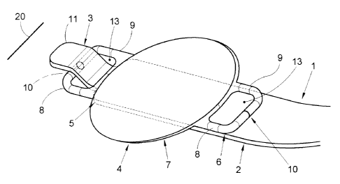
The inserting instrument 1 shown in Figs. 1 and 2 is for inserting an
intraocular
lens 4 into an eye via an incision 20 (schematically shown in Fig. 2) in the
cornea. The
instrument 1 has an elongate inserting member 2 projecting from a grip 14 and,
at a distal
end of the member 2, a hook 3 projecting transversally from the member 2. The
lens 4 to
be implanted using the instrument 1 has haptics 5, 6 radially projecting from
opposite sides
of an optical portion 7 of the lens 4. The optical portion 7 is deformable.
The haptics 5, 6
are each formed by a pair of arms 8, 9 for clamping iris tissue between
surfaces of the arms
8, 9 facing eacl-i other in a clamping area 10 and are arranged for supporting
the optical
portion 7 in a position parallel to and against an anterior iris surface plane
when the lens 4
is in implanted condition. In Fig. 2, one of the haptics 5 is located at a
side of the optical
portion 6 facing the distal end of the inserting member 2 of the instrument 1
and the other
one of the haptics 6 is located at a side of the optical portion 7 facing the
proximal end of
the inserting member 2 of the instrument 1. The haptics 5, 6 project radially
from the
optical portion 7 for holding the lens 4 with its optical portion 7 in a
position essentially
CA 02422167 2007-01-12
8
parallel to a plane formed by the anterior surface of the iris when in
implanted condition.
Apertures 13 are bounded by the haptics 5, 6 and the optical portion 7 is of a
transparent,
deformable material.
The hook 3 engages the haptic 5 facing the distal end of the inserting member
2.
The distal end portion of the inserting member 2 includes wide portions 11, 12
having a
width for engaging the lens 4 in laterally spaced apart positions.
In preparation of insertion of the lens 4 into an eye, the lens 4 is
positioned with the
optical portion 7 against the inserting member 2 and a portion of the haptic
5, which is
located distally from the optical portion 7, is engaged by the hook 3. The
inserting member
2 then supports the lens engaged by the hook 3.
More specifically, the lens 4 is engaged by the wide portions 11, 12 of the
distal
end portion in at least laterally spaced apart positions. This counteracts
tilting of the lens 4
about the inserting member 2, so that control over the orientation of the lens
4 before and
after insertion is improved. This, in turn, is advantageous for facilitating
insertion and for
avoiding contact between the lens and sensitive tissue in the eye. The support
of the lens 4
in laterally spaced apart positions results in the lens being supported in at
least three
positions, so that its position relative to the inserting member is in
principle fully
controlled.
The width of the wide portions is preferably at least one millimeter.
According to the present example, one of the wide portions 11, 12 is a support
plateau 12 closely adjacent the hook 3. This wide portion 12 supports the lens
4 engaged
by the hook 3. A particular advantage of providing a support plateau, which
may have a
closed or an open structure, is that lens 4 is easily held in position along
the inserting
member 2. This effect can be obtained by gravity if the lens 4 is located on
top of the
inserting member. In addition or alternatively, visco-elastic liquid such as
HPMC
(HydroxyPropylMethylCellulose) or Sodiumhyaluron - for instance of the type
which is
usually injected into the eye to maintain the volume of the anterior chamber -
may be
applied (preferably liberally) to the lens 4 and/or to the inserting member 2.
Such a
substance causes the lens 4 to stick to the inserting member 2 and this
sticking effect is
particularly effective if the substance is located between the relatively
large surface formed
by the wide portion 12 of the inserting member 2 and the lens 4. The substance
also forms
a lubricant between the lens 4 and the inserting member 2 reducing friction
between the
CA 02422167 2007-01-12
9
lens 4 and the inserting member 2 if the lens 4 is slid over the inserting
member 2 and
reducing the risl: of damage to the lens 4 and in particular the optical
portion 7 of the lens
4.
The wide portion 12 of the inserting member 2 thus defines a plane against
which
the lens 4 retained closely adjacent the hook 3 such that the inserting
instrument reliably
supports the lens 4 engaged by the hook 3 prior to insertion of the lens 4 in
a well
controlled orientation essentially parallel to the wide portion 12. In this
example, the
width of the wicie portion 12 is about two to four millimeters.
During insertion of the lens 4 into the eye, the optical portion 7 of the lens
4 is
deformed to a shape which is elongate in the direction of insertion, since the
hook 3 pulls
the lens 4 through a relatively small incision. After the optical portion 7
has passed the
incision 20, it unfolds again and regains its original shape in the anterior
chamber of the
eye. This allows the optical portion 7 to pass through an incision 20 which is
too small for
allowing passage of the optical portion 7 in undeformed condition.
After the lens 4 has entered the eye, the wide portion 12 shields the iris
and, where
applicable, the natural lens from the lens 4 and particularly from the haptics
5, 6, so that
the risk of causing damage to these internals of the eye is particularly low.
The hook 3 includes a first section 15 projecting transversely from the
inserting
member 2 and a second section 16 projecting distally from the first section
15. The second
section 16 of the hook 3 includes another one 11 of the wide portions 11, 12.
The haptics
5, 6, or at least stiff portions thereof, have a higher stiffness against
bending about an axis
in longitudinal direction from one haptic 5 to the other haptic 6 than the
optical portion 7,
at least prior to insertion of the lens. For this purpose, the optical portion
of the lens
according to this example is made from a material which has a higher specific
deformability and a lowe specific stiffness than the material of the haptics
5, 6. Examples
of materials for the optical portions are sillicone material and hydrophilic
or hydrophobic
acrylate. It is generally advantageous if such deformable materials for the
optical portion
allow an elastic elongation of at least about 50% and more preferably at least
about 75%.
However, it is also possible to achieve the relatively low stiffness of the
optical portion
about an axis in longitudinal direction from one haptic to the other haptic by
suitably
dimensioning the optical portion and the haptics, while the haptics and the
optical portion
are made of the same material of materials having similar specific stiffness.
For instance,
CA 02422167 2007-01-12
the optical portion can be substantially thinner than the dimensions of the
haptics in the
direction of the optical axis of the optical portion.
As is best shown in Fig. 18, the relatively inflexible portion has a width a
measured
parallel to the support plane defined by the haptics 5, 6 and perpendicular to
the
5 longitudinal direction, which is smaller than the width b, measured in the
same direction,
of the optical portion 7. The stiff portions of the haptics preferably have a
width a
transverse to the radial direction in which they project smaller than 4 mm and
smaller than
80% and more preferably 60% of the width b (measured in the same direction) of
the
optical; portion.
10 When the lens 4 is engaged by the hook 3, the wide second section 16 of the
hook 3
engages the haptic in positions spaced apart transversally to the longitdinal
direction of the
inserting member 2 and thereby prevents the haptic 5 from tilting about the
longitudinal
axis of the inserting member 2. Since the haptic 5 is relatively stiff, the
forces exerted by
the hook 3 onto the haptic 5 are effectively transferred to the deformable
optical portion 7
and define a zone 17 longitudinally in-line with the haptic 5 in which flexing
of the optical
portion 7 is counteracted. Thus, if the optical portion 7 is deformed prior to
or during
insertion into the eye, the flexural deformation is restrained mainly to
lateral zones 18, 19
located laterally of the central zone 17. Thereby, the orientation of the
central zone 17
- and since the haptics 5, 6 and the zone 17 in which the optical portion is
least flexed are
retained along the inserting member 2 also of the whole lens 4 - when the lens
regains its
original shape is very predictable. The width of the second section 16 of the
hook 3
according to this example is 1.5 to 2.5 millimeter.
The second section 16 of the hook 3 is formed by a flat lip. Thus, the end of
the
inserting member 2 is relatively blunt which reduces the risk of inflicting
damage to eye
tissue. Moreover, this features facilitates insertion of the hook 3 in the
opening 13 bounded
by the haptic 5 to be engaged by the hook 3 and the hook 3 can be manufactured
easily, for
instance by bending plate material or by injection moulding.
The first section 15 of the hook structure 3 extends from a neighboring
portion of
the inserting member 2 in a direction with a distal component. This allows the
hook 3 to be
withdrawn easily from the opening 13 in the haptic 5 by simply retracting the
inserting
member 2 backward in its longitudinal direction, for instance through the
insertion 20 after
the lens 4 has been inserted in the eye. The angle between the longitudinal
direction of the
CA 02422167 2007-01-12
11
inserting member or at least the portion thereof adjacent the hook 3 and the
first portion of
the hook 3 projecting therefrom can for instance be at least 20 or at most 70
.
According to the present example, the inserting member 2 is a flat strip of
plate
material. This allows the inserting member 2 to be manufactured in a simple
manner and
provides sufficient rigidity and flexibility for controlling and maneuvering
the lens 4 while
occupying very little of the cross-sectional surface of the incision 20 during
insertion of
the lens 4 into the eye.
As is best seen in Fig. 1, a shoulder portion 21 of the inserting member 2
closely
adjacent the hook structure 3 has a larger width than the hook structure 3.
This prevents
the lens 4 engaged by the hook 3 of the inserting member 2 projecting through
an opening
13 bounded by the haptic 5 from sliding along the inserting member 2 in the
direction of
the grip. The shoulder 21 forms an end of a portion of the inserting member 2
having a
width larger than the width of the opening 13 and is therefore prevented from
passing into
the opening 13.
In Fig. 3 an inserting member 102 of an inserting instrument is shown which
has a
narrow section 122 adjacent the hook structure 103, the narrow section 122
being narrower
than the wide portion 112. The narrow section 122 is located where the optical
portion of
the lens is bent when it is inserted into the eye and interferes less with the
bending of the
optical portion and occupies less space than if the narrow section is as wide
as the wide
portion 112 so that room for folded portions of an optical portion of a lens
engaged by the
inserting member is obtained. This further facilitates passage of the optical
portion of the
lens through the incision.
As is shown in Figs. 4 and 5 the inserting instrument may further include an
engagement member 223 on a side of the inserting member 202, the engagement
member
223 and the hook 203 being located on the same side of the inserting member
202. The
engagement member 223 is adapted for engaging a haptic 206 of a lens 204
engaged by the
hook 203 and projecting away from the hook 203. Thus, the engagement member
223 can
retain the haptic 206 facing away from the hook 203 (and from the haptic 205
engaged
thereby) closely to or against the inserting member 202, so that an even more
positive
control over the lens 204 is obtained. The inserting member 202 according to
this example
is formed by a flexible strip of metal and can easily be bent away from the
engagement
member 223. The engagement member 223 can then easily be slipped into the
opening 213
CA 02422167 2007-01-12
12
in the haptic 206 facing the engagement member 202 by moving the lens 204 in
longitudinal direction of the inserting member 202. Engagement between the
lens 204 and
the hook 203 may have been established beforehand, but may also be established
simultaneously or afterwards. When the inserting member 202 is allowed to flex
back, the
arms of the haptic 206 are retained between the inserting member 202 and the
engagement
member 223.
To facilitate disengagement of the lens 204 from the engagement member 223
after
insertion into the eye, it can be provided that the engagement member 223 can
be lifted
from the inserting member 202 to release the haptic 206 engaged thereby. To
this end, the
engagement member 223 can for instance be moveable in longitudinal direction
224 along
a portion of the inserting member which extends at an angle to the portion of
the
engagement member 202 in the area where the haptic 206 is held by the
engagement
member 223.
In Fig. 6 an inserting member 302 of yet a further example of an inserting
instrument according to the invention is shown. According to this example,
adjacent the
hook structure 303 and at the same side of the inserting member 302 as the
hook 303, the
inserting member 302 has a section having a projecting central zone 326. The
projecting
central zone 326 supports flexing of the optical portion of the lens in a
predetermined
direction with the lateral portions of the optical portion towards the
inserting member 302
when the optical portion is forced through a narrow passage, such as the
incision in the
eye. A similar effect, but in the opposite sense can be achieved by providing
that the
central portion is recessed. The lateral portions of the optical portion of
the lens are then
urged to flex away from the inserting member.
In Figs. 7-9 an embodiment of the invention is shown in which the inserting
instrument furtlier includes a feeder tube 427 having a length smaller than
the length of the
inserting member 402. The feeder tube has an inner channel 428 for receiving a
portion of
the inserting member 402 and a lens 404 of which the haptic 405 is engaged by
the hook
403 and a funnel 429 for compressing the lens 404 during entry into the tube
427. The
funnel 429 is removably mounted to a distal end of the tube 427.
In use, the lens 404 is first brought in engagement with the hook 403 of the
inserting member 402 projecting from the tube 427 and the funnel 429. Then the
lens 404
is pulled into the tube 427, for which purpose for instance suction can be
applied to the
CA 02422167 2007-01-12
13
proximal end of'the tube 427 or a pulling shank 430 having a hook 431 at its
distal end and
a cross-section smaller than the internal cross-section of the feeder tube 427
as shown in
Figs. 7 and 8 can be used. With the lens 404, the inserting member 402 is
entrained due the
engagement of the hook 403 to the lens 404. The width of the channe1428 of the
tube is
smaller than the width of the optical portion of the lens 404, so the optical
portion has to
be deformed during entry into the channel 428 to accommodate to the width of
the channel
428. This is facilitated by the funnel 429. After the lens has been pulled
into the channel
428, the funnel 429 is removed from the tube 427 to reduce the cross-section
of the portion
of the instrument to be inserted through the incision in the cornea. Then, the
distal end of
the tube is inserted into the eye via the incision in the cornea. Next, the
inserting member
402 is pushed outward so that the lens is pulled out of the tube 427 and
emerges from the
distal end of the tube 427 in the eye. Although the use of a tube to maintain
deformation of
the lens while it is passing through the incision in the cornea entails that
part of the
effective cross-section of the incision is occupied by the tube, it brings
about the
advantage, that relatively large forces can be applied to deform the lens and
that the forces
applied for defarming the lens are not exerted on tissue around the incision
in the cornea.
It is also possible to hold the tube 427 closely to and in front of the
incision through which
the lens is to be inserted and to then drive the lens 504 out of the tube and
through the
incision. 34. After the lens 404 is forced out of a distal end of the tube 427
by the inserting
instrument 402, it temporarily remains engaged to the inserting instrument
after being
released from the tube 427. Accordingly, the lens 404 is engaged to the
inserting member
402 at least while it begins to regain its original form, so the position of
the lens 404
remains controlled as it is released from the tube and the risk of the lens
404 reaching an
undesirable position or uncontrolled touching of internal tissue of the eye by
the lens 404
after being released is substantially reduced.
As is best seen in Fig. 9, the tube 427 has an elongate cross-section. This
allows an
important reduction of the dimensions of the lens 404 transverse to the
direction in which
the tube extends and in which the lens is to be inserted and the elongate
cross-section can
be inserted relatively easily through a line-shaped incision.
In Figs. 10-13 a distal portion of another embodiment of an inserting
instrument
including a tube 527 in which the lens 504 is inserted is shown. In Figs. 10
and 11 the lens
504 is shown in a position in font of the funnel 529, in which position it is
held by the
CA 02422167 2007-01-12
14
insertion member 502. The funne1529 is integrally formed with the tube 527. In
Figs. 12
and 13, the lens 504 is shown after introduction into the tube 527 in the
direction of arrow
532 by pushing the inserting member 502 of which the hook 503 engages the lens
504 via
the funne1529 into the narrowest portion of the tube 527. In the condition
shown in Figs.
12 and 13, the lens 504 is ready for insertion. This is accomplished by
inserting the end of
the tube 527 remote from the funnel 529 into the incision in the cornea of the
eye and
subsequently pulling the lens 504 out of the distal end of the tube 527 remote
from the
funnel 529 by moving the inserting member 502 further through the tube 527 in
the
direction of the arrow 532. Also the tube 527 can be held closely to and in
front of the
incision througli which the lens is to be inserted as the lens 504 is driven
out of the tube
527 and through the incision. The lens 504 then unfolds as it passes through
the incision.
In Figs. 14-17 a lens 604 and a distal end portion of an embodiment of an
inserting
instrument is shown which further includes a cap 627. The cap 627 has a width
for
receiving a portion of the inserting member 602 adjacent the hook 603 with
some play.
When the cap 627 is positioned over the inserting member 602 in a direction
transverse to
the longitudinal direction of the inserting member 602 (arrow 630), lateral
portions of the
optical portion of the lens 604 engaged by the inserting member 602 are bent
around side
edges of the inserting member 602. After the cap 627 is positioned over the
lens 604 and
the inserting member 602, the lens 604 which is held in deformed condition by
the cap 627
is inserted into the eye. Next, the cap 627 is pulled back from the eye,
thereby releasing the
lens 604. Finally, the inserting member is also pulled back from the eye,
leaving the lens in
the eye for fixation to the iris.
Next, details of the lens shown in Fig. 18 and subsequently of the lenses in
Figs.
19-24 are described and discussed. The dimensions of the apertures 13 of the
lens 4 shown
in Fig. 18 measured parallel to the plane and perpendicular to the radial
direction are larger
than the dimensions of the apertures 13 measured in the radial direction. This
substantially
limits the freedom of rotation of the lens 4 about the first portion 15 of the
hook 3, so that
the lens 4 engaged by the hook 3 is reliably held in a position essentially
aligned with the
inserting member 2.
The apertures 13 in the haptics 5, 6 are each bounded by and located between
flexible, pincer-like arms 8, 9 of the haptics which arm define a clamping
slit 10 between
the arms for pinching and fixating an anterior surface portion of iris tissue
without
CA 02422167 2007-01-12
penetrating to the posterior surface of the iris. Thus, the apertures 13
between the arms 8, 9
for pinching iris tissue are also used for the purpose of engaging and
retaining the lens 4 to
the inserting member before and during insertion of the lens 4 into the eye
and no separate,
additional apertures or constructional elements are required for this purpose.
5 In Fig. 19 a lens 704 is shown of which one haptic 706 includes a hole 733
in
addition to the aperture 713 between the arms 708, 709. The hole 733 is
adapted for
engagement by a hook such as the hook 431 and smaller than the aperture 713.
Preferably,
the hole 733 has a diameter smaller than 1 mm. Another feature of the lens
shown in Fig.
19 is that one of the arms 708, 709 is thicker than the other one. This
provides room for
10 the additional hole 733. Another advantage of one arm being thicker than
the other is, that
during the introduction of a plea of iris tissue in the clamping slit between
the clamping
arms, essentially only the thinner arm flexes so the other arm can be gripped
for accurately
holding the lens 704 in place. However, a hole 833 for engagement by a hook as
the hook
431 can also be provided in a symmetrical haptic 806 as is illustrated by Fig.
20.
15 In Figs. 21 and 22 a lens is shown which, for illustrative purposes, has
two
different haptics. In practice it is usually preferred to have the same
haptics on both sides
of the lens. As shown in Fig. 22, the haptics 905, 1005 project posteriorly
from the optical
portion 907. Of' each of the apertures 913, 1013, a portion 934, 1034 most
remote from the
optical portion 907 is located posteriorly from a portion 935, 1035 nearest to
the optical
portion 907. This facilitates insertion of the hook 3 and of the engagement
member 223
into the apertures, since it allows to insertion thereof in a direction
essentially parallel to
the plane of the optical portion 907. That the haptics 905, 1005 project
posteriorly from the
optical portion 907 is also advantageous for keeping the optical portion
elevated form the
plane defined by the anterior surface of the iris, when in implanted
condition. This is
advantageous for allowing aqueous flow through the pupil.
The optical portion 907 has a concave posterior surface 937, such that the
concave
surface 937 bounds a dome-shaped space between the optical portion 907 and the
plane
936. One of the haptics 1005 has a lateral side gate 1038 which intersects the
concave
posterior surface 937 and communicates with the dome-shaped space. Thus, the
risk of
inhibiting aqueous flow too much is reduced. Even if the posterior peripheral
edge of the
optical portion 907 is in contact with the iris surface 936, for instance
because the haptic
1005 is attached to the iris in the area of a recess in the iris surface, such
a lateral side gate
CA 02422167 2007-01-12
16
1038 will generally remain open. When such side gates are provided in an
optical portion
of easily deformable material, as is used for foldable or collapsible lenses,
the optical
properties in the area adjacent the lateral side gate may easily be affected
unfavorably.
With a haptic 1005 according to the present example, the likelihood of such
effect is
reduced, because a portion 1039 of the haptic 1005 extends along the side gate
1038 and
stabilizes the optical portion 907 in the area of the lateral side gate 1038.
To achieve a strong bond between the haptic 905, 1005 of a relatively rigid
material and the optical portion 907 of a relatively resilient material, while
keeping the
area occupied by the connection between the haptic 905, 1005 and the optical
portion
narrow to avoicl optical hindrance and to obtain a lens of a compact design,
the haptics
905, 1005 are bonded to the optical portion. In this example the bonding is
achieved by an
adhesive, but direct bonding for instance obtained during injection moulding
about an
insert. The adhesive is at least partially located in a groove 940, 1040 in
the optical portion
907. In additiori or alternatively, it is also possible to arrange the
adhesive in a groove in
the haptic if the haptic and the optical portion are designed accordingly.
To facilitate mounting of the haptics 905, 1005 and to increase the strength
of the
connection between the haptics 905, 1005 and the optical portion 907, flanges
943, 1043
are provided. The flanges 943 are integrated in the optical portion 907 of
which the
concave posterior surface 937 extends to the outer ends of the flanges. As can
be seen in
Fig. 22, this constructional feature results in side gates 951 in the optical
portion 907 along
the portions of the circumference of the optical portion 907 between the
haptics.
The flanges 1043 project from the optical portion 907. The flanges 943, 1043
are
provided with bosses 944, 1044 co-operating with recesses 945, 1045 in the
haptics. This
further reinforces the connection and further provides a snapping action
during mounting
of the haptics 905, 1005 which facilitates assembly of the lens 904. It is
also possible to
arrange bosses on the haptics and recesses in the flanges of the optical
portion.
Also in Figs. 23 and 24 different haptics 1105, 1205 are shown on opposite
sides of
the lens 1104 for illustrative purposes. Also in this lens, the haptics 1105,
1205 are bonded
to the optical portion 1107 by an adhesive. In this lens 1104, the optical
portion 1107 and
the haptics 1105, 1205 each have a proximal end portion 1142, 1242 enclosed
peripherally
by positioning portions 1146, 1246 of the flanges 1143, 1243. The adhesive is
at least
partially located between the haptic 1105, 1205 and the positioning portions
1146, 1246,
CA 02422167 2007-01-12
17
so that a particularly reliable connection is obtained. The flanges 1143, 1243
are integrated
in the optical portion 1107 of which the concave posterior surface extends
along posterior
surface portions of the flanges. As can be seen in Fig. 24, this
constructional feature results
in side gates 1151, 1251 in the optical portion 1107 along the portions of the
circumference of the optical portion 1107 between the haptics.
A particular feature of the haptic 1105 is, that it includes a lateral
aperture 1147 in
the optical portion 1107 in addition to the aperture 1113 bounded by the
clamping arms
1108, 1109. The aperture 1147 in the optical portion 1107 communicates with
the dome
shaped space bounded by the posterior concave surface of the optical portion
1107. Thus,
aqueous flow in the pupillary area is ensured particularly reliably. To
further provide
passages for aqueous flow in the pupillary area, a lateral port 1148
interconnecting the
lateral aperture 1147 in the optical portion 1107 and the aperture 1113
bounded by the
clamping arms 1108, 1109. A particularly stable fixation of the lens 1104 to
the anterior
surface of the iris is obtained, because the haptic 1105 furthermore has
support surfaces
1149 defining a plane 1136 essentially parallel to the optical plane 1150 of
the optical
portion 1107.
Lenses and inserting instruments or members according to the invention are
preferably provided in combination as eye treatment kits including an
instrument and a
lens, the instrument being dimensioned to engage the stiff portion of the lens
in the
aperture. It is then automatically ensured that the instrument used for
implanting the lens
fits to the lens.
To further facilitate the implantation, the lens is preferably provided
premounted in
a position retained by the instrument or at least the inserting member thereof
and packaged
and sterilized with the instrument or at least the inserting member in a
common package.
Thus, the need of separately sterilizing the instrument or at least the
inserting member is
avoided and the risk of contamination of the lens and the inserting member
during
mounting of the lens to the inserting member is reduced. To reduce waste, used
inserting
members can be returned to be cleaned, repackaged and sterilized with other
lenses to be
implanted.