Note: Descriptions are shown in the official language in which they were submitted.
CA 02422231 2003-03-11
1
Surgical Staple
This invention relates to a surgical staple.
Staples have been used in general surgery for many
years, mainly for anastomosing tissue. Examples
include skin staplers used to close a skin incision in
place of the standard manual suturing process, and end
to-end and end-to-side bowel stapling instruments which
are generally one shot devices used during bowel
reconstruction procedures.
The staples used with these devices are generally
manufactured from a metal or metal alloy material such
as stainless steel or titanium. The majority are
constructed from round profile wire and generally
produced in a generally 'U'-shaped configuration. The
ends of the 'U'-shape are normally pointed or sharpened
so as to ensure easy tissue penetration. Examples of
prior art in this area include US Patents 4,505,273,
5,026,390 and 4,719,917.
In clinical use the staples are delivered using a
stapler device which generally consists of an anvil
component positioned inside the 'U' between staple legs
and in contact with the staple. A former component is
positioned on the other side of the staple base, the
gap between the forming arms of the former being
approximately the width of the anvil plus two times the
diameter of the staple wire. The head of the stapler
1
w
CA 02422231 2003-03-11
2
device is normally positioned centrally across the slit
or opening which is to be closed.
On activation of the device the staple legs are
advanced forward so that they penetrate the tissue on
both sides of the slit or opening. As the former is
advanced further the legs of the staple bend around the
anvil causing the tips of the legs to advance along an
arcuate path toward each other so that the staple
ultimately assumes a generally rectangular shape
thereby compressing the tissue which has been trapped
between the staple legs. This compression of tissue is
the mechanism by which a closure is effected.
Depending on the length of the incision or opening a
series of staples will be delivered along its length in
order to ensure a blood tight closure.
While this method of closing an incision is effective
when a series of staples are used along the length of
the incision it is less effective when it is desirable
to close the opening with the minimum number of
staples. For example for an incision of 5-6mm in
length one round wire staple positioned centrally along
the incision is insufficient to effect a closure as the
compression due to the staple legs only acts in a
limited area towards the centre of the incision,
leaving the extremities open.
Also in situations where the tissue is soft and friable
the narrow staple leg will have a tendency to tear
through the tissue as they are bent around the anvil
CA 02422231 2003-03-11
r
3
thereby decreasing the level of compression between the
staple legs and causing unnecessary damage to the
vessel wall.
In order to avoid complications such as clot formation,
it is important to retain the staple legs within the
vessel wall, i.e. avoid the penetration of the internal
wall on the introduction of a foreign body into the
lumen of the vessel. If the staple legs penetrate into
the lumen of the vessel there is the added danger that
excessive pressure from the staple gun may cause the
vessel to collapse, which can lead to the legs
penetrating the opposing vessel wall, i.e. stapling the
vessel walls together and blocking the lumen of the
vessel.
Therefore there is a need for an improved surgical
staple which will more effectively close an incision,
thereby requiring fewer staples to close an incision.
In addition it would be advantageous to profile the
staple legs so that they are less inclined to tear
through softer tissue. Furthermore, it would be
desirable to limit the depth of penetration of the
staple legs to prevent the legs entering the lumen of
the vessel.
Accordingly, the present invention provides a surgical
staple comprising a base and a pair of legs each
extending from an opposite end of the base, each leg
having a penetrative portion terminating at a tip, the
staple being deformable to bend each leg relative to
CA 02422231 2003-03-11
r
4
the base causing each tip to approach the other leg
along a substantially arcuate path lying in a plane,
wherein each leg further comprises a compressive
portion located intermediate the base and the
penetrative portion, the compressive portion having a
height greater than that of the penetrative portion,
said heights being measured in the direction
perpendicular to the plane defined by the arcuate path.
The advantage of the invention is that the improved
surgical staple delivers a significantly increased area
of compression between the staple legs once the staple
has been deformed in use. The increased area of
compression is achieved by providing the compression
portion which tends to increase the contact area
between the staple and the tissue against which it is
bearing.
The invention is particularly useful in applications
where the staple is permanently implanted inside the
body. In such cases it is desirable to minimise the
amount of metal which is needed to effect a positive
closure. With existing stapler devices a series of
staples need to be positioned along the length of the
slit or tissue edges being anastomosed. Staples are
normally positioned close together as any one staple
will only compress a small amount of tissue on either
side. Using staples with an improved compression
capacity, as provided by this invention, will mean that
a significantly lower number of staples is required to
close any one incision.
CA 02422231 2003-03-11
The invention also has particular relevance in the area
of vascular puncture closure. During this percutaneous
procedure it is desirable to close the arterial
puncture preferably with one staple. Again it is
5 desirable that the staple contains the minimum amount
of metal. However, it is important that once delivered
the staple has generated enough compression along the
length of the slit or hole to prevent any blood
leakage. The direction of height of the compression
portion normal to the plane of closure of the legs
corresponds in use to the direction of length along the
incision.
Embodiments of the invention will now be described, by
way of example, with reference to the accompanying
drawings, in which:
Fig. 1 is a perspective view of a conventional surgical
staple.
Fig. 2a is a sectional view of an unformed staple in a
vessel wall.
Fig. 2b is a sectional view of a partially formed
staple in a vessel wall.
Fig. 2c is a sectional view of a staple fully formed in
a vessel wall.
Fig. 3a is a plan view of a staple before and after
forming.
CA 02422231 2003-03-11
6
Fig. 3b is an enlarged view of a staple leg before and
after forming.
Fig. 4 is a plan view of a staple in position across a
tissue opening.
Figs. 5 to 9 are perspective views of embodiments of
the invention.
Fig. 10 is a plan view of the staple of Fig. 9 in
position across a tissue opening.
Figs. 11 and 12, Figs. 13 and 14, and Figs. 15 and 16,
respectively, are further perspective views of three
additional embodiments of staple, each shown before and
after forming.
In the figures the same reference numerals have been
used to indicate the same or equivalent components.
Referring first to Fig. 1, a conventional round wire
surgical staple is of a generally 'U'-shaped
configuration, consisting of a base 10 and a pair of
"L"-shaped legs 12 each having a proximal portion 14
forming a linear extension of the base before use (as
shown in Fig. 1) and a distal portion 16 projecting
substantially perpendicularly from the proximal
portion.
CA 02422231 2003-03-11
7
The free ends 18 of the staple legs are generally
sharpened so as to ensure easy tissue penetration. In
addition to penetrating the tissue the staple is also
formed in use, to bring the free ends of the legs
together and thereby hold closed a wound. By forming
the staple, the staple is transformed from a generally
"U"-shaped configuration to a generally rectangular
shaped configuration during the delivery process. This
occurs by bending the legs 12 through 90° relative to
the base 10 of the staple at the point where the
proximal portions of the legs meet the base (known as
the bend points and denoted as points X and Y in the
drawings) at points relative to the central portion
10b.
Figs. 2(a) to 2(c) are a sequence of views showing the
process by which the conventional staple is deployed
and deformed from a generally "U"-shape to a generally
rectangular shape to effect a closure of a puncture
hole or slit 20 in a vessel or other tissue 22. In
Fig. 2a the staple has been advanced from the delivery
device (not shown) such that the distal portions of the
staple legs 16 have punctured the tissue 22 and the
staple base 10 and proximal portions 14 are lying
against the outer surface of the tissue. In Fig. 2b
the forming process has begun and the staple is being
deformed around bend points X and Y causing the
proximal portions 14 and distal portions 16 to arc
through an angle of approximately 90° thereby
compressing the tissue which is being captured between
both staple legs. In Fig. 2c the staple has been
CA 02422231 2003-03-11
8
fully formed into a rectangular shape, the tissue
contained within the rectangle being compressed as a
result of the staple legs having arced through
approximately 90°.
In Figs. 3a and 3b, the staple is shown prior to
forming (dashed lines) and after forming (solid lines).
As seen particularly in Fig. 3b, it can be seen that
prior to forming and following penetration of the
staple leg 12 into the tissue wall that there is an
area of tissue captive in the region (c). After the
forming process, i.e. when proximal portion 14 and
distal portion 16 have arced through 90° at the bend
point Y, the tissue which was previously captive at
point (c) has now moved to point (d). The same process
of compression occurs on the opposite leg of the staple
thereby creating compressed tissue 24 (Fig. 4) within
the rectangular shape of the formed staple.
In Fig. 4, the same compression process can be seen in
plan view, the tissue which was captive inside the legs
12 at points (c) prior to forming has been moved to
point (d) as a result of the staple forming process.
However, the level of compression which has transferred
to the hole 16 in the tissue is related to the area of
surface contact between the staple leg and the tissue
at points (c) and (d). With conventional round wire
staples this contact area is quite small and therefore
delivers a limited amount of compression over the
length of the hole opening or slit in the tissue.
Also, with round wire and leg to cut its way through
CA 02422231 2003-03-11
9
softer tissue as opposed to compressing the tissue
ahead of it.
The invention solves this problem by increasing the
height of a portion of the legs 12 (i.e. the height
being the dimension perpendicular to the plane in which
the staple legs bend during forming), in order to
increase the effective contact area between the staple
legs and the tissue as the staple is being deformed.
Increasing the contact area in this way will help
prevent the staple leg from tearing its way through the
tissue but more importantly will create a much greater
area of compression within the rectangle of the formed
staple and radiating from it, so that this compression
will be transferred over a much greater length of the
slit or opening 20 in the tissue.
Fig. 5 is a perspective view of a first embodiment of
surgical staple according to the invention. Here the
proximal portion 14 of the legs of the conventional
round wire staple described above have been deformed
from a round to a flat, rectangular cross-section,
providing a compressive portion located between the
penetrative portion of the legs (which in this case is
the entire proximal portion 16). As mentioned, the
purpose of this compressive portion is to increase the
surface contact area between the staple legs and tissue
in the direction in which the tissue is being
compressed as the staple leg arcs through approximately
90° at its bend point.
CA 02422231 2003-03-11
Fig. 6 shows another embodiment, in which the staple
legs have been divided along the axis of the proximal
portions and the opposite divisions 14' and 14"
deformed apart so as to significantly increase the
5 overall height of the proximal portions of the legs.
In Fig. 7 another round wire embodiment of the staple
is shown. In this staple the wire in the proximal
portion 14 of each leg is bent sinusoidally out of
10 alignment with the base 10 to provide a compressive
portion whose height H is significantly greater than
the height h of the penetrative portion of the leg 16,
again for the purpose of increasing the area of
compression, and preventing the proximal portion from
entering the wound. In the latter regard, it can be
seen that the leading section 28 of the sinusoidally
bent proximal portion extends generally at right angles
from the penetrative portion 16. This provides a
slightly rounded step or shoulder to act as a depth
stop, defining the end of the penetrative portion of
the leg and the start of the compressive portion.
Fig. 8 shows an embodiment which consists of a standard
round wire staple with flat plates or wings 30 attached
to the proximal portion of the legs. Fig. 9 shows
another staple similar to that of Fig. 9 which is
manufactured from flat metal stock and bent. Again the
staple legs include wings 30 such that the height of
these wings is significantly greater than the height of
the penetrative portion 16 of the staple legs.
CA 02422231 2003-03-11
11
The process by which these improved staples achieve
greater areas of compression over the length of an
opening in body tissue is illustrated in Fig. 10. Fig.
11 shows a staple of the kind illustrated in Fig. 9 but
the same principle applies to all the staples of Figs.
5 to 9. It can be seen that as the staple legs move
from their open position at 'A' to their closed
position at 'B' tissue 24 is compressed ahead of the
wings 30 and this compression radiates over a much
greater length of the slit or opening 20 than would be
the case if the wings were not attached to the staple
legs.
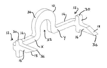
Fig. 11 shows a staple stamped from a flat sheet and
bent into its initial configuration (rather than a wire
staple as previously described). The base 10 of the
staple is horseshoe shaped rather than a flat linear
base. The horseshoe shape defines a "U"-shaped opening
32 which allows the staple to sit on top of a blood
locator tube extending from the end of a stapler. Such
a stapler is described in WO 02/19922.
The stapler of WO 02/19922 takes the form of a hollow
shaft and a blood locator tube slidable axially within
the shaft. The tube projects beyond the end of the
shaft to enter a puncture site in a blood vessel, and
blood flowing back through the tube and exiting the
device indicates to the surgeon that the tip of the
shaft (where the stapling head is located) is at the
incision in the vessel. A surgical staple straddles
the tube and is slidable thereon forwardly towards an
CA 02422231 2003-03-11
12
anvil against which the staple may be deformed to
staple together the opposite edges of the puncture
site. A cam mechanism drives the staple forwardly
along the tube into deforming engagement with the anvil
and at the same time retracts the tube into the shaft
in time to allow the legs of the staple to close onto
the puncture site.
The staple of Fig. 11 is adapted for use with such a
device in that "U"-shaped opening 32 is adapted to
straddle and slide on the blood locator tube.
The staple has a pair of legs 12 extending from the
ends of the base 10. Each leg is generally "L"-shaped
in plan view and comprises a proximal portion 14 and a
distal portion 16 terminating at a pointed tip 18. In
use (see also Fig. 12), the base 10 is held by an anvil
(not shown) while forming arms of the stapler (not
shown) push the proximal portions forwardly deforming
the staple at bend points X and Y. The blood locator
tube is withdrawn during this formation to ensure that
as the tips 18 approach one another (ultimately coming
to rest in the configuration of Fig. 12), they do not
catch the locator tube.
Located on the distal portion 16 is a compressive
portion 30 in the form of a bar extending at right
angles to the distal portion. In this staple,
therefore, the compressive portion and the penetrative
portion are both located on the distal portion of the
"L"-shaped leg. The penetrative portion is the part of
CA 02422231 2003-03-11
13
the leg extending from the bar 30 to the tip 18. The
forward surface 30a of the bar provides a shoulder
acting as a depth stop to prevent the leg penetrating
the vessel wall too deeply. This feature can be used
to ensure that the tip will not penetrate into the
lumen of a blood vessel by designing the staple such
that the distance between the front surface 30a and the
extremity of the tip 18 is less than the vessel wall
thickness. The bar also serves as a compressive
feature spreading the compressive forces provided by
the staple along a length of the incision corresponding
to the height H of the bar 30 (Fig. 12) as opposed to
just the lesser height h of the penetrative portion.
The compression is also increased by the relatively
small distance between the bars 30 when the staple is
closed.
By making the staple from a sheet material rather than
from wire, another significant advantage is obtainable.
The thickness of the material of the base (measured
between the internal surface of the opening 32 and the
corresponding external surface 34) is not constant but
instead increases to a maximum at the apex of the
horseshoe. This strengthens the structure against a
tendency for the curve to distort as the staple is
being formed. It has been found that the action of the
former and anvil bending the legs relative to the base
tends to cause the horseshoe curve to open out or
flatten somewhat. It will be appreciated that this can
lead to the staple deploying incorrectly, as the legs
tend to deviate from the "straight-ahead" orientation
CA 02422231 2003-03-11
14
during closure. Adding extra material to the curve
toward the top selectively reinforces the curve at this
point of maximum strain during forming and counteracts
the tendency to distort.
Another important feature of the staple of Figs. 11 and
12 is that the staple is not symmetrical about the
centre line. The penetrative portions 16 are staggered
vertically relative to one another so that one is
disposed slightly above the line of the proximal
portions and the other slightly below this line.
In addition, the respective tips 18 are bevelled
oppositely to one another so that the leading edge 36
of the tip on the left-hand penetrative portion (as
viewed in Fig. 11) is significantly above the leading
edge 36 of the right-hand tip. This double offset
(staggering the respective penetrative portions and
reversing the bevelling of the tips) allows the two
legs to close completely, so that the tips approach one
another and pass one another when the staple is formed,
providing greater compression and more reliable
closure.
Figs. 13 and 14 show a further embodiment of staple in
open and closed configurations. The staple again has a
base 10 with a leg 12 extending from each end. The
base is horseshoe shaped, but in this case rather than
there being additional material at the apex of the
horseshoe curve, the curve assumes a slight omega (S2)
CA 02422231 2003-03-11
shape with the ends of the curve pointing inwards to
counteract straightening tendencies.
Each leg 12 branches to a pair of tips 18 each having a
5 penetrative portion 38. The two penetrative portions
on each leg extend from the ends of a respective
compressive portion 30 in the form of a curved bar
which is generally perpendicular to both the proximal
portion 14 and the penetrative portions 38. The bar 30
10 provides a shoulder acting as a depth stop and acts to
spread the compressive forces of the staple along its
length.
It can be seen from Fig. 14 that the legs are once
15 again asymmetrical with respect to one another. The
penetrative portions 38 of the left-hand leg are both
longer and further separated from one another than
those of the right-hand leg. Again this ensures that
the two legs do not interfere with one another during
closure and that the staple can form a fully closed
structure when viewed in plan (see by comparison Fig.
2C in which there is a gap between the respective tips
of the prior art staple, and the curve appears open in
plan as a consequence.
The reason for the curvature of the bars 30 in the
embodiment of Figs. 13 and 14 is that the stapler for
which it is designed has a round profile. In general
it is desired to make the cross-sectional area of the
stapler shaft as small as possible to minimise trauma
arising from the introduction of the stapler. The
CA 02422231 2003-03-11
16
shape of this embodiment of staple therefore allows the
staple to fit in a rounded shaft while allowing the
compressive portions (bars 30) to grip the sides of the
wound as widely as possible, as will be appreciated
with reference to Fig. 10.
Figs. 15 and 16 provide yet another embodiment in which
the penetrative portions 38 of the legs 12 extend
between the tip 18 and a shoulder of a compressive
portion of the leg which in this case is provided by a
disk 40 mounted on the distal portion of each leg.
The embodiments described herein have "L"-shaped legs
with a roughly 90° angle between proximal and distal
sections. It will be noted that the compressive
section can be on either the proximal section or the
distal section. Furthermore, the legs need not take
this "L"-shape and can instead be curved (e.g. in a
quarter-circle), with the portion of leg adjacent the
tip defining a penetrative portion and a compressive
structure being located further along the curve towards
the base.
To aid in staple formation the point at which the legs
join the base can be weakened or provided by a notch,
but in most cases this is unnecessary as the
deformation between the anvil and former will cause the
legs to bend correctly at the junction with the base.
CA 02422231 2003-03-11
17
The invention is not limited to the embodiments
described herein which may be modified or varied
without departing from the scope of the invention.