Note: Descriptions are shown in the official language in which they were submitted.
CA 02422286 2003-03-13
WO 02/28302 PCT/USO1/31228
EPIDURAL THERMAL POSTERIOR ANNULOPLASTY
TECHNICAL FIELD
The present invention is directed to a treatment for injured or
degenerated intervertebral discs. Specifically, the present invention is a
method and
apparatus for strengthening an injured or degenerated intervertebral disc and
relieving
pain. The treatment may allow a spine surgeon to avoid a discectomy and
removal of
the nucleus pulposus during laminectomy operations and may reduce
postoperative
discogenic pain.
BACKGROUND ART
As shown in FIG. 1, each intervertebral disc 10 is a cushionlike pad
with fop and bottom endplates adjoining the bone surfaces on each adjacent
vertebral
body 20. As shown in FIG. 2, each disc has an inner sphere, the nucleus
pulposus 30,
which acts as a cushion for compressive stress. Around the nucleus pulposus is
an
outer collar of approximately 12 concentric rings, the annulus fibrosis 40,
which limits
the expansion of the nucleus pulposus when the spine is compressed. The rings
of the
annulus fibrosis also bind the successive vertebrae together, resist torsion
of the spine,
and assist the nucleus pulposus in absorbing compressive forces.
The grains of collagen fibers in adjacent rings of the annulus fibrosis
40 run in different directions so that the grains cross like an X. This
arrangement of
the collagen layers allows the spine to withstand twisting, shear forces.
FIG. 2 shows an exemplary injury to an intervertebral disc. A
herniated or prolapsed disc is commonly called a "slipped disc." Severe or
sudden
trauma to the spine or nontraumatic pathology such as degenerative spine
disease may
cause a bulge, rupture, degeneration, or other area of injury ("injury") 50 in
one or
more intervertebral discs. The annulus fibrosis 40 is thinnest posteriorly in
the
CA 02422286 2003-03-13
WO 02/28302 PCT/USO1/31228
2
general direction of the spinous process 60, so the nucleus pulposus 30
usually
herniates in that direction. The injury usually proceeds posterolaterally
instead of
directly posteriorly because the posterior longitudinal ligament strengthens
the
annulus fibrosis at the posterior sagittal midline of the annulus. The terms
"posterior"
and "posteriorly" mean the general posterior and posterolateral aspects of the
disc as
distinguished from the anterior aspects of the disc. The posterior aspect of
the
annulus fibrosis is also the location of vulnerable nerve tissues, including
but not
limited to the cauda equina 70 and spinal nerve roots 80.
A posterior injury of the nucleus pulposus often impinges on the spinal
nerve roots $0 exiting the spinal canal 90. The resulting pressure on these
nerve roots
often leads to pain and/or numbness in the lower extremities. Injured
intervertebral
discs are treated with bed rest, physical therapy, modified activities, and
painkillers
over time. If these treatments are ineffective, the injured and usually
protruding disc
is often surgically removed.
Current treatments offer only limited success in avoiding surgical
removal of injured intervertebral discs that do not heal themselves over time.
A few
treatments are adopted for use on an intervertebral disc from broad methods to
shrink
collagen in various other parts of the body. Several treatments attempt to
reduce
discogenic pain.
Several exemplary prior art references disclose using heat to shrink
collagen. U.S. Patent Nos. 5,374,265 and 5,484,432 to Sand (the "Sand
references")
are directed to methods for shrinking collagen with an infrared laser. The
collagen
shrinkage in the Sand references is generally accomplished in an
ophthalmological
context. Laser light that is optimally absorbed by collagen tissue is applied
to a
corneal stoma resulting in collagen shrinkage and reshaping of the cornea for
vision
correction. Although the Sand method generally applies to shrinkage of
collagen, it
CA 02422286 2003-03-13
WO 02/28302 PCT/USO1/31228
3
only contemplates applying relatively small amounts of energy to delicate eye
tissue.
No provision is made for protecting vulnerable tissue near collagen in other
parts of
the body. The amount of energy needed to shrink collagen in synovial joints or
the
spine is greater than the amount needed for eye tissue and may damage
vulnerable
tissue near the collagen being treated.
U.S. Patent Nos. 5,458,596 and 5,569,242 to Lax et al. (the "Lax
references") are directed to broad methods and apparatuses fox controlled
contraction
of soft tissue. The Lax references disclose the application of radio frequency
energy
through an electrode to tissue containing collagen. Such an application of
energy as
envisioned by the Lax references to an intervertebral disc would damage
vulnerable
tissues near the application site. The Lax references do not disclose the use
of energy
other than radio frequency. The shape of the Lax electrode is not designed for
use on
the spine. Also, because the Lax electrode is a general applicator, it does
not protect
vulnerable tissues during application of energy and therefore would not be
suitable for
applications involving the spine.
U.S. Patent No. 5,954,716 to Sharkey et al. (the "Sharkey'716
reference") is directed to a method and device for modifying the length of a
ligament.
In the Sharkey'716 reference, radio frequency energy is applied to one
ligament in a
set of opposing ligaments. Only radio frequency energy is disclosed. The radio
frequency energy shrinks one ligament, restoring equal length and a balance of
function to the set of opposing ligaments. Although the Sharkey'716 treatment
uses
radio frequency energy to shrink a ligament, it would not work on an
intervertebral
disc because an intervertebral disc is surrounded by vulnerable tissues.
Because
intervertebral discs lie close to the spinal canal and spinal nerve roots,
application
without thermal protection of radio frequency energy suitable for shrinking a
ligament
might harm vulnerable nerve tissues.
CA 02422286 2003-03-13
WO 02/28302 PCT/USO1/31228
4
Heating an intervertebral disc for relief of discogenic pain is disclosed
in U.S. Patent Nos. 5,433,739 and 5,571,147 to Sluijter et al. (the "Sluijter
references"). In the Sluijter references, probes are inserted into an
intervertebral disc
by puncturing the annulus fibrosis. Radio frequency or direct current energy
is
delivered through probes to heat the nucleus pulposus of a disc to
approximately 60°
C to 70° C. The heat travels to the outer perimeter of the disc being
treated so that the
entire disc is heated. The applied heat relieves back pain by denervating fine
nerve
endings in the disc. Although the probes of the Sluijter references may
relieve back
pain, the Sluijter probes invade the disc and are not intended to shrink
collagen or
repair a bulging, ruptured, or injured intervertebral disc. Since the entire
disc is
heated to approximately 60° C to 70° C, the heat may harm
vulnerable tissues near the
disc and have other thermally detrimental side effects. Some recent studies
have
shown that the amount of thermal energy provided to the posterior annulus by
the
IDET procedure is insufficient to cause either shrinkage/strengthening of the
posterior
annulus or ablation of the pain-sensing posterior annular nerve endings.
Several prior art references disclose methods for applying energy to the
interior of an intervertebral disc by invading the disc with a needle or
catheter. For
example, U.S. Patent No. 5,865,833 to Daikuzono is directed to a device for
laser
treatment. The Daikuzono device is for a discectomy procedure and for removal
of
intervertebral disc tissue, not to avoid a discectomy or to preserve disc
tissue or ablate
posterior annulus pain-sensing nerve endings. The Daikuzono method uses a
hollow
needle that is advanced into the center of an intervertebral disc, and then
disc tissue is
vaporized with laser energy and the vapor removed through the hollow needle.
The
hollow needle invasively punctures the disc.
U.S. Patent Nos. 6,007,570, 6,073,051, 6,095,149, and 6,122,549 to
Sharkey et al. (the "Sharkey references") are directed to methods for treating
an
CA 02422286 2003-03-13
WO 02/28302 PCT/USO1/31228
intervertebral disc and to devices with tip portions for performing various
functions on
a disc. Externally guidable catheters having one lumen or several lumina
puncture the
annulus fibrosis of an intervertebral disc and are inserted into the nucleus
pulposus at
the center of the disc. Functional tips on the distal ends of the catheters
add or remove
5 material or deliver energy. The Sharkey references also disclose injecting a
sealant
into fissures in the annulus fibrosis. The methods and devices of the Sharkey
references have the advantage of treating an intervertebral disc from the
inside,
thereby using the annulus fibrosis of a disc as thermal insulation from the
spinal canal.
The Sharkey methods and devices, however, have the disadvantage of not being
able
to reach many types of bulges, ruptures, or areas of injury in or near the
outer layers of
the annulus fibrosis. Further, because they puncture the disc, the Sharkey
catheters
are invasive and larger puncture holes are needed in order to use larger
functional tips.
The Sharkey methods and devices do not provide a noninvasive external approach
to
disc repair, and require maneuvering a catheter inside an intervertebral disc.
They
also do not ablate nerve endings in the posterior annulus and do not
shrinklstrengthen
the posterior annulus.
Known prior art methods for treating an injured intervertebral disc are
invasive to the disc, do not shrink/strengthen the posterior annulus, do not
ablate the
pain-sensing nerve endings in the posterior annulus, and may be thermally
unsafe to
vulnerable tissues around the spine.
DISCLOSURE OF THE INVENTION
The present invention provides a method and apparatus for shrinking
and strengthening the cartilaginous or collagenous material ("collagen") near
an injury
in the annulus fibrosis or the nucleus pulposus of one or more intervertebral
discs.
The present invention may allow a spine surgeon to avoid a discectomy and
removal
of the nucleus pulposus during a laminectomy operation.
CA 02422286 2003-03-13
WO 02/28302 PCT/USO1/31228
6
The present invention's epidural and extradiscal approach to repairing a
disc prevents the invasion of a disc with a needle or catheter. Needle and
catheter
methods puncture the intervertebral disc being treated, thereby exacerbating
the very
condition sought to be cured or may introduce infection into the nerve space.
The present invention may eliminate or greatly reduce discogenic pain
by thermally destroying nerve endings that transmit pain sensation from the
posterior
annulus. The surface area of the posterior annulus that can be treated for the
reduction
of discogenic pain is not limited as in prior art methods that deliver energy
from a
device inside the disc.
During thermal treatment by the present invention, vulnerable tissues
near a disc undergoing treatment may be thermally insulated or cooled and/or
displaced away from the thermal energy and thereby protected from potentially
destructive heat. Laser embodiments of the present invention may achieve
thermocoagulation of disc tissue by short laser bursts that confine heating to
the disc.
This thermal confinement combined with posterior displacement of neural
structures
may protect these vulnerable tissues near a disc without requiring insulation
or
cooling of the vulnerable tissues.
The present invention's strengthening of collagen may result in the
reduction of future incidents of disc herniation, reduction of spinal nerve-
root
impingement, and reduction of discogenic pain arising from nerve endings in
posterior
annulus.
The present invention is directed to an apparatus for thermally treating
intervertebral discs using an energy application head having an energy
application
region and a tissue protecting region. A control member is operationally
connected to
the energy application head to control the energy application head during
treatment of
an intervertebral disc.
The present invention also includes a method for thermally treating an
injured intervertebral disc while thermally protecting vulnerable tissues. The
method
includes gaining access to a vertebral column, epidurally approaching the
posterior
aspect of an injured intervertebral disc, and evaluating the extent of disc
injury. The
CA 02422286 2003-03-13
WO 02/28302 PCT/USO1/31228
evaluation preferably includes calculating an amount of energy needed to
thermally
refurbish the intervertebral disc. Energy is applied to the posterior aspect
of the
injured intervertebral disc while maintaining a safe temperature in vulnerable
tissues
near the disc. The energy delivered is monitored and the shrinkage and
strengthening
of the disc may be observed to determine if additional energy is required by
the disc or
adjacent discs. Further energy may be applied to other posterior areas of the
disc to
reduce pain. The steps of this method may be performed in alternate order.
Steps that
are unnecessary in a specific surgery may be omitted.
The foregoing and other objectives, features, and advantages of the
invention will be more readily understood upon consideration of the following
detailed description of the invention, taken in conjunction with the
accompanying
drawings.
BRIEF DESCRIPTION OF THE DRAWINGS
FIG. 1 is a posterolateral view of two adjacent lumbar vertebrae.
FIG. 2 is a transverse cross-section through the spine showing a
ruptured intervertebral disc, the spinal canal and cauda equina, and the bony
processes
of a vertebra.
FIG. 3 is a flowchart of a preferred method of the present invention for
thermal treatment of a bulging, ruptured, or injured intervertebral disc.
FIG. 4 is a transverse cross-section through a spine with an exemplary
disc refurbishes head positioned near the posterior annulus.
FIG. 5 is a top view of an exemplary head and part of an exemplary
control member of one preferred embodiment of the disc refurbishes of the
present
invention.
FIG. 6 is a side view of the exemplary head and a cut-away view of
part of the exemplary control member of FIG. 5.
FIG. 7 is a side view of an exemplary head of one preferred
embodiment of the disc refurbishes of the present invention showing an
expanded
head shape.
CA 02422286 2003-03-13
WO 02/28302 PCT/USO1/31228
FIG. 8 is a side view of an exemplary head of one preferred
embodiment of the disc refurbishes of the present invention showing a
contracted head
shape.
FIG. 9 is a side view of an exemplary head of one preferred
embodiment of the disc refurbishes of the present invention showing a flat
energy
application region.
FIG. 10 is a side view of an exemplary head of one preferred
embodiment of the disc refurbishes of the present invention showing a concave
energy
application region.
FIG. 11 is a side view of an exemplary head of one preferred
embodiment of the disc refurbishes of the present invention showing a convex
energy
application region.
FIG. 12 is a side view of an exemplary head of one preferred
embodiment of the disc refurbishes of the present invention showing a
malleable
energy application region.
FIG. 13 is a side and partial cut-away view of a section of an
exemplary control member of one preferred embodiment of the present invention
showing optional operational members.
FIG. 14 is a cross-sectional side view of an exemplary head of one
preferred embodiment of the disc refurbishes of the present invention showing
control
members operationally connected to a head.
FIG. 15 is a cross-sectional side view of an exemplary head of one
preferred embodiment of the disc refurbishes of the present invention showing
an
integrated defocused laser as a energy applicator.
FIG. 16 is a cross-sectional side view of an exemplary head of one
preferred embodiment of the disc refurbishes of the present invention showing
an
external defocused laser as an energy applicator.
FIG. 17 is a cross-sectional side view of an exemplary head of one
preferred embodiment of the disc refurbishes of the present invention showing
a lens
as an energy applicator.
CA 02422286 2003-03-13
WO 02/28302 PCT/USO1/31228
9
FIG. 1~ is a cross-sectional side view of an exemplary head of one
preferred embodiment of the disc refurbishes of the present invention showing
electrodes as energy applicators.
FIG. 19 is a cross-sectional side view of an exemplary head of one
preferred embodiment of the disc refurbishes of the present invention showing
a wire
as an energy applicator.
FIG. 20 is a cross-sectional side view of an exemplary head of one
preferred embodiment of the disc refurbishes of the present invention showing
a light
bulb as an energy applicator.
FIG. 21 is a cross-sectional side view of an exemplary head of one
preferred embodiment of the disc refurbishes of the present invention showing
a
resistive heating element as an energy applicator.
FIG. 22 is a cross-sectional side view of an exemplary head of one
preferred embodiment of the disc refurbishes of the present invention showing
an
ultrasonic transducer as an energy applicator.
FIG. 23 is a cross-sectional side view of an exemplary head of one
preferred embodiment of the disc refurbishes of the present invention showing
an
integrated defocused laser for slowly applying thermal energy.
FIG. 24 is a cross-sectional side view of an exemplary head of one
preferred embodiment of the disc refurbishes of the present invention showing
an
external defocused laser for slowly applying thermal energy.
FIG. 25 is a cross-sectional side view of an exemplary head of one
preferred embodiment of the disc refurbishes of the present invention showing
an
integrated collimated laser for applying thermally confined energy.
FIG. 26 is a cross-sectional side view of an exemplary head of one
preferred embodiment of the disc refurbishes of the present invention showing
an
external collimated laser for applying thermally confined energy.
FIG. 27 is a cross-sectional side view of an exemplary head of one
preferred embodiment of the disc refurbishes of the present invention showing
optional optical devices for visualizing a treatment area.
CA 02422286 2003-03-13
WO 02/28302 PCT/USO1/31228
FIG. 28 is a cross-sectional side view of an exemplary head of one
preferred embodiment of the disc refurbishes of the present invention showing
an
exemplary instrument for performing a physical measurement.
5 BEST MODES FOR CARRYING OUT THE INVENTION
Epidural thermal posterior annuloplasty is a method for shrinking and
strengthening the collagen at an injury in the annulus fibrosis of the nucleus
pulposus
of one or more intervertebral discs. This method of the present invention
differs from
previous methods by treating one or more intervertebral discs from an
epidural,
10 extradiscal approach while protecting vulnerable tissue near the disc.
Central to the
method of the present invention is a disc refurbishes device having an energy
source-
for example, a defocused laser-that heats the injured tissue without
vaporizing if. The
heating may cause shrinkage of the collagen resulting in a stronger, tighter
intervertebral disc and desirable destruction of microscopic pain-causing
nerve
endings in the intervertebral disc being treated. The intervertebral disc is
not
physically invaded, and tissues surrounding the disc remain safe.
Description of a Preferred Method of the Present Invention
A preferred embodiment of a method of the present invention is an
epidural, extradiscal, thermal treatment for repairing an injured
intervertebral disc that
protects vulnerable tissues near the disc and does not physically invade the
disc.
Several adjacent discs may be treated by manipulating a disc refurbishes in
the
epidural space.
FIG. 3 shows an exemplary method of treatment. As shown, access is
gained to the vertebral column through surgical means 100. An injury in or
near the
annulus fibrosis of an intervertebral disc is approached epidurally 110. The
extent of
disc injury is evaluated and the amount of energy needed to thermally
refurbish the
disc is calculated 120. Energy is then applied to the injured intervertebral
disc from a
posterior position 130. The application of energy 130 may be carried out by an
instrument. FIG. 4 shows one preferred embodiment of an instrument for
treating an
CA 02422286 2003-03-13
WO 02/28302 PCT/USO1/31228
11
intervertebral disc ("disc refurbishes") 200 that may be used in the preferred
method.
The disc refurbishes is inserted epidurally from a posterior approach,
remaining
outside the rings of the annulus fibrosis 40 of the intervertebral disc, and
is used to
apply energy to an exemplary injury 50 in the annulus fibrosis 40. The amount
of
energy delivered in the preferred method may be monitored simultaneously with
the
temperature of vulnerable tissues around the disc 140. Sensors and instruments
including but not limited to thermometers, thermistors, thyristors, phosphor-
coated
optic fibers, and temperature-sensitive crystals may monitor temperatures and
pressures of delivered energy at the energy application site. Instruments such
as
micro-forceps, biopsy samplers, and aspirators may be inserted through a lumen
in the
disc refurbishes. Matter and bodily tissues, such as vascular lesion tissue,
sequestrated
disc fragments, and synovial cyst tissue may be removed through a lumen in the
disc
refurbishes. The amount of shrinkage and strengthening of the collagen in and
around
the injury is observed and evaluated to determine the intensity and duration
of further
energy delivery 150. The observation and evaluation of shrinkage and
strengthening
may be made using unaided vision. Alternately, at least one lens, mirror,
camera,
fiber-optic device, or other optical device may be used. Observation and
evaluation
could also be made with a mechanical probe. The mechanical success of the
thermal
disc refurbishment is preferably verified 160. Further energy for deadening
sensory
nerve endings in the annulus may be delivered to as much of the posterior
annulus as
is feasible or necessary to reduce discogenic pain 170. The surgical access
site or sites
are closed. The steps may be performed in alternate order.
Description of a Preferred Apparatus of the Present Invention
A preferred apparatus embodiment of the present invention is a disc
refurbishes that may be used intraoperatively, but not necessarily for
percutaneous
spine surgery. A disc refurbishes has a shape for approaching an
intervertebral disc
epidurally. In one preferred embodiment, the disc refurbishes has an energy
delivery
system for treating at least one intervertebral disc. In an alternate
preferred
CA 02422286 2003-03-13
WO 02/28302 PCT/USO1/31228
12
embodiment the disc refurbisher has additional thermal protection features for
safeguarding tissues that surround an intervertebral disc.
Shape and Physical Geometry of Preferred Apparatus Embodiments
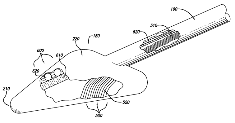
The exemplary disc refurbisher shown in FIGS. 5 and 6 has an energy
applicator such as a head 180 operationally connected to a control member 190
such
as a longitudinal shaft member.
A preferred energy application head ("head") of the disc refurbisher is
preferably shaped so that the approach to an injured intervertebral disc may
be
epidural. To approach epidurally, the surface of the exemplary head 180 of the
instrument is preferably smoothly contoured to glide over the posterior
annulus and
reach the injury site without snagging or tearing the nearby nerve roots,
epidural blood
vessels, dura, and thecal sac. The smooth, rounded edges 210 of the anterior
portion
of the head lift and displace the dura to epidurally gain access to the site
of injury at
the annulus fibrosis and reduce the thermal effect on the dura and neural
structures.
The head is preferably thinner at its smooth rounded edges 210 than at its
exemplary
domed center 220, allowing easy insertion between tissue layers and separation
of
tissues as the instrument is advanced to the injury site or moved from side to
side.
The shape facilitates treating adjacent discs by manipulating the disc
refurbisher in the
epidural space.
A wedge-shaped cross-sectional or longitudinal geometry of an
exemplary head, such as the head shown in FIGS. 5 and 6, separates and thereby
insulates the nerve roots, dura, and thecal sacs of the spinal canal on one
side of the
instrument from the energy delivery occurring at the surface of the
intervertebral disc
on another side of the instrument. The smooth, rounded edges 210 of the
anterior
portion of the exemplary head are relatively thin and slope to a relatively
thick region
under the exemplary domed center 220 creating a wedge-shaped head geometry.
The wedge-shaped exemplary head has a maximum wedge thickness
that may automatically lift vulnerable tissues a calculated safe distance away
from a
site of energy application as the instrument is moved. A calculated safe
distance may
CA 02422286 2003-03-13
WO 02/28302 PCT/USO1/31228
13
be proportional to the amount and duration of energy being applied or
proportional to
temperatures induced in the disc. In one variation shown in FIGS. 7 and 8, the
maximum wedge thickness of a head is variable and automatically expands 300 or
contracts 310 in proportion to the amount of energy being delivered. The
variation in
thickness may be accomplished mechanically or by using an inflatable top
portion that
expands under air or liquid pressure.
In FIGS. 9-12, the energy application regions of preferred head
embodiments may be flat 320, concave 330, convex 340, or malleable 350.
Initially,
an embodiment with a concave energy application region may be used to
approximate
the contour of a bulging area of disc, followed by an embodiment with a flat
energy
application region to impart a finished surface to the shrunken and tightened
collagen.
Each embodiment is operationally connected to at least one controlling member
190.
A head embodiment may have a diameter of approximately five
millimeters, but a wider head could be used for tissue shielding or a wider
application
of energy. Alternately, a set of disc refurbishers may have heads of various
useful
shapes and sizes. Still another alternative disc refurbishes may have a head
that varies
in size using mechanical means.
In FIGS. 5 and 6, an operational steering and controlling member
("control member" 190) such as an exemplary longitudinal shaft member may be
attached to a disc refurbishes head 180 at an angle from the plane of the head
of
between 0° and 180°, shown as 25°. Alternately, the
control member 190 may be
rotatably connected to the head. The control member 190 may be stiff,
flexible,
malleable, or articulated to provide physical control of disc refurbishes
movement. In
FIG. 13, a portion of a control member 190 is shown as optionally containing
operational members, such as at least one wire 360, fiber-optic strand 370,
hollow
tube 380, or radio control device 390. The hollow tube 380 or lumen rnay allow
instruments such as micro-forceps, biopsy samplers, and aspirators to be
inserted
through the disc refurbishes to the site of treatment. Matter and bodily
tissues, such as
blood, irrigation fluid, vascular lesion tissue, sequestrated disc fragments,
and
synovial cyst tissue may be removed through the hollow tube 380 in the disc
CA 02422286 2003-03-13
WO 02/28302 PCT/USO1/31228
14
refurbisher. Several lumina may be used to provide irrigation to the site of
treatment.
The control member 190 may also contain a moving mechanical link, such as a
rotating inner shaft 400 or an oscillating inner member. Alternately, as shown
in FIG.
14, control members 410 may be one or more wires, radio control mechanisms,
beams
of light, or any other control mechanism. One or more control members 410 may
be
attached in various useful configurations and at various useful angles.
Energy Application Using Preferred Apparatus Embodiments of the Present
Invention
As shown in FIG. 6, a disc refurbisher embodiment of the present
invention may deliver energy to an intervertebral disc from an energy
applicator on an
energy application region, shown as the bottom side 500, of the instrument's
head 180.
Other surfaces than the shown bottom side could be used as the energy
application
region in other embodiments. Energy applicators may be positioned on, consist
of, or
deliver energy through an energy application region depending on the type of
energy
applicator being used. As shown in FIGS. 15-22, energy applicators may include
one
or more lasers 420, fiber-optic strands 430, lenses 440, electrodes 450, wires
460,
light bulbs 470, heating elements 480, and ultrasound transducers 490. A disc
refurbisher may have more than one energy-delivering side and each energy-
delivering
side may have more than one energy application region.
The energy applicator may be supplied with energy from a source
external to the head, for example laser energy transmitted by optical fibers
from an
external laser to the head. Alternately, the energy applicator may generate or
convert
energy within the head, for example electric current from an external source
carried to
a resistive heating element within the head. If energy is supplied to the
head,
transmission of energy through a control member may be through any energy
transmission means, such as wire, lumen, thermal conductor, or fiber-optic
strand. In
FIG. 6, an exemplary fiber-optic bundle 510 fans out 520 into a useful pattern
at the
energy application region, shown as the flat bottom 500 of the head.
The disc refurbisher may deliver electromagnetic energy, including but
not limited to radio waves, microwaves, infrared light, visible light, and
ultraviolet
CA 02422286 2003-03-13
WO 02/28302 PCT/USO1/31228
light. The electromagnetic energy may be in incoherent or laser form. The
energy in
laser form may be collimated or defocused. The energy delivered to a disc may
also
be electric current, ultrasound waves, or thermal energy from a heating
element.
5 Laser Application of Energy
One exemplary preferred embodiment uses laser energy. The
interaction of laser energy with the collagen of an intervertebral disc has
photothermal, photomechanical, and photochemical components. The present
invention takes advantage of all three effects.
10 Photothermally, photons absorbed by a disc heat the disc and thermally
coagulate the collagen ("thermocoagulation"). Thermocoagulation may be
achieved
by applying energy with a continuous or long-pulse laser using microsecond or
millesecond pulses. FIGS. 23 and 24 show preferred embodiments of a disc
refurbisher in which defocused lasers are used to provide a relatively slow,
areawide
15 application of heat. FIG. 23 shows an integrated defocused laser 530. FIG.
24 shows
an external defocused laser 535 that may use a fiber-optic bundle in the
transmission
of defocused energy. Since lasers are monochromatic, wavelengths may be
selected
that would efficiently match the peak absorption range of collagen. To
optimize the
relatively slow application of heat using a defocused Iaser embodiment, a
photosensitive chemical reagent that would enhance or modify the absorption of
selected laser energy could be painted or sprayed onto the target and exposed
to the
laser output.
Alternately, laser energy for thermocoagulation may be collimated.
FIGS. 25 and 26 show preferred embodiments in which the energy applicator of a
preferred disc refurbisher is a collimated laser. In FIG. 25, an integrated
laser 540
generates the collimated laser energy. In FIG. 26, an external laser 550
generates
collimated laser energy that is focused into optical fibers 555 for delivery
to the
treatment site and optionally focused to very small areas by at least one lens
560. The
laser light may be short-pulsed, which would make the delivery of relatively
large
amounts of energy, such as gigawatts, possible in very short time periods,
such as
CA 02422286 2003-03-13
WO 02/28302 PCT/USO1/31228
16
nanoseconds. Short-pulsed laser bursts may achieve thermal confinement, the
desirable rapid buildup of heat in a treatment site before thermal diffusion
can
dissipate the heat, preventing the heating of vulnerable tissues near the
disc.
Photomechanically, the laser may be used for vaporizing undesirable
tissues or spallation of the surface layer of the disc, in addition to overall
thermocoagulation of the collagen. Spallation achieves surface modification by
removing only superficial collagen layers. During disc surgery, at the free
boundary
r
of an air/collagen interface, collagen expands at the surface when exposed to
a rapid
laser pulse, then snaps back with elastic force. The expansion creates
positive
pressure, but the recoil creates negative pressure. If the negative pressure
exceeds the
strength of the collagen, then the surface layer breaks. A thin layer of
collagen is
ejected. Such breaks or spall planes induced in the surface of the collagen
could be
used to shape a finished surface of the disc or to eject unwanted bulge
material at the
beginning of a disc refurbishment procedure. A vaporizing laser may be used to
remove undesirable tissues, such as excess collagen, vascular lesion tissue,
sequestrated disc fragments, and synovial cysts.
Photochemically, the therapeutic application of energy to an
intervertebral disc may cause several physiological changes. Once the
delivered laser
or other energy is translated into thermal energy in the collagen in or near
an injury in
the annulus fibrosis, a desirable microscopic breakdown of pain-causing nerve
ending
tissue may occur if so desired by the practitioner. The therapeutic
destruction of nerve
endings begins to occur when the temperature of the annulus reaches
approximately
45° C. At temperatures above 60° C, changes in the cross-linked
structure of the
collagen near an injury in the annulus begin to occur. The thermal denaturing
of the
collagen protein molecules causes thermocoagulation and desirable shrinkage of
the
bulging, ruptured, or injured annulus fibrosis. The thermocoagulation is
accompanied
by a simultaneous strengthening of the annulus fibrosis.
Photochemical welding of injuries, for example tears, in the annulus
fibrosis may also be accomplished by using a chemical reagent containing an
adhesive
photoactivated by laser light.
CA 02422286 2003-03-13
WO 02/28302 PCT/USO1/31228
17
Thermal Protection of Vulnerable Tissues by Preferred Apparatus Embodiments
The manner of energy delivery may forestall the need to protect
vulnerable tissues, as when a pulsed laser achieves thermocoagulation with
thermal
confinement to the disc. In FIG. 6, when thermal protection is needed because
of the
type of energy applicator being used, thermally vulnerable tissues near a site
of energy
application to a disc may be protected by an exemplary tissue protecting
region 600 of
an energy application head 180. A tissue protecting region 600 may contain a
thermal
protector, including but not limited to at least one optional insulation layer
610, and/or
an optional cooling system 620. One or more optional layers of insulation 610
or a
cooling system 620 in a preferred embodiment of a disc refurbishes may
thermally
separate an energy applying side of a disc refurbishes head from a tissue
protecting
side. The tissue protecting region may also result from a disc refurbishes
shape that
lifts vulnerable tissues away from a site of energy application.
A disc refurbishes may be cooled by internal or external airflow, or by
fluid or liquid pumped from a cooling reservoir such as a controlled
temperature bath.
As shown in FIG. 6, the cooling system of one preferred embodiment of a disc
refurbishes may incorporate internal cooling tubes 620. Refrigerants may be
used in
the cooling tubes to provide mild or aggressive cooling. Cooling may be
controlled by
mechanism or computer to counteract a proportional amount of heat being
generated
by a disc refurbishes. Alternately, the cooling may be accomplished by at
least one
thermocouple in contact with the tissue protecting region of a head
embodiment. The
thermocouple may constitute all or part of the material surface of a tissue
protecting
region. As another alternative, heat-pipe technology currently used to cool
state-of-
the-art microprocessor chips may also be used as a preferred thermal
protector. The
tissue protecting side may incorporate metals such as aluminum alloys or other
materials having high heat conductivity and heat-sinking properties to
transfer heat to
a cooling system.
A preferred embodiment of a disc refurbishes, shown in FIG. 6, has an
energy delivery system, shown as a fiber-optic bundle 510 and 520, surmounted
by a
CA 02422286 2003-03-13
WO 02/28302 PCT/USO1/31228
1~
cooling system shown as a layer of insulation 610, cooling tubes 620, and a
thermally
conductive metal outer surface.
Sensors and other instruments including but not limited to
thermometers, thermistors, thyristors, phosphor-coated optic fibers, and
temperature-
s sensitive crystals may monitor temperatures at the energy application and
tissue
protecting regions of a disc refurbisher and adjust the energy applicators and
cooling
systems to maintain selected temperatures. The control of heating and cooling
may be
by thermostat, electronic circuit, computer, or any other mechanism able to
dynamically adjust temperature.
Other Embodiments and Features of a Disc Refurbisher
All embodiments of a disc refurbisher may be robotically manipulable.
At least one robotic mechanism may be used to place a disc refurbisher at the
surface
of a disc, to apply energy, and to move a disc refurbisher around the
posterior annulus
of a disc. A disc refurbisher under robotic control may apply computer-
controlled
amounts of energy in computer-controlled patterns and amounts.
A disc refurbisher may optionally contain at least one tube or lumen for
transmitting material to and from a treatment site. A lumen may transmit a gas
or
fluid such as compressed air or water to the treatment site for uses including
but not
limited to irrigation, clearing away debris, and cooling. The lumen may also
be a
suction channel for vacuuming debris from the treatment site. The lumen may
allow
instruments such as micro-forceps, biopsy samplers, aspirators, and other
surgical
tools to be inserted through the disc refurbisher to the site of treatment.
Matter and
bodily tissues, such as blood, irrigation fluid, vascular lesion tissue,
sequestrated disc
fragments, synovial cyst tissue, and vaporized tissue may be removed through
one or
more lumina.
In FIG. 27, preferred embodiments of a disc refurbisher may optionally
incorporate at least one tissue visualizing instrument. Optics for visualizing
the
treatment site-for example, at least one camera 700, mirror 710, fiber-optic
bundle
720, or lens 730-may be incorporated into the energy application head. The
optics
CA 02422286 2003-03-13
WO 02/28302 PCT/USO1/31228
19
may transmit human-readable visual images from the treatment site or may
transmit
machine-readable feedback about energy being delivered and its effect on
tissue. The
disc refurbishes may transmit human-readable images that are displayable on a
monitor or other medical imaging equipment.
At least one physical measuring instrument may be added to preferred
disc refurbishes embodiments. FIG. 28 shows an exemplary instrument 740
integrated
into an energy application head that may measure pressures, distances, areas,
or
volumes with a human body. Other physical measurements may be performed by a
disc refurbishes using instruments integrated or external to a disc
refurbishes head.
The terms and expressions that have been employed in the foregoing
specification are used as terms of description and not of limitation and are
not
intended to exclude equivalents of the features shown and described or
portions of
them. The scope of the invention is defined and limited only by the claims
that
follow.