Note: Descriptions are shown in the official language in which they were submitted.
CA 02422722 2003-03-17
WO 02/22053 PCT/US01/29166
-1-
APPARATUS FOR DELIVERING ENDOLUMINAL PROSTHESES AND
METHODS OF MAKING AND USING THEM
FIELD OF THE INVENTION
The present invention relates generally to apparatus and methods for
delivering
endoluminal prostheses within body lumens of a patient, and more particularly
to
apparatus for delivering tubular prostheses or "stents" within a patient's
vasculature for
treating stenoses or other lesions, for example, within the coronary and
carotid arteries,
and to methods of making and using such apparatus.
BACKGROUND
In recent years, a number of minimally invasive technologies have been
developed
for treating diseases, such as atherosclerosis, that result in narrowing of
blood vessels, for
example, within the coronary or carotid arteries. Tubular prostheses or
"stents" have been
developed for maintaining the patency of a blood vessel, for example,
following
angioplasty or other procedures used to treat a stenosis, occlusion, or other
lesion within
the blood vessel. The stent may be implanted across a treatment site to
scaffold the site
and prevent it from subsequently contracting or otherwise becoming obstructed.
Generally, the stent may be placed upon a catheter in a contracted condition,
and
the catheter advanced endoluminally to the treatment site until the stent is
positioned
across the stenosis. The stent may then be deployed and substantially anchored
at the
treatment site. The stent may be self-expanding, i.e., may be biased to expand
to an
enlarged condition upon release from the delivery catheter, thereby
automatically
substantially anchoring the stent at the treatment site. Alternatively, the
stent may be
plastically deformable, i.e., may be expanded with the aid of a balloon, which
may
underlie the stent on the catheter. The balloon may be inflated to expand the
stent from
the contracted condition to the enlarged condition wherein the stent
substantially engages
the wall of the treatment site. A balloon, for example, on a separate balloon
catheter, may
also be used to further expand and/or anchor a self-expanding stent.
Similarly, for ablation procedures and the like, a catheter including an array
of
electrodes, for example, on an expandable basket assembly, may be provided.
The device
CA 02422722 2003-03-17
WO 02/22053 PCT/US01/29166
-2-
may be introduced into a body lumen, e.g., through the patient's vasculature
into the heart,
to treat conditions, such as heart arrhythmia.
With any of these devices, a sheath may be provided over the catheter to
protect the
elements on the distal end of the catheter, such as a stent, a balloon, and/or
an array of
electrodes. The sheath may be advanced distally over the proximal end of the
catheter
until it covers the distal end and the element(s) thereon, or the distal end
of the catheter
may be introduced into the sheath, and advanced until it is proximate the
distal end of the
sheath. The distal end of the catheter, with the overlying sheath thereon, may
then be
introduced into a patient and positioned at a treatment site, whereupon the
sheath may be
retracted to expose the distal end of the catheter. After treatment, the
sheath may be
advanced back over the distal end of the catheter, and the entire device
withdrawn from the
patient.
One of the problems associated with these devices is that they may have
substantially blunt distal ends that may scrape along the wall of a vessel
during
advancement therethrough, possibly damaging the wall and/or dislodging embolic
material
from the wall. To facilitate atraumatic advancement, particularly through
tortuous
anatomy, transition tips have been suggested for these devices.
For example, a conical or tapered nosepiece may be provided on the distal end
of
the catheter. A sheath may be disposed over the catheter, for example, to
substantially
cover the stent or other underlying element, such that the nosepiece extends
distally from
the end of the sheath, a distal edge of the sheath abutting the nosepiece. The
nosepiece
may facilitate advancement of the device through a narrow region of a blood
vessel,
although it may also risk catching on the wall of the vessel and/or dislodging
embolic
material, e.g., between the distal edge of the sheath and the nosepiece.
Following delivery
of a stent from the device, the nosepiece is generally positioned distal to
the treated lesion.
If the nosepiece is withdrawn directly, the proximal edge of the nosepiece may
catch on
the stent struts, resulting in the potential for trauma and embolic debris
release.
Alternatively, the sheath may be re-advanced across the treatment site to
"recapture" the
nosepiece, although in this approach the distal edge of the sheath may also
catch on the
stent struts.
As an alternative to a tapered nosepiece, a sheath having a rounded distal end
has
been suggested, as disclosed in U.S. Patent No. 5,593,412 issued to Martinez
et al.
CA 02422722 2003-03-17
WO 02/22053 PCT/US01/29166
-3-
Weakened areas or slits are provided in the distal end, thereby defining
sections that may
be softened upon introduction of warm saline solution. Once the sections are
softened, the
sheath may be retracted from an underlying balloon catheter to expose and
implant a stent
mounted on the catheter. Introduction of saline or other liquids into a
patient's
vasculature, however, may be undesirable, but is necessary in order to soften
the sections
on the distal end of the sheath and allow the stent to be deployed from the
sheath.
Another problem associated with such delivery systems is that the sheaths
and/or
catheters may buckle during insertion, because of the distal force applied
from the
proximal end to advance them through the patient's vasculature. In addition,
because of
their tubular nature, they may kink when advanced through tortuous'anatomy,
possibly
damaging the device or an element within the device.
Another problem associated with self-expanding stents is the stent embedding
within the delivery apparatus. With this type of stent, the delivery apparatus
generally
includes an overlying sheath that prevents the stent from expanding
prematurely. A distal
end of the delivery apparatus, with the sheath over the stent, may be
introduced into a
patient and positioned at a treatment site, whereupon the sheath may be
retracted to expose
the stent. The stent may then automatically expand to engage and/or open the
treatment
site.
During storage or otherwise before use, however, the stent may partially embed
itself into a wall of the sheath. Because of its inherent bias to expand, the
stent may exert
an outward force on the sheath and, over time, cause the wall of the sheath to
deform,
creating a pocket within which the stent may nest. During use, the stent may
resist being
removed from this pocket, and "stick" to the sheath as the sheath is retracted
at a treatment
site within a patient's vasculature. Retraction of the sheath despite this may
compress the
stent axially, possibly crushing or damaging the stent within the lumen.
Alternatively, as
the stent is compressed axially, forces may build within the stent until they
overcome the
frictional engagement with the pocket, and cause the stent to spring distally
out of the
pocket. This may cause the stent to move unpredictably within the lumen, to be
ejected
from the lumen suddenly, or cause an unusual tactile feedback to the user, all
of which
may contribute to inaccurate delivery of the stent.
U.S. Patent No. 6,019,778, issued to Wilson et al., attempts to address this
problem
by providing a braided mesh within the wall of a sheath of a delivery
apparatus. Because a
CA 02422722 2006-09-18
50336-58
-4-
stent is generally not a continuous smooth-walled tube, but
may include many edges or corners, such a braided mesh may
not prevent edges or corners of the stent from nesting into
pockets between strands in the braided mesh. In addition,
because reinforcing structures, such as the braided mesh of
the Wilson et al. patent, are generally embedded within a
wall of a sheath, the portion of the wall between the
reinforcing structure and the stent may remain at risk of
being deformed and creating a pocket.
Accordingly, it is believed that delivery systems
that facilitate delivery of a stent through a patient's
vasculature and/or that overcome the problems discussed
above would be considered useful.
SUMMARY OF THE INVENTION
The present invention is directed to apparatus for
delivering treatment elements, such as tubular prostheses or
"stents", within a body lumen of a patient, for example, for
treating stenoses or other lesions within the coronary
arteries, the carotid arteries, or other blood vessels, and
to methods of maki_ng such apparatus. The present invention
is also directed t:o methods for preparing such apparatus
before introduction into a patient, and to methods for using
such apparatus to deliver prostheses or otherwise treat a
patient.
In accordance with one aspect of the present
invention, there i_s provided an apparatus for delivering a
prosthesis into a blood vessel of a patient, comprising: an
elongate tubular member having a proximal end, a distal end,
and a lumen extending between the proximal and distal ends,
the distal end having a size for endoluminal insertion into
a blood vessel and terminating in a substantially atraumatic
CA 02422722 2006-09-18
50336-58
-4a-
distal portion comprising a plurality of flexible leaflets
integrally molded thereto, the leaflets being deflectable
from a closed position wherein the leaflets engage one
another to an open position wherein the leaflets define an
opening communicating with the lumen; a tubular prosthesis
disposed within the lumen proximate the distal portion; and
an elongate bumper member having a proximal end and a distal
end, the bumper member being slidably disposed within the
lumen of the elongate tubular member, the distal end of the
bumper member having a blunt edge that engages the proximal
end of the prosthesis for preventing axial displacement of
the prosthesis upon retraction of the tubular member with
respect to the bumper member; wherein the bumper member
comprises a helical coil.
In accordance with another aspect of the present
invention, there is provided an apparatus for delivering a
prosthesis into a blood vessel of a patient, comprising: an
elongate tubular member having a proximal end, a distal end,
and a lumen extending between the proximal and distal ends,
the distal end having a size for endoluminal insertion into
a blood vessel; a tubular prosthesis disposed within the
lumen proximate the distal end; and an elongate bumper
member comprising a helical coil having a proximal end and a
distal end, the bumper member being slidably disposed within
the lumen of the elongate tubular member, the distal end of
the bumper member having a blunt distal edge that engages
the proximal end of the prosthesis for preventing axial
displacement of the prosthesis upon retraction of the
tubular member with respect to the bumper member.
In accordance with a further aspect of the present
invention, an apparatus is provided for delivering a
prosthesis into a blood vessel of a patient that includes an
elongate tubular member having a proximal end, a distal end,
CA 02422722 2006-09-18
50336-58
-4b-
and a lumen extending between the proximal and distal ends.
The distal end has a size for endoluminal insertion into a
blood vessel and terminates in a substantially atraumatic
distal portion including a plurality of flexible leaflets
integrally molded thereto.
The leaflets are deflectable from a closed
position wherein the leaflets engage one another to an open
position wherein the leaflets define an opening
communicating with the lumen. Preferably, the leaflets
define a substantially rounded bullet shape in the closed
position, although alternatively, the leaflets may define a
substantially conical shape in the closed position. The
leaflets are preferably substantially flexible and
independently deflectable at a temperature less than body
temperature, and are biased towards the closed position, but
are resiliently deflectable to the open position. Adjacent
leaflets may be separated by a slit, or may be connected to
one another by weakened regions, the
CA 02422722 2003-03-17
WO 02/22053 PCT/US01/29166
-5-
weakened regions being tearable upon retraction of the tubular member with
respect to the
prosthesis to allow the leaflets to be deflected towards the open position.
In a preferred embodiment, a tubular prosthesis is disposed within the lumen
proximate the distal portion. An elongate bumper member having a proximal end
and a
distal end is also provided, the bumper member being slidably disposed within
the lumen
of the sheath. The distal end of the bumper member has a blunt edge disposed
adjacent to
the proximal end of the prosthesis for preventing axial displacement of the
prosthesis upon
retraction of the tubular member with respect to the bumper member and/or the
prosthesis.
Preferably, the prosthesis is a self-expanding stent, such as a coiled-sheet
stent, the
stent being biased to assume an expanded condition having a cross-section
larger than the
lumen of the tubular member, and being compressible to a contracted condition
to
facilitate insertion into the lumen.
In accordance with another aspect of the present invention, an apparatus for
delivering a prosthesis into a blood vessel of a patient is provided that
includes an elongate
tubular member, such as that described above, having a proximal end, a distal
end, and a
lumen extending between the proximal and distal ends, the distal end having a
size for
endoluminal insertion into a blood vessel. A tubular prosthesis is disposed
within the
lumen proximate the distal end. An elongate bumper member is also provided
that
includes a helical coil having a proximal end and a distal end, the bumper
member being
slidably disposed within the lumen of the sheath. The distal end of the bumper
member
has a blunt distal edge disposed adjacent a proximal end of the prosthesis for
preventing
axial displacement of the prosthesis upon retraction of the tubular member
with respect to
the bumper member.
In a preferred embodiment, the bumper member includes a helical wire
compression coil, preferably a solid height coil, extending between its
proximal and distal
ends. A plastic bumper element extends from a distal end of the helical coil,
the bumper
element including the blunt distal edge thereon. An extension element extends
distally
from the bu.inper element, the extension element having a cross-section
substantially
smaller than the bumper element, whereby the extension element may extend
through the
prosthesis disposed within the lumen of the tubular member. The helical coil,
bumper
element, and/or the extension element include a lumen extending axially
therethrough for
receiving a guidewire therethrough.
CA 02422722 2003-03-17
WO 02/22053 PCT/US01/29166
-6-
In accordance with yet another aspect of the present invention, a method for
making a sheath for delivering a treatment element within a body lumen of a
patient is
provided. A tubular member is provided that is formed from a substantially
flexible
material, the tubular member having a proximal end, a distal end, and a lumen
extending
axially between the proximal and distal ends, the distal end having a size for
endoluminal
insertion into a body lumen.
A die is provided having a bore therein, the bore having a tapered shape. The
die is
heated to a temperature in excess of a melting point of the flexible material
from which the
tubular member is formed. The distal end of the tubular member is inserted
into the bore
of the heated die until a distal portion of the tubular member is softened and
deformed into
a tapered shape substantially enclosing the distal end. One or more slits are
then created in
the distal portion of the tubular member after it is deformed into the tapered
shape, the slits
defining a plurality of leaflets. A treatment element may be inserted into the
lumen of the
tubular member until it is disposed proximate the distal portion.
In a preferred method, a bullet having a tapered shape distal end is inserted
into the
distal end of the tubular member before inserting the distal end of the
tubular member into
the bore. Preferably, the bullet and the bore have corresponding substantially
rounded
shapes defining a mold cavity therebetween when the distal end of the tubular
member is
inserted into the bore.
In another preferred method, the treatment element is a tubular prosthesis for
implantation within a body lumen of a patient. Preferably, the prosthesis is a
self-
expanding stent biased to assume an expanded condition having a cross-section
larger than
the lumen, and compressible to a contracted condition before being inserted
into the lumen
of the tubular member. The prosthesis may be inserted into the lumen of the
tubular
member before inserting the distal end of the tubular member into the bore,
e.g., inserted
into the lumen from the distal end of the tubular member. Alternatively, the
prosthesis
may be inserted into the lumen from the proximal end of the tubular member,
e.g., either
before or after the leaflets are formed on the distal portion of the tubular
member.
An elongate bumper member may be inserted into the lumen of the tubular
member, the bumper member being slidably disposed within the lumen of the
tubular
member, the distal end having a blunt distal edge for abutting a proximal end
of the
prosthesis. To make the bumper member, an elongate helical coil may be
provided having
CA 02422722 2003-03-17
WO 02/22053 PCT/US01/29166
-7-
a proximal end and a distal end. A tubular bumper element may be attached to
the distal
end of the helical coil to provide the bumper member, the bumper element
including the
blunt distal edge of the bumper element. Preferably, the bumper element is
formed from
plastic, and is attached to the helical coil by heating the bumper element
until it is
softened, and then directing the softened bumper element over the distal end
of the helical
coil. A tubular extension element may be attached to the bumper element, the
extension
element having a cross-section substantially smaller than the bumper element.
In accordance with yet another aspect of the present invention, an apparatus
is
provided for delivering a prosthesis into a body lumen of a patient. The
apparatus includes
a tubular member including a proximal portion, a distal portion having a size
for insertion
into a body lumen, and an intermediate portion between the proximal and distal
portions.
Each portion includes a wall defining a lumen extending between the proximal
and distal
portions. The wall of the distal portion may allow observation of the lumen
within the
distal portion, e.g., may be formed from substantially transparent material,
and/or material
free from reinforcing elements.
A prosthesis, preferably a self-expanding stent, is disposed within the
tubular
member proximal to the distal portion, and a bumper member is slidably
disposed within
the lumen of the tubular member. A distal end of the bumper member is disposed
adjacent
a proximal end of the prosthesis for preventing axial displacement of the
prosthesis upon
retraction of the tubular member. Thus, the prosthesis may be moved at least
partially into
the distal portion, whereupon the prosthesis may be observed through the wall
of the distal
portion.
Alternatively, the prosthesis may be disposed within the tubular member
proximal
to the distal portion, which may or may not be substantially transparent. The
bumper
member may be slidably disposed within the lumen of the tubular member, and an
actuator
may be coupled to the tubular member and the bumper member. The actuator,
e.g., a
handle device, may be configured for retracting the tubular member relative to
the buinper
member a predetermined distance to pre-load the prosthesis, and/or to maintain
the tubular
member under tension when the tubular member is retracted.
Before using the apparatus, the tubular member may be retracted proximally
with
respect to the prosthesis to pre-load or "bump forward" the prosthesis.
Retraction may be
ceased when at least a portion of the prosthesis is observed within the distal
portion of the
CA 02422722 2003-03-17
WO 02/22053 PCT/US01/29166
-8-
tubular member. For example, the tubular member may be retracted relative to
the bumper
member a distance equal to or greater than a length of the prosthesis. Thus,
the prosthesis
may be bumped forward to remove the prosthesis from a location where it has
become
nested or partially embedded within a wall of the tubular member. This may
facilitate
subsequent movement of the tubular member relative to the prosthesis, i.e.,
allowing the
prosthesis to slide freely within the lumen.
In addition, a proximal tension may be applied to the tubular member during
retraction, thereby removing any substantial slack in the tubular member. The
proximal
tension may be maintained after retraction is ceased, thereby preventing any
substantial
slaclc from returning into the tubular member.
After the apparatus has been pre-loaded, it may be used to deliver the
prosthesis
within a patient, e.g., within a body lumen, such as a carotid artery,
cerebral artery,
coronary artery, or other blood vessel. The distal portion of the tubular
member may be
introduced into the patient's body, e.g., percutaneously into a peripheral
vessel, and
advanced into a target body lumen. The tubular member may be retracted to
deploy the
prosthesis from the apparatus and into the body lumen.
Upon deployment, the prosthesis preferably automatically expands to
substantially
engage tissue surrounding the body lumen and/or dilate a stenosis or other
lesion within
the body lumen. Alternatively or in addition, a balloon or other expandable
member may
be used to further expand the prosthesis, e.g., to dilate a stenosis or
otherwise anchor the
prosthesis in place.
Other objects and features of the present invention will become apparent from
consideration of the following description taken in conjunction with the
accompanying
drawings.
BRIEF DESCRIPTION OF THE DRAWINGS
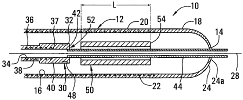
FIG. 1A is a cross-sectional side view of a sheath having a rounded distal
tip, in
accordance with the present invention.
FIG. 1B is a cross-sectional side view of an apparatus for delivering a stent,
including the sheath of FIG. 1A.
FIGS. 2A and 2B are end views of the sheath of FIGS. lA and 1B, showing
leaflets closed and partially open, respectively.
CA 02422722 2003-03-17
WO 02/22053 PCT/US01/29166
-9-
FIGS. 3A-3E are cross-sectional views showing a method for forming a rounded
distal tip on a sheath, such as that shown in FIG. 1A.
FIGS. 4A and 4B are cross-sectional side views of the apparatus of FIGS. 1A
and
1B, showing the stent being pre-loaded into the distal portion of the sheath,
before delivery
of the stent.
FIGS. 5A and 5B are cross-sectional views of a body lumen, showing a method
for
implanting a stent, in accordance with the present invention.
DETAILED DESCRIPTION OF THE PREFERRED EMBODIMENTS
Turning now to the drawings, FIGS. 1A-2B show a preferred embodiment of an
apparatus 10 for delivering a stent or other tubular prosthesis 50 into a
blood vessel or
other body lumen of a patient (not shown). Generally, the apparatus 10
includes an
elongate tubular sheath 12 having a proximal end (not shown), a distal end 14,
and a
lumen 16 extending generally therebetween. The tubular sheath 12 may be formed
from a
substantially flexible or semi-rigid material that may facilitate its
advancement within a
body lumen of a patient, preferably within the vasculature of a patient.
For example, the sheath 12 may be formed from a polymer, such as pebax,
polyethylene, urethane, nylon, or other plastic material, that may be extruded
or molded
into elongate tubing of a desired length. Preferably, the tubing has a wall
thiclcness of
between about 0.003-0.006 inch (0.075-0.150 mm), and has a substantially
uniform outer
diameter appropriate for the size of the stent being implanted, for example,
between about
1.5-2.5 mm. The sheath 12 may have a substantially uniform construction along
its length,
or the sheath 12 may include portions along its length having varying degrees
of
flexibility.
In a preferred embodiment, the sheath 12 includes a distal portion 18 formed
entirely from a substantially flexible material, such as pebax. Preferably,
the distal portion
18 is formed from a material that allows observation of the lumen 16 within
the distal
portion 18. For example, the distal portion 18 is formed from substantially
transparent
pebax, that may be free from reinforcing elements, thereby facilitating direct
visual
observation through the wall of the distal portion 18 into the lumen 16.
The sheath 12 also includes an intermediate portion 20 formed from pebax
including a reinforcing or stiffening element 20 therein. For example, the
intermediate
CA 02422722 2003-03-17
WO 02/22053 PCT/US01/29166
-10-
portion 20 may include a braid or mesh, e.g., of stainless steel, laid over a
Teflon liner,
with pebax tubing formed over the braid. Alternatively, the reinforcing
element 22 may be
a helical wire coil and the like molded or otherwise formed in the tubing. The
reinforcing
element 22 may enhance a rigidity of the intermediate portion 20, for example,
to reduce
the risk of the intermediate portion 20 buckling or kinking, while still
providing flexibility
transverse to the longitudinal axis 28, e.g., to accommodate advancement
through tortuous
anatomy. The intermediate portion 20 may be translucent or substantially
opaque.
Alternatively, the intermediate portion 20 may be substantially transparent
and one or
more visual markers (not shown) may be provided on the intermediate portion 20
and/or
the distal portion 18 to facilitate pre-loading of the stent 50.
Preferably, the sheath 12 also includes a proximal portion (not shown) that is
formed from a more rigid material, such as nylon tubing, that may include a
stiffening
element as described above. In a preferred embodiment, the distal portion 18
has a length
of between about ten and twenty centimeters (10-20 cm), the intermediate
portion 20 has a
length of between about twenty and thirty centimeters (20-30 cm), and the
proximal
portion has a length of between about eight five and one hundred twenty
centimeters (85-
120 cm), more preferably about one hundred centimeters (100 cm) or more.
The distal portion 18 of the sheath 12 preferably has a rounded bullet shape
defined
by a plurality of flexible leaflets 24 that are integrally formed thereon. The
leaflets 24 are
preferably deflectable from a closed position, wherein adjacent leaflets 24
abut one
another, to an open position. In the closed position, the leaflets 24
substantially close the
lumen 16, as shown in FIG. 2A. Preferably, in the closed position, the
leaflets 24 define a
relatively small opening 25 where their apices meet. In the open position (the
leaflets 24
are shown only partially open in FIG. 2B), the leaflets 24 are spread apart to
define an
opening 26 communicating with the lumen 16. Preferably, in the open position,
the
leaflets 24 are oriented substantially axially such that the opening 26 has a
cross-section
similar to the lumen 16. In the preferred embodiment shown in FIGS. 2A and 2B,
three
leaflets 24 are provided, although additional leaflets may be provided if
desired.
As best seen in FIG. 1A, in the closed position, the leaflets 24 preferably
define a
substantially atraumatic distal portion 18 that may facilitate advancement of
the sheatli 12
endoluminall.y within a patient's vasculature with minimal risk of dislodging
embolic
material from and/or otherwise damaging the wall of a body lumen through which
the
CA 02422722 2003-03-17
WO 02/22053 PCT/US01/29166
-11-
sheath 12 is advanced. In the preferred embodiment shown, the leaflets 24
define a
substantially rounded bullet shape in the closed position. Alternatively,
leaflets 24
defining a substantially conical shape (not shown) in the closed position may
be provided,
with the leaflets 24 preferably biased to the closed position, as described
below.
The leaflets 24 are substantially flexible and independently deflectable
substantially independent of the temperature to which the leaflets 24 are
exposed, e.g., at a
temperature substantially less than body temperature. In a preferred
embodiment, the
leaflets 24 are biased towards the closed position, but are resiliently
deflectable to the open
position. This may ensure that the opening 26 remains substantially closed
until time of
deployment of an element, such as stent 50, from within the lumen 16, and/or
that the
leaflets 24 do not catch on anything and open inadvertently. This may be
particularly
important when the apparatus 10 is advanced through tortuous anatomy, as
described
further below. Alternatively, the leaflets 24 may be at least partially
plastically deformed
when they are deflected from the closed position to the open position. In this
alternative,
the leaflets 24 may not return completely to the closed position when released
from the
fully open position, e.g., after the stent 50 is deployed from the apparatus
10.
Preferably, adjacent leaflets 24 are separated by a relatively narrow slit 28,
although alternatively, the leaflets 24 may partially overlap with one another
in the closed
position. In a further alternative, adjacent leaflets may be separated by a
thin-walled or
weakened region (not shown) that may be easily tearable upon retraction of the
sheath 12
with respect to a stent or other element being deployed from within the lumen
16. Once
the weakened regions are torn, the leaflets may be freely deflected towards
the open
position as the element is being deployed.
In addition, the leaflets 24 may have a thickness that is substantially
thinner than a
wall thickness of the rest of the distal portion 18, preferably tapering
towards their distal
tips 24a as shown in FIGS. 1A and 1B, thereby enhancing the flexibility of the
leaflets 24.
The tapering thickness may also ensure that the leaflets 24 are biased towards
the closed
position, yet may deflect easily to accommodate a guidewire (not shown),
bumper
extension element, and the like, as described further below.
Returning to FIG. 1B, in a preferred embodiment, the apparatus 10 also
includes an
elongate bumper member 30 that is slidably disposed within the sheath 12. The
bumper
member 30 preferably includes a proximal end (not shown), a distal end 32, and
a lumen
CA 02422722 2003-03-17
WO 02/22053 PCT/US01/29166
-12-
34 that extends therebetween. The bumper member 30 preferably has a
substantially
uniform outer diameter slightly smaller than the interior lumen 16 of the
sheath 12,
preferably by about 0.003-0.005 inch (0.075-0.125 mm) to create a close
sliding, but not
interfering, fit between the bumper member 30 and the sheath 12. The lumen 34
has a
diameter sufficiently large to accommodate a guidewire (not shown)
therethrough,
preferably between about 0.015-0.020 inch (0.375-0.500 mm), and more
preferably about
0.016 inch (0.400 mm).
In a preferred form, the bumper member 30 is formed from a helical wire
compression coi136, e.g., having adjacent turns that substantially abut one
another. The
coi136 may be formed from flat or round wire, e.g., of stainless steel and the
like, that is
continuously helically wound along the length of the bumper member 30,
preferably a
solid height coil. A relatively thin layer of Teflon 38 and the like may be
provided around
the outside of the coi136 to enhance a sliding relationship between the bumper
member 30
and the sheath 12. Because of the coi136, the bumper member 30 may be
substantially
resistant to buckling or kinking, while facilitating bending of the bumper
member 30
transverse to the longitudinal axis 28.
A substantially rigid tubular segment (not shown) may be attaclied to or
otherwise
extend from the proximal end of the coi136. Preferably, the tubular segment is
a section
of hypotube having an inner lumen (not shown) similar to the lumen 34 of the
coil 36, and
more preferably a two-stage length of hypotube that has a greater outer
diameter on its
proximal-most end. The tubular segment may facilitate distal advancement of
the bumper
member 30 into the sheath 12 with minimal risk of buckling and/or may provide
enllanced
tactile perception of relative movement of the bumper member 30 and the sheath
12. A
valve or other seal (not shown), e.g., for accommodating a guidewire
theretlirough while
maintaining a fluid-tight seal, may also be provided on the proximal end of
the tubular
segment.
The bumper member 30 also includes a tubular bumper element 40 on a distal end
37 of the coi136 that includes a substantially blunt distal edge 42. The
bumper element 40
is preferably formed from pebax or other plastic material. A plastic bumper
element 40
ensures no metal-to-metal contact, e.g., between the coi136 of the bumper
member 30 and
the stent 50 that may lead to corrosion of the stent material. In addition,
pebax and other
substantially flexible materials may deform slightly, e.g., when the sheath 12
is retracted,
CA 02422722 2003-03-17
WO 02/22053 PCT/US01/29166
-13-
to enhance contact between the blunt distal edge 42 of the bumper element 40
and the stent
50. The bumper element 40 is preferably attached to the distal end 37 of the
coil 36, e.g.,
by heating the bumper element 40 to soften it and directing it over the distal
end 37, such
that the bumper element is fused into the coils adjacent the distal end 37.
Alternatively, the bumper element 40 may be eliminated and the distal end 37
may
be substantially blunt to abut the stent 50. If metal-to-metal contact is to
be avoided, the
distal end 37 may be coated with an inert film or coating (not shown).
The bumper member 30 may also include a radiopaque or other marker 48 thereon
for identifying a location of the bumper member 30 using external imaging,
such as
fluoroscopy. Preferably, a platinum iridium ring 48 is provided on the bumper
element 40
immediately adjacent the blunt distal edge 42, thereby identifying a position
of the
proximal end 52 of the stent 50. Alternatively, a marker (not shown) may be
provided
elsewhere on the apparatus 10 in addition to or instead of the marker 48, such
as on the
sheath 12 or the stent 50 itself. Thus, the marker 48 may facilitate
positioning of the
apparatus 10, and more particularly the stent 50 or other element therein,
axially within a
body lumen (not shown) before deploying the element from within the sheath 12,
as
described further below.
The bumper member 30 may also include a tubular extension element 44 that is
thermally bonded or otherwise attached to and extends distally from the bumper
element
40. The extension element 44 has an outer diameter that is substantially
smaller than the
bumper element 40 For example, the extension element 44 may be partially
inserted into
the bumper element 40 as it is thermally bonded thereto so as not to interfere
with the
blunt edge 42 of the bumper element 40. Preferably, the extension element 44
has an outer
diameter of about 0.66 mm (0.026 inch) to facilitate its insertion through the
stent 50, an
2.5 inner diameter of about 0.41 mm (0.016 inch) to accommodate a guidewire
therethrough,
and a length of about 25 mm (1 inch). The extension element 44 may be
appropriately
sized larger or smaller to accommodate a guidewire, for example, between about
0.009-
0.038 in (0.225-0.95 mm). The extension element 44 is preferably substantially
flexible
and has a substantially smooth outer surface to provide a low-friction,
sliding contact with
3 0 an element disposed within the sheath 12.
In a preferred embodiment, a stent 50 or other tubular prosthesis or graft may
be
disposed within the lumen 16 of the sheath 12 proximate the distal portion 18.
The stent
CA 02422722 2009-02-26
52884-15
14
50 preferably is expandable between a contracted conditionthat facilitates its
loading into
the lumen 16 of the sheath 12, and an enlarged condition for engagi.ng a wall
of a blood
vessel or other body lumen (not shown). In a preferred embodiment, the stent
50 is a
coiled-sheet- stent, such as that disciosed in U.S. Patent No. 5,443,400
issued to Sigwai-t,
and/or in U.S. Patent No. 6,290,720, filed September 28, 1999. 'The stent
50 may be self-expanding, i.e., may be biased to assume the enlarged
condition, but may be compressed and constrained in the contracted condition,
for
ea.axnple; by the lumen 16 of the sheath 12. Al.ternatively, the stent 50 may
be plastically
deformable, i.e., may be substantially relaxed in the contracted condition,
but may be
forcibly expanded to the enlarged condition, for example, using a balloon
catheter, as is
laioarn in the art.
Generally, the apparatus 10 is provided pre-assembled with the stent 50
disposed
within the -lumen 16 of the sheath 12 adjacent the distal portion 18 of the
sheath in its
contracted cond'ztion. Preferably, the stent 50 is provided proximally to the
distal portion
18, e.g., such that the stent 50 is located entirely within the intermediate
portion 20. More
preferably, the stent 50 has a length, L, and is disposed at a distance equal
to or greater
than the length, L, from the distal end.14 of the sheath 12, explained further
below.
The biuraper member 30 is also disposed within the lumen 16 such that the
blunt
edge 42 of the bumper element 40 is adjacent.a proximal end 52 of the stent
50. The
extension element 44 preferably extends distally tbrough the stent 50 and
through the
leaflets 24, as best seen in FIGS. IB and 2B: The extension element 44 may
facalitate -
insertion of a guidewire (not shown) through the apparatus 10, i.e., through
the lumen 16
of the sheath 12 into the lumen 34 of the bumper member 30 to a proximal end
of the
apparatus 10. Preferably, the opening 25 at the apices of the leaflets 24
accommodates the
extension element 44 therethrough without causing the leaflets 24 to partially
buckle or
bulge_
.Alteznatively, the extension element 44 may be eliminated, either alone or
along
with the bumper element 40. In these alternatives, the distal end 37 of the
coi136 may
3 0 include an inlet port (not shown) communicating "Alith the Iumen 34, e.g.,
for backloading
a guidewire (not shown) into the lumen 34, as explained further below.
CA 02422722 2003-03-17
WO 02/22053 PCT/US01/29166
-15-
The apparatus 10 may be used to implant the stent 50 within a body lumen,
preferably within a carotid artery, a coronary artery, a cerebral artery, a
renal artery, or
other blood vessel, as described fiuther below. In a further alternative, the
apparatus 10
may incorporate "rapid exchange" configurations where a guidewire may exit
from the
lumens 16, 34 of the sheath 12 and/or bumper member 30 through side ports (not
shown)
at a location along their lengths, i.e., at an intermediate location, rather
than at their
proximal ends, as is known to those skilled in the art. To accommodate a
guidewire
between the sheath 12 and the bumper member 30 during retraction, a
longitudinal slot
(not shown) may be provided in either the inner surface of the sheath or the
outer surface
of the bumper adjacent the side ports.
Turning to FIGS. 3A-3E, a method is shown for forming a rounded bullet-shaped
distal portion 18 on a tubular sheath 12 and the like. A tubular sheath 12 is
provided that
is formed from substantially flexible plastic material, such as those
described above,
preferably pebax, and that has a lumen 16 therein extending from the distal
end 14 towards
the proximal end (not shown). The sheath 12 initially has a distal end 14 that
terminates in
a substantially blunt distal edge 19 (FIG. 3A).
In a preferred embodiment, the sheath 12 has a plurality of segments having
varying degrees of flexibility, for example, including a distal portion 18, an
intermediate
portion 20, and a proximal portion (not shown). Preferably, the distal portion
18 is a
predetermined length of substantially transparent pebax tubing that is thermal
bonded, e.g.,
butt bonded to the intermediate portion, which is a predetermined length of
pebax tubing
reinforced by a stainless steel braid, such as the lengths described above.
The intermediate
portion 20, in turn, is thermally bonded to a predetermined length of nylon
tubing.
Alternatively, an adhesive, connectors, and the like may be used to attach two
or more of
the portions to one another, in addition to or instead of butt bonding.
Preferably, the sheath 12 is pre-assembled, i.e., with the distal portion 18,
intermediate portion 20, and proximal portion bonded to one another before the
distal
portion 18 is formed into its bullet shape, as described below. Alternatively,
the distal
portion 18 may be formed into its bullet shape and/or other steps of the
method performed
before the distal portion 18 is attached to the intermediate portion 20.
A stent 50 or other prosthesis may be disposed within the lumen 16, preferably
a
predetermined distance greater than the length, L, of the stent 50 from the
distal end 14 of
CA 02422722 2003-03-17
WO 02/22053 PCT/US01/29166
-16-
the sheath 12. Preferably, the stent 50 is constrained in its contracted
condition, and
inserted into the distal end 14 of the sheath 12 before the distal portion 18
is formed into
its bullet shape. Alternatively, the stent 50 may be provided in its
contracted condition,
and introduced into the lumen 16 from the proximal end of the sheath 12, e.g.,
either
before or after the distal portion 18 is formed into its bullet shape.
In a preferred embodiment, the stent 50 is a self-expanding tubular member
formed
from Nitinol having a transition temperature between ambient and body
temperatures. The
stent 50 may be formed into its enlarged condition in its austenitic phase
(e.g. by hand
rolling for a coiled-sheet stent) and heat treated to set the enlarged
condition in its shape
memory. The stent 50 may then be chilled to its martensitic phase, e.g., at a
temperature
below ambient temperature, and preferably between about zero to ten degrees
Celsius (0-
10 C), for example, by blowing liquid Nitrogen onto the stent 50.
The stent 50 may then be pulled through one or more draw-down fixtures, i.e.,
tapered tubular dies (not shown), which may be chilled, to plastically
compress the stent
50 into a contracted condition. In the contracted condition, the stent 50
preferably has a
diameter substantially smaller than the lumen 16 of the sheath 12. The stent
50 may then
be pulled from the draw-down fixture into the lumen 16 of the sheath 12. In a
preferred
method, a Teflon tubular guide or sheath (not shown) may be used to facilitate
sliding the
stent 50 through one or more of the draw-down fixtures. The stent 50 may be
pulled into
the Teflon guide as it enters a draw-down fixture, the Teflon guide being
split or otherwise
removed from the stent 50 before it is pulled into the sheath 12.
The bumper member 30 (not shown in FIGS. 3A-3C) may be inserted into the
lumen 16 of the sheath 12 until the extension element 44 approaches, but does
not extend
from, the distal end 14 of the sheath 12. For example, the blunt edge 42 of
the bumper
element 40 may abut the proximal end 52 of the stent 50, with the extension
element 44
extending therethrough. Alternatively, the bumper member 30 may not be
extended
distally to abut the stent 50 until after the distal portion 18 is formed into
its bullet shape.
In a fixrther alternative, the bumper member 30 may not be introduced into the
sheath 12
until after the distal portion 18 is formed into its bullet shape.
Returning to FIGS. 3A-3C, a die 60, e.g., a spherically shaped "hot die," is
provided having a bore or other recess 62 therein. The bore 62 has an entry 64
with a
cross-section substantially similar to the cross-section of the sheath 12, a
rounded inner
CA 02422722 2003-03-17
WO 02/22053 PCT/US01/29166
-17-
end 66 having a tapered shape corresponding to the desired shape of the
rounded distal
portion 18 (FIG. 3C), and a relatively narrow aperture 67 extending distally
from the inner
end 66 through the die 60. The die 60 may be coupled to a heating element in a
conventional manner such that the die 60 may be heated to a desired
temperature, as is
well known in the art. In a preferred method, the die 60 is heated to a
temperature in
excess of a melting point of the material from which the distal portion 18 of
the sheath 12
is formed, for example, between about 150-200 degrees Celsius (about 300-400
degrees
Fahrenheit), and preferably about 160 degrees Celsius (320 degrees
Fahrenheit).
As seen in FIG. 3A, a bullet 70 is inserted a predetermined distance into the
distal
end 14 of the sheath 12, i.e., such that the bullet 70 does not contact the
stent 50 (shown in
FIG. 3B) but provides sufficient sheath material beyond a distal end 72 of the
bullet 70 to
form the bullet-shaped distal portion 18. Preferably, a wire or other filament
73 is attached
to the bullet 70 that extends distally from the distal end 72 of the bullet
70. The bullet 70
and die 60 may be formed from like materials, preferably a hardened and
polished tool
steel. The distal end 72 of the bullet 70 has a predetermined curved shape
corresponding
to the rounded inner end 66 of the bore 62 in the die 60.
In preparation for molding the distal portion 18 of the sheath 12, the
filament 73 is
guided through the aperture 67, maintaining sufficient tension to keep the
filament 73 taut,
but without pulling the bullet 70 from the tubular member 12. As shown in FIG.
3B, the
distal portion 18 of the tubular member 12 is inserted into the bore 62 of the
heated die 60
until the distal portion 18 of the tubular member 12 is softened and deformed
to fill the
cavity defined between the distal end 72 of the bullet 70 and the rounded
inner end 66 of
the bore 62.
Thus, the distal portion 18 is molded into a rounded bullet shape, the molded
shape
being defined by the distal end 72 of the bullet 70 and the rounded inner end
66 of the bore
62 in the die 60. Preferably, only slight pressure, e.g., mere hand pressure,
preferably
between about one to two pounds (1-2 lbs.), is applied axially to the sheath
12 to fill the
cavity defined by the bullet 70 and the bore 62 and ensure that there are no
discontinuities
in the resulting bullet shaped distal portion 18. Because of the filament 73,
the resulting
bullet shaped distal portion 18 includes the relatively small opening 25 (not
shown in FIG.
3B) therethrough corresponding to the filament 73 for accommodating a
guidewire or
bumper extension element (not shown).
CA 02422722 2003-03-17
WO 02/22053 PCT/US01/29166
-18-
As shown in FIG. 3C, once the rounded bullet-shaped distal portion 18 is
formed,
the sheath 12 may be removed from the bore 62 of the die 60, and allowed to
cool for
sufficient time to substantially solidify the sheath, i.e., to return to its
flexible, but solid
state.
One or more slits 34 (not shown, see FIG. 2A) may then be formed in the
tapered
region 19 of the distal portion 18. Preferably, a cutting device (not shown)
is used that
includes three cutting wires or blades that are equally spaced radially about
a central axis.
The cutting device may be aligned with the longitudinal axis 28 of the sheath
12 and
forced into the enclosed distal portion 18 until the cutting device cuts
completely through
the material of the enclosed distal portion 18. The cutting device may then be
withdrawn,
thereby providing a plurality of substantially independently flexible leaflets
24 (not shown,
see FIG. 2A) on the distal portion 18.
As shown in FIGS. 3D, the bullet 70 may be removed from the distal portion 18,
e.g., by pulling on the filament 73 to deflect the leaflets 24 and withdraw
the bullet 70
through the opening 26. The leaflets 24 preferably resiliently return to their
closed
position upon removal of the bullet 70, as shown in FIG. 3E, thereby defining
the opening
25.
Alternatively, the filament 73 and aperture 67 may be eliminated from the
bullet 70
and die 60, and the bullet 70 withdrawn from the formed sheath 12 using other
methods.
For even numbers of symmetrical slits, a cutting device including a single
blade or wire
(not shown) may be oriented substantially perpendicular to the longitudinal
axis 28 of the
sheath 12, and a plurality of individual transverse slits may be cut into the
distal portion
18. In alternative methods, individual leaflets may be formed using a multi-
cavity tool,
and the leaflets may be shaped into a final position, as will be appreciated
by those skilled
in the art.
Once the leaflets 24 are formed, the bumper member 30 may be advanced further
distally to push the stent 50 into a desired position within the lumen 16 of
the sheath 12, as
shown in FIG. 3E (in which the bumper element 40 and extension element 44 have
been
eliminated for convenience). The stent 50 may be positioned proximate the
bullet-shaped
distal portion 18, and/or the extension element 44 (not shown) may be extended
through
the stent 50 and through the opening 25. Preferably, during this stage, the
stent 50 remains
entirely within the intermediate portion 20, but may be in close proximity to
the distal
CA 02422722 2003-03-17
WO 02/22053 PCT/US01/29166
-19-
portion 18 of the sheath 12. Alternatively, the intermediate portion 20 may be
formed
from substantially transparent material, and may include markers (not shown)
for
providing visual indicators of the proper position for the stent 50. The
apparatus 10 may
then be packaged, shipped, or other otherwise provided to users to introduce
and implant
the stent 50 within a body lumen of a patient, as described further below.
In an alternative method, the stent 50 may be inserted into the sheath 12 from
its
proximal end after the distal portion 18 is formed into its bullet shape. For
example, the
stent 50 may be constrained in its contracted condition, and advancing it
through the
lumen 16 of the tubular member 12 to the distal portion 18. The stent may be
released,
i.e., unconstrained, once introduced into the lumen 16, whereupon the stent
may partially
expand to engage the wall of the lumen 16. Preferably, the stent remains
slidable within
the lumen 16 such that the stent 50 may be advanced to a location proximate
the distal
portion 18 and/or easily deployed through the opening 26. The bumper member 30
may
be inserted into the proximal end of the sheath 12 and directed distally to
advance the stent
50 to the desired position.
Turning to FIGS. 4A-5B, the apparatus 10 may be used to implant the stent 50
or
otlier prosthesis within a body lumen 100 of a patient, such as within a
coronary, carotid,
cerebral, renal artery, or other blood vessel. Initially, the apparatus 10 may
be stored in a
configuration as shown in FIG. 4A, e.g., during shipping or other pre-use
handling. In this
storage configuration, the stent 50 may be disposed entirely within the
intermediate
portion 20, i.e., such that the distal end 54 of the stent 50 is located
proximal to the distal
portion 18 of the sheath 12, as described above.
As is shown in FIG. 4A, due its bias to expand, the stent 50 may partially
embed
itself into the sheath 12, creating a pocket 17, even though the intermediate
portion 20 may
include a reinforcing element 22, such as a braid or mesh. To remove the stent
50 from
this pocket 17, the sheath 12 may be retracted relative to the bumper member
30. The
distal end 34 of the bumper member 30 holds the stent 50 and prevents it from
being
displaced proximally along with the sheath 12. Consequently, the stent 50 may
be directed
into the distal portion 18 of the sheath 12, as shown in FIG. 4B.
Preferably, because of the substantially transparent material of the distal
portion
18, entry of the stent 50 into the distal portion 18 may be directly observed
through the
wall of the distal portion 18. More preferably, the sheath 12 is retracted a
distance equal to
CA 02422722 2009-02-26
-52884-15
or greater than the length, L, of the stent 50, thereby ensuring that the
stent- 50 is removed
entirely from the poclcet 17. This may ensure that subsequent retraction of
the sheath 12
allows the stent 50 to-freely slide along the inner wall of the sheath 12.
If the distal end 54 of the stent 50 is initially disposed immediately
adjacent to the
5 distal portion 18, the sheath 12 may be retracted uatil.the entixe stent 50
is observed within
the distal portion 18. Alternatively, the distal end 54 of the stent 50 may be
initially
disposed a distance equal to or greater than its length, L, from the distal
portion 18. Tn.this
case, the sheath 12 may be retracted until the. distal end 54 of the stent 50
is observed
entering the distal portion 18. Ta a finther aiternative, if the distal
portion 18 is not
10 substantially transparent; the sheath 12 may be retracted a predetermined
distance equal to
or greater than the length, L, which may be-monitored from the proximal end
(not shown)
of the apparatus 10.
To faci.litate retracting the sheath 12, -a handle. device (not shown) inay be
coupled
to the .proxunal ends (not shown) of the sheath 12 and the buniper member 30.
The handie
15 device may include an actuator mechanism (also not shown) for moving the
sheath 12
axially relative to the bumper member. Preferably, the actuator miechanism_
only allows
the sheath 12 to be retracted proximally and does not allow the sheath 12 -to
be retuined
distally relative to the bumper member 30. Such a device is disclosed in.
U.S. Patent No. 6,527,779, filed Ju-ly 10, 2000.
Such a handle device and/or actuator mechanism may maintain a constaut tension
on the sheath 12, e.g., for-eliminating any slack or baeklash that may be
encountered due
to slight.longitudinal eiasticity_of the sheath .12. In addition, such a uni-
directional device
may prevent the sheath 12 -from being advanced over the bumper member after
delivery of
: 2-5 the stent 50.
Turning to FIGS. 5A and 5B, -once the stent 50 is pre-loaded to'a desired
position;
the apparatus 10 may be percutaneously introduced into the patient's
vasculature. For
example, the distal portion 18 may be introduced into a peripheral vessel,
such as a
femoral or carotid artery (not shown) and advanced endoluminally to a.target
treatment
region 102, e:g., witlun a carotid, cerebral, or coronary artery. Preferably,
the apparatus 10
is advanced over a guidewire 104 ah-eady placed across the treafinent region
102 using
conventional methods. The guidewire 104 may be backloaded through the
extension
CA 02422722 2003-03-17
WO 02/22053 PCT/US01/29166
-21-
element 44,and through the bumper member 30 to the proximal end (not shown) of
the
apparatus 10, as described above.
The rounded distal portion 18 of the sheath 12 substantially protects the
stent 50
during advancement and/or allows atraumatic advancement of the apparatus 10.
Preferably, as explained above, the leaflets 24 are resiliently flexible and
biased to the
closed position, causing the leaflets 24 to hug the guidewire 104 during
advancement,
particularly through tortuous anatomy. For example, if the leaflets 24 are
flexible and
biased to the closed position, the leaflet(s) 24 on the outside of a sharp
bend may hug the
guidewire 104, rather than deflecting away from the guidewire 104 and risking
catching on
the wall of the vessel, and possibly damaging the wall and/or dislodging
embolic material
from the wall. In addition, the rounded distal portion 18 may facilitate
advancement of the
apparatus 10 through the treatment region 100.
Once the apparatus 10 is advanced into the body lumen 100, the stent 50 may be
positioned across the treatment region 102, as shown in FIG. 5A, for example,
by
monitoring the marker 48 using fluoroscopy and the like. Preferably, the
treatment region
102 is a stenotic or occluded region of a blood vessel, although other lesions
or damaged
vessel segments may be treated, as will be appreciated by those skilled in the
art.
Once the stent 50 is properly positioned, the bumper member 30 may be held
stationary, and the sheath 12 retracted to deploy the stent 50 from the lumen
16, as shown
in FIG. 5B. Because of their flexible nature, the leaflets 24 easily deflect
outward to allow
the stent 50 to be deployed through the opening 26, and slide over the stent
50 and/or over
the bumper member 30. Once the stent 50 is deployed, the apparatus 10 may be
withdrawn from the body lumen 100 and from the patient (not shown). The sheath
12 may
remain in its retracted position without requiring advancement back over the
bumper
element 40 and/or the extension element 44 before removing the apparatus 10.
The
leaflets 24 preferably hug the outside of the bumper member 30, thereby
facilitating
substantially atraumatic withdrawal of the apparatus 10.
Preferably, the stent 50 is self-expanding, and therefore automatically
expands
upon deployment to engage the body lumen 100 at the treatment location 102.
The stent
50 may trap embolic material between itself and the body lumen 100 and/or may
dilate and
hold the body lumen 100 open. If desired to further expand the stent 50, an
expansion
device, such as a catheter (not shown) may be introduced into the body lumen
100, e.g.,
CA 02422722 2003-03-17
WO 02/22053 PCT/US01/29166
-22-
upon removal of the apparatus 10, and positioned within the stent 50. A
balloon or other
expandable member (also not shown) on the catheter may be expanded to engage
and
further expand the stent 50 to a predetermined diameter, e.g., corresponding
substantially
to the unobstructed diameter of the body lumen 100.
In an alternative embodiment (not shown), the stent 50 may be plastically
expandable, and may be mounted onto a catheter that is inserted into a sheath
12 in
acco,rdance with the present invention. The catheter may include a balloon or
other
expandable member over which the stent may be mounted. Once the sheath is
retracted to
deploy the stent, for example, at a target treatment region, the expandable
member may be
expanded, e.g., by inflating the balloon, to plastically deform the stent and
expand it to
engage the body lumen at the treatment region. Once the stent has been
expanded to a
desired size, the expandable member may be deflated, and the apparatus
withdrawn from
the body lumen and the patient.
In further alternatives, other deployable devices may be provided within a
sheath in
accordance with the present invention, such as an electrode device, e.g., an
array of
electrodes on an expandable basket assembly and the like. Once a desired
location is
reached, such as a chamber of a heart, the sheath may be retracted with
respect to the
underlying device, until one or more elements on the device are deployed from
the sheath.
A procedure may be completed at the location, e.g., an ablation procedure, and
then the
sheath and device may be withdrawn from the location.
While the invention is susceptible to various modifications, and alternative
forms,
specific examples thereof have been shown in the drawings and are herein
described in
detail. It should be understood, however, that the invention is not to be
limited to the
particular forms or methods disclosed, but to the contrary, the invention is
to cover all
modifications, equivalents and alternatives falling within the spirit and
scope of the
appended claims.