Note: Descriptions are shown in the official language in which they were submitted.
. a
CA 02423016 2003-03-20
1
A DEVICE FOR THE IDENTIFICATION OF THE EPIDURAL SPACE
The present invention relates to a device that aids injection into the
epidural space, by clearly
visually indicating when the needle enters this cavity.
In medical practice, identification of the epidural space is required for
therapeutic and
anaesthetic procedures.
The currently used techniques rely on high levels of manual skill and
dexterity and require
specialist training. These techniques are not uncommonly associated with
technical
difficulties or complications. Trainees have a high complication rate that
decreases with
experience. It can take up to 2 years to learn the techniques involved. The
current techniques
rely on a kinetic "feel" endpoint.
An object of this invention is to provide the operator of this procedure with
a device that
achieves a high success rate, a steep training/learning curve, and a minimal
complication rate.
Several devices to aid in the detection of the epidural space have been
described previously.
Many of these devices use the feature of low pressure in the epidural space to
identify when
the needle enters the space. EP0091846 and US5188594 both describe devices
that contain a
balloon, which is inflated prior to the needle entering the epidural space. As
the pressure
within the epidural space is lower, the balloon deflates when the needle
enters the epidural
space. This results in the air being injected into the epidural space, which
can cause side
effects as air bubbles are formed which prevent the even spread of the
medication
administered.
CA 02423016 2003-03-20
2
EP0538259 discloses an electronic means of detecting changes in pressure
within the liquid
inside the syringe, and can provide a visual and/or aural end point. However
this device
increases the length of the equipment used, which provides a larger axis of
movement. It also
makes the device cumbersome, so only allows the operator to have one hand on
the needle.
The electronic equipment needs to be calibrated and is prone to failure.
US5902273 and US 5258003 describe devices that make use of a spring-loaded
mechanical
gauge to indicate the loss in pressure. These gauges are not well suited for
detecting the low
pressures encountered during the insertion of an epidural needle. In practice
these devices
failed due to the complexity of the device, mechanical stickiness and also the
increase in
weight.
US4215699 discloses a device that incorporates a membrane which is displaced
inwardly or
outwardly in response to a decrease or increase in pressure respectively. This
device however
cannot detect small pressure changes.
Therefore a small, simple lightweight device that clearly indicates when a
needle enters the
epidural space is needed to improve epidural injection techniques.
Thus in one aspect, the present invention provides an injection device
comprising a
diaphragm, adapted for pressurisation, so that said diaphragm bulges outwards
when the
device is pressurised, and wherein said device is adapted to connect to a
needle.
CA 02423016 2003-03-20
3
The term " adapted for pressurisation" as used herein means that the device
can be
pressurised prior to use, so that the diaphragm bulges outwards. This provides
a visual
indication of when the needle enters a space which has a lower pressure, such
as the epidural
cavity.
The device can be pressurised with a fluid, wherein the term "fluid" refers to
a gas or a liquid.
In one preferred embodiment the fluid is saline or air.
In another preferred embodiment the device is adapted for connection to the
needle by means
of a connection port. In another embodiment the device further comprises an
injection port
for the injection of pressurising fluid.
The device can be adapted for pressurisation by the presence of one or more
valves. A valve,
in particular a one way valve, may be present in the injection port, so that
the pressure is
maintained until the tip of the needle enters an area of low pressure within
the body, such as
the epidural space.
The means for connecting the device to the needle may also contain a valve.
The valve is shut
to enable the device to be pressurised. Once the device is connected to the
needle, and the
needle has been inserted into the body, the valve is opened so that any
decrease in pressure
experienced at the tip of the needle is detected by the retraction of the
diaphragm.
In one preferred embodiment the needle is an epidural needle.
CA 02423016 2003-03-20
4
The invention allows for pressurisation of the device and automatic injection
of air or saline
at the point of entry to the epidural space. However, in comparison to the
balloon devices
described in EP0091846 and US5188594 a greater and more accurate displacement
of the
membrane is achieved with a smaller volume of air entering the epidural space,
and thus
causing less side effects. Alternatively the use of a fluid such as saline to
pressurise the
chamber eliminates the problem of the introduction of air into the epidural
space.
The present device provides an instant clear visual aid for identification of
the point of entry
into the epidural space. It allows for full concentration and undivided
attention of the operator
on the advancement of the epidural needle. It also frees her/his hands
allowing for a bi-
manual grip of the epidural needle wings and a steady control on its movement.
Previous
devices have only allowed one hand to be used, which in combination with
lengthy apparatus
causes an undesirably large axis of movement.
Furthermore due to the simple nature of the device it can be easily made by
injection
moulding.
In another aspect the present invention provides the use of an injection
device as described
herein for use in medicine.
In a further aspect the present invention provides a method of injection into
a body cavity of a
patient which has an internal pressure less than atmospheric pressure
comprising the
following steps:
a) attaching an injection device as defined herein to a needle;
CA 02423016 2003-03-20
b) pressurising the device with a fluid until the diaphragm bulges
outwards;and
c) advancing the needle into a patient until the diaphragm retracts.
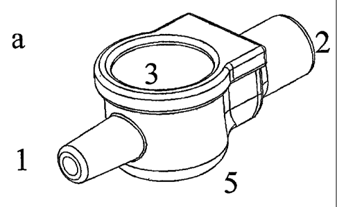
5 The invention will now be described with reference to the accompanying
drawings in which:
Figures l and 2 show line-draw of side and front views of the device
respectively.
Figures 3 and 4 show side and front line-draw views of alternative designs for
the device.
Figure 5 shows two different views of the device. (a) Side perspective view
from the right;
(b) Cross section through device showing one embodiment of the valve at the
end of the
injection port.
Figure 6 show the device in use attached to the needle and a syringe.
The device (5) is connected to the epidural needle via end 1. End 2 is the
injection port
through which the fluid can be injected to pressurise the cavity. It
incorporates a one-way
valve which may be made of, for example, flaps of elastic material such as
rubber (2 flaps are
shown in figures 1,2, and 3.) The elastic diaphragm (3) is designed to bulge
outwardly when
the cavity is pressurised by a fluid, for example air or saline. The diaphragm
(3) flattens when
the pressure is lost suddenly, for example when the needle enters the epidural
space,
providing a visual end point. This diaphragm may be made of thin elastic
material such as
rubber or other imitative synthetic fibre. The cavity (4), which could have
different shapes
such as cylindrical or cubical, may be pressurised by injection of air or
saline. The body of
CA 02423016 2003-03-20
6
the device (5), excluding the diaphragm and the valve, is made of hard
material for example
plastic.
The same components of this invention may be rearranged in an alternative
design such as
shown in the side and front line-draw views of figures 3 and 4 respectively.
All features of each of the aspects of the present invention apply to all
other aspects mutatis
mutandis.