Note: Descriptions are shown in the official language in which they were submitted.
CA 02424038 2003-03-28
WO 02/26281 PCT/USO1/30519
COATED MEDICAL DEVICES
CROSS REFERENCE TO RELATED APPLICATIONS
This application is a continuation-in-part application of U.S. Application
Serial Number09/887,464 filed June 22, 2001, a continuation-in-part
application of US Application Serial Number 09/675,882, filed September 29,
2000,a continuation-in-part application of U.S. Application Serial Number
09/884,729 filed June 19, 2001 and a continuation-in-part of U.S. Application
Serial Number 09/850,482 filed May 7, 2001.
BACKGROUND OF THE INVENTION
1. Field of the Invention
The present invention relates to the local administration of drug/drug
combinations for the prevention and treatment of vascular disease, and more
particularly to intraluminal medical devices for the local delivery of
drug/drug
combinations for the prevention and treatment of vascular disease caused by
injury and methods for maintaining the drug/drug combinations on the
intraluminal medical devices. The present invention also relates to medical
devices having drugs, agents or compounds affixed thereto to minimize or
substantially eliminate a biological organism's reaction to the introduction
of the
medical device to the organism.
2. Discussion of the Related Art
Many individuals suffer from circulatory disease caused by a progressive
blockage of the blood vessels that perfuse the heart and other major organs
with nutrients. More severe blockage of blood vessels in such individuals
often
leads to hypertension, ischemic injury, stroke, or myocardial infarction.
Atherosclerotic lesions, which limit or obstruct coronary blood flow, are the
1
CA 02424038 2003-03-28
WO 02/26281 PCT/USO1/30519
major cause of ischemic heart disease. Percutaneous transluminal coronary
angioplasty is a medical procedure whose purpose is to increase blood flow
through an artery. Percutaneous transluminal coronary angioplasty is the
predominant treatment for coronary vessel stenosis. The increasing use of this
procedure is attributable to its relatively high success rate and its minimal
invasiveness compared with coronary bypass surgery. A limitation associated
with percutaneous transluminal coronary angioplasty is the abrupt closure of
the vessel which may occur immediately after the procedure and restenosis
which occurs gradually following the procedure. Additionally, restenosis is a
chronic problem in patients who have undergone saphenous vein bypass
grafting. The mechanism of acute occlusion appears to involve several factors
and may result from vascular recoil with resultant closure of the artery
and/or
deposition of blood platelets and fibrin along the damaged length of the newly
opened blood vessel.
IS
Restenosis after percutaneous transluminal coronary angioplasty is a
more gradual process initiated by vascular injury. Multiple processes,
including
thrombosis, inflammation, growth factor and cytokine release, cell
proliferation,
cell migration and extracellular matrix synthesis each contribute to the
restenotic process.
While the exact mechanism of restenosis is not completely understood,
the general aspects of the restenosis process have been identified. In the
normal arterial wall, smooth muscle cells proliferate at a low rate,
approximately less than 0.1 percent per day. Smooth muscle cells in the
vessel walls exist in a contractile phenotype characterized by eighty to
ninety
percent of the cell cyfioplasmic volume occupied with the contractile
apparatus.
Endoplasmic reticulum, Golgi, and tree ribosomes are few and are located in
the perinuclear region. Extracellular matrix surrounds the smooth muscle cells
and is rich in heparin-like glycosylaminoglycans which are believed to be
responsible for maintaining smooth muscle cells in the contractile phenotypic
state (Campbell and Campbell, 1985).
2
CA 02424038 2003-03-28
WO 02/26281 PCT/USO1/30519
Upon pressure expansion of an intracoronary balloon catheter during
angioplasty, smooth muscle cells within the vessel wall become injured,
initiating a thrombotic and inflammatory response. Cell derived growth factors
such as platelet derived growth factor, basic fibroblast growth factor,
epidermal
growth factor, thrombin, etc., released from platelets, invading macrophages
and/or leukocytes, or directly from the smooth muscle cells provoke a
proliferative and migratory response in medial smooth muscle cells. These
cells undergo a change from the contractile phenotype to a synthetic
phenotype characterized by only a few contractile filament bundles, extensive
rough endoplasmic reticulum, Golgi and free ribosomes. Proliferation/migration
usually begins within one to two days post-injury and peaks several days
thereafter (Campbell and Campbell, 1987; Clowes and Schwartz, 1985).
Daughter cells migrate to the intimal layer of arterial smooth muscle and
continue to proliferate and secrete significant amounts of extracellular
matrix
proteins. Proliferation, migration and extracellular matrix synthesis continue
until the damaged endothelial layer is repaired at which time proliferation
slows
within the intima, usually within seven to fourteen days post-injury. The
newly
formed tissue is called neointima. The further vascular narrowing that occurs
over the next three to six months is due primarily to negative or constrictive
remodeling.
Simultaneous with local proliferation and migration, inflammatory cells
adhere to the site of vascular injury. Within three to seven days post-injury,
inflammatory cells have migrated to the deeper layers of the vessel wall. In
animal models employing either balloon injury or stenfi implantation,
inflammatory cells may persist at the site of vascular injury for at least
thirty
days (Tanaka et al., 1993; Edelman et al., 1998). Inflammatory cells therefore
are present and may contribute to both the acute and chronic phases of
restenosis.
Numerous agents have been examined for presumed anti-proliferative
actions in restenosis and have shown some activity in experimental animal
3
CA 02424038 2003-03-28
WO 02/26281 PCT/USO1/30519
models. Some of the agents which have been shown to successfully reduce
the extent of intimal hyperplasia in animal models include: heparin and
heparin
fragments (Clowes, A.W. and Karnovsky M., Nature 265: 25-26, 1977; Guyton,
J.R. et al., Circ. Res., 46: 625-634, 1980; Clowes, A.W. and Clowes, M.M.,
Lab. Invest. 52: 611-616, 1985; Clowes, A.W. and Clowes, M.M., Circ. Res. 58:
839-845, 1986; Majesky et al., Circ. Res. 61: 296-300, 1987; Snow et al., Am.
J. Pathol. 137: 313-330, 1990; Okada, T. et al., Neurosurgery 25: 92-98,
1989),
colchicine (furrier, J.W. et al., Circ. 80: 11-66, 1989), taxol (Sollot, S.J.
et al.,
J. Clin. Invest. 95: 1869-1876, 1995), angiotensin converting enzyme (ACE)
inhibitors (Powell, J.S, et al., Science, 245: 186-188, 1989), angiopeptin
(Lundergan, C.F, et al. Am. J. Cardiol. 17(Suppl. B):132B-136B, 1991),
cyclosporin A (Jonasson, L. et al., Proc. Natl., Acad. Sci., 85: 2303, 1988),
goat-anti-rabbit PDGF antibody (Ferns, G.A.A., et al., Science 253: 1129-1132,
1991 ), terbinafine (Nemecek, G.M, et al., J. Pharmacol. Exp. Thera. 248: 1167-
1174, 1989), trapidil (Liu, M.W, et al., Circ. 81: 1089-1093, 1990), tranilast
(Fukuyama, J. et al., Eur. J. Pharmacol. 318: 327-332, 1996), interferon-
gamma (Hansson, G.K. and Holm, J., Circ. 84: 1266-1272, 1991 ), rapamycin
(Marx, S.O. et al., Circ. Res. 76: 412-417, 1995), steroids (Colburn, M.D. et
al.,
J. Vasc. Surg. 15: 510-518, 1992), see also Berk, B.C. et al., J. Am. Coll.
Cardiol. 17: 111 B-117B, 1991 ), ionizing radiation (Weinberger, J. et al.,
Int. J.
Rad. Onc. Biol. Phys. 36: 767-775, 1996), fusion toxins (Farb, A. et al.,
Circ.
Res. 80: 542-550, 1997) antisense oligionucleotides (Simons, M. et al., Nature
359: 67-70, 1992) and gene vectors (Chang, M.W. et al., J. Clin. Invest. 96:
2260-2268, 1995). Anti-proliferative action on smooth muscle cells in vitro
has
been demonstrated for many of these agents, including heparin and heparin
conjugates, taxol, tranilast, colchicine, ACE inhibitors, fusion toxins,
antisense
oligionucleotides, rapamycin and ionizing radiation. Thus, agents with diverse
mechanisms of smooth muscle cell inhibition may have therapeutic utility in
reducing intimal hyperplasia.
However, in contrast to animal models, attempts in human angioplasty
patients to prevent restenosis by systemic pharmacologic means have thus far
been unsuccessful. Neither aspirin-dipyridamole, ticlopidine, anti-coagulant
4
CA 02424038 2003-03-28
WO 02/26281 PCT/USO1/30519
therapy (acute heparin, chronic warfarin, hirudin or hirulog), thromboxane
receptor antagonism nor steroids have been effective in preventing restenosis,
although platelet inhibitors have been effective' in preventing acute
reocclusion
after angioplasty (Mak and Topol, 1997; Lang et al., 1991; Popma et al., 1991
).
The platelet GP Ilb/Illa receptor, antagonist, Reopro~ is still under study
but
Reopro~ has not shown definitive results for the reduction in restenosis
following angioplasty and stenting. Other agents, which have also been
unsuccessful in the prevention of restenosis, include the calcium channel
antagonists, prostacyclin mimetics, angiotensin converting enzyme inhibitors,
serotonin receptor antagonists, and anti-proliferative agents. These agents
must be given systemically, however, and attainment of a therapeutically
effective dose may not be possible; anti-proliferative (or anti-restenosis)
concentrations may exceed the known toxic concentrations of these agents so
that levels sufficient to produce smooth muscle inhibition may not be reached
(Mak and Topol, 1997; Lang et al., 1991; Popma et al., 1991 ).
Additional clinical trials in which fihe effectiveness for preventing
restenosis utilizing dietary fish oil supplements or cholesterol lowering
agents
has been examined showing either conflicting or negative results so that no
pharmacological agents are as yet clinically available to prevent post-
angioplasty restenosis (Mak and Topol, 1997; Franklin and Faxon, 1993:
Serruys, P.W, et al., 1993). Recent observations suggest that the
antilipid/antioxident agent, probucol, may be useful in preventing restenosis
but
this work requires confirmation (Tardif et al., 1997; Yokoi, et al., 1997).
Probucol is presently not approved for use in the United States and a thirty-
day
pretreatment period would preclude its use in emergency angioplasty.
Additionally, the application of ionizing radiation has shown significant
promise
in reducing or preventing restenosis after angioplasty in patients with stents
(Teirstein et al., 1997). Currently, however, the most effective treatments
for
restenosis are repeat angioplasty, atherectomy or coronary artery bypass
grafting, because no therapeutic agents currently have Food and Drug
Administration approval for use for the prevention of post-angioplasty
restenosis.
5
CA 02424038 2003-03-28
WO 02/26281 PCT/USO1/30519
Unlike systemic pharmacologic therapy, stents have proven useful in
significantly reducing restenosis. Typically, stents are balloon-expandable
slotted metal tubes (usually, but not limited to, stainless steel), which,
when
expanded within the lumen of an angioplastied coronary artery, provide
structural support through rigid scaffolding to the arterial wall. This
support is
helpful in maintaining vessel lumen patency. In two randomized clinical
trials,
stents increased angiographic success after percutaneous transluminal
coronary angioplasty, by increasing minimal lumen diameter and reducing, but
IO not eliminating, the incidence of restenosis at six months (Serruys et al.,
1994;
Fischman et al., 1994).
Additionally, the heparin coating of stents appears to have the added
benefit of producing a reduction in sub-acute thrombosis after stent
IS implantation (Serruys et al., 1996). Thus, sustained mechanical expansion
of a
stenosed coronary artery with a stent has been shown to provide some
measure of restenosis prevention, and the coating of stenfis with heparin has
demonstrated both the feasibility and the clinical usefulness of delivering
drugs
locally, at the site of injured tissue.
As stated above, the use of heparin coated stenfis demonstrates the
feasibility and clinical usefulness of local drug delivery; however, the
manner in
which the particular drug or drug combination is affixed to the local delivery
device will play a role in the efficacy of this type of treatment. For
example, the
processes and materials utilized to affix the drug/drug combinations to the
local
delivery device should not interfere with the operations of the drug/drug
combinations. In addition, the processes and materials utilized should be
biocompatible and maintain the drug/drug combinations on the local device
through delivery and over a given period of time. For example, removal of the
drug/drug combination during delivery of the local delivery device may
potentially cause failure of the device.
6
CA 02424038 2003-03-28
WO 02/26281 PCT/USO1/30519
Accordingly, there exists a need for drug/drug combinations and
associated local delivery devices for the prevention and treatment of vascular
injury causing intimal thickening which is either biologically induced, for
example atherosclerosis, or mechanically induced, for example, through
percutaneous transluminal coronary angioplasty. In addition, there exists a
need for maintaining the drugldrug combinations on the local delivery device
through delivery and positioning as well as ensuring that the drug/drug
combination is released in therapeutic dosages over a given period of time.
A variety of stent coatings and compositions have been proposed for the
prevention and treatment of injury causing intimal thickening. The coatings
may be capable themselves of reducing the stimulus the stent provides to the
injured lumen wall, thus reducing the tendency towards thrombosis or
restenosis. Alternately, the coating may deliver a pharmaceutical/therapeutic
agent or drug to the lumen that reduces smooth muscle tissue proliferation or
restenosis. The mechanism for delivery of the agent is through difFusion of
the
agent through either a bulk polymer or through pores that are created in the
polymer structure, or by erosion of a biodegradable coating.
Both bioabsorbable and biostable compositions have been reported as
coatings for stents. They generally have been polymeric coatings that either
encapsulate a pharmaceutical/therapeutic agent or drug, e.g. rapamycin, taxol
etc., or bind such an agent to the surface, e.g. heparin-coated scents. These
coatings are applied to the stent in a number of ways, including, though not
limited to, dip, spray, or spin coating processes.
One class of biostable materials that has been reported as coatings for
stents is polyfluoro homopolymers. Polytetrafluoroethylene (PTFE)
homopolymers have been used as implants for many years. These
homopolymers are not soluble in any solvent at reasonable temperatures and
therefore are difficult to coat onto small medical devices while maintaining
important features of the devices (e.g. slots in stems).
7
CA 02424038 2003-03-28
WO 02/26281 PCT/USO1/30519
Stents with coatings made from polyvinylidenefluoride homopolymers
and containing pharmaceutical/therapeutic agents or drugs for release have
been suggested. However, like most crystalline polyfluoro homopolymers, they
are difficult to apply as high quality films onto surfaces without subjecting
them
to relatively high temperatures, that correspond to the melting temperature of
the polymer.
It would be advantageous to develop coatings for implantable medical
devices that will reduce thrombosis, restenosis, or other adverse reactions,
that
may include, but do not require, the use of pharmaceutical or therapeutic
agents or drugs to achieve such affects, and that possess physical and
mechanical properties effective for use in such devices even when such coated
devices are subjected to relatively low maximum temperatures.
SUMMARY OF THE INVENTION
The drug/drug combination therapies, drug/drug combination carriers
and associated local delivery devices of the present invention provide a means
for overcoming the difficulties associated with the methods and devices
currently in use, as briefly described above. In addition, the methods for
maintaining the drug/drug combinations and drug/drug combination carriers on
the local delivery device ensure that the drug/drug combination therapies
reach
the target site.
In accordance with one aspect, the present invention is directed to a
medical device for implantation into a treatment site of a living organism.
The
device comprises a biocompatible vehicle affixed to at least a portion of the
medical device, and at least one agent in therapeutic dosages incorporated
into the biocompatible vehicle for the treatment of reactions by the living
organism caused by the medical device or the implantation thereof.
In accordance with another aspect, the present invention is directed to a
medical device for implantation into a treatment site of a living organism.
The
s
CA 02424038 2003-03-28
WO 02/26281 PCT/USO1/30519
device comprises a biocompatible vehicle affixed to at least a portion of the
medical device, at least one agent in therapeutic dosages incorporated into
the
biocompatible vehicle for the treatment of reactions by the living organism
caused by the medical device or the implantation thereof, and a material for
preventing the at least one agent from separating from the medical device
prior
to and during implantation of the medical device at the treatment site, the
material being affixed to at least one of the medical devices or a delivery
system for the medical device.
In accordance with another aspect, the present invention is directed to a
medical device for implantation into a treatment site of a living organism.
The
device comprises a stent, a biocompatible vehicle affixed to at least a
portion of
the stent, and at least one agent in therapeutic dosages incorporated into the
biocompatible vehicle for the treatment of reactions by the living organism
caused by the medical device or the implantation thereof.
In accordance with another aspect, the present invention is directed to a
medical device for implantation info a treatment site of a living organism.
The
device comprises a stent having a substantially tubular member having open
ends, and a first diameter for insertion into a lumen of a vessel and a second
diameter for anchoring in the lumen of the vessel, a biocompatible vehicle
affixed to at least a portion of the stent, at least one agent in therapeutic
dosages incorporated into the biocompatible vehicle for the treatment of
reactions by the living organism caused by the medical device or the
implantation thereof, and a material for preventing the at least one agent
from
separating from the medical device prior to and during implantation of the
medical device at the treatment site, the material being affixed to at least
one
of the medical devices or a delivery system for the medical device.
In accordance with another aspect, the present invention is directed to a
focal drug delivery device. The device comprises a stent having a
substantially
tubular member having open ends, and a first diameter for insertion into a
lumen of a vessel and a second diameter for anchoring in the lumen of a
9
CA 02424038 2003-03-28
WO 02/26281 PCT/USO1/30519
vessel, a biocompatible polymeric vehicle affixed to at least a portion of the
stent, and rapamycin, in therapeutic dosages, incorporated into the
biocompatible polymeric vehicle .
In accordance with another aspect, the present invention is directed to a
method of coating a medical device with a therapeutic agent. The method
comprises the steps of creating a polymer utilizing vinylidene fluoride and
hexafluoropropylene in a batch emulsion polymerization process, priming the
medical device with the polymer utilizing a dip coating process, creating a
polymer and therapeutic agent mixture, applying the polymer and therapeutic
agent mixture on the primer layer utilizing a spin coating process, and drying
the medical device in a vacuum oven for approximately sixteen hours at a
temperature in the range of fifty to sixty degrees centigrade.
In accordance with another aspect, the present invention is directed to a
medical device for implantation into a treatment site of a living organism.
The
medical device comprises a biocompatible vehicle affixed to at least a portion
of the medical device, and at least one agent incorporated into the
biocompatible vehicle. The at least one agent being designed to react with one
or more chemicals produced by the living organism to treat reactions by the
living organism caused by the medical device or the implantation thereof.
In accordance with another aspect, the present invention is directed to a
medical device for implantation into the vasculature of a living organism. The
medical device comprises a self-expanding stent, a biocompatible vehicle
affixed to at least a portion of the stent, and rapamycin, in therapeutic
dosages,
incorporated info the biocompatible vehicle for the prevention of restenosis.
In accordance with another aspect, the present invention is directed to a
method of coating a medical device with a therapeutic agent. The method
comprises the steps of creating a polymer utilizing vinylidene fluoride and
hexafluoropropylene, adding one or more therapeutic agents to the polymer to
CA 02424038 2003-03-28
WO 02/26281 PCT/USO1/30519
create a polymer and therapeutic agent mixture, and applying the polymer and
therapeutic agent mixture to the medical device.
In accordance with another aspect, the present invention is directed to a
medical device for implantation into a treatment site of a living organism.
The
medical device comprises a biocompatible vehicle affixed to at least a portion
of the medical device, at least one agent in therapeutic dosages incorporated
into the biocompatible vehicle for the treatment of disease proximate the
implantation site.
In accordance with another aspect, the present invention is directed to a
medical device for implantation into a treatment site of a living organism.
The
medical device comprises a biocompatible vehicle afFixed to at least a portion
of the medical device, at least one agent in therapeutic dosages incorporated
into the biocompatible vehicle for the treatment of disease remote from the
implantation site.
The medical devices, drug coatings and methods for maintaining the
drug coatings or vehicles thereon of the present invention utilizes a
combination of materials to treat disease, and reactions by living organisms
due to the implantation of medical devices for the treatment of disease or
other
conditions. The local delivery of drugs, agents or compounds generally
substantially reduces the potential toxicity of the drugs, agents or compounds
when compared to systemic delivery while increasing their efficacy.
Drugs, agents or compounds may be affixed to any number of medical
devices to treat various diseases. The drugs, agents or compounds may also
be affixed to minimize or substantially eliminate the biological organism's
reaction to the introduction of the medical device utilized to treat a
separate
condition. For example, stents may be introduced to open coronary arteries or
other body lumens such as biliary ducts. The introduction of these stents
cause a smooth muscle cell proliferation effect as well as inflammation.
11
CA 02424038 2003-03-28
WO 02/26281 PCT/USO1/30519
Accordingly, the stents may be coated with drugs, agents or compounds to
combat these reactions.
The drugs, agents or compounds will vary depending upon the type of
medical device, the reaction to the introduction of the medical device and/or
the disease sought to be treated. The type of coating or vehicle utilized to
immobilize the drugs, agents or compounds to the medical device may also
vary depending on a number of factors, including the type of medical device,
the fiype of drug, agent or compound and the rate of release thereof.
!n order to be effective, the drugs, agents or compounds should
preferably remain on the medical devices during delivery and implantation.
Accordingly, various coating techniques for creating strong bonds between the
drugs, agents or compounds may be utilized. In addition, various materials
may be utilized as surface modifications to prevent the drugs, agents or
compounds from coming off prematurely.
BRIEF DESCRIPTION OF THE DRA1NINGS
The foregoing and other features and advantages of the invention will
be apparent from the following, more particular description of preferred
embodiments of the invention, as illustrated in the accompanying drawings.
Figure 1 is a view along the length of a scent (ends not shown) prior to
expansion showing the exterior surface of the stent and the characteristic
banding pattern.
Figure 2 is a view along the length of the stent of Figure 1 having
reservoirs in accordance with the present invention.
Figure 3 indicates the fraction of drug released as a function of time
from coatings of the present invention over which no topcoat has been
disposed.
12
CA 02424038 2003-03-28
WO 02/26281 PCT/USO1/30519
Figure 4 indicates the fraction of drug released as a function of time
from coatings of the present invention including a topcoat disposed thereon.
Figure 5 indicates the fraction of drug released as a function of time
from coatings of the present invention over which no topcoat has been
disposed.
Figure 6 indicates in vivo scent release kinetics of rapamycin from
poly(VDF/HFP)
Figure 7 is a cross-sectional view of a band of the scent of Figure 1
having drug coatings thereon in accordance with a first exemplary embodiment
of the invention.
Figure 8 is a cross-sectional view of a band of the stent of Figure 1
having drug coatings thereon in accordance with a second exemplary
embodiment of the invention.
Figure 9 is a cross-sectional view of a band of the stent of Figure 1
having drug coatings thereon in accordance with a third exemplary
embodiment of the present invention.
Figure 10 is a perspective view of an exemplary stent in its compressed
state which may be utilized in conjunction With the present invention.
Figure 11 is a sectional, flat view of the stent shown in Figure 10.
Figure 12 is a perspective view of the stent shown in Figure 10 but
showing it in its expanded state.
Figure 13 is an enlarged sectional view of the scent shown in Figure 12.
13
CA 02424038 2003-03-28
WO 02/26281 PCT/USO1/30519
Figure 14 is an enlarged view of section of the scent shown in Figure 11.
Figure 15 is a view similar to that of Figure 11 but showing an alternate
embodiment of the stmt.
Figure 16 is a perspective view of the scent of Figure 10 having a
plurality of markers attached to the ends thereof in accordance with the
present
invention.
Figure 17 is a cross-sectional view of a marker in accordance with the
present invention,
Figure 13 is an enlarged perspective view of an end of the stent with the
markers forming a substantially straight line in accordance with the present
invention.
Figure 19 is a simplified partial cross-sectional view of a stent delivery
apparatus having a scent loaded therein, which can be used with a stent made
in accordance with the present invention.
Figure 20 is a view similar to that of Figure 19 but showing an enlarged
view of the distal end of the apparatus.
Figure 21 is a perspective view of an end of the scent with the markers in
a partially expanded form as it emerges from the delivery apparatus in
accordance with the present invention.
Figure 22 is a cross-sectional view of a balloon having a lubricious
coating affixed thereto in accordance with the present invention.
Figure 23 is a cross-sectional view of a band of the stent in Figure 1
having a lubricious coating affixed thereto in accordance with fihe present
invention.
14
CA 02424038 2003-03-28
WO 02/26281 PCT/USO1/30519
Figure 24 is a cross-sectional view of a self-expanding stent in a delivery
device having a lubricious coating in accordance with the present invention.
Figure 25 is a cross-sectional view of a band of the scent in Figure 1
having a modified polymer coating in accordance with the present invention.
Figure 26 illustrates an exemplary balloon-expandable stent having an
alternative arrangement of "N" and "J" links between sets of strut members,
represented on a flat, two-dimensional plan view in accordance with the
present invention.
DETAILED DESCRIPTION OF THE PREFERRED EMBODIMENTS
The drug/drug combinations and delivery devices of the present
invention may be utilized to effectively prevent and treat vascular disease,
and
in particular, vascular disease caused by injury. Various medical treatment
devices utilized in the treatment of vascular disease may ultimately induce
further complications. For example, balloon angioplasty is a procedure
utilized
to increase blood flow through an artery and is the predominant treatment for
coronary vessel stenosis. However, as stated above, the procedure typically
causes a certain degree of damage to the vessel wall, thereby potentially
exacerbating the problem at a point later in time. Although other procedures
and diseases may cause similar injury, exemplary embodiments of the present
invention will be described with respect to the treatment of restenosis and
related complications following percutaneous transluminal coronary angioplasty
and other similar arterial/venous procedures.
While exemplary embodiments of the invention will be described with
respect to the treatment of restenosis and related complications following
percutaneous transluminal coronary angioplasty, it is important to note that
the
local delivery of drug/drug combinations may be utilized to treat a wide
variety
of conditions utilizing any number of medical devices, or to enhance the
CA 02424038 2003-03-28
WO 02/26281 PCT/USO1/30519
function and/or life of the device. For example, intraocular lenses, placed to
restore vision after cataract surgery is often compromised by the formation of
a
secondary cataract. The latter is often a result of cellular overgrowth on the
lens surface and can be potentially minimized by combining a drug or drugs
with the device. Other medical devices which often fail due to tissue in-
growth
or accumulation of proteinaceous material in, on and around the device, such
as shunts for hydrocephalus, dialysis grafts, colostomy bag attachment
devices, ear drainage tubes, leads for pace makers and impfantable
defibrillators can also benefit from the device-drug combination approach.
'Devices which serve to improve the structure and function of tissue or
organ may also show benefits when combined with the appropriate agent or
agents. For example, improved osteointegration of orthopedic devices to
enhance stabilization of the implanted device could potentially be achieved by
combining it with agents such as bone-morphogenic protein. Similarly other
surgical devices, sutures, staples, anastomosis devices, vertebral disks, bone
pins, suture anchors, hemostatic barriers, clamps, screws, plates, clips,
vascular implants, tissue adhesives and sealants, tissue scaffolds, various
types of dressings, bone substitutes, intraluminal devices, and vascular
supports could also provide enhanced patient benefit using this drug-device
combination approach. Essentially, any type of medical device may be coated
in some fashion with a drug or drug combination which enhances treatment
over use of the singular use of the device or pharmaceutical agent.
In addition to various medical devices, the coatings on these devices
may be used to deliver fiherapeutic and pharmaceutic agents including:
antiproliferative/antimitotic agents including natural products such as vinca
alkaloids (i.e. vinblastine, vincristine, and vinorelbine), paclitaxel,
epidipodophyllotoxins (i.e. etoposide, teniposide), antibiotics (dactinomycin
(actinomycin D) daunorubicin, doxorubicin and idarubicin), anthracyclines,
mitoxantrone, bleomycins, plicamycin (mithramycin) and mitomycin, enzymes
(L-asparaginase which systemically metabolizes L-asparagine and deprives
cells which do not have the capacity to synthesize their own asparagine);
16
CA 02424038 2003-03-28
WO 02/26281 PCT/USO1/30519
antiplatelet agents such as G(GP)Ilbl I la inhibitors and vitronectin receptor
antagonists; antiproliferative/antimitotic alkylating agents such as nitrogen
mustards (mechlorethamine, cyclophosphamide and analogs, melphalan,
chlorambucil), ethylenimines and methylmelamines (hexamethylmelamine and
thiotepa), alkyl sulfonates-busulfan, nirtosoureas (carmustine (BCNU) and
analogs, streptozocin), trazenes - dacarbazinine (DTIC);
antiproliferative/antimitotic antimetabolites such as folic acid analogs
(methotrexate), pyrimidine analogs (fluorouracil, floxuridine, and
cytarabine),
purine analogs and related inhibitors (mercaptopurine, thioguanine,
pentostatin
IO and 2-chlorodeoxyadenosine ~cladribine}); platinum coordination complexes
(cisplatin, carboplatin), procarbazine, hydroxyurea, mitotane,
aminoglutethimide; hormones (i.e. estrogen); anticoagulants (heparin,
synthetic
heparin salts and other inhibitors of thrombin); fibrinolytic agents (such as
tissue plasminogen activator, streptokinase and urokinase), aspirin,
I5 dipyridamole, ticlopidine, clopidogrel, abciximab; antimigratory;
antisecretory
(breveldin); antiinflammatory: such as adrenocortical steroids (cortisol,
cortisone, fludrocortisone, prednisone, prednisolone, 6a-methylprednisolone,
triamcinolone, betamethasone, and dexamethasone), non-steroidal agents
(salicylic acid derivatives i.e. aspirin; para-aminophenol derivatives i.e.
20 acetominophen; indole and indene acetic acids (indomethacin, sulindac, and
etodalac), heteroaryl acetic acids (tolmetin, diclofenac, and ketorolac),
arylpropionic acids (ibuprofen and derivatives), anthranilic acids (mefenamic
acid, and meclofenamic acid), enolic acids (piroxicam, tenoxicam,
phenylbutazone, and oxyphenthatrazone), nabumetone, gold compounds
25 (auranofin, aurothioglucose, gold sodium thiomalate); immunosuppressives:
(cyclosporine, tacrolimus (FK-506), sirolimus (rapamycin), azathioprine,
mycophenolate mofetil); angiogenic agents: vascular endothelial growth factor
(VEGF), fibroblast growth factor (FGF); angiotensin receptor blocker; nitric
oxide donors; anti-sense oligionucleotides and combinations thereof; cell
cycle
30 inhibitors, mTOR inhibitors, and growth factor signal transduction kinase
inhibitors.
17
CA 02424038 2003-03-28
WO 02/26281 PCT/USO1/30519
As stated previously, the implantation of a coronary stent in conjunction
with balloon angioplasty is highly effective in treating acute vessel closure
and
may reduce the risk of restenosis. Intravascular ultrasound studies (Mintz et
al., 1996) suggest that coronary scenting effectively prevents vessel
constriction and that most of the fate luminal loss after stent implantation
is due
to plaque growth, probably related to neointimal hyperplasia. The late luminal
loss after coronary stenting is almost two times higher than that observed
after
conventional balloon angioplasty. Thus, inasmuch as stents prevent at least a
portion of the restenosis process, a combination of drugs, agents or
compounds which prevents smooth muscle cell proliferation, reduces
inflammation and reduces coagulation or prevents smooth muscle cell
proliferation by multiple mechanisms, reduces inflammation and reduces
coagulation combined with a stent may provide the most efficacious treatment
for post-angioplasty restenosis. The systemic use of drugs, agents or
compounds in combination with the local delivery of the same or different
drug/drug combinations may also provide a beneficial treatment option.
The local delivery of drug/drug combinations from a stent has the
following advantages; namely, the prevention of vessel recoil and remodeling
through the scaffolding action of the stent and the prevention of multiple
components of neointimal hyperplasia or restenosis as well as a reduction in
inflammation and thrombosis. This local administration of drugs, agents or
compounds to stented coronary arteries may also have additional therapeutic
benefit. For example, higher tissue concentrations of the drugs, agents or
compounds may be achieved utilizing local delivery, rather than systemic
administration. In addition, reduced systemic toxicity may be achieved
utilizing
local delivery rather than systemic administration while maintaining higher
tissue concentrations. Also in utilizing local delivery from a stent rather
than
systemic administration, a single procedure may suffice with better patient
compliance. An additional benefit of combination drug, agent, and/or
compound therapy may be to reduce the dose of each of the therapeutic drugs,
agents or compounds, thereby limiting their toxicity, while still achieving a
reduction in restenosis, inflammation and thrombosis. Local stent-based
18
CA 02424038 2003-03-28
WO 02/26281 PCT/USO1/30519
therapy is therefore a means of improving the therapeutic ratio
(efficacy/toxicity) of anti-restenosis, anti-inflammatory, anti-thrombotic
drugs,
agents or compounds.
There are a multiplicity of different stents that may be utilized following
percutaneous transluminal coronary angioplasty. Although any number of
stents may be utilized in accordance with the present invention, for
simplicity, a
limited number of scents will be described in exemplary embodiments of the
present invention. The skilled artisan will recognize that any number of
stents
may be utilized in connection with the present invention. In addition, as
stated
above, other medical devices may be utilized.
A stent is commonly used as a tubular structure left inside the lumen of
a duct to relieve an obstruction. Commonly, stents are inserted into the lumen
in a non-expanded form and are then expanded autonomously, or with the aid
of a second device in situ. A typical method of expansion occurs through the
use of a catheter-mounted angioplasty balloon which is inflated within the
stenosed vessel or body passageway in order to shear and disrupt the
obstructions associated with the wall components of the vessel and to obtain
an enlarged lumen.
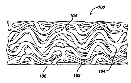
Figure 1 illustrates an exemplary stent 100 which may be utilized in
accordance with an exemplary embodiment of the present invention. The
expandable cylindrical scent 100 comprises a fenestrated structure for
placement in a blood vessel, duct or lumen to hold the vessel, duct or lumen
open, more particularly for protecting a segment of artery from restenosis
after
angioplasty. The scent 100 may be expanded circumferentially and maintained
in an expanded configuration, that is circumferentially or radially rigid. The
stent 100 is axially flexible and when flexed at a band, the stent 100 avoids
any
externally-protruding component parts.
The stent 100 generally comprises first and second ends with an
intermediate section therebetween. The stent 100 has a longitudinal axis and
19
CA 02424038 2003-03-28
WO 02/26281 PCT/USO1/30519
comprises a plurality of longitudinally disposed bands 102, wherein each band
102 defines a generally continuous wave along a line segment parallel to the
longitudinal axis. A plurality of circumferentially arranged links 104
maintain
the bands 102 in a substantially tubular structure. Essentially, each
longitudinally disposed band 102 is connected at a plurality of periodic
locations, by a short circumferentially arranged link 104 to an adjacent band
102. The wave associated with each of the bands 102 has approximately the
same fundamental spatial frequency in the intermediate section, and the bands
102 are so disposed that the wave associated with them are generally aligned
so as to be generally in phase with one another. As illustrated in the figure,
each longitudinally arranged band 102 undulates through approximately two
cycles before there is a link to an adjacent band 102.
The stent 100 may be fabricated utilizing any number of methods. For
example, the stent 100 may be fabricated from a hollow or formed stainless
steel tube that may be machined using lasers, electric discharge milling,
chemical etching or other means. The stent 100 is inserted into the body and
placed at the desired site in an unexpended form. In one exemplary
embodiment, expansion may be effected in a blood vessel by a balloon
catheter, where the final diameter of the stent 100 is a function of the
diameter
of the balloon catheter used.
It should be appreciated that a stent 100 in accordance with the present
invention may be embodied in a shape-memory material, including, for
example, an appropriafie alloy of nickel and titanium or stainless steel.
Structures formed from stainless steel may be made self expanding by
configuring the stainless steel in a predetermined manner, for example, by
twisting it into a braided configuration. In this embodiment after the stent
100
has been formed it may be compressed so as to occupy a space sufficiently
small as to permit its insertion in a blood vessel or other tissue by
insertion
means, wherein the insertion means include a suitable catheter, or flexible
rod.
On emerging from the catheter, the stent 100 may be configured to expand into
CA 02424038 2003-03-28
WO 02/26281 PCT/USO1/30519
the desired configuration where the expansion is automatic or triggered by a
change in pressure, temperature or electrical stimulation.
Figure 2 illustrates an exemplary embodiment of the present invention
utilizing the stent 100 illustrated in Figure 1. As illustrated, the stent 100
may
be modified to comprise one or more reservoirs 106. Each of the reservoirs
106 may be opened or closed as desired. These reservoirs 106 may be
specifically designed to hold the drug/drug combinations to be delivered.
Regardless of the design of the scent 100, it is preferable to have the
drug/drug
combination dosage applied with enough specificity and a sufficient
concentration to provide an effective dosage in the lesion area. In this
regard,
the reservoir size in the bands 102 is preferably sized to adequately apply
the
drug/drug combination dosage at the desired location and in the desired
amount.
In an alternate exemplary embodiment, the entire inner and outer
surface of the stent 100 may be coated with drug/drug combinations in
therapeutic dosage amounts. A detailed description of a drug for treating
restenosis, as well as exemplary coating techniques, is described below. It
is,
however, important to note that the coating techniques may vary depending on
the drug/drug combinations. Also, the coating techniques may vary depending
on the material comprising the stent or other intraluminal medical device.
Figure 26 illustrates another exemplary embodiment of a balloon
expandable stent. Figure 26 illustrates the stent 900 in its crimped, pre
deployed state as it would appear if it were cut longitudinally and then (aid
out
into a flat, two-dimensional configuration. The stent 900 has curved end
struts
902 and diagonal struts 904 with each set of strut members 906 connected by
sets of flexible links 908, 910 or 912. In this exemplary embodiment, three
different types of flexible links are used. A set of "N" links 910 comprising
six
circumferentially spaced "N" links 914 and a set of inverted "N" links 912
comprising six circumferentially spaced inverted "N" links 916 each connect to
adjacent sets of strut members 906 at the ends of the stem 900. A set of
21
CA 02424038 2003-03-28
WO 02/26281 PCT/USO1/30519
inverted "J" links 918 comprising six circumferentially spaced inverted "J"
links
908 are used to connect the adjacent sets of strut members 906 in the center
of the stent 900. The shape of the "N" links 914 and inverted "N" links 916
facilitate the links' ability to lengthen and shorten as the stent bends
around a
curve during delivery into the human body. This ability to lengthen and
shorten
helps to prevent the sets of strut members from being pushed or pulled off the
balloon during delivery into the body and is particularly applicable to short
stents which tend to have relatively poor stent retention onto an inflatable
balloon. The stent 900 with its greater strength at its central region would
advantageously be used for comparatively short stenoses that have a tough,
calcified central section. It should also be understood that a regular "J"
link
could be used for the stent 900 in place of the inverted "J" link 908. Other
exemplary embodiments of balloon expandable stents may be found in U.S.
Patent No. 6,190,403 B1 issued on February 20, 2001 and,which is
incorporated by reference herein.
Rapamycin is a macrocyclic triene antibiotic produced by Streptomyces
hygroscopicus as disclosed in U.S. Patent No. 3,929,992. It has been found
that rapamycin among other things inhibits the proliferation of vascular
smooth
muscle cells in vivo. Accordingly, rapamycin may be utilized in treating
intimal
smooth muscle cell hyperplasia, restenosis, and vascular occlusion in a
mammal, particularly following either biologically or mechanically mediated
vascular injury, or under conditions that would predispose a mammal to
suffering such a vascular injury. Rapamycin functions to inhibit smooth muscle
cell proliferation and does not interfere with the re-endothelialization of
the
vessel walls.
Rapamycin reduces vascular hyperplasia by antagonizing smooth
muscle proliferation in response to mitogenic signals that are released during
an angioplasty induced injury. Inhibition of growth factor and cytokine
mediated smooth muscle proliferation at the late G1 phase of the cell cycle is
believed to be the dominant mechanism of action of rapamycin. However,
rapamycin is also known to prevent T-cell proliferation and differentiation
when
22
CA 02424038 2003-03-28
WO 02/26281 PCT/USO1/30519
administered systemically. This is the basis for its immunosuppresive activity
and its ability to prevent graft rejection.
As used herein, rapamycin includes rapamycin and all analogs,
derivatives and congeners that find FKBP12, and other immunophilins, and
possesses the same pharmacologic properties as rapamycin.
Although the anti-proliferative effects of rapamycin may be achieved
through systemic use, superior results may be achieved through the local
delivery of the compound. Essentially, rapamycin works in the tissues, which
are in proximity to the compound, and has diminished effect as the distance
from the delivery device increases. In order to take advantage of this effect,
one would want the rapamycin in direct contact with the lumen walls.
Accordingly, in a preferred embodiment, the rapamycin is incorporated onto the
surface of the stent or portions thereof. Essentially, the rapamycin is
preferably
incorporated into the scent 100, illustrated in Figure 1, where the stent 100
makes contact with the lumen wall.
Rapamycin may be incorporated onto or affixed to the stent in a number
of ways. In the exemplary embodimenfi, the rapamycin is directly incorporated
into a polymeric matrix and sprayed onto the outer surface of the stent. The
rapamycin elutes from the polymeric matrix over time and enters the
surrounding tissue. The rapamycin preferably remains on the stent for at least
three days up to approximately six months, and more preferably between
seven and thirty days.
Any number of non-erodible polymers may be utilized in conjunction with
the rapamycin. In one exemplary embodiment, the polymeric matrix comprises
two layers. The base layer comprises a solution of polyethylene-co-
vinylacetate) and polybutylmethacrylate. The rapamycin is incorporated into
this base layer. The outer layer comprises only polybutylmethacrylate and acts
as a diffusion barrier to prevent the rapamycin from eluting too quickly. The
thickness of the outer layer or top coat determines the rate at which the
23
CA 02424038 2003-03-28
WO 02/26281 PCT/USO1/30519
rapamycin elutes from the matrix. Essentially, the rapamycin elutes from the
matrix by diffusion through the polymer matrix. Polymers are permeable,
thereby allowing solids, liquids and gases to escape therefrom. The total
thickness of the polymeric matrix is in the range from about one micron to
about twenty microns or greater. It is important to note that primer layers
and
metal surface treatments may be utilized before the polymeric matrix is
affixed
to the medical device. For example, acid cleaning, alkaline (base) cleaning,
salinization and parylene deposition may be used as part of the overall
process
described below.
The polyethylene-co-vinylacetate), polybutylmethacrylate and
rapamycin solution may be incorporated into or onto the stent in a number of
ways. For example, the solution may be sprayed onto the stent or the stent
may be dipped into the solution. Other methods include spin coating and RF-
plasma polymerization. In one exemplary embodiment, the solution is sprayed
onto the stent and then allowed to dry. In another exemplary embodiment, the
solution may be electrically charged to one polarity and the stent
electrically
changed to the opposite polarity. In this manner, the solution and stent will
be
attracted to one another. In using this type of spraying process, waste may be
reduced and more precise control over the thickness of the coat may be
achieved.
In another exemplary embodiment, the rapamycin or other therapeutic
agent may be incorporated into a film-forming poiyfluoro copolymer comprising
an amount of a first moiety selected from the group consisting of polymerized
vinylidenefluoride and polymerized tetrafluoroethylene, and an amount of a
second moiety other than the first moiety and which is copolymerized with the
first moiety, thereby producing the polyfluoro copolymer, the second moiety
being capable of providing toughness or elastomeric properties to the
polyfluoro copolymer, wherein the relative amounts of the first moiety and the
second moiety are effective to provide the coating and film produced therefrom
with properties effective for use in treating implantable medical devices.
24
CA 02424038 2003-03-28
WO 02/26281 PCT/USO1/30519
The present invention provides polymeric coatings comprising a
polyfluoro copolymer and implantable medical devices, for example, stents
coated with a film of the polymeric coating in amounts effective to reduce
thrombosis and/or restenosis when such stems are used in, for example,
angioplasty procedures. As used herein, polyfluoro copolymers means those
copolymers comprising an amount of a first moiety selected from the group
consisting of polymerized vinylidenefluoride and polymerized
tetrafluoroethylene, and an amount of a second moiety other than the first
moiety and which is copolymerized with the first moiety to produce the
polyfluoro copolymer, the second moiety being capable of providing toughness
or elastomeric properties to the polyfluoro copolymer, wherein the relative
amounts of the first moiety and the second moiety are effective to provide
coatings and film made from such polyfluoro copolymers with properties
effective for use in coating implantable medical devices.
The coatings may comprise pharmaceutical or therapeutic agents for
reducing restenosis, inflammation and/or thrombosis, and stents coated with
such coatings may provide sustained release of the agents. Films prepared
from certain polyfluoro copolymer coatings of the present invention provide
the
physics( and mechanical properties required of conventional coated medical
devices, even where maximum temperature, to which the device coatings and
films are exposed, are limited to relatively low temperatures. This is
particularly
important when using the coating/film to deliver pharmaceutical/therapeutic
agents or drugs that are heat sensitive, or when applying the coating onto
temperature-sensitive devices such as catheters. When maximum exposure
temperature is not an issue, for example, where heat-stable agents such as
itraconazole are incorporated into the coatings, higher melting thermoplastic
polyfluoro copolymers may be used and, if very high elongation and adhesion
is required, elastomers may be used. If desired or required, the polyfluoro
elastomers may be crosslinked by standard methods described in, e.g.,
Modern Fluoropol,r~mers, (J. Shires ed.) John Wiley & Sons, New York, 1997,
pp. 77-87.
CA 02424038 2003-03-28
WO 02/26281 PCT/USO1/30519
The present invention comprises polyfluoro copolymers that provide
improved biocompatible coatings or vehicles for medical devices. These
coatings provide inert biocompatible surfaces to be in contact with body
tissue
of a mammal, for example, a human, sufficient to reduce resfienosis, or
thrombosis, or other undesirable reactions. While many reported coatings
made from polyfluoro homopolymers are insoluble and/or require high heat, for
example, greater than about one hundred twenty-five degrees centigrade, to
obtain films with adequate physical and mechanical properties for use on
implantable devices, for example, stents, or are not particularly tough or
elastomeric, films prepared from the polyfluoro copolymers of the present
invention provide adequate adhesion, toughness or elasticity, and resistance
to
cracking when formed on medical devices. In certain exemplary embodiments,
this is the case even where the devices are subjected to relatively low
maximum temperatures.
The polyfluoro copolymers used for coatings according to the present
invention are preferably film-forming polymers that have molecular weight high
enough so as not to be waxy or tacky. The polymers and films formed
therefrom should preferably adhere to the stent and not be readily deformable
after deposition on the stent as to be able to be displaced by hemodynamic
stresses. The polymer molecular weight should preferably be high enough to
provide sufficient toughness so that films comprising the polymers will not be
rubbed off during handling or deployment of the stent. In certain exemplary
embodiments the coating will not crack where expansion of the stent or other
medical devices occurs.
Coatings of the present invention comprise polyfluoro
copolymers, as defined hereinabove. The second moiety polymerized with the
first moiety to prepare the polyfluoro copolymer may be selected from those
polymerized, biocompatible monomers that would provide biocompatible
polymers acceptable for implantation in a mammal, while maintaining sufficient
elastomeric film properties for use on medical devices claimed herein. Such
monomers include, without limitation, hexafluoropropylene (HFP),
26
CA 02424038 2003-03-28
WO 02/26281 PCT/USO1/30519
tetrafluoroethylene (TFE), vinylidenefluoride, 1-hydropentafluoropropylene,
perfluoro(methyl vinyl ether), chlorotrifluoroethylene (CTFE),
pentafluoropropene, trifluoroethylene, hexafluoroacetone and
hexafluoroisobutylene.
Polyfluoro copolymers used in the present invention typically comprise
vinylidinefluoride copolymerized with hexafluoropropylene, in the weight ratio
in
the range of from about fifty to about ninety-two weight percent
vinylidinefluoride to about fifty to about eight weight percent HFP.
Preferably,
polyfluoro copolymers used in the present invention comprise from about fifty
to about eighty-five weight percent vinylidinefluoride copolymerized wifih
from
about fifty to about fifteen weight percent HFP. More preferably, the
polyfluoro
copolymers will comprise from about fifty-five to about seventy weight percent
vinylidineflyoride copolymerized with from about forty-five to about thirty
weight
percent HFP. Even more preferably, polyfluoro copolymers comprise from
about fifty-five to about sixty-five weight percent vinylidinefluoride
copolymerized with from about forty-five to about thirty-five weight percent
HFP. Such polyfluoro copolymers are soluble, in varying degrees, in solvents
such as dimethylacetamide (DMAc), tetrahydrofuran, dimethyl formamide,
dimethyl sulfoxide and n-methyl pyrrolidone. Some are soluble in
methylethylketone (MEK), acetone, methanol and other solvents commonly
used in applying coatings to conventional implantable medical devices.
Conventional polyfiuoro homopolymers are crystalline and difficult to
apply as high quality films onto metal surfaces without exposing the coatings
to
relatively high temperatures that correspond to the melting temperature (Tm)
of
the polymer. The elevated temperature serves to provide films prepared from
such PVDF homopolymer coatings that exhibit sufficient adhesion of the film to
the device, while preferably maintaining sufficient flexibility to resist film
cracking upon expansion/contraction of the coated medical device. Certain
films and coatings according to the present invention provide these same
physical and mechanical properties, or essentially the same properties, even
when the maximum temperatures to which the coatings and films are exposed
27
CA 02424038 2003-03-28
WO 02/26281 PCT/USO1/30519
is less than about a maximum predetermined temperature. This is particularly
important when the coatings/films comprise pharmaceutical or therapeutic
agents or drugs that are heat sensitive, for example, subject to chemical or
physical degradation or other heat-induced negative affects, or when coating
heat sensitive substrates of medical devices, for example, subject to heat-
induced compositional or structural degradation.
Depending on the particular device upon which the coatings and
films of the present invention, are to be applied and the particular
use/result
required of the device, polyfluoro copolymers used to prepare such devices
may be crystalline, semi-crystalline or amorphous.
Where devices have no restrictions or limitations with respect to
exposure of same to elevated temperatures, crystalline polyfluoro copolymers
may be employed. Crystalline polyfluoro copolymers tend to resist the
tendency to flow under applied stress or gravity when exposed to temperatures
above their glass transition (Tg) temperatures. Crystalline polyfluoro
copolymers provide tougher coatings and films than their fully amorphous
counterparts. In addition, crysfialline polymers are more lubricious and more
easily handled through crimping and transfer processes used to mount self
expanding scents, for example, nitinol stents.
Semi-crystalline and amorphous polyfluoro copolymers are
advantageous where exposure to elevated temperatures is an issue, for
example, where heat-sensitive pharmaceutical or therapeutic agents are
incorporated into the coatings and films, or where device design, structure
and/or use preclude exposure to such elevated temperatures. Semi-crystalline
polyfluoro copolymer elastomers comprising relatively high levels, for
example,
from about thirty to about forty-five weight percent of the second moiety, for
example, HFP, copolymerized with the first moiety, for example, VDF, have the
advantage of reduced coefficient of friction and self-blocking relative to
amorphous polyfluoro copolymer elastomers. Such characteristics may be of
significant value when processing, packaging and delivering medical devices
28
CA 02424038 2003-03-28
WO 02/26281 PCT/USO1/30519
coated with such polyfluoro copolymers. In addition, such polyfluoro copolymer
elastomers comprising such relatively high content of the second moiety serves
to control the solubility of certain agents, for example, rapamycin, in the
polymer and therefore controls permeability of the agent through the matrix.
Polyfluoro copolymers utilized in the present inventions may be
prepared by various known polymerization methods. For example, high
pressure, free-radical, semi-continuous emulsion polymerization techniques
such as those disclosed in Fluoroelastomers-dependence of relaxation
phenomena on compositions, POLYMER 30, 2180, 1989, by Ajroldi, et al., may
be employed to prepare amorphous polyfluoro copolymers, some of which may
be elastomers. In addition, free-radical batch emulsion polymerization
techniques disclosed herein may be used to obtain polymers that are semi-
crystalline, even where relatively high levels of the second moiety are
included.
As described above, stents may comprise a wide variety of materials
and a wide variety of geometrics. Stents may be made of biocomptible
materials, including biostable and bioabsorbable materials. Suitable
biocompatible metals include, but are not limited to, stainless steel,
tantalum,
titanium alloys (including nitinol), and cobalt alloys (including cobalt-
chromium
nickel alloys). Suitable nonmetallic biocompatible materials include, but are
not
limited to, polyamides, polyolefins (i.e. polypropylene, polyethylene etc.),
nonabsorbable polyesters (i.e. polyethylene terephthalate), and bioabsorbable
aliphatic polyesters (i.e. homopolymers and copolymers of lactic acid,
glycolic
acid, lactide, glycolide, para-dioxanone, trimethylene carbonate, s-
caprolactone, and blends thereof).
The film-forming biocompatible polymer coatings generally are applied
to the stent in order to reduce local turbulence in blood flow through the
scent,
as well as adverse tissue reactions. The coatings and films formed therefrom
also may be used to administer a pharmaceutically active material to the site
of
the stent placement. Generally, the amount of polymer coating to be applied to
the stent will vary depending on, among other possible parameters, the
29
CA 02424038 2003-03-28
WO 02/26281 PCT/USO1/30519
particular polyfluoro copolymer used to prepare the coating, the stent design
and the desired effect of the coating. Generally, the coated stent will
comprise
from about 0.1 to about fifteen weight percent of the coating, preferably from
about 0.4 to about ten weight percent. The polyfluoro copolymer coatings may
be applied in one or more coating steps, depending on the amount of
polyfluoro copolymer to be applied. Different polyfluoro copolymers may be
used for different layers in the stent coating. In fact, in certain exemplary
embodiments, it is highly advantageous to use a diluted first coating solution
comprising a polyfluoro copolymer as a primer to promote adhesion of a
subsequent polyfluoro copolymer coating layer that may include
pharmaceufiically active materials. The individual coatings may be prepared
from different polyfluoro .copolymers.
Additionally, a top coating may be applied to delay release of the
pharmaceutical agent, or they could be used as the matrix for the delivery of
a
different pharmaceutically active material. Layering of coatings may be used
to
stage release of the drug or to control release of different agents placed in
different layers.
Blends of polyfluoro copolymers may also be used to control the release
rate of different agents or to provide a desirable balance of coating
properties,
i.e. elasticity, toughness, etc., and drug delivery characteristics, for
example,
release profile. Polyfluoro copolymers with different solubilities in solvents
may
be used to build up different polymer layers that may be used to deliver
different drugs or to control the release profile of a drug. For example,
polyfluoro copolymers comprising 85.5/14.5 (wt/wt) of
poly(vinylidinefluoride/HFP) and 60.6/39.4 (wt/wt) of poly(vinylidinefluoride
/HFP) are both soluble in DMAc. However, only the 60.6/39.4 PVDF polyfluoro
copolymer is soluble in methanol. So, a first layer of the 85.5/14.5 PVDF
polyfluoro copolymer comprising a drug could be over coated with a topcoat of
the 60.6/39.4 PVDF polyfluoro copolymer made with the methanol solvent. The
top coating may be used to delay the drug delivery of the drug contained in
the
first layer. Alternately, the second layer could comprise a different drug to
CA 02424038 2003-03-28
WO 02/26281 PCT/USO1/30519
provide for sequential drug delivery. Multiple layers of different drugs could
be
provided by alternating layers of first one polyfluoro copolymer, then the
other.
As will be readily appreciated by those skilled in the art, numerous layering
approaches may be used to provide the desired drug delivery.
Coatings may be formulated by mixing one or more therapeutic agents
with the coating polyfluoro copolymers in a coating mixture. The therapeutic
agent may be present as a liquid, a finely divided solid, or any other
appropriate
physical form. Optionally, the coating mixture may include one or more
additives, for example, nontoxic auxiliary substances such as diluents,
carriers,
excipients, stabilizers or the like. Other suitable additives may be
formulated
with the polymer and pharmaceutically active agent or compound. For example,
a hydrophilic polymer may be added to a biocompatible hydrophobic coating to
modify the release profile, or a hydrophobic polymer may be added to a
hydrophilic coating to modify the release profile. One example would be adding
a hydrophilic polymer selected from the group consisting of polyethylene
oxide,
polyvinyl pyrrolidone, polyethylene glycol, carboxylmethyl cellulose, and
hydroxymethyl cellulose to a polyfluoro copolymer coating to modify the
release
profile. Appropriate relative amounts may be determined by monitoring the in
vitro and/or in vivo release profiles for the therapeutic agents.
The best conditions for the coating application are when the polyfluoro
copolymer and pharmaceutic agent have a common solvent. This provides a
wet coating that is a true solution. Less desirable, yet still usable, are
coatings
that contain the pharmaceutical agent as a solid dispersion in a solution of
the
polymer in solvent. Under the dispersion conditions, care must be taken to
ensure that the particle size of the dispersed pharmaceutical powder, both the
primary powder size and its aggregates and agglomerates, is small enough not
to cause an irregular coating surface or to clog the slots of the stent that
need
to remain essentially free of coating. In cases where a dispersion is applied
to
the stent and the smoothness of the coating film surface requires improvement,
or to be ensured that all particles of the drug are fully encapsulated in the
polymer, or in cases where the release rate of the drug is to be slowed, a
clear
31
CA 02424038 2003-03-28
WO 02/26281 PCT/USO1/30519
(polyfluoro copolymer only) topcoat of the same polyfluoro copolymer used to
provide sustained release of the drug or another polyfluoro copolymer that
further restricts the diffusion of the drug out of the coating may be applied.
The
topcoat may be applied by dip coating with mandrel to clear the slots. This
method is disclosed in United States Patent No. 6,153,252. Other methods for
applying the topcoat include spin coating and spray coating. Dip coating of
the
topcoat can be problematic if the drug is very soluble in the coating solvent,
which swells the polyfluoro copolymer, and the clear coating solution acts as
a
zero concentration sink and redissolves previously deposited drug. The time
spent in fihe dip bath may need to be limited so that the drug is not
extracted
out into the drug-free bath. Drying should be rapid so that the previously
deposited drug does not completely diffuse into the topcoat.
The amount of therapeutic agent will be dependent upon the particular
drug employed and medical condition being treated. Typically, the amount of
drug represents about 0.001 percent to about seventy percent, more typically
about 0.001 percent to about sixty percent.
The quantity and type of polyfluoro copolymers employed in the coating
film comprising the pharmaceutic agent will vary depending on the release
profile
desired and the amount of drug employed. The product may contain blends of
the same or different polyfluoro copolymers having different molecular weights
to
provide the desired release profile or consistency to a given formulation.
Polyfluoro copolymers may release dispersed drug by diffusion. This can
result in prolonged delivery (over, say approximately one to two-thousand
hours,
preferably two to eight-hundred hours) of effective amounts (0.001 ~g/cm2-min
to
1000 ~.g/cm2-min) of the drug. The dosage may be tailored to the subject being
treated, the severity of the affliction, the judgment of the prescribing
physician,
and the like.
Individual formulations of drugs and polyfluoro copolymers may be tested
in appropriate in vitro and in vivo models to achieve the desired drug release
32
CA 02424038 2003-03-28
WO 02/26281 PCT/USO1/30519
profiles. For example, a drug could be formulated with a polyfluoro copolymer,
or
blend of polyfluoro copolymers, coated onto a stent and placed in an agitated
or
circulating fluid system, for example, twenty-five percent ethanol in water.
Samples of the circulating fluid could be taken to determine the release
profile
(such as by HPLC, UV analysis or use of radiotagged molecules). The release
of a pharmaceutical compound from a stent coating into the inferior wall of a
lumen could be modeled in appropriate animal system. The drug release profile
could then be monitored by appropriate means such as, by taking samples at
specific times and assaying the samples for drug concentration (using HPLC to
defect drug concentration). Thrombus formation can be modeled in animal
models using the In-platelet imaging methods described by Hanson and Harker,
Proc. Natl. Acad. Sci. USA 85:3184-3188 (1988). Following this or similar
procedures, those skilled in the art will be able to formulate a variety of
stent
coating formulations.
While not a requirement of the present invention, the coatings and films
may be crosslinked once applied to the medical devices. Crosslinking may be
affected by any of the known crosslinking mechanisms, such as chemical, heat
or light. In addition, crosslinking initiators and promoters may be used where
applicable and appropriate. In those exemplary embodiments utilizing
crosslinked films comprising pharmaceutical agents, curing may affect the
rafie at
which the drug diffuses from the coating. Crosslinked polyfluoro copolymers
films and coatings of the present invention also may be used without drug to
modify the surface of implantable medical devices.
EXAMPLES
Example 1:
A PVDF homopolymer (Solef~ 1008 from Solvay Advanced Polymers,
Houston, TX, Tm about 175°G) and polyf(uoro copolymers of
poly(vinylidenefluoride/HFP), 92/8 and 91/9 weight percent
vinylidenefluoride/HFP as determined by F'9 NMR, respectively (eg: Soief~
11010 and 11008, Solvay Advanced Polymers, Houston, TX, Tm about 159
33
CA 02424038 2003-03-28
WO 02/26281 PCT/USO1/30519
degrees C and 160 degrees C, respectively) were examined as potential
coatings for stents. These polymers are soluble in solvents such as, but not
limited to, DMAc, N,N-dimethylformamide (DMF), dimethyl sulfoxide (DMSO),
N-methylpyrrolidone (NMP), tetrahydrofuran (THF) and acetone. Polymer
coatings were prepared by dissolving the polymers in acetone, at five weight
percent as a primer, or by dissolving the polymer in 50/50 DMAc/acetone, at
thirty weight percent as a topcoat. Coatings that were applied to the stents
by
dipping and dried at 60 degrees C in air for several hours, followed by 60
degrees C for three hours in a <100 mm Hg vacuum, resulted in white foamy
films. As applied, these films adhered poorly to the stent and flaked off,
indicating they were too brittle. When stents coated in this manner were
heated above 175 degrees C, i.e. above the melting temperature of the
polymer, a clear, adherent film was formed. Since coatings require high
temperatures, for example, above the melting temperature of the polymer, to
achieve high quality films. As mentioned above, the high temperature heat
treatment is unacceptable for the majority of drug compounds due to their
thermal sensitivity.
Example 2:
A polyfluoro copolymer (Solef~ 21508) comprising 85.5 weight percent
vinylidenefluoride copolymerized with 14.5 weight percent HFP, as determined
by F'9 NMR, was evaluated. This copolymer .is less crystalline than the
polyfluoro homopolymer and copolymers described in Example 1. It also has a
lower melting point reported to be about 133 degrees C. Once again, a coating
comprising about twenty weight percent of the polyfluoro copolymer was
applied from a polymer solution in 50/50 DMAc/MEK. After drying (in air) at 60
degrees C for several hours, followed by 60 degrees C for three hours in a
<100 mtorr Hg vacuum, clear adherent films were obtained. This eliminated
the need for a high temperature heat treatment to achieve high quality films.
Coatings were smoother and more adherent than those of Example 1. Some
coated stents that underwent expansion show some degree of adhesion loss
and "tenting" as the film pulls away from the metal. Where necessary,
modification of coatings containing such copolymers may be made, e.g. by
34
CA 02424038 2003-03-28
WO 02/26281 PCT/USO1/30519
addition of plasticizers or the like to the coating compositions. Films
prepared
from such coatings may be used to coat stents or ofiher medical devices,
particularly where those devices are not susceptible to expansion to the
degree
of the stents.
The coating process above was repeated, this time with a coating
comprising the 85.5114.6 (wtlwt) (vinylidenefluoride/HFP) and about thirty
weight percent of rapamycin (Wyeth-Ayerst Laboratories, Philadelphia, PA),
based on total weight of coating solids. Clear films that would occasionally
crack or peel upon expansion of the coated stents resulted. It is believed
that
inclusion of plasticizers and the like in the coating composition will result
in
coatings and films for use on stents and other medical devices that are not
susceptible to such cracking and peeling.
Example 3:
Polyfluoro copolymers of still higher HFP content were then examined.
This series of polymers were not semicrystalline, but rather are marketed as
elastomers. One such copolymer is FluorelT"" FC2261Q (from Dyneon, a 3M-
Hoechst Enterprise, Oakdale, MN), a 60.6/39.4 (wt/wt) copolymer of
vinylidenefluoride/HFP. Although this copolymer has a Tg well below room
temperature (Tg about minus twenty degrees C) it is not tacky at room
temperature or even at sixty degrees C. This polymer has no detectable
crystallinity when measured by Differential Scanning Calorimetry (DSC) or by
wide angle X-ray diffraction. Films formed on stents as described above were
non-tacky, clear, and expanded without incident when the stents were
expanded.
The coating process above was repeated, this time with coatings
comprising the 60.6/39.4 (wt/wt) (vinylidenefiuoride/HFP) and about nine,
thirty
and fifty weight percent of rapamycin (Wyeth-Ayerst Laboratories,
Philadelphia,
PA), based on total weight of coating solids, respectively. Coatings
comprising
about nine and thirty weight percent rapamycin provided white, adherent, tough
CA 02424038 2003-03-28
WO 02/26281 PCT/USO1/30519
films that expanded without incident on the stem. Inclusion of fifty percent
drug, in the same manner, resulted in some loss of adhesion upon expansion.
Changes in the comonomer composition of the polyfluoro copolymer also
can affect the nature of the solid state coating, once dried. For example, the
semicrystalline copolymer, Solef~ 21508, containing 85.5 percent
vinylidenefluoride polymerized with 14.5 percenfi by weight HFP forms
homogeneous solutions with about 30 percent rapamycin (drug weight divided
by total solids weight, for example, drug plus copolymer) in DMAc and 50/50
DMAc/MEK. When the film is dried (60 degrees C/16 hours followed by 60
degrees C/3 hours in vacuum of 100 mm Hg) a clear coating, indicating a solid
solution of the drug in the polymer, is obtained. Conversely, when an
amorphous copolymer, FluoreITM FC2261Q, of PDVF/HFP at 60.6/39.5 (wt/wt)
forms a similar fihirty percent solution of rapamycin in DMAc/MEK and is
similarly
dried, a white film, indicating phase separation of the drug and the polymer,
is
obtained. This second drug containing film is much slower to release the drug
into an in vitro test solution of twenty-five percent ethanol in water than is
the
former clear film of crystalline Solef~ 21508. X-ray analysis of both films
indicates that the drug is present in a non-crystalline form. Poor or very low
solubility of the drug in the high HFP containing copolymer results in slow
permeation of the drug through the thin coating film. Permeability is the
product
of diffusion rate of the diffusing species (in this case the drug) through the
film
(the copolymer) and the solubility of the drug in the film.
Example 4: In vitro release results of rapamycin from coating.
Figure 3 is a plot of data for the 85.5/14.5 vinylidenefluoride/HFP
polyfluoro copolymer, indicating fraction of drug released as a function of
time,
with no topcoat. Figure 4 is a plot of data for the same polyfluoro copolymer
over which a topcoat has been disposed, indicating that most effect on release
rate is with a clear topcoat. As shown therein, TC150 refers to a device
comprising one hundred fifty micrograms of topcoat, TC235 refers to two
hundred thirty-five micrograms of topcoat, etc. The stents before topcoating
had an average of seven hundred fifty micrograms of coating containing thirty
36
CA 02424038 2003-03-28
WO 02/26281 PCT/USO1/30519
percent rapamycin. Figure 5 is a plot for the 60.6/39.4 vinylidenefluoride/HFP
polyfluoro copolymer, indicating fraction of drug released as a function of
time,
showing significant control of release rate from the coating without the use
of a
topcoat. Release is controlled by loading of drug in the film.
Example 5: In vivo stent release kinetics of rapamycin from poly(VDF/HFP).
Nine New Zealand white rabbits (2.5-3.0 kg) on a normal diet were given
aspirin twenty-four hours prior to surgery, again just prior to surgery and
for the
remainder of the study. At the time of surgery, animals were premedicated
with Acepromazine (0.1-0.2 mg/kg) and anesthetized with a Ketamine/Xylazine
mixture (40 mg/kg and 5 mg/kg, respectively). Animals were given a single
intraprocedural dose of heparin (150 IU/kg, i.v.)
Arteriectomy of the right common carotid artery was performed and a 5
F catheter introduces (Cordis, Inc.) placed in the vessel and anchored with
ligatures. Iodine contrast agent was injected to visualize the right common
carotid artery, brachlocephalic trunk and aortic arch. A steerable guide wire
(0.014 inch/180 cm, Cordis, Inc.) was inserted via the introduces and advanced
sequentially into each iliac artery to a location where the artery possesses a
diameter closest to 2 mm using the angiographic mapping done previously.
Two stents coated with a film made of poly(VDF/HFP): (60.6/39.4) with thirty
percent rapamycin were deployed in each animal where feasible, one in each
iliac artery, using 3.0 mm balloon and inflation to 8-10 ATM for thirty
seconds
followed after a one minute interval by a second inflation to 8-10 ATM for
thirty
seconds. Follow-up angiographs visualizing both iliac arteries are obtained to
confirm correct deployment position of the scent.
At the end of procedure, the carotid artery was ligated and the skin is
closed with 3/0 vicryl suture using a one layered interrupted closure. Animals
were given butoropanol (0.4 mg/kg, s.c.) and gentamycin (4 mg/kg, i.m.).
Following recovery, the animals were returned to their cages and allowed free
access to food and water.
37
CA 02424038 2003-03-28
WO 02/26281 PCT/USO1/30519
Due to early deaths and surgical difficulties, two animals were not used
in this analysis. Stented vessels were removed from the remaining seven
animals at the following time points: one vessel (one animal) at ten minutes
post implant; six vessels (three animals) between forty minutes and two hours
post-implant (average, 1.2 hours); two vessels (two animals) at three days
post
implant; and two vessels (one animal) at seven days post-implant. In one
animal at two hours, the stent was retrieved from the aorta rather than the
iliac
artery. Upon removal, arteries were carefully trimmed at both the proximal and
distal ends of the stent. Vessels were then carefully dissected free of the
stent,
flushed to remove any residual blood, and both stent and vessel frozen
immediately, wrapped separately in foil, labeled and Kept frozen at minus
eighty degrees C. When all samples had been collected, vessels and stents
were frozen, transported and subsequently analyzed for rapamycin in tissue
and results are illustrated in Figure 4.
Example 6: Purifying the polymer.
The FluoreITM FC2261 Q copolymer was dissolved in MEK at about ten
weight percent and was washed in a 50/50 mixture of ethanol/water at a 14:1 of
ethanol/water to the MEK solution ratio. The polymer precipitated out and was
separated from the solvent phase by centrifugation. The polymer again was
dissolved in MEK and the washing procedure repeated. The polymer was dried
after each washing step at sixty degrees C in a vacuum oven (<200 mtorr) over
night.
Example 7: In vivo testing of coated stents in porcine coronary arteries.
CrossFlex~ stents (available from Cordis, a Johnson & Johnson
Company) were coated with the "as received" FluoreITM FC2261Q PVDF
copolymer and with the purified polyfluoro copolymer of Example 6, using the
dip
and wipe approach. The coated stents were sterilized using ethylene oxide and
a
standard cycle. The coated stems and bare metal stents (controls) were
implanted in porcine coronary arteries, where they remained for twenty-eight
days.
38
CA 02424038 2003-03-28
WO 02/26281 PCT/USO1/30519
Angiography was performed on the pigs at implantation and at twenty-
eight days. Angiography indicated that the control uncoated stent exhibited
about
twenty-one percent restenosis. The polyfluoro copolymer "as received"
exhibited
about twenty-six percent restenosis(equivalent to the control) and the washed
copolymer exhibited about 12.5 percent restenosis.
Histology results reported neointimal area at twenty-eight days to be
2.89~0.2, 3.57~0.4 and 2.75~0.3, respectively, for the bare metal control, the
unpurified copolymer and the purified copolymer.
Since rapamycin acts by entering the surrounding tissue, it is preferably
only affixed to the surface of the stent making contact with one tissue.
Typically, only the outer surface of the stent makes contact with the tissue.
Accordingly, in one exemplary embodiment, only the outer surface of the stent
is coated with rapamycin.
The circulatory system, under normal conditions, has to be self sealing,
otherwise continued blood loss from an injury would be life threatening.
Typically, all but the most catastrophic bleeding is rapidly stopped though a
process known as hemostasis. Hemostasis occurs through a progression of
steps. At high rates of flow, hemostasis is a combination of events involving
platelet aggregation and fibrin formation. Platelet aggregation leads to a
reduction in the blood flow due to the formation of a cellular plug while a
cascade of biochemical steps leads to the formation of a fibrin clot.
Fibrin clots, as stated above, form in response to injury. There are
certain circumstances where blood clotting or clotting in a specific area may
pose a health risk. For example, during percutaneous transluminal coronary
angioplasty, the endothelial cells of the arterial walls are typically
injured,
thereby exposing the sub-endothelial cells. Platelets adhere to these exposed
cells. The aggregating platelets and the damaged tissue initiate further
biochemical process resulting in blood coagulation. Platelet and fibrin blood
clots may prevenfi the normal flow of blood to critical areas. Accordingly,
there
39
CA 02424038 2003-03-28
WO 02/26281 PCT/USO1/30519
is a need to control blood clotting in various medical procedures. Compounds
that do not allow blood to clot are called anti-coagulants. Essentially, an
anti
coagulant is an inhibitor of thrombin formation or function. These compounds
include drugs such as heparin and hirudin. As used herein, heparin includes
all direct or indirect inhibitors of thrombiri or Factor Xa.
In addition to being an effective anti-coagulant, heparin has also been
demonstrated to inhibit smooth muscle cell growth in vivo. Thus, heparin may
be effectively utilized in conjunction with rapamycin in the treatment of
vascular
disease. Essentially, the combination of rapamycin and heparin may inhibit
smooth muscle cell growth via two different mechanisms in addition to the
heparin acting as an anti-coagulant.
Because of its multifunctional chemistry, heparin may be immobilized or
affixed to a scent in a number of ways. For example, heparin may be
immobilized onto a variety of surfaces by various methods, including the
photolink methods set forth in U.S. Patent Nos. 3,959,078 and 4,722,906 to
Quire et al. and U.S. Patent Nos. 5,229,172; 5,308,641; 5,350,800 and
5,415,938 to Cahalan et al. Heparinized surfaces have also been achieved by
controlled release from a polymer matrix, for example, silicone rubber, as set
forth in U.S. Patent Nos. 5,837,313; 6,099,562 and 6,120,536 to Ding et al.
In one exemplary embodiment, heparin may be immobilized onto the
stent as briefly described below. The surface onto which the heparin is to be
affixed is cleaned with ammonium peroxidisulfate. Once cleaned, alternating
layers of polyethylenimine and dextran sulfate are deposited thereon.
Preferably, four layers of the polyethylenimine and dextran sulfate are
deposited with a final layer of polyethylenimine. Aldehyde-end terminated
heparin is then immobilized to this final layer and stabilized with sodium
cyanoborohydride. This process is set forth in U.S. Patent Nos. 4,613,665;
4,810,784 to Larm and 5,049,403 to Larm et al.
CA 02424038 2003-03-28
WO 02/26281 PCT/USO1/30519
Unlike rapamycin, heparin acts on circulating proteins in the blood and
heparin need only make contact with blood to be effective. Accordingly, if
used
in conjunction with a medical device, such as a stent, it would preferably be
only on the side that comes into contact with the blood. For example, if
heparin were to be administered via a stent, it would only have to be on the
inner surface of the stent to be effective.
In an exemplary embodiment of the invention, a stent may be utilized in
combination with rapamycin and heparin to treat vascular disease. In this
exemplary embodiment, the heparin is immobilized to the inner surface of the
stent so that it is in contact with the blood and the rapamycin is immobilized
to
the outer surface of the stent so that it is in contact with the surrounding
tissue.
Figure 7 illustrates a cross-section of a band 102 of the stent 100
illustrated in
Figure 1. As illustrated, the band 102 is coated with heparin 108 on its inner
surface 110 and with rapamycin 112 on its outer surface 114.
In an alternate exemplary embodiment, the stent may comprise a
heparin layer immobilized on its inner surface, and rapamycin and heparin on
its outer surface. Utilizing current coating techniques, heparin tends to form
a
stronger bond with the surface it is immobilized to then does rapamycin.
Accordingly, it may be possible to firsfi immobilize the rapamycin to the
outer
surface of the stent and then immobilize a layer of heparin to the rapamycin
layer. In this embodiment, the rapamycin may be more securely affixed to the
stent while still effectively eluting from its polymeric matrix, through the
heparin
and into the surrounding tissue. Figure 8 illustrates a cross-section of a
band
102 of the stent 100 illustrated in Figure 1. As illustrated, the band 102 is
coated with heparin 108 on its inner surface 110 and with rapamycin 112 and
heparin 108 on its outer surface 114.
There are a number of possible ways to immobilize, i.e., entrapment or
covalent linkage with an erodible bond, the heparin layer to the rapamycin
layer. For example, heparin may be introduced into the top layer of the
polymeric matrix. In other embodiments, different forms of heparin may be
41
CA 02424038 2003-03-28
WO 02/26281 PCT/USO1/30519
directly immobilized onto the top coat of the polymeric matrix, for example,
as
illustrated in Figure 9. As illustrated, a hydrophobic heparin layer 116 may
be
imPnobilized onto the top coat layer 118 of the rapamycin layer 112. A
hydrophobic form of heparin is utilized because rapamycin and heparin
coatings represent incompatible coating application technologies. Rapamycin
is an organic solvent-based coating and heparin, in its native form, is a
water-
based coating.
As staffed above, a rapamycin coating may be applied to stents by a dip,
spray or spin coating method, and/or any combination of these methods.
Various polymers may be utilized. For example, as described above,
polyethylene-co-vinyl acetate) and polybutyl methacrylate blends may be
utilized. Other polymers may also be utilized, but not limited to, for
example,
polyvinylidene fluoride-co-hexafluoropropylene and polyethylbutyl
methacrylate-co-hexyl methacrylate. Also as described above, barrier or top
coatings may also be applied to modulate the dissolution of rapamycin from the
polymer matrix. In the exemplary embodiment described above, a thin layer of
heparin is applied to the surface of the polymeric matrix. Because these
polymer systems are hydrophobic and incompatible with the hydrophilic
heparin, appropriate surface modifications may be required.
The application of heparin to the surface of the polymeric matrix may be
performed in various ways and utilizing various biocompatible materials. For
example, in one embodiment, in water or alcoholic solutions, polyethylene
imine may be applied on the stents, with care not to degrade the rapamycin
(e.g., pH < 7, low temperature), followed by the application of sodium
heparinate in aqueous or alcoholic solutions. As an extension of this surface
modification, covalent heparin may be finked on polyethylene imine using
amide-type chemistry (using a carbondiimide activator, e.g. EDC) or reductive
amination chemistry (using CBAS-heparin and sodium cyanoborohydride for
coupling). In another exemplary embodiment, heparin may be photolinked on
the surface, if it is appropriately grafted with photo initiator moieties.
Upon
application of this modified heparin formulation on the covalent stent
surface,
42
CA 02424038 2003-03-28
WO 02/26281 PCT/USO1/30519
light exposure causes cross-linking and immobilization of the heparin on the
coating surface. In yet another exemplary embodiment, heparin may be
complexed with hydrophobic quaternary ammonium salts, rendering the
molecule soluble in organic solvents (e.g. benzalkonium heparinate,
troidodecylmethylammonium heparinate). Such a formulation of heparin may
be compatible with the hydrophobic rapamycin coating, and may be applied
directly on the coating surface, or in the rapamycin/hydrophobic polymer
formulation.
It is important to note that the stent, as described above, may be formed
from any number of materials, including various metals, polymeric materials
and ceramic materials. Accordingly, various technologies may be utilized to
immobilize the various drugs, agent, compound combinations thereon.
Specifically, in addition to the polymeric matricies, described above,
biopolymers may be utilized. Biopolymers may be generally classified as
natural polymers, while the above-described polymers may be described as
synthetic polymers. Exemplary biopolymers, which may be utilized include
agarose, alginate, gelatin, collagen and elastin. In addition, the drugs,
agents
or compounds may be utilized in conjunction with other percutaneously
delivered medical devices such as grafts and perfusion balloons.
In addition to utilizing an anti-proliferative and anti-coagulant, anti-
inflammatories may also be utilized in combination therewith. One example of
such a combination would be the addition of an anti-inflammatory
corticosteroid
such as dexamethasone with an anti-proliferative, such as rapamycin,
cladribine, vincristine, taxol, or a nitric oxide donor and an anti-coagulant,
such
as heparin. Such combination therapies might result in a better therapeutic
effect, i.e., less proliferation as well as less inflammation, a stimulus for
proliferation, than would occur with either agent alone. The delivery of a
stent
comprising an anti-proliferative, anti-coagulant, and an anti-inflammatory to
an
injured vessel would provide the added therapeutic benefit of limiting fihe
degree of focal smooth muscle cell proliferation, reducing a stimulus for
43
CA 02424038 2003-03-28
WO 02/26281 PCT/USO1/30519
proliferation, i.e., inflammation and reducing the effects of coagulation thus
enhancing the restenosis-limiting action of the stent.
In other exemplary embodiments of the inventions, growth factor
inhibitor or cytokine signal transduction inhibitor, such as the ras
inhibitor,
8115777 or P38 kinase inhibitor RWJ67657, or a tyrosine kinase inhibitor,
such as tyrphostin, might be combined with an anti-proliferative agent such as
taxol, vincristine or rapamycin so that proliferation of smooth muscle cells
could
be inhibited by different mechanisms. Alternatively, an anti-proliferative
agent
such as taxol, vincristine or rapamycin could be combined with an inhibitor of
extracellular matrix synthesis such as halofuginone. In the above cases,
agents acting by different mechanisms could act synergistically to reduce
smooth muscle cell proliferation and vascular hyperplasia. This invention is
also intended to cover other combinations of two or more such drug agents.
As mentioned above, such drugs, agents or compounds could be administered
systemically, delivered locally via drug delivery catheter, or formulated for
delivery from the surface of a stent, or given as a combination of systemic
and
local therapy.
In addition to anti-proliferatives, anti-inflammatories and anti-coagulants,
other drugs, agents or compounds may be utilized in conjunction with the
medical devices. For example, immunosuppressants may be utilized alone or
in combination with these other drugs, agents or compounds. Also gene
therapy delivery mechanisms such as modified genes (nucleic acids including
recombinant DNA) in viral vectors and non-viral gene vectors such as plasmids
may also be introduced locally via a medical device. In addition, the present
invention may be utilized with cell based therapy.
In addition to all of the drugs, agents, compounds and modified genes
described above, chemical agents that are not ordinarily therapeutically or
biologically active may also be utilized in conjunction with the present
invention. These chemical agents, commonly referred to as pro-drugs, are
agents that become biologically active upon their introduction into the living
44
CA 02424038 2003-03-28
WO 02/26281 PCT/USO1/30519
organism by one or more mechanisms. These mechanisms include the
addition of compounds supplied by the organism or the cleavage of
compounds from the agents caused by another agent supplied by the
organism. Typically, pro-drugs are more absorbable by the organism. In
addition, pro-drugs may also provide some additional measure of time release.
The coatings and drugs, agents or compounds described above may be
utilized in combination with any number of medical devices, and in particular,
with implantable medical devices such as stents and stent-grafts. Other
devices such as vena cava filters and anastomosis devices may be used with
coatings having drugs, agents or compounds therein. The exemplary stent
illustrated in Figures 1 and 2 is a balloon expandable stent. Balloon
expandable stents may be utilized in any number of vessels or conduits, and
are particularly well suited for use in coronary arteries. Self-expanding
stents,
on the other hand, are particularly well suited for use in vessels where crush
recovery is a critical factor, for example, in the carotid artery.
Accordingly, it is
important to note that any of the drugs, agents or compounds, as well as the
coatings described above, may be utilized in combination with self-expanding
stents such as those described below.
There is illustrated in Figures 10 and 11, a stent 200, which may be
utilized in connection with the present invention. Figures 10 and 11
illustrate
the exemplary stent 200 in its unexpanded or compressed state. The stent
200 is preferably made from a superelastic alloy such as Nitinol. Most
preferably, the stent 200 is made from an alloy comprising from about fifty
percent (as used herein these percentages refer to weight percentages) Ni to
about sixty percent Ni, and more preferably about 55,8 percent Ni, with the
remainder of the alloy being Ti. Preferably, the stent 200 is designed such
that
it is superelastic at body temperature, and preferably has an Af in the range
from about twenty-tour degrees C to about thirty-seven degrees C. The
superelastic design of the stent 200 makes it crush recoverable which, as
discussed above, makes it useful as a stent or frame for any number of
vascular devices in different applications.
CA 02424038 2003-03-28
WO 02/26281 PCT/USO1/30519
Stent 200 is a tubular member having front and back open ends 202
and 204 and a longitudinal axis 206 extending therebetween. The tubular
member has a first smaller diameter, Figures 10 and 11, for insertion into a
patient and navigation through the vessels, and a second larger diameter,
Figures 12 and 13, for deployment into the target area of a vessel. The
tubular
member is made from a plurality of adjacent hoops 208, Figure 10 showing
hoops 208(a) - 208(d), extending between the front and back ends 202 and
204. The hoops 208 include a plurality of longitudinal struts 210 and a
plurality
of loops 212 connecting adjacent struts, wherein adjacent struts are connected
at opposite ends so as to form a substantially S or Z shape pattern. The loops
212 are curved, substantially semi-circular with symmetrical sections about
their centers 214.
Stent 200 further includes a plurality of bridges 216 which connect
adjacent hoops 208 and which can best. be described in detail by referring to
Figure 14. Each bridge 216 has two ends 218 and 220. The bridges 216 have
one end attached to one strut and/or loop, and another end attached to a strut
and/or loop on an adjacent hoop. The bridges 216 connect adjacent struts
together at bridge to loop connection points 222 and 224. For example, bridge
end 218 is connected to loop 214(a) at bridge to loop connection point 222,
and bridge end 220 is connected to loop 214(b) at bridge to loop connection
point 224. Each bridge to loop connection point has a center 226. The bridge
to loop connection points are separated angularly with respect to the
longitudinal axis. That is, the connection points are not immediately opposite
each other. Essentially, one could not draw a straight line between the
connection points, wherein such line would be parallel to the longitudinal
axis
of the stent.
The above described geometry helps to better distribute strain
throughout the stent, prevents metal to metal contact when the stent is bent,
and minimizes the opening size between the struts, loops and bridges. The
number of and nature of the design of the struts, loops and bridges are
46
CA 02424038 2003-03-28
WO 02/26281 PCT/USO1/30519
important factors when determining the working properties and fatigue life
properties of the stent. It was previously thought that in order to improve
the
rigidity of the stent, that struts should be large, and therefore there should
be
fewer struts per hoop. However, it has now been discovered that stents having
smaller struts and more struts per hoop actually improve the construction of
the
stent and provide greater rigidity. Preferably, each hoop has between twenty-
four to thirty-six or more struts. It has been determined that a stent having
a
ratio of number of struts per hoop to strut length L (in inches) which is
greater
than four hundred has increased rigidity over prior art stents, which
typically
have a ratio of under two hundred. The length of a strut is measured in its
compressed state parallel to the longitudinal axis 206 of the stent 200 as
illustrated in Figure 10.
As seen from a comparison of Figures 70 and 12, the geometry of the
stent 200 changes quite significantly as the stent 200 is deployed from its un-
expended state to its expanded state. As a stent undergoes diametric change,
the strut angle and strain levels in the loops and bridges are afFected.
Preferably, all of the stent features will strain in a predictable manor so
that the
stent is reliable and uniform in strength. In addition, it is preferable to
minimize
the maximum strain experienced by struts loops and bridges, since Nitinol
properties are more generally limited by strain rather than.by stress. As will
be
discussed in greater detail below, the stent sits in the delivery system in
its un-
expended state as shown in Figures 19 and 20. As the stent is deployed, it is
allowed to expand towards its expanded state, as shown in Figure 12, which
preferably has a diameter which is the same or larger than the diameter of the
target vessel. Nitinol scents made from wire deploy in much the same manner,
and are dependent upon the same design constraints, as laser cut stents.
Stainless steel stents deploy similarly in terms of geometric changes as they
are assisted by forces from balloons or other devices.
In trying to minimize the maximum strain experienced by features of the
stent, the present invention utilizes structural geometries which distribute
strain
to areas of the stent which are less susceptible to failure than others. For
47
CA 02424038 2003-03-28
WO 02/26281 PCT/USO1/30519
example, one of the most vulnerable areas of the stent is the inside radius of
the connecting loops. The connecting loops undergo the most deformation of
all the stent features. The inside radius of the loop would normally be the
area
with the highest level of strain on the stent. This area is also critical in
that it is
usually the smallest radius on the stent. Stress concentrations are generally
controlled or minimized by maintaining the largest radii possible. Similarly,
we
want to minimize local strain concentrations on the bridge and bridge
connection points. One way to accomplish this is to utilize the largest
possible
radii while maintaining feature widths which are consistent with applied
forces.
Another consideration is to minimize the maximum open area of the stent.
Efficient utilization of the original tube from which the stent is cut
increases
stent strength and its ability to trap embolic material.
Many of these design objectives have been accomplished by an
exemplary embodiment of the present invention, illustrated in Figures 10, 11
and 14. As seen from these figures, the most compact designs which maintain
the largest radii at the loop to bridge connections are non-symmetric with
respect to the centerline of the strut connecting loop. That is, loop to
bridge
connection point centers 226 are offset from the center 214 of the loops 212
to
which they are atfiached. This feature is particularly advantageous for stents
having large expansion ratios, which in turn requires them to have extreme
bending requirements where large elastic strains are required. Nitinol can
withstand extremely large amounts of elastic strain deformation, so the above
features are well suited to stents made from this alloy. This feature allows
for
maximum utilization of Ni-Ti or other material properties to enhance radial
strength, to improve stent strength uniformity, to improve fatigue life by
minimizing local strain levels, to allow for smaller open areas which enhance
entrapment of embolic material, and to improve stent apposition in irregular
vessel wall shapes and curves.
As seen in Figure 14, stent 200 comprises strut connecting loops 212
having a width W1, as measured at the center 214 parallel to axis 206, which
are greater than the strut widths W2, as measured perpendicular to axis 206
48
CA 02424038 2003-03-28
WO 02/26281 PCT/USO1/30519
itself. In fact, it is preferable that the thickness of the loops vary so that
they
are thickest near their centers. This increases strain deformation at the
strut
and reduces the maximum strain levels at the extreme radii of the loop. This
reduces the risk of stent failure and allows one to maximize radial strength
properties. This feature is particularly advantageous for stents having large
expansion ratios, which in turn requires them to have extreme bending
requirements where large elastic strains are required. Nitinol can withstand
extremely large amounts of elastic strain deformation, so the above features
are well suited to stents made from this alloy. As stated above, this feature
allows for maximum utilization of Ni-Ti or other material properties to
enhance
radial strength, to improve stent strength uniformity, to improve fatigue life
by
minimizing local strain levels, to allow for smaller open areas which enhance
entrapment of embolic material, and to improve stent apposition in irregular
vessel wall shapes and curves.
As mentioned above, bridge geometry changes as a stent is deployed
from its compressed state to its expanded state and vise-versa. As a stem
undergoes diametric change, strut angle and loop strain is affected. Since the
bridges are connected to either the loops, struts or both, they are affected.
Twisting of one end of the stent with respect to the other, while loaded in
the
stent delivery system, should be avoided. Local torque delivered to the bridge
ends displaces the bridge geometry. If the bridge design is duplicated around
the stent perimeter, this displacement causes rotational shifting of the two
loops being connected by the bridges. If the bridge design is duplicated
throughout the stent, as in the present invention, this shift will occur down
the
length of the stent. This is a cumulative effect as one considers rotation of
one
end with respect to the other upon deployment. A stent delivery system, such
as the one described below, will deploy the distal end first, then allow the
proximal end to expand. It would be undesirable to allow the distal end to
anchor into the vessel wall while holding the stent fixed in rotation, then
release
the proximal end. This could cause the stent to twist or whip in rotation to
equilibrium after it is at least partially deployed within the vessel. Such
whipping action may cause damage to the vessel.
49
CA 02424038 2003-03-28
WO 02/26281 PCT/USO1/30519
However, one exemplary embodiment of the present invention, as
illustrated in Figures 10 and 11, reduces the chance of such events happening
when deploying the stent. By mirroring the bridge geometry longitudinally
down the stent, the rotational shift of the Z-sections or S-sections may be
made
to alternate and will minimize large rotational changes between any two points
on a given scent during deployment or constraint. That is, the bridges 216
connecting loop 208(b) to loop 208(c) are angled upwardly from left to right,
while the bridges connecting loop 208(c) to loop 208(d) are angled downwardly
from left to right. This alternating pattern is repeated down the length of
the
stent 200. This alternating pattern of bridge slopes improves the torsional
characteristics of the scent so as to minimize any twisting or rotation of the
stent with respect to any two hoops. This alternating bridge slope is
particularly beneficial if the stent starts to twist in vivo. As the stent
twists, the
diameter of the stent will change. Alternating bridge slopes tend to minimize
this effect. The diameter of a sfient having bridges which are all sloped in
the
same direction will tend to grow if twisted in one direction and shrink if
twisted
in the other direction. With alternating bridge slopes this effect is
minimized
and localized.
The feature is particularly advantageous for stents having large
expansion ratios, which in turn requires them to have extreme bending
requirements where large elastic strains are required. Nitinol, as stated
above,
can withstand extremely large amounts of elastic strain deformation, so the
above features are well suited to stents made from this alloy. This feature
allows for maximum utilization of Ni-Ti or other material properties to
enhance
radial strength, to improve stent strength uniformity, to improve fatigue life
by
minimizing local strain levels, to allow for smaller open areas which enhance
entrapment of embolic material, and to improve stent apposition in irregular
vessel wall shapes and curves.
Preferably, scents are laser cut from small diameter tubing. For prior art
stents, this manufacturing process led to designs with geometric features,
such
CA 02424038 2003-03-28
WO 02/26281 PCT/USO1/30519
as struts, loops and bridges, having axial widths W2, W1 and W3, respectively,
which are larger than the tube wall thickness T (illustrated in Figure 12).
When
the scent is compressed, most of the bending occurs in the plane that is
created if one were to cut longitudinally down the scent and flatten it out.
However, for the individual bridges, loops and struts, which have widths
greater
than their thickness, there is a greater resistance to this in-plane bending
than
to out-of plane bending. Because of this, the bridges and sfirufis tend to
twist,
so that the stent as a whole may bend more easily. This twisting is a buckling
condition which is unpredictable and can cause potentially high strain.
However, this problem has been solved in an exemplary embodiment of
the present invention, as illustrated in Figures 10-14. As seen from these
figures, the widths of the struts, hoops and bridges are equal to or less than
the
wall thickness of the tube. Therefore, substantially all bending and,
therefore,
all strains are "out-of-plane." This minimizes twisting of the stem which
minimizes or eliminates buckling and unpredictable strain conditions. This
feature is particularly advantageous for stents having large expansion ratios,
which in turn requires them to have extreme bending requirements where large
elastic strains are required. Nitinol, as stated above, can withstand
extremely
large amounts of elastic strain deformation, so the above features are well
suited to stents made from this alloy. This feature allows for maximum
utilization of Ni-Ti or other material properties to enhance radial strength,
to
improve stent strength uniformity, to improve fatigue life by minimizing local
strain levels, to allow for smaller open areas which enhance entrapment of
embolic material, and to improve stent apposition in irregular vessel wall
shapes and curves.
An alternate exemplary embodiment of a stent that may be utilized in
conjunction with the present invention is illustrated in Figure 15. Figure 15
shows stent 300 which is similar to stent 200 illustrated in Figures 10-14.
Stent
300 is made from a plurality of adjacent hoops 302, Figure 15 showing hoops
302(a) - 302(d). The hoops 302 include a plurality of longitudinal struts 304
and a plurality of loops 306 connecting adjacent struts, wherein adjacent
struts
51
CA 02424038 2003-03-28
WO 02/26281 PCT/USO1/30519
are connected at opposite ends so as to form a substantially S or Z shape
pattern. Stent 300 further includes a plurality of bridges 308 which connect
adjacent hoops 302. As seen from the figure, bridges 308 are non-linear and
curve between adjacent hoops. Having curved bridges allows the bridges to
curve around the loops and struts so that the hoops can be placed closer
together which in turn, minimizes the maximum open area of the stent and
increases its radial strength as well. This can best be explained by referring
to
Figure 13. The above described scent geometry attempts to minimize the
largest circle which could be inscribed between the bridges, loops and struts,
when the stent is expanded. Minimizing the size of this theoretical circle,
greatly improves the stent because it is then better suited to trap embolic
material once it is inserted into the patient.
As mentioned above, it is preferred that the stent of the present
invention be made from a superelastic alloy and most preferably made of an
alloy material having greater than 50.5 atomic percentage Nickel and the
balance Titanium. Greater than 50.5 atomic percentage Nickel allows for an
alloy in which the temperature at which the martensite phase transforms
completely to the austenite phase (the Af temperature) is below human body
temperature, and preferably is about twenty-four degrees C to about thirty-
seven degrees C, so that austenite is the only stable phase at body
temperature.
In manufacturing the Nitinol scent, the material is first in the form of a
tube. Nitinol tubing is commercially available from a number of suppliers
including Nitinol Devices and Components, Fremont CA. The tubular member
is then loaded into a machine which will cut the predetermined pattern of the
stent into the tube, as discussed above and as shown in the figures. Machines
for cutting patterns in tubular devices to make stents or the like are well
known
to those of ordinary skill in the art and are commercially available. Such
machines typically hold the metal tube between the open ends while a cutting
laser, preferably under microprocessor control, cuts the pattern. The pattern
dimensions and styles, laser positioning requirements, and other information
52
CA 02424038 2003-03-28
WO 02/26281 PCT/USO1/30519
are programmed into a microprocessor which controls all aspects of the
process. After the stent pattern is cut, the stent is treated and polished
using
any number of methods or combination of methods well known to those skilled
in the art. Lastly, the stent is then cooled until it is completely
martensitic,
crimped down to its un-expanded diameter and then loaded into the sheath of
the delivery apparatus.
As stated in previous sections of this application, markers having a
radiopacity greater than that of the superelastic alloys may be utilized to
facilitate more precise placement of the stem within the vasculature. In
addition, markers may be utilized to determine when and if a stent is fully
deployed. For example, by determining the spacing between the markers, one
can determine if the deployed stent has achieved its maximum diameter and
adjusted accordingly utilizing a tacking process. Figure 16 illustrates an
exemplary embodiment of the stent 200 illustrated in Figures 10-14 having at
least one marker on each end thereof. In a preferred embodiment, a stent
having thirty-six struts per hoop can accommodate six markers 800. Each
marker 800 comprises a marker housing 802 and a marker insert 804. The
marker insert 804 may be formed from any suitable biocompatible material
having a high radiopacity under X-ray fluoroscopy. In other words, the marker
inserts 804 should preferably have a radiopacity higher than that of the
material comprising the stent 200. The addition of the marker housings 802 to
the stent necessitates that the lengths of the struts in the last two hoops at
each end of the stent 200 be longer than the strut lengths in the body of the
stent to increase the fatigue life at the stent ends. The marker housings 802
are preferably cut from the same tube as the scent as briefly described above.
Accordingly, the housings 802 are integral to the stent 200. Having the
housings 802 integral to the stent 200 serves to ensure that the markers 800
do not interfere with the operation of the scent
Figure 17 is a cross-sectional view of a marker housing 802. The
housing 802 may be elliptical when observed from the outer surface as
illustrated in Figure 16. As a result of the laser cutting process, the hole
806 in
53
CA 02424038 2003-03-28
WO 02/26281 PCT/USO1/30519
the marker housing 802 is conical in the radial direction with the outer
surface
808 having a diameter larger than the diameter of the inner surface 810, as
illustrated in Figure 17. The conical tapering in the marker housing 802 is
beneficial in providing an interference fit between the marker insert 804 and
the
marker housing 802 to prevent the marker insert 804 from being dislodged
once the stent 200 is deployed. A detailed description of the process of
locking
the marker insert 804 into the marker housing 802 is given below.
As set forth above, the marker inserts 804 may be made from any
suitable material having a radiopacity higher than the superelastic material
forming the stent or other medical device. For example, the marker insert 804
may be formed from niobium, tungsten, gold, platinum or tantalum. In the
preferred embodiment, tantalum is utilized because of its closeness to nickel-
titanium in the galvanic series and thus would minimize galvanic corrosion. In
addition, the surface area ratio of the tantalum marker inserts 804 to the
nickel-
titanium is optimized to provide the largest possible tantalum marker insert,
easy to see, while minimizing the galvanic corrosion potential. For example,
it
has been determined that up to nine marker inserts 804 having a diameter of
0.010 inches could be placed at the end of the stent 200; however, these
marker inserts 804 would be less visible under X-ray fluoroscopy. On the other
hand, three to four marker inserts 804 having a diameter of 0.025 inches could
be accommodated on the stent 200; however, galvanic corrosion resistance
would be compromised. Accordingly, in the preferred embodiment, six
tantalum markers having a diameter of 0.020 inches are utilized on each end of
the stent 200 for a total of twelve markers 800.
The tantalum markers 804 may be manufactured and loaded into the
housing utilizing a variety of known techniques. In the exemplary embodiment,
the tantalum markers 804 are punched out from an annealed ribbon stock and
are shaped to have the same curvature as the radius of the marker housing
802 as illustrated in Figure 17. Once the tantalum marker insert 804 is loaded
into the marker housing 802, a coining process is used to properly seat the
marker insert 804 below the surface of the housing 802. The coining punch is
54
CA 02424038 2003-03-28
WO 02/26281 PCT/USO1/30519
also shaped to maintain the same radius of curvature as the marker housing
802. As illustrated in Figure 17, the coining process deforms the marker
housing 802 material to lock in the marker insert 804.
As stated above, the hole 806 in the marker housing 802 is conical in
the radial direction with the outer surface 808 having a diameter larger than
the
diameter of the inner surface 810 as illustrated in Figure 17. The inside and
outside diameters vary depending on the radius of the tube from which the
stem is cut. The marker inserts 804, as stated above, are formed by punching
a tantalum disk from annealed ribbon stock and shaping it to have the same
radius of curvature as the marker housing 802. It is important to note that
the
marker inserts 804, prior to positioning in the marker housing 804, have
straight edges. In other words, they are not angled to match the hole 806. The
diameter of the marker insert 804 is between the inside and outside diameter
of
the marker housing 802. Once the marker insert 804 is loaded into the marker
housing, a coining process is used to properly seat the marker insert 804
below
the surface of the housing 802. In the preferred embodiment, the thickness of
the marker insert 804 is less than or equal to the thickness of the tubing and
thus the thickness or height of the hole 806. Accordingly, by applying the
proper pressure during the coining process and using a coining tool that is
larger than the marker insert 804, the marker insert 804 may be seated in the
marker housing 802 in such a way that it is locked into position by a radially
oriented protrusion 812. Essentially, the applied pressure, and size and shape
of the housing tool forces the marker insert 804 to form the protrusion 812 in
the marker housing 802. The coining tool is also shaped to maintain the same
radius of curvature as the marker housing. As illustrated in Figure 17, the
protrusion 812 prevents the marker insert 804 from becoming dislodged from
the marker housing.
It is important to note that the marker inserts 804 are positioned in and
locked into the marker housing 802 when the stent 200 is in its unexpanded
state. This is due to the fact that it is desirable that the tube's natural
curvature
CA 02424038 2003-03-28
WO 02/26281 PCT/USO1/30519
be utilized. If the stent were in its expanded state, the coining process
would
change the curvature due to the pressure or force exerted by the coining tool.
As illustrated in Figure 18, the marker inserts 804 form a substantially
solid line that clearly defines the ends of the stent in the stent delivery
system
when seen under fluoroscopic equipment. As the stent 200 is deployed from
the scent delivery system, the markers 800 move away from each other and
flower open as the scent 200 expands as illustrated in Figure 16. The change
in the marker grouping provides the physician or other health care provider
with
the ability to determine when the stent 200 has been fully deployed from the
stent delivery system.
It is important to note that the markers 800 may be positioned at other
locations on the stent 200.
It is believed that many of the advantages of the present invention can
be better understood through a brief description of a delivery apparatus for
the
scent, as shown in Figures 19 and 20. Figures 19 and 20 show a self
expanding stent delivery apparatus 10 for a stent made in accordance with the
present invention. Apparatus 10 comprises inner and outer coaxial tubes.
The inner tube is called the shaft 12 and the outer tube is called the sheath
14.
Shaft 12 has proximal and distal ends. The proximal end of the shaft 12
terminates at a luer lock hub 16. Preferably, shaft 12 has a proximal portion
18
which is made from a relatively stiff material such as stainless steel,
Nitinol, or
any other suitable material, and a distal portion 20 which may be made from a
polyethylene, polyimide, Pellethane, Pebax, Vestamid, Cristamid, Grillamid or
any other suitable material known to those of ordinary skill in the art. The
two
portions are joined together by any number of means known to those of
ordinary skill in the art. The stainless steel proximal end gives the shaft
the
necessary rigidity or stiffness it needs to effectively push out the stent,
while
the polymeric distal portion provides the necessary flexibility to navigate
tortuous vessels.
56
CA 02424038 2003-03-28
WO 02/26281 PCT/USO1/30519
The distal portion 20 of the shaft 12 has a distal tip 22 attached thereto.
The distal tip 22 has a proximal end 24 whose diameter is substantially the
same as the outer diameter of the sheath 14. The distal tip 22 tapers to a
smaller diameter from its proximal end to its distal end, wherein the distal
end
26 of the distal tip 22 has a diameter smaller than the inner diameter of the
sheath 14. Also attached to the distal portion 20 of shaft 12 is a stop 28
which
is proximal to the distal tip 22. Stop 28 may be made from any number of
materials known in the art, including stainless steel, and is even more
preferably made from a highly radiopaque material such as platinum, gold or
tantalum. The diameter of stop 28 is substantially the same as the inner
diameter of sheath 14, and would actually make frictional contact with the
inner
surface of the sheath. Stop 28 helps to push the stent out of the sheath
during
deployment, and helps keep the stent from migrating proximally into the sheath
14.
A stent bed 30 is defined as being that portion of the shaft between the
distal tip 22 and the stop 28. The stent bed 30 and the stent 200 are coaxial
so
that the distal portion 20 of shaft 12 comprising the scent bed 30 is located
within the lumen of the stent 200. However, the stent bed 30 does not make
any contact with stent 200 itself. Lastly, shaft 12 has a guidewire lumen 32
extending along its length from ifs proximal end and exiting through ifs
distal tip
22. This allows the shaft 12 to receive a guidewire much in the same way fihat
an ordinary balloon angioplasty catheter receives a guidewire. Such
guidewires are well known in art and help guide catheters and other medical
devices through the vasculature of the body. .
Sheath 14 is preferably a polymeric catheter and has a proximal end
terminating at a sheath hub 40. Sheath 14 also has a distal end which
terminates at the proximal end 24 of distal tip 22 of the shaft 12, when the
stent
is in its fully un-deployed position as shown in the figures. The distal end
of
sheath 14 includes a radiopaque marker band 34 disposed along its outer
surface. As will be explained below, the stent is fully deployed from the
delivery apparatus when the marker band 34 is lined up with radiopaque stop
57
CA 02424038 2003-03-28
WO 02/26281 PCT/USO1/30519
28, thus indicating to the physician that it is now safe to remove the
apparatus
from the body. Sheath 14 preferably comprises an outer polymeric layer
and an inner polymeric layer. Positioned between outer and inner layers is a
braided reinforcing layer. Braided reinforcing layer is preferably made from
S stainless steel. The use of braided reinforcing layers in other types of
medical
devices can be found in U.S. Patent No. 3,585,707 issued to Stevens on June
22, 1971, U.S. Patent No. 5,045,072 issued to Castillo et al. on September 3,
1991, and U.S. Patent No. 5,254,107 issued to Soltesz on October 19, 1993.
10 Figures 19 and 20 illustrate the stent 200 as being in its fully un-
deployed position. This is the position the stent is in when the apparatus 10
is
inserted into the vasculature and ifs distal end is navigated to a target
site.
Stent 200 is disposed around stenfi bed 30 and at the distal end of sheath 14.
The distal tip 22 of the shaft 12 is distal to the distal end of the sheath
14, and
the proximal end of the shaft 12 is proximal to the proximal end of the sheath
14. The stent 200 is in a compressed state and makes frictional contact with
the inner surface 36 of the sheath 14.
When being inserted into a patient, sheath 14 and shaft 12 are locked
together at their proximal ends by a Tuohy Borst valve 38. This prevents any
sliding movement between the shaft and sheath which could result in a
premature deployment or partial deployment of the stent 200. When the stent
200 reaches its target site and is ready for deployment, the Tuohy Borst valve
38 is opened so that that the sheath 14 and shaft 12 are no longer locked
together.
The method under which the apparatus 10 deploys the scent 200 is
readily apparent. The apparatus 10 is first inserted into the vessel until the
radiopaque stent markers 800 (front 202 and back 204 ends, see Figure 16)
are proximal and distal to the target lesion. Once this has occurred the
physician would open the Tuohy Borst valve 38. The physician would then
grasp hub 16 of shaft 12 so as to hold it in place. Thereafter, the physician
would grasp the proximal end of the sheath 14 and slide it proximal, relative
to
58
CA 02424038 2003-03-28
WO 02/26281 PCT/USO1/30519
the shaft 12. Stop 28 prevents the stent 200 from sliding back with the sheath
14, so that as the sheath 14 is moved back, the stent 200 is pushed out of the
distal end of the sheath 14. As stent 200 is being deployed the radiopaque
stent markers 800 move apart once they come out of the distal end of sheath
14. Stent deployment is complete when the marker 34 on the outer sheath 14
passes the stop 28 on the inner shaft 12. The apparatus 10 can now be
withdrawn through the stent 200 and removed from the patient.
Figure 21 illustrates fihe stent 200 in a partially deployed state. As
illustrated, as the stent 200 expands from the delivery device 10, the markers
800 move apart from one another and expand in a flower like manner.
It is important to note that any of the above-described medical devices
may be coated with coatings that comprise drugs, agents or compounds or
simply with coatings that contain no drugs, agents or compounds. In addition,
the entire medical device may be coated or only a portion of the device may be
coated. The coating may be uniform or non-uniform. The coating may be
discontinuous. However, the markers on the stent are preferably coated in a
manner so as to prevent coating buildup which may interfere with the operation
of the device.
In a preferred exemplary embodiment, the self expanding stents,
described above, may be coated with a rapamycin containing polymer. In this
embodiment, the polymeric coated stent comprises rapamycin in an amount
ranging from about fifty to one-thousand micrograms per square centimeter
surface area of the vessel that is spanned by the stent. The rapamycin is
mixed with the polyvinylidenefluoride-hexaffuoropropylene polymer (described
above) in the ratio of drug to polymer of about thirty/seventy. The polymer is
made by a batch process using the two monomers, vinylidene fluoride and
hexafluoropropylene under high pressure by an emulsion polymerization
process. )n an alternate exemplary embodiment, the polymer may be made
utilizing a batch dispersion process. The polymeric coating weight itself is
in
the range from about two-hundred to about one thousand seven hundred
59
CA 02424038 2003-03-28
WO 02/26281 PCT/USO1/30519
micrograms per square centimeter surface area of the vessel that is spanned
by the stent.
The coated stent comprises a base coat, commonly referred to as a
primer layer. The primer layer typically improves the adhesion of the coating
layer that comprises the rapamycin. The primer also facilitates uniform
wetting
of the surface thereby enabling the production of a uniform rapamycin
containing coating. The primer layer may be applied using any of the above-
described techniques. It is preferably applied utilizing a dip coating
process.
The primer coating is in the range from about one to about ten percent of the
total weight of the coating. The next layer applied is the rapamycin
containing
layer. The rapamycin containing layer is applied by a spin coating process and
subsequently dried in a vacuum oven for approximately sixteen hours at a
temperature in the range from about fifty to sixty degrees centigrade. After
drying or curing, the stent is mounted onto a stent delivery catheter using a
process similar to the uncoated stent. The mounted stent is then packaged
and sterilized in any number of ways. In one exemplary embodiment, the stent
is sterilized using ethylene oxide.
As described above, various drugs, agents or compounds may be
locally delivered via medical devices. For example, rapamycin and heparin
may be delivered by a scent to reduce restenosis, inflammation, and
coagulation. Various techniques for immobilizing the drugs, agents or
compounds are discussed above, however, maintaining the drugs, agents or
compounds on the medical devices during delivery and positioning is critical
to
the success of the procedure or treatment. For example, removal of the drug,
agent or compound coating during delivery of the scent can potentially cause
failure of the device. For a self-expanding stent, the retraction of the
restraining sheath may cause the drugs, agents or compounds to rub off the
stent. For a balloon expandable stent, the expansion of the balloon may cause
the drugs, agents or compounds to simply delaminate from the stent through
contact with the balloon or via expansion. Therefore, prevention of this
CA 02424038 2003-03-28
WO 02/26281 PCT/USO1/30519
potential problem is important to have a successful therapeutic medical
device,
such as a stent.
There are a number of approaches that may be utilized to substantially
reduce the above-described concern. In one exemplary embodiment, a
lubricant or mold release agent may be utilized. The lubricant or mold release
agent may comprise any suitable biocompatible lubricious coating. An
exemplary lubricious coating may comprise silicone. In this exemplary
embodiment, a solution of the silicone base coating may be introduced onto
the balloon surFace, onto the polymeric matrix, and/or onto the inner surface
of
the sheath of a self-expanding stent delivery apparatus and allowed to air
cure.
Alternately, the silicone based coating may be incorporated into the polymeric
matrix. It is important to note, however, that any number of lubricious
materials
may be utilized, with the basic requirements being that the material be
biocompatible, that the material not interfere with the actions/effectiveness
of
the drugs, agents or compounds and that the material not interfere with the
materials utilized to immobilize the drugs, agents or compounds on the medical
device. It is also important to note that one or more, or all of the above-
described approaches may be utilized in combination.
Referring now to Figure 22, there is illustrated a balloon 400 of a balloon
catheter that may be utilized to expand a stent in situ. As illustrated, the
balloon 400 comprises a lubricious coating 402. The lubricious coating 402
functions to minimize or substantially eliminate the adhesion between the
balloon 400 and the coating on the medical device. In the exemplary
embodiment described above, the lubricious coating 402 would minimize or
substantially eliminate the adhesion between the balloon 400 and the heparin
or rapamycin coating. The lubricious coating 402 may be attached to and
maintained on the balloon 400 in any number of ways including but not limited
to dipping, spraying, brushing or spin coating of the coating material from a
solution or suspension followed by curing or solvent removal step as needed.
61
CA 02424038 2003-03-28
WO 02/26281 PCT/USO1/30519
Materials such as synthetic waxes, e.g. diethyleneglycol monostearate,
hydrogenated castor oil, oleic acid, stearic acid, zinc stearate, calcium
stearate,
ethylenebis (stearamide), natural products such as paraffin wax, spermaceti
wax, carnuba wax, sodium alginate, ascorbic acid and flour, fluorinated
compounds such as perfluoroalkanes, perfluorofatty acids and alcohol,
synthetic polymers such as silicones e.g. polydimethylsiloxane,
polytetrafluoroethylene, polyfluoroethers, polyalkylglycol e.g. polyethylene
glycol waxes, and inorganic materials such as talc, kaolin, mica, and silica
may
be used to prepare these coatings. Vapor deposition polymerization e.g.
parylene-C deposition, or RF-plasma polymerization of perfluoroalkenes and
perfluoroalkanes can also be used to prepare these lubricious coatings.
Figure 23 illustrates a cross-section of a band 102 of the stent 100
illustrated in Figure 1. In this exemplary embodiment, the lubricious coating
500 is immobilized onto the outer surface of the polymeric coating. As
described above, the drugs, agents or compounds may be incorporated into a
polymeric matrix. The stent band 102 illustrated in Figure 23 comprises a base
coat 502 comprising a polymer and rapamycin and a top coat 504 or diffusion
layer 504 also comprising a polymer. The lubricious coating 500 is affixed to
the top coat 502 by any suitable means, including but not limited to spraying,
brushing, dipping or spin coating of the coating material from a solution or
suspension with or without the polymers used to create the top coat, followed
by curing or solvent removal step as needed. Vapor deposition polymerization
and RF-plasma polymerization may also be used to affix those lubricious
coating materials that lend themselves to this deposition method, to the top
coating. In an alternate exemplary embodiment, the lubricious coating may be
directly incorporated into the polymeric matrix.
If a self-expanding stent is utilized, the lubricious coating may be affixed
to the inner surface of the restraining sheath. Figure 24 illustrates a self
expanding stent 200 (Figure 10) within the lumen of a delivery apparatus
sheath 14. As illustrated, a lubricious coating 600 is affixed to the inner
surfaces of the sheath 14. Accordingly, upon deployment of the stent 200, the
62
CA 02424038 2003-03-28
WO 02/26281 PCT/USO1/30519
lubricious coating 600 preferably minimizes or substantially eliminates the
adhesion between the sheath 14 and the drug, agent or compound coated
stent 200.
In an alternate approach, physical and/or chemical cross-linking
methods may be applied to improve the bond strength between the polymeric
coating containing the drugs, agents or compounds and the surface of the
medical device or between the polymeric coating containing the drugs, agents
or compounds and a primer. Alternately, other primers applied by either
traditional coating methods such as dip, spray or spin coating, or by RF-
plasma
polymerization may also be used to improve bond strength. For example, as
shown in Figure 25, the bond strength can be improved by first depositing a
primer layer 700 such as vapor polymerized parylene-C on the device surface,
and then placing a second layer 702 which comprises a polymer that is similar
in chemical composition to the one or more of the polymers that make up the
drug-containing matrix 704, e.g., polyethylene-co-vinyl acetate or polybutyl
methacrylate but has been modified to contain cross-linking moieties. This
secondary layer 702 is then cross-linked to the primer after exposure to ultra-
violet light. It should be noted that anyone familiar with the art would
recognize
that a similar outcome could be achieved using cross-linking agents that are
activated by heat with or without the presence of an activating agent. The
drug-containing matrix 704 is then layered onto the secondary layer 702 using
a solvent that swells, in part or wholly, the secondary layer 702. This
promotes
the entrainment of polymer chains from the matrix into the secondary layer 702
and conversely from the secondary layer 702 into the drug-containing matrix
704. Upon removal of the solvent from the coated layers, an interpenetrating
or interlocking network of the polymer chains is formed between the layers
thereby increasing the adhesion strength between them. A top coat 706 is
used as described above.
A related difficultyoccurs in medical devices such as stents. In the drug-
coated stents crimped state, some struts come into contact with each other and
when the stent is expanded, the motion causes the polymeric coating
63
CA 02424038 2003-03-28
WO 02/26281 PCT/USO1/30519
comprising the drugs, agents or compounds to stick and stretch. This action
may potentially cause the coating to separate from the stent in certain areas.
The predominant mechanism of the coating self-adhesion is believed to be due
to mechanical forces. When the polymer comes in contact with itself, its
chains
can tangle causing the mechanical bond, similar to hook and loop fasteners
such as Velcro. Certain polymers do not bond with each other, for example,
fluoropolymers. For other polymers, however, powders may be utilized. In
other words, a powder may be applied to the one or more polymers
incorporating the drugs, agents or other compounds on the surfaces of the
medical device to reduce the mechanical bond. Any suitable biocompatible
material which does not interfere with the drugs, agents, compounds or
materials utilized to immobilize the drugs, agents or compounds onto the
medical device may be utilized. For example, a dusting with a water soluble
powder may reduce the tackiness of the coatings surface and this will prevent
the polymer from sticking to itself thereby reducing the potential for
delamination. The powder should be water- soluble so that it does not
present an emboli risk. The powder may comprise an anti-oxidant, such as
vitamin C, or it may comprise an anti-coagulant, such as aspirin or heparin.
An
advantage of utilizing an anti-oxidant may be in the fact that the anti-
oxidant
may preserve the other drugs, agents or compounds over conger periods of
time.
It is important to note that crystalline polymers are generally not sticky or
tacky. Accordingly, if crystalline polymers are utilized rather than amorphous
polymers, then additional materials may not be necessary. It is also important
to note that polymeric coatings without drugs, agents, and/or compounds may
improve the operating characteristics of the medical device. For example, the
mechanical properties of the medical device may be improved by a polymeric
coating, with or without drugs, agents and/or compounds. A coated stent may
have improved flexibility and increased durability. In addition, the polymeric
coating may substantially reduce or eliminate galvanic corrosion between the
different metals comprising the medical device.
64
CA 02424038 2003-03-28
WO 02/26281 PCT/USO1/30519
Any of the above-described medical devices may be utilized for the local
delivery of drugs, agents and/or compounds to other areas, not immediately
around the device itself. In order to avoid the potential complications
associated with systemic drug delivery, the medical devices of the present
invention may be utilized to deliver therapeutic agents to areas adjacent to
the
medical device. For example, a rapamycin coated stent may deliver the
rapamycin to the tissues surrounding the stent as well as areas upstream of
the stent and downstream of the stent. The degree of tissue penetration
depends on a number of factors, including the drug, agent or compound, the
concentrations of the drug and the release rate of the agent.
The drug, agent and/or compound/carrier or vehicle compositions
described above may be formulated in a number of ways. For example, they
may be formulated utilizing additional components or constituents, including a
variety of excipient agents and/or formulary components to affect
manufacturability, coating integrity, sterilizability, drug stability, and
drug
release rate. Within exemplary embodiments of the present invention,
excipient agents and/or formulary components may be added to achieve both
fast-release and sustained-release drug elution profiles. Such excipient
agents
may include salts and/or inorganic compounds such as acids/bases or buffer
components, anti-oxidants, surfactants, polypeptides, proteins, carbohydrates
including sucrose, glucase or dextrose, chelating agents such as EDTA,
glutathione or other excipients or agents.
Although shown and described is what is believed to be the most
practical and preferred embodiments, it is apparent that departures from
specific designs and methods described and shown will suggest themselves to
those skilled in the art and may be used without departing from the spirit and
scope of the invention. The present invention is not restricted to the
particular
constructions described and illustrated, but should be constructed to cohere
with all modifications that may fall within the scope of the appended claims.