Note: Descriptions are shown in the official language in which they were submitted.
CA 02425120 2003-04-11
WO 02/22014 PCT/USO1/28680
ASSESSING THE CONDITION OF A JOINT AND
DEVISING TREATMENT
This invention was supported in part by a National Institute of Health Grant
No. PAR-97-
014, and the government may have rights in this invention.
BACKGROUND OF THE INVENTION
15
FIELD OF INVENTION
This invention relates to assessing the condition of a joint and the use of
the assessment in
aiding in prevention of damage to the joint or treatment of diseased cartilage
in the joint.
BACKGROUND
Osteoarthritis is the most conunon condition to affect human joints as well as
a
frequent cause of locomotor pain and disability. More particularly,
osteoarthritis (OA) of
the knee occurs in a substantial portion of the population over the age of
fifty.
In spite of its societal impact and prevalence, however, there is a paucity of
information on the factors that cause osteoarthritis to progress more rapidly
in some
individuals and not in others. Previously considered a "wear and tear"
degenerative disease
with little opportunity for therapeutic intervention, osteoarthritis is now
increasingly viewed
as a dynamic process with potential for new pharmacologic and surgical
treatment
modalites such as cartilage transplantation, osteochondral allo- or
autografting, osteotomies
and tibial corticotomies with angular distraction.
However, the appropriate deployment and selection of treatment interventions
for
OA is dependent on the development of better methods for the assessment of the
condition
of a patient's joint and the degeneration process.
There is, therefore, a need for improved methods for examining the factors
that
influence as well as quantification of the progression of the disease.
Magnetic resonance imaging (MRI) is an accurate non-invasive imaging technique
for visualization of articular cartilage in osteoarthritis, particularly in
knees. However,
1
CA 02425120 2003-04-11
WO 02/22014 PCT/USO1/28680
current MRI techniques camiot provide information on the relationship between
the location
of the cartilage loss and variations in the load bearing areas during the
walking cycle. This
information is important since it has been shown that dynamic loads during
walking are
related to the progression of knee OA. Thus, the ability to locate cartilage
defects or areas of
cartilage thinning relative to the load bearing areas of the knee could be
valuable in
evaluating factors influencing the progression of osteoarthritis.
REFERENCES
1. Alexander EJ: Estimating the motion of bones from markers on the skin
[Doctoral
Dissertation]. University of Illinois at Chicago; 1998.
2. Alexander EJ, Andriacchi TP: Correcting for deformation in skin-based
marker
systems. Proceedings of the 3rd Annual Gait and Clinical Movement Analysis
Meeting, San Diego, CA, 1998.
3. Alexander EJ, Andriacchi TP: Internal to external correspondence in the
analysis of
lower limb bone motion. Proceedings of the 1999 ASME Summer Bioengineering
Conference, Big Sky, Montana, 1999.
4. Alexander EJ, Andriacchi TP: State estimation theory in human movement
analysis.
Proceedings of the 1998 ASME International Mechanical Engineering Congress,
1998.
5. Alexander EJ, Andriacchi TP, Lang PK: Dynamic functional imaging of the
musculoskeletal system. ASME Winter International Congress and Exposition,
Nashville, Tennessee, 1999.
6. Alexander EJ, Andriacchi TP, Naylor DL: Optimization techniques for skin
deformation correction. International Symposium on 3-D Human Movement
Conference, Chattanooga, TN, 1998.
7. Allen PR, Denham R.A, Swan AV: Late degenerative changes after
meniscectomy:
factors affecting the knee after operations. J Bone Joint Surg 1984; 66B: 666-
671.
8. Alley MT, Shifrin RY, Pelc NJ, Herfl~ens RJ: Ultrafast contrast-enhanced
three
dimensional MR angiography: state of the art. Radiographics 1998; 18: 273-285.
9. Andriacchi TP: Dynamics of knee malalignment. Orthop Clin North Am 1994;
25:
395-403.
2
CA 02425120 2003-04-11
WO 02/22014 PCT/USO1/28680
10. Andriacchi TP, Alexander EJ, Toney MK, Dyrby CO, Sum J: A point cluster
method for in vivo motion analysis: applied to a study of knee kinematics. J
Biomech Eng 1998; 120(12): 743-749.
11. Andriacchi TP, Lang P, Alexander E, Hurwitz D: Methods for evaluating the
progression of osteoarthritis. J Rehab Res Develop 2000; 37, 2: 163-170.
12. Andriacchi TP, Sen K, Toney MK, Yoder D: New developments in
musculoskeletal
testing. Proceedings of the Canadian Society of Biomechanics, 1994.
13. Andriacchi TP, Strickland AB: Gait analysis as a tool to assess joint
kinetics
biomechanics of normal and pathological human articulating joints. Nijhoff,
Series
E 1985; 93: 83-102.
14. Andriacchi TP, Toney MK: In vivo measurement of six-degrees-of freedom
knee
movement during functional testing. Transactions of the Orthopedic Research
Society 1995: 698.
15. Beaulieu CF, Hodge DK, Bergman AG: Glenohumeral relationships during
physiological shoulder motion and stress testing: initial experience with open
MRI
and active scan-plane registration. Radiology 1999: accepted for publication.
16. Beaulieu CF, Hodge DK, Thabit G, Lang PK, Bergman AG: Dynamic imaging of
glenohumeral instability with open MRI. Int. Society for Magnetic Resonance in
Medicine, Sydney, Australia, 1998.
17. Benedetti MG, Cappozzo A: Anatomical landmark definition and
identification in
computer aided movement analysis in a rehabilitation context II (Internal
Report). U
Degli Studi La Sapienza 1994: 1-31.
18. Bergman AG, Beaulieu CF, Pearle AD, et al.: Joint motion: assessment by
upright
interactive dynamic near-real time MR imaging. Radiological Society of North
America, 83rd Scientific Assembly and Annual Meeting, Chicago, IL, 1997.
19. Biswal S, Hastie T, Andriacchi T, Bergman G, Dillingham MF, Lang P: The
rate of
progressive cartilage loss at the knee is dependent on the location of the
lesion: a
longitudinal MRI study in 43 patients. Arthritis&Rheumatism 2000: submitted
for
publication.
20. Bobic V: Arthoscopic osteochondral autograft transplantation in anterior
cruciate
ligament reconstruction: a preliminary clinical study. Knee Surg Sports
Traumatol
Arthrosc 1996; 3 (4): 262-264.
21. Boe S, Hansen H: Arthroscopic partial meniscectomy in patients aged over
50. J
Bone Joint Surg 1986; 68B: 707.
3
CA 02425120 2003-04-11
WO 02/22014 PCT/USO1/28680
22. Bregler C, Hertzmann A, Biermann H: Recovering non-rigid 3D shape from
image
streams. Proc. IEEE Conference on Computer Vision and Pattern Recognition
2000:
m press.
23. Brittberg M, Lindahl A, Homrninga G, Nilsson A, Isaksson O, Peterson L: A
critical
S analysis of cartilage repair. Acta Orthop Scand 1997; 68 (2)186-191.
24. Brittberg M, Lindahl A, Nilsson A, Ohlsson C, Isaksson O, Peterson L:
Treatment of
deep cartilage defects in the knee with autologous chondrocyte
transplantation. N
Engl J Med 1994; 331 (14): 889-895.
25. Broderick LS, Turner DA, Renfrew DL, Schnitzer TJ, Huff JP, Harris C:
Severity of
articular cartilage abnormality in patients with osteoarthritis: evaluation
with fast
spin-echo MR vs arthroscopy. AJR 1994; 162: 99-103.
26. Butts K, Pauly JM, Kerr AB, Bergman AG, Beaulieu CF: Real-Time MR imaging
of
joint motion on an open MR imaging scanner. Radiological Society of North
America, 83rd Scientific Assembly and Annual Meeting, Chicago, IL, 1997.
1S 27. Cohen ZA, McCarthy DM, Kwak, SD, Legrand P, Fogarasi F, Ciaccio EJ,
Ateshian
GA: Knee cartilage topography, thickness, and contact areas from MRI: in-vitro
calibration and in-vivo measurements. Osteoarthritis and Cartilage 1999; 7: 95-
109.
28. Daniel B, Butts K, Glover G, Herfkens R: Breast cancer: gadolinium-
enhanced MR
imaging with a O.ST open imager and three-point Dixon technique. Radiology
1998;
207 (1): 183-190.
29. Disler DG: Fat-suppressed three-dimensional spoiled gradient-recalled MR
imaging:
assessment of articular and physeal hyaline cartilage. AJR 1997; 169: 1117-
1123. .
30. Disler DG, McCauley TR, Kelinan CG, et al.: Fat-suppressed three-
dimensional
spoiled gradient-echo MR imaging of hyaline cartilage defects in the knee:
2S comparison with standard MR imaging and arthroscopy. AJR 1996; 167: 127-
132.
31. Disler DG, McCauley TR, Wirth CR, Fuchs MD: Detection of knee hyaline
cartilage
defects using fat-suppressed three-dimensnional spoiled gradient-echo MR
imaging:
comparison with standard MR imaging and correlation with arthrosocpy. AJR
1995;
165: 377-382.
32. Doherty M, Hutton C, Bayliss MT: Osteoarthritis. In: Maddison PJ, Isenberg
DA,
Woo P, et al., eds. Oxford Textbook of Rheumatology, vol 1. Oxford, New York,
Tokyo: Oxford University Press, 1993; 9S9-983.
33. Dougados M, Gueguen A, Nguyen M, et al.: Longitudinal radiologic
evaluation of
osteoarthritis of the knee. J Rheumatol 1992; 19: 378-384.
4
CA 02425120 2003-04-11
WO 02/22014 PCT/USO1/28680
34. Du YP, Parker DL, Davis WL: Vessel enhancement filtering in three-
dimensional
MR angiography. J Magn Res Imaging 1995; 5: 151-157.
35. Du YP, Parker DL, Davis WL, Cao G: Reduction of partial-volume artifacts
with
zero-filled interpolation in three-dimensional MR angiography. J Magn Res
Imaging
1994; 4: 733-741.
36. Dumoulin CL, Souza SP, Darrow RD: Real-time position monitoring of
invasive
devices using magnetic resonance. Magn Reson Med 1993; 29: 411-5.
37. Dyrby CO: The three-dimensional kinematics of knee joint motion:
functional
differences in two populations [Master's Thesis]. University of Illinois at
Chicago;
1998.
38. Eckstein F, Westhoff J, Sittek H, et al.: In vivo reproducibility of three-
dimensional
cartilage volmne and thickness measurements with MR imaging. AJR 1998; 170(3):
593-597.
39. Elting JJ, Hubbell JC: Unilateral frame distraction: proximal tibial
valgus osteotomy
for medial gonarthritis. Contemp Orthop 1993; 27(6): 522-524.
40. Falcao AX, Udupa JK, Samarasekera S, Sharma S: User-steered image
segmentation
paradigms: Live wire and live lane. Graphical Models and Image Processing
1998;
60: 233-260.
41. Felson DT, Zhang Y, Anthony JM, Naimark A, Anderson JJ: Weight loss
reduces
the risk for symptomatic knee osteoarthritis in women: the Framingham study.
Ann
Intern Med 1992; 116: 535-539.
42. Garrett JC: Osteochondral allografts for reconstruction of articular
defects of the
knee. Instr Course Lect 1998; 47: 517-522.
43. Ghosh S, Newitt DC, Majumdar S: Watershed segmentation of high resolution
articular cartilage image. International Society for Magnetic Resonance in
Medicine,
Philadelphia, 1999.
44. Gouraud H: Continuous shading of curved surfaces. IEEE Trans on Computers
1971; C-20(6).
45. Gray A: Modern Differential Geometry of Curves and Surfaces. 1993: CRC
Press,
Inc.
46. Hargreaves BA, Gold GE, Conolly SM, Nishimura DG: Technical considerations
for DEFT imaging. International Society for Magnetic Resonance in Medicine,
Sydney, Australia, April 17-24, 1998.
5
CA 02425120 2003-04-11
WO 02/22014 PCT/USO1/28680
47. Hargreaves BA, Gold GE, Lang PK, Bergman G, Conolly SM, Nishimura DG:
Imaging of articular cartilage using driven equilibrium. International Society
for
Magnetic Resonane in Medicine, Sydney, Australia, April 17-24, 1998.
48. Hayes C, Conway W: Evaluation of articular cartilage: radiographic and
cross-
sectional imaging techniques. Radiographics 1992; 12: 409-428.
49. Henkelinan RM, Stanisz G, Kim J, Bronskill M: Anisotropy of NMR properties
of
tissues. Magn Res Med 1994; 32: 592-601.
50. Hoppenfeld S, Huton R: Physical Examination of the Knee. In: Hoppenfeld S,
ed.
Physical Examination of the Spine and Extremities: Appleton-Century-Crofts/
Prentice-Hall, 1976; 171-196.
51. Hyhlik-Durr A, Faber S, Burgkart R, et al.: Precision of tibial cartilage
morphometry
with a coronal water-excitation MR sequence. European Radiology 2000; 10 (2):
297-303.
52. Irarrazabal P, Nishimura DG: Fast three-dimensional magnetic resonance
imaging.
Mag Res Med 1995; 33: 656-662.
53. Johnson F, Leitl S, Waugh W: The distribution of load across the knee. A
comparison of static and dynamic measurements. J Bone Joint Surg 1980; 62B:
346-
349.
54. Johnson TS: In vivo contact kinematics of the knee joint: Advancing the
point
cluster technique. Ph.D. thesis, University of Minnesota 1999.
55. Johnson TS, Andriacchi TP, Laurent M: Development of a knee wear method
based
on prosthetic in vivo slip velocity. Transactions of the Orthopedic Research
Society,
46th Annual Meeting, Maxch, 2000.
56. LaFortune MA, Cavanagh PR, Sommer HJ, Kalenak A: Three dimensional
kinematics of the human knee during walking. J. Biomechanics 1992; 25: 347-
357.
57. Lang P, Alexander E, Andriacchi T: Funcional joint imaging: a new
technique
integrating MRI and biomotion studies. International Society for Magnetic
Resonance in Medicine, Denver, 4/18/00-4/24100, 2000.
58. Lang P, Biswal S, Dillingham M, Bergman G, Hastie T, Andriacchi T: Risk
factors
for progression of cartilage loss: a longitudinal MRI study. European Society
of
Musculoskeletal Radiology, 6th Annual Meeting, Edinburgh, Scotland, 1999.
59. Lang P, Hargreaves BA, Gold G, et al.: Cartilage imaging: comparison of
driven
equilibrium with gradient-echo, SPGR, and fast spin-echo sequences.
International
Society for Magnetic Resonance in Medicine, Sydney, Australia, April 17-24,
1998.
6
CA 02425120 2003-04-11
WO 02/22014 PCT/USO1/28680
60. Ledingham J, Regan M, Jones A, Doherty M: Factors affecting radiographic
progression of knee osteoarthritis. Ann Rheum Dis 1995; 54: 53-58.
61. Lorensen WE, Cline HE: Marching cubes: a high resolution 3d surface
construction
algorithm. Comput Graph 1987; 21: 163-169.
62. Losch A, Eckstein F, Haubner M, Englmeier KH: A non-invasive technique for
3-
dimensional assessment of articular cartilage thickness based on MRI part 1:
development of a computational method. Magn Res Imaging 1997; 15, 7: 795-804.
63. Lu TW, O'Connor JJ: Bone position estimation from skin marker co-ordinates
using
globals optimisation with joint constraints. J Biomechanics 1999; 32: 129 -
134.
64. Lucchetti L, Cappozzo A, Cappello A, Della Croce U: Skin movement artefact
assessment and compensation in the estimation of knee joint kinematics. J
Biomechanics 1998; 31: 977-984.
65. Lynch JA, Zaim S, Zhao J, Stork A, Genant HIS: Cartilage segmentation of
3D MRI
scans of the osteoarthritic knee combining user knowledge and active contours.
Proc. SPIE 3979 Medical Imaging, San Diego, Februaxy 2000.
66. Maki JH, Johnson GA, Cofer GP, MacFall JR: SNR improvement in NMR
microscopy using DEFT. J Mag Res 1988.
67. Meyer CH, Pauly JM, Macovski A, Nishimura DG: Simultaneous spatial and
spectral selective excitation. Magn Res Med 1990; 15: 287-304.
68. Mollica Q, Leonardi W, Longo G, Travaglianti G: Surgical treatment of
arthritic
varus k~.zee by tibial corticotomy and angular distraction with an external
fixator. Ital
J Orthop Traumatol 1992; 18 (1): 17-23.
69. Nizard RS: Role of tibial osteotomy in the treatment of medial
femorotibial
osteoarthritis. Rev Rhum Engl Ed 1998; 65 (7-9): 443-446.
70. Noll DC, Nishimura D, Macovski A: Homodyne detection in magnetic resonance
imaging. IEEE Trans Med ImaglO 1991; 10 (2): 154-163.
71. Ogilvie-Harris DJ, Fitsialos DP: Arthroscopic management of the
degenerative knee.
Arthroscopy 1991; 7: 151-157.
72. Pearle A, Bergman AG, Daniels B, et al.: Use of an external MR-tracking
coil for
active scan plane registration during dynamic musculoskeletal MR imaging in a
vertically open MRT unit. American Roentgen Ray Society, San Francisco, CA,
1998.
73. Pearle AD, Daniel BL, Bergman AG: Joint motion in an open MR unit using MR
tracking. JMRI 1999; 10 (10): 1566-1576.
7
CA 02425120 2003-04-11
WO 02/22014 PCT/USO1/28680
74. Peterfy C, van Dijke C, Lu Y, et al.: Quantification of the volume of
articular
cartilage in the metacarpophalangeal joints of the hand: accuracy and
precision of
three-dimensional MR imaging. AJR 1995; 165: 371-375.
75. Peterfy CG, Majumdar S, Lang P, van Dijke C, Sack K, Genant HK: MR imaging
of
the arthritic knee: improved discrimination of cartilage, synovium, and
effusion with
pulsed saturation transfer and fat-suppressed T1-weighted sequences. Radiology
1994; 191(2): 413-419.
76. Peterfy CG, van Dijke CF, Janzen DL, et al.: Quantification of articular
cartilage in
the knee with pulsed saturation transfer subtraction and fat-suppressed MR
imaging:
optimization and validation. Radiology 1994; 192(2): 485-491.
77. Piplani MA, Disler DG, McCauley TR, Holmes TJ, Cousins JP: Articular
cartilage
volume in the knee: semiautomated determination from three-dimensional
reformations of MR images. Radiology 1996; 198: 855-859.
78. Potter HG, Linklater JM, Allen AA, Hannafin JA, Haas SB: Magnetic
resonance
imaging of articular cartilage in the knee: an evaluation with use of fast-
spin-echo
imaging. J Bone Joint Surg 1998; 80-A(9): 1276-1284.
79. Prodromos CC, Andriacchi TP, Galante JO: A relationship between gait and
clinical
changes following high tibial osteotomy. J Bone Joint Surg 1985; 67A: 1188-
1194.
80. Radin EL, Burr DB, Caterson B, Fyhrie D, Brown TD, Boyd RD: Mechanical
determinants of osteoarthrosis. Sem Arthr Rheum 1991; 21(3): 12-21.
81. Radin EL, Burr DB, Fyhrie D: Characteristics of joint loading as it
applies to
osteoarthrosis. In: Mow VC, Woo S-Y, Ratcliffe T, eds. Symposium on
Biomechanics of Diarthrodial Joints, vol 2. New York, NY: Springer-Verlag,
1990;
437-451.
82. Recht MP, Piraino DW, Paletta GA, Schils JP, Belhobek GH: Accuracy of fat-
suppressed three-dimensional spoiled gradient-echo FLASH MR imaging in the
detection of patellofemoral articular cartilage abnormalities. Radiology 1996;
198:
209-2I2.
83. Recht MP, Resnick D: MR imaging of articular cartilage: current status and
future
directions. AJR 1994; 163: 283-290.
84. Bitter MA, Faris PM, Keating EM, Meding JB: Postoperative alignment of
total
knee replacement. Clin Orthop 1994; 299: 153-156.
8
CA 02425120 2003-04-11
WO 02/22014 PCT/USO1/28680
8S. Saito T, Toriwaki J-I: New algorithms for Euclidean distance
transformation of an n-
dimensional digitized picture with applications. Pattern Recognition 1994; 27
(11):
1551-1 S6S.
86. Schipplein OD, Andriacchi TP: Interaction between active and passive knee
S stabilizers during level walking. J Orthop Res 1991; 9: 113-119.
87. Schouten JSAG, van den Ouweland FA, Valkenburg HA: A 12 year follow up
study
in the general population on prognostic factors of cartilage loss in
osteoarthritis of
the knee. Ann Rheum Dis 1992; Sl: 932-937.
88. Sharif M, George E, Shepstone L, et al.: Serum hyaluronic acid level as a
predictor
of disease progression in osteoarhritis of the knee. Arthritis Rheum 1995; 38:
760-
767.
89. Sharma L, D.E. H, Thonar EJMA, et al.: Knee adduction moment, serum
hyaluronic
acid level, and disease severity in medial tibiofemoral osteoarthritis.
Arthritis and
Rheumatism 1998; 41(7): 1233-40.
1S 90. Shoup RR, Becker ED: The driven equilibrium Fourier transform NMR
technique:
an experimental study. J Mag Res 1972; 8.
91. Slemenda C, Mazzuca S, Brandt K, Katz B: Lower extremity lean tissue mass
and
strength predict increases in pain and in functional impairment in knee
osteoarthritis.
Arthritis Rheum 1996; 39(suppl): 5212.
92. Slemenda C, Mazzuca S, Brandt K, Katz B: Lower extremity strength, lean
tissue
mass and bone density in progression of knee osteoarthritis. Arthritis Rheum
1996;
39(suppl): 5169.
93. Solloway S, Hutchinson CE, Waterton JC, Taylor CJ: The use of active shape
models for making thickness measurements of articular cartilage from MR
images.
2S Mag Res Med 1997; 37:943-952.
94. Spoor CW, Veldpas FE: Rigid body motion calculated from spatial
coordinates of
markers. J Biomechanics 1980; I3: 391-393.
95. Stammberger T, Eckstein F, Englineier KH, Reiser M: Determination of 3D
cartilage thickness data from MR imaging: computational method and
reproducibility in the living. Mag Res Med 1999; 41: S29-536.
96. Stammberger T, Eckstein F, Michaelis M, Englmeier KH, Reiser M:
Tnterobserver
reproducibility of quantitative cartilage measurements: Comparison of B-spline
snakes and manual segmentation. Mag Res Imaging 1999; 17:1033-1042.
9
CA 02425120 2003-04-11
WO 02/22014 PCT/USO1/28680
97. Steines D, Berger F, Cheng C, Napel S, Lang P: 3D thickness maps of
articular
cartilage for quantitative assessment of osteoarthritis. To be presented at
ACR 64th
Annual Scientific Meeting, Philadelphia, October 2000.
98. Steines D, Cheng C, Wong A, Berger F, Napel S, Lang P: Segmentation of
osteoarthritic femoral cartilage from MR images. CARS - Computer-Assisted
Radiology and Surgery, p. 578-583, San Francisco, 2000.
99. Steines D, Napel S, Lang P: Measuring volume of articular cartilage
defects in
osteoarthritis using MRI. To be presented at ACR 64th Annual Scientific
Meeting,
Philadelphia, October 2000.
100. Stevenson S, Dannucci GA, Sharkey NA, Pool RR: The fate of articular
cartilage
after transplantation of fresh and cryopreserved tissue-antigen-matched and
mismatched osteochondral allografts in dogs. J Bone Joint Surg 1989; 71 (9):
1297-
1307.
101. Tieschky M, Faber S, Haubner M, et al.: Repeatability of patellar
cartilage thickness
patterns in the living, using a fat-suppressed magnetic resonance imaging
sequence
with short acquisition time and three-dimensional data processing. J Orthop
Res
1997; 15(6): 808-813.
102. Tomasi C, Kanade T: Shape and motion from image streams under orthography-
--a
factorization method. Proc Nat Acad Sci 1993; 90(21): 9795-9802.
103. Tsai J, Ashj aee S, Adalsteinsson E, et al.: Application of a flexible
loop-gap
resonator for MR imaging of articulax cartilage at 3.0T. International Society
for
Magnetic Resonance in Medicine, Denver, 4118/00-4/24/00, 2000.
104. Wang JW, Kuo KN, Andriacchi TP, Galante JO: The influence of walking
mechanics and time on the results of proximal tibial osteotomy. J Bone Joint
Surg
1990; 72A: 905-909.
105. Waterton JC, Solloway S, Foster JE, Keen MC, Gandy S, Middleton BJ,
Maciewicz
RA, Watt I, Dieppe PA, Taylor CJ: Diurnal variation in the femoral articular
cartilage of the knee in young adult humans. Mag Res Med 2000, 43: 126-132.
106. Woolf SD, Chesnick F, Frank J, Lim K, Balaban R: Magnetization transfer
contrast:
MR imaging of the knee. Radiology 1991; 179: 623-628.
I07. Worring M, Smeulders AWM: Digital curvature estimation. CVGIP: Image
Understanding, 1993. 58(3): p. 366-382.
CA 02425120 2003-04-11
WO 02/22014 PCT/USO1/28680
108. Yan CH: Measuring changes in local volumetric bone density: new
approaches to
quantitative computed tomography, Ph.D. thesis, 1998, Dept. of Electrical
Engineering, Stanford University
109. Yao L, Gentili A, Thomas A: Incidental magnetization transfer contrast in
fast spin-
echo imaging of cartilage. J Magn Reson Imaging 1996; 6 (1): 180-184.
110. Yao L, Sinha S, Seeger L: MR imaging of joints: analytic optimization of
GRE
techniques at 1.5 T. AJR 1992; 158(2): 339-345.
11I. Yasuda K, T. M, Tsuchida T, Kameda K: A 10 to 15 year follow up
observation of
high tibial osteotomy in medial compartment osteoarthritis. Clin Orthop 1992;
282:
186-195.
112. Kass M, Witkin A, Terzopoulos D: Snakes: Active contour models. Int J
Comput
Vision 1988; 1:321-331
113. Falcao AX, Udupa TK,Samarasekera S, Sharma S, Hirsch BE, Lotufo R.A: User-
steered image segmentation paradigms: Live wire and live lane. GMIP 1998; 60,
233-260
114. Steines, D., et al., Segmentation of osteoarthritic femoral cartilage
using live wire,
ISMRM Eight Scientific Meeting, Denver Colorado, 2000
SUMMARY OF THE INVENTION
This invention relates to assessing the condition of a joint of a mammal,
particularly
a human subject, using the assessment to treat and monitor the subject as
needed for
cartilage degeneration problems. While the numerous aspects of the invention
are useful for
joints generally, they are particularly suited for dealing with the human
knee. Some aspects
related the static images and degeneration patterns of a cartilage, while
others relate to the
interaction of such images and patterns to provide a better means of assessing
the condition
of a cartilage.
One aspect of this invention is a method for assessing the condition of a
cartilage.
The method comprises obtaining an image of a cartilage, (preferably a magnetic
resonance
image), converting the image to a three-dimensional degeneration pattern, and
evaluating
the degree of degeneration in a volume of interest of the cartilage. By
performing this
method at an initial time T, and a later time Tz, one can determine the change
in the volume
of interest and evaluate what steps to take for treatment.
11
CA 02425120 2003-04-11
WO 02/22014 PCT/USO1/28680
Another aspect of this invention is a method of estimating the loss of
cartilage in a
joint. The method comprises obtaining a three-dimensional map of the cartilage
at an initial
time and calculating the thickness or regional volume of a region thought to
contain
degenerated cartilage so mapped at the initial time, obtaining a three-
dimensional map of
the cartilage at a later time, and calculating the thickness or regional
volume of the region
thought to contain degenerated cartilage so mapped at the later time, and
determining the
loss in thickness or regional volume of the cartilage between the later and
initial times. The
3D map may be a thickness map, a biochemical map or a combination.
Another aspect of the invention is a method for assessing the condition of
cartilage
in a joint of a human, which method comprises electronically transferring an
electronically-
generated image of a cartilage of the joint from a transferring device to a
receiving device
located distant from the transferring device; receiving the transferred image
at the distant
location; converting the transferred image to a degeneration pattern of the
cartilage; and
transmitting the degeneration pattern to a site for analysis.
Another aspect of the invention is a method for determining the volume of
cartilage
loss in a region of a cartilage defect of a cartilage in joint of a mammal.
The method
comprises (a) determining the thickness, Drr, of the normal cartilage near the
cartilage
defect; (b) obtaining the thickness of the cartilage defect, DD, of the
region; (c) subtracting
DD from Drr to give the thickness of the cartilage loss, DL; and (d)
multiplying the DL value
times the area of the cartilage defect, AD, to give the volume of cartilage
loss.
Still another aspect of the invention is a method of estimating the change of
a region
of cartilage in a joint of a mammal over time. The method comprises (a)
estimating the
width or area or volume of a region of cartilage at an initial time Tl, (b)
estimating the
width or area or volume of the region of cartilage at a later time T2, and (c)
determining the
change in the width or area or volume of the region of cartilage between the
initial and the
later times.
Still another aspect of the invention is a method of estimating the loss of
cartilage in
a joint. The method comprises (a) defining a 3D object coordinate system of
the joint at an
initial time, Tl; (b) identifying a region of a cartilage defect within the 3D
object coordinate
system; (c) defining a volume of interest around the region of the cartilage
defect whereby
the volume of interest is larger than the region of cartilage defect, but does
not encompass
the entire articular cartilage; (d) defining the 3D object coordinate system
of the joint at a
second timepoint, TZ; (e) placing the identically-sized volume of interest
into the 3D object
coordinate system at timepoint T2 using the obj ect coordinates of the volume
of interest at
12
CA 02425120 2003-04-11
WO 02/22014 PCT/USO1/28680
timepoint TI; (f) and measuring any differences in cartilage volume within the
volume of
interest between timepoints TI and T2.
Another aspect of this invention is a method for providing a biochemically-
based
map of joint cartilage. The method comprises measuring a detectable
biochemical
component throughout the cartilage, determining the relative amounts of the
biochemical
component throughout the cartilage; mapping the amounts of the biochemical
component
through the cartilage; and determining the areas of cartilage deficit by
identifying the areas
having an altered amount of the biochemical component present.
Once a map is obtained, it can be used in assessing the condition of a
cartilage at an
initial time and over a time period. Thus, the biochemical rnap may be used in
the method
aspects of the invention in a manner similar to the cartilage thickness map.
Another aspect of this invention is a method for assessing the condition of
cartilage
in a joint from a distant location. The method comprises electronically
transferring an
electronically-generated image of a cartilage of the joint from a transfernng
device to a
receiving device located distant from the transferring device; receiving the
transferred
image at the distant location; converting the transferred image to a
degeneration pattern of
the cartilage; and transmitting the degeneration pattern to a site for
analysis.
Another aspect of the invention is a kit for aiding in assessing the condition
of
cartilage in a joint of a mammal, which kit comprises a software program,
which when
installed and executed on a computer reads a cartilage degeneration pattern
presented in a
standard graphics format and produces a computer readout showing a cartilage
thickness
map of the degenerated cartilage.
Another aspect of this invention is a method for assessing the condition of a
subject's cartilage in a joint, the method comprises obtaining a three
dimensional
biochemical representation of the cartilage, obtaining a morphological
representation of the
cartilage, and merging the two representations, and simultaneously displaying
the merged
representations on a medium. The merged representations are then used to
assess the
condition of a cartilage, estimate the loss of cartilage in a joint,
determining the volume of
cartilage loss in a region of cartilage defect, or estimating the change of a
region of cartilage
at a particular point in time or over a period of time.
A method for correlating cartilage image data, bone image data, and opto-
electrical
image data for the assessment of the condition of a joint, which method
comprises (a)
obtaining the bone image data of the joint with a set of skin reference
markers positioned in
externally near the joint, (b) obtaining the opto-electrical image data of the
joint with a set
13
CA 02425120 2003-04-11
WO 02/22014 PCT/USO1/28680
of skin reference markers positioned in the same manner as (a), and (c) using
the skin
reference markers to correlate the images obtained in (a) and (b) with each
other, wherein
each skin reference marker is detectable in the bone data and the opto-
electrical data. The
method also can be used to further evaluate cartilage image data that is
obtained using a
similarly positioned set of skin reference markers.
Another aspect of the invention is a skin reference marker that comprises (a)
a
material detectable by an imaging technique; (b) a container for holding the
material, (c) a
material that causes the container to adhere to the skin of a human, and (d) a
reflective
material placed on the surface of the container.
Another aspect of the invention is a biochemical map of a cartilage that
comprises a
three-dimensional representation of the distribution of the amount of the
biochemical
component throughout the cartilage.
Another aspect of the invention is a method for providing a biochemically-
based
map of joint cartilage of a mammal, wherein the joint comprises cartilage and
associated
bones on either side of the joint, which method comprises (a) measuring a
detectable
biochemical component throughout the cartilage; (b) determining the relative
amounts of
the biochemical component throughout the cartilage; (c) mapping the amounts of
the
biochemical component in three dimensions through the cartilage; and (d)
determining the
areas of abnormal joint cartilage by identifying the areas having altered
amounts of the
biochemical component present.
Another aspect of the invention is a method for deriving the motion of bones
about a
joint from markers placed on the skin, which method comprises (a) placing at
least three
external markers on the patient's limb segments surrounding the joint, (b)
registering the
location of each marker on the patient's limb while the patient is standing
completely still
and while moving the limb, (c) calculating the principal axis, principal
moments and
deformation of rigidity of the cluster of markers, and (d) calculating a
correction to the
artifact induced by the motion of the skin markers relative to the underlying
bone.
Another aspect of the invention is a system for assessing the condition of
cartilage in
a joint of a human, which system comprises (a) a device for electronically
firansfernng a
cartilage degeneration pattern for the joint to a receiving device located
distant from the
transferring device; (b) a device for receiving the cartilage degeneration
pattern at the
remote location; (c) a database accessible at the remote location for
generating a movement
pattern for the joint of the human wherein the database includes a collection
of movement
patterns of human joints, which patterns are organized and can be accessed by
reference to
14
CA 02425120 2003-04-11
WO 02/22014 PCT/USO1/28680
characteristics such as type of joint, gender, age, height, weight, bone size,
type of
movement, and distance of movement; (d) a device for generating a movement
pattern that
most closely approximates a movement pattern fox the human patient based on
the
characteristics of the human patient; (e) a device for correlating the
movement pattern with
the cartilage degeneration pattern; and (f) a device for transmitting the
correlated movement
pattern with the cartilage degeneration pattern back to the source of the
cartilage
degeneration pattern.
A method for assessing the condition of the knee joint of a human patient,
wherein
the knee joint comprises cartilage and associated bones on either side of the
joint, which
method comprises (a) obtaining the patient's magnetic resonance imaging (MRI)
data of the
knee showing at least the bones on either side of the joint, (b) segmenting
the MRI data
from step (a), (c) generating a geometrical representation of the bone of the
joint from the
segmented MRI data, (d) assessing the patient's gait to determine the load
pattern or the
cartilage contact pattern of the articular cartilage in the joint during the
gait assessment, and
(e) correlating the load pattern or cartilage contact pattern obtained in step
(d) with the
geometrical representation obtained in step (c).
Another aspect of the invention is a method of assessing the rate of
degeneration of
cartilage in the joint of a mammal, wherein the joint comprises cartilage and
the bones on
either side of the cartilage, which method comprises (a) obtaining a cartilage
degeneration
pattern of the joint that shows an area of greater than normal degeneration,
(b) obtaining a
movement pattern of the joint that shows where the opposing cartilage surfaces
contact, (c)
comparing the cartilage degeneration pattern with the movement pattern of the
joint, and (d)
determining if the movement pattern shows contact of one cartilage surface
with a portion
of the opposing cartilage surface showing greater than normal degeneration in
the cartilage
degeneration pattern.
Another aspect of the invention is a method for monitoring the treatment of a
degenerative joint condition in a mammal, wherein the joint comprises
cartilage and
accompanying bones on either side of the joint, which method comprises (a)
comparing the
movement pattern of the joint with the cartilage degeneration pattern of the
joint; (b)
determining the relationship between the movement pattern and the cartilage
degeneration
pattern; (c) treating the mammal to minimize further degeneration of the joint
condition; and
(d) monitoring the treatment to the mammal.
Still another aspect of the invention is a method of assessing the condition
of a joint
in a mammal, wherein the joint comprises cartilage and accompanying bones on
either side
CA 02425120 2003-04-11
WO 02/22014 PCT/USO1/28680
of the joint, which method comprises (a) comparing the movement pattern of the
joint with
the cartilage degeneration pattern of the joint; and (b) determining the
relationship between
the movement pattern and the cartilage degeneration pattern.
Other aspects of the invention may be apparent upon further reading the
specification and claims of the patent application.
BRIEF DESCRIPTION OF THE DRAWINGS
In the accompanying drawings:
Figure 1 shows an overview schematic representation of some aspects of the
invention of
this application.
Figure 2 shows a DEFT pulse sequence.
Figure 3 shows the signal levels for cartilage and synovial fluid with RARE
and DEFT
pulse sequences, both TE = 14 miliseconds.
Figure 4 shows the mean contrast to noise ratio (CNR) of cartilage to joint
fluid for various
MRI pulse sequences.
Figure 5 shows the mean contrast for cartilage and joint fluid for various MRI
pulse
sequences.
Figure 6 shows a DEFT acquisition using non-selective refocusing pulses to
maximize the
SNR efficiency and a partial K- Echo-Plainer acquisition gradients in order to
minimize the
required scan time for 3D volume.
Figure 7 shows four sample images acquired with a DEFT pulse sequence combined
with a
partial K- Echo-Plainer acquisition in order to provide efficient 3D coverage.
Figures 8A and 8B show a 3-point Dixon GRE image of the articular cartilage of
medial
fermorotibial compartment in a normal 35-year old volunteer. Figure 13A has
the subject in
supine position and Figure 13B has the subject in an upright position.
16
CA 02425120 2003-04-11
WO 02/22014 PCT/USO1/28680
Figures 9A -9C show patient position and application of imaging coil and
tracker coil for
kinetic MR imaging of the knee. Patient is in upright weight-bearing position
for active
flexion and extension study of the knee.
Figure 9B is a 2D cartilage thickness map demonstrating abrupt decrease in
cartilage
thickness in the area of the defect (arrows). The 0 thickness between the
neighboring pixels
can be use to define the borders of the cartilage defect. Note defused
cartilage thinning in
the area enclosed by the asterisks (*).
Figures l0A-lOC show a 3D surface registration of femoral condyles based on T1-
weighted spin-echo MR images. Figure 6A is a baseline with a knee and neutral
position.
6B is a follow-up with knee and external rotation with a 3D view that is the
identical to the
one used in 6A but the difference in knee rotation is apparent. In Figure 6C,
transformation
and re-registration of Scan B into the object coordinate system of Scan A
shows the
anatomic match to A can be excellent.
Figure 11A shows a 2D cartilage thickness map where a proton density fast spin-
echo MR
image demonstrates a focal cartilage defect in the posterior lateral fermoral
condyle (black
arrows). White arrows indicate endpoints of the thickness map.
Figure 12 shows the anatomic coordinate system in the femur and in the tibia.
Figure 13 shows calculation of the anatomic coordinate system from palpable
bony
landmarks.
Figure 14 shows additional marker names and locations for MR to optical cross
registration.
Figure 15 shows the marker names and locations for the standard point-cluster
technique
protocol.
Figure 16 shows the error in the tibial location estimate for the rigid body
model and the
intrical deformation correction technique.
17
CA 02425120 2003-04-11
WO 02/22014 PCT/USO1/28680
Figure 17 shows the error in tibial orientation estimate for the rigid body
model and the
interval deformation correction technique.
Figure 18A - 18I show functional joint imaging.
Figure 19 shows the superimposition of the tibiofemoral contact line onto the
3D cartilage
thickness map.
Figure 20 shows the determination of the natural line of curvature as the
cutting plain is
rotated about the transepicondyear reference, the cartilage-plain intersection
results in a
curve.
Figure 21 shows the determination of the tibiofemoral contact line through the
proximity
detection and approach algorithm.
Figures 22A and 22B show a 2D MRI (3D SPQR) and 3D cartilage thickness map.
Figures 23A - E show the matching of 3D thickness maps generated from MR
images
obtained with a knee neutral position and external rotation.
SPECIFIC DESCRIPTION
Overview
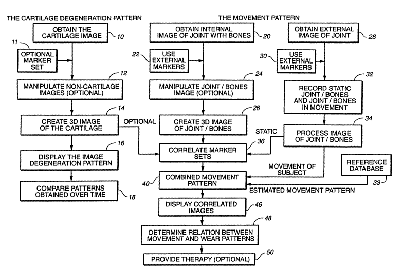
Figure 1 is a schematic overview of some of the various aspects of the
invention.
While a complete description of the many aspects of the invention is found in
the
specification and claims, the schematic overview gives some of the broad
aspects of the
invention.
This invention relates to assessing the condition of a joint in a mammal. One
aspect
is a method for such an assessment. The assessment can be done using internal
images, or
maps, of the cartilage alone or in combination with a movement pattern of the
joint. If used
alone, a map obtained at an initial time is compared with a map obtained at a
later time to
provide a view of the change in cartilage over time. Another aspect is a
method is
comparing the movement pattern for a joint of a subject being studied with the
cartilage
18
CA 02425120 2003-04-11
WO 02/22014 PCT/USO1/28680
degeneration pattern of the subj ect, then determining the relationship
between the
movement pattern and the degeneration pattern. If, in determining the
relationship between
the two patterns, one finds that the movement pattern has caused the
degeneration pattern or
will continue to adversely affect the degeneration pattern, therapy can be
prescribed to
minimize the adverse effects, such as fixrther degeneration or inflammation.
In overview, some of the systems and methods of this invention are illustrated
by the
flow chart in the attached Figure 1. Figure 1 is based on the full range of
processes,
preferably applied to a knee and surrounding cartilage.
In Figure 1, the first step 10 represents obtaining an image of the cartilage
itself.
This is typically achieved using MRI techniques to take an image of the entire
knee and
then, optionally, manipulating (e.g., "subtracting out" or "extracting") the
non-cartilage
images as shown in step 12. Non-cartilage images typically come from bone and
fluid.
Preferably, the MRI is taken using external markers to provide reference
points to the MRI
image (step 11).
If the cartilage is imaged with a 2D MRI acquisition technique, the resulting
stack of
2D images so obtained can be combined into a 3D image, as indicated in step
14. A
preferred alternative is to use 3D MRI acquisition techniques to acquire a 3D
image
directly. In either case, the same "non-cartilage image extraction techniques
referred to in
step 12 can be used.
With a full 3D image captured, various "maps" or displays of the cartilage can
be
constructed to give a cartilage degeneration pattern. This is represented by
step 16. One
such display can, for example, be a color-coding of a displayed image to
reflect the
thickness for the cartilage. This will allow easy visual identification of
actual or potential
defects in the cartilage.
Together with or independently of the cartilage imaging, and as represented by
parallel step 20, a 3D image of the knee joint is taken, again preferably
using MRI. Many of
the same techniques as applied in steps 10 to 14 are used to do this. However,
as illustrated
by sub-step 22, it is useful to define and register a skin-external frame of
reference around
the joint. This is achieved by placing fiduciary markers on the skin around
the outside of
the knee (step 22) prior to taking the image.
In addition to an image extraction technique (as described above in step 12),
an
image is manipulated to enhance the image of the position of the markers (step
24). The
resulting manipulated image is used to give a 3D image of the joint and
associated bones
(step 26).
19
CA 02425120 2003-04-11
WO 02/22014 PCT/USO1/28680
With the markers in place, and as shown by step 30, an additional set of
markers is
placed on the skin along the outside of the leg, and an external image of the
limb is
obtained. Using at least two cameras, images are then taken of the subject in
a static state.
In addition, images are also taken of the subject while moving. This is shown
collectively
by step 32. The images obtained are then processed to relate the movement of
the skin
relative to the bone. In addition, certain calculations are performed, for
example, the center
of mass is calculated. These manipulations are shown in Step 34. Further, as
the fiduciary
markers are still in place during the video image capture, a correlation
between the fiduciary
and the additional set of markers can be made. This is shown in step 36.
Once this marker-to-marker correlation is made, the static 3D image of the
joint
(with associated f duciary markers) and the movement images of the leg bones
(also with
fiduciary markers in place) can be combined. The fiduciary markers, therefore,
serve as
baseline references. The combination (step 40) of 3D cartilage image (from
step 14), 3D
knee joint image (step 26), and the moving leg co-ordinates (step 34) will,
after appropriate
corrections, result in a displayable, 3D motion image of the joint moving as
per step 46.
The moving images, showing the contact areas of the knee joint can be used in
conjunction
with the various "maps" or displays generated at step 16 to provide a visual
indication of
potential or actual cartilage defects and help in determining their relation
between
movement and degeneration patterns. This is shown in step 48.
Furthermore, as the various images are supported by actual mathematical
quantification, real measurements (such as cartilage thickness) can be taken
and compared
with later or earlier measurements and/or imaging. This allows the tracking of
the
progression of a defect, or conversely, continued tracking of healthy
cartilage. This aids a
health worker in providing therapy for the patients. The method allows
monitoring and
evaluation of remedial actions as well as possible treatment prescriptions.
Thus, this invention discloses, for example, a method to examine the
relationship
between articular cartilage morphology and the functional load bearing areas
of a knee joint
measured during movement. The method includes enhanced imaging techniques to
reconstruct the volumetric and biochemical parameters of the articular
cartilage in three
dimensions; and a method for in vivo kinematic measurements of the knee. The
kinematic
measurement permits direct ih vivo measurements of complete six-degrees of
freedom
motion of the femur or the tibia or associated bones during normal activities.
This permits
the study of load bearing of articular cartilage during movement. In
particular, this method
can aid in locating cartilage defects relative to the changing load bearing
areas of the knee
CA 02425120 2003-04-11
WO 02/22014 PCT/USO1/28680
joint during daily activities. While the various aspects of the invention are
useful in
mammals generally, they are particularly useful fox human patients.
Obtaining the Cartilage Degeneration Pattern
Ifyaagirag Ar-ticular Cartilage
In general, the joint of a patient is that place of union, more or less
movable,
between two or more bones. A joint comprises cartilage and other elements such
as the
accompanying bones on either side of the joint, fluid, and other anatomical
elements. Joints
are classified into three general morphological types: fibrous, cartilaginous,
and synovial.
This invention is particularly useftil for assessing synovial joints,
particularly the knee.
In obtaining an image of the cartilage of a joint in a mammal, a number of
internal
imaging techniques known in the art are useful for electronically generating a
cartilage
image. These include magnetic resonance imaging (MRI], computed tomography
scanning
(CT, also known as computerized axial tomography or CAT), and ultrasound
imaging
techniques. Others may be apparent to one of skill in the art. MRI techniques
are preferred.
MRI, with its superior soft tissue contrast, is the best technique available
for
assessing tissue and its defects, for example articular cartilage and
cartilage lesions, to
obtain a cartilage degeneration can provide morphologic information about the
area of
damage. Specifically, changes such as fissuring, partial or full thickness
cartilage loss, and
signal changes within residual cartilage can be detected.
The reason MR imaging techniques are particularly suitable for cartilage is
because
they can provide accurate assessment of cartilage thickness, demonstrate
internal cartilage
signal changes, evaluate the subchondral bone for signal abnormalities, and
demonstrate
morphologic changes of the cartilage surface.
MRI provides several important advantages over other techniques in this
invention.
One advantage is good contrast between cartilage, bone, joint fluid,
ligaments, and muscle
in order to facilitate the delineation and segmentation of the data sets.
Another is the
coverage of the entire region of interest in a single scan within acceptable
acquisition times.
For a brief discussion of the basic MRI principles and techniques, see MRI
Basic Principles
and Applications, Second Edition, Mark A. Brown and Richard C. Semelka, Wiley
Liss,
Inc. (1999).
MRI employs pulse sequences that allow for better contrast of different parts
of the
area being imaged. Different pulse sequences are better fitted for
visualization of different
21
CA 02425120 2003-04-11
WO 02/22014 PCT/USO1/28680
anatomic areas, for example, hyaline cartilage or joint fluid. More than one
pulse sequence
can be employed at the same time. A brief discussion of different types of
pulse sequences
is provided below.
High Resolution 3D lIIRI Pulse Sequences
Routine MRI pulse sequences available for imaging tissue, such as cartilage,
include
conventional T1 and T2-weighted spin-echo imaging, gradient recalled echo
(GRE)
imaging, magnetization transfer contrast (MTC) imaging, fast spin-echo (FSE)
imaging,
contrast enhanced imaging, rapid acquisition relaxation enhancement, (RARE)
imaging,
gradient echo acquisition in the steady state, (GRASS), and driven equilibrium
Fourier
transform (DEFT) imaging. As these imaging techniques are well known to one of
skill in
the art, e.g. someone having an advanced degree in imaging technology, each is
discussed
only generally hereinafter. While each technique is useful for obtaining a
cartilage
degeneration pattern, some are better than others.
Conventional TI arzd T2-Weighted Spin-Echo Imaging
Conventional Tl and T2-weighted MRI depicts articular cartilage, and can
demonstrate defects and gross morphologic changes. T1-weighted images show
excellent
intra-substance anatomic detail of hyaline ,cartilage. However, T1-weighted
imaging does
not show significant contrast between joint effusions and the cartilage
surface, making
surface irregularities difficult to detect. T2-weighted imaging demonstrates
joint effusions
and thus surface cartilage abnormalities, but since some components of
cartilage have
relatively short T2 relaxation times, these are not as well depicted as other
preferred
imaging.
Gradient-Recalled Eeho Imaging
Gradient-recalled echo imaging has 3D capability and ability to provide high
resolution images with relatively short scan times. Fat suppressed 3D spoiled
gradient echo
(FS-3D-SPGR) imaging has been shown to be more sensitive than standard MR
imaging for
the detection of hyaline cartilage defects in the knee.
22
CA 02425120 2003-04-11
WO 02/22014 PCT/USO1/28680
Magnetization Transfer Cofztrast Izzzagifzg
Cartilage, as well as other ordered tissues, demonstrate the effects of
magnetization
transfer. Magnetization transfer imaging can be used to separate articular
cartilage from
S adjacent joint fluid and inflamed synovium.
Fast Spin-Echo Izzzagizzg
Fast spin-echo imaging is another useful pulse sequence to evaluate articular
cartilage. Incidental magnetization transfer contrast contributes to the
signal characteristics
of articular cartilage on fast spin-echo images and can enhance the contrast
between
cartilage and joint fluid. Sensitivity and specificity of fast spin-echo
imaging have been
reported to be 87% and 94% in a study with arthroscopic correlation.
Contrast Eyzhanced Imaging
The use of gadolinium for imaging of axticular cartilage has been applied in
several
different forms. Direct magnetic resonance (MR) arthrography, wherein a dilute
solution
containing gadolinium is injected directly into the joint, improves contrast
between cartilage
and the arthrographic fluid. Indirect MR arthrography, with a less invasive
intravenous
injection, can also been applied. Gadolinium enhanced imaging has the
potential to monitor
glycosaminoglycan content within the cartilage, which may have implications
for
longitudinal evaluations of injured cartilage.
Driven Equilibrium Fourier Transform
Another 3D imaging method that has been developed is based on the driven
equilibrium fourier transform (DEFT) pulse sequence (U.S. Patent No.
5,671,741), and is
specifically designed for cartilage imaging. DEFT provides an effective
tradeoff between
T2/T1 weighting and spin density contrast that delineates the structures of
interest in the
knee. Contrast-to-noise ratio between cartilage and joint fluid is greater
with DEFT than
with spoiled gradient echo (SPGR). DEFT is an alternative approach to SPGR.
DEFT
contrast is very well suited to imaging articular cartilage. Synovial fluid is
high in signal
intensity, and articular cartilage intermediate in signal intensity. Bone is
dark, and lipids axe
23
CA 02425120 2003-04-11
WO 02/22014 PCT/USO1/28680
suppressed using a fat saturation pulse. Hence, cartilage is easily
distinguished from all of
the adjacent tissues based on signal intensity alone, which will greatly aid
segmentation and
subsequent volume calculations.
The basic DEFT pulse sequence is shown in Fig. 2. A conventional spin echo
pulse
sequence was followed by an additional refocusing pulse to form another echo,
and then a
reversed, negated, excitation pulse to return any residual magnetization to
the +z axis. This
preserved the magnetization of longer T2 species, such as synovial fluid.
Typical MRI
parameters for cartilage are a T1-relaxation time of 900 Milliseconds (ms) and
a T2-
relaxation time of 40 ms, while synovial fluid has a Tl-relaxation time of
3000 ms and a
T2-relaxation time of 200 ms. In addition, synovial fluid has a 30% greater
proton density
than cartilage. The signal levels of cartilage and synovial fluid were plotted
in Fig. 3 for a
RARE pulse sequence and for DEFT, and show that DEFT maintains excellent
contrast for
any relaxation time (TR). It achieves this contrast while maintaining a signal-
to-noise ratio
(SNR) efficiency (SNR/(Taequisiti°n)) that is equal to or better than
other methods with much
lower contrast, such as T1-weighted GRASS.
DEFT was compared with a fast spin-echo (FSE), a gradient-echo (GRE), and a
spoiled gradient-echo (SPGR) sequence with parameters similar to the ones
published by
Disler et al. The patella was scanned in I O normal volunteer knees using a
1.5T whole-body
system (GE Signa) with a 3 inch surface coil. All images were acquired with
field of view
(FOV) IOxlO cm, matrix 256x256 elements, slice thickness 4 mm using fat-
saturation.
DEFT (400/15 [TR/TE in msec], 2 NEX (number of excitations), FSE (3500/15,
echo train
length [ETL] 8, 2 NEX (number of excitations), FSE (3500/15, ETL 4, 2 NEX),
GRE
(400/20, 30 , 2 NEX), and SPGR (50115, 30 [flip angle], 2 NEX) images were
obtained.
Contrast-to-noise ratios (CNR) between cartilage and joint fluid were
calculated as:
CNR = ~ (SIJoint Fluid - SICartilage) / SIl3ackground Noise I [Eq. 1 ]
Contrast (C) between cartilage and joint fluid was calculated as:
C = I [(SIJoint Fluid - SICartilage) / Sl7oint Fluid ] x 100 ~ [Eq. 2]
In the equations SI is signal intensity. DEFT demonstrated greater contrast-to-
noise
ratio and contrast between cartilage and joint fluid than SPGR, GRE, and FSE
sequences
(Figs. 4 & 5). Cartilage had intermediate signal intensity with DEFT, while
joint fluid was
high in signal intensity. The difference in CNR between DEFT and SPGR was
statistically
significant (p<0.001). Cartilage morphology, i.e. cartilage layers, were
consistently best
24
CA 02425120 2003-04-11
WO 02/22014 PCT/USO1/28680
delineated with the DEFT sequence. At the resolution used in this study, FSE
sequences
suffered from image blurring. Blurring was improved with ETL 4 when compared
to ETLB;
nonetheless, even with ETL 4, cartilage morphology seen on FSE images was
inferior to the
DEFT sequence. In light of these results, DEFT imaging is a preferred MRI
technique.
Another Application of DEFT
DEFT was combined with a partial k-space echo-planar data acquisition. This
pulse
sequence is illustrated in Fig. 6 above. A slab selective pulse in z defines
the imaging
volume, which is then resolved with phase-encoding gradients in the y and z
axes, and an
oscillating EPI gradient in the x axis.
Example images acquired with this approach are shown in Fig. 7. This case was
optimized for resolution, in order to image the patellar cartilage. The EPI
readout acquired 5
echoes for each DEFT sequence. Partial k-space acquisition collected only 60%
of the data
along the x-axis. Correction for the missing data was performed using a
homodyne
reconstruction. The image matrix was 192x192x32, with a resolution of
O.Sx0.5x2.5 mm,
resulting in a 1Ox10x8 cm FOV. The echo time TE was 22 ms, and the TR was 400
ms. Fat
was suppressed with a fat presaturation pulse. The total scan time for this
acquisition was 5
minutes.
Additional image studies that can be performed using this approach may require
greater spatial coverage, but one can permit slightly less spatial resolution,
and a longer
scan time similar to the one used with the 3D SPGR approach. If one relaxes
the resolution
to 0.75x0.75x1.5 mm, and doubles the z slab thickness and z phase encodes, the
result will
be a FOV of 15x15x16 cm, and a total scan time of approximately 15 minutes,
which
exactly fits the desired scan protocol. Similar to the 3D SPGR acquisition,
one can acquire a
first 3D DEFT scan in the sagittal plane with fat saturation. The 3D DEFT
acquisition can
then be repeated without fat saturation using the identical parameters and
slice coordinates
used during the previous acquisition with fat saturation. The resultant non-
fat-saturated 3D
DEFT images can be used for 3D rendering of the femoral and tibial bone
contours.
In summary, Driven Equilibrium Fourier Transform is a pulse sequence preferred
for cartilage imaging that provides higher contrast-to-noise ratios and
contrast between
cartilage and joint fluid than SPGR, GRE, and FSE sequences. Cartilage
morphology is
better delineated with DEFT sequences than with SPGR, GRE, and FSE images. The
combination of high anatomic detail and high cartilage joint fluid CNR and
contrast may
CA 02425120 2003-04-11
WO 02/22014 PCT/USO1/28680
render this sequence particularly useful for longitudinal studies of cartilage
in patients with
osteoarthritis.
A Representative Example of MR Imaging is described below:
A MR image can be performed using a whole body magnet operating at a field
strength of 1.5 T (GE Signa , for example, equipped with the GE SR-120 high
speed
gradients [2.2 Gauss/cm in 184~,sec risetimes]). Prior to MR imaging, external
markers
filled with Gd-DTPA (MagnevistC~, Berlex Inc., Wayne, N.J.) doped water (T1
relaxation
time approximately 1.0 sec) can be applied to the skin around the knee joint
and optionally
at the same positions used for gait analysis in a biomotion laboratory
(discussed below).
The external markers can be included in the field of view of all imaging
studies. Patients
can be placed in the scanner in supine position. After an axial scout
sequence, coronal and
sagittal T1-weighted images of the femur can be acquired using the body coil
(spin-echo,
TR=SOOmsec, TE=l5msec, 1 excitation (NEX), matrix 256x128 elements, field of
view
(FOV) 48 cm, slice thickness 7 mm, interstice spacing 1 mm). The scanner table
can then be
moved to obtain coronal and sagittal images of the knee joint and tibia using
the same
sequence parameters. These T1-weighted scans can be employed to identify axes
through
the femur and tibia which can be used Later for defining the geometry of the
knee joint. The
knee can then be placed in the knee coil with the joint space located in the
center of the coil.
The knee can be secured in the coil with padding. Additionally, the foot and
ankle region
can be secured in neutral position to the scanner table using adhesive tape in
order to
minimize motion artifacts. A rapid scout scan can be acquired in the axial
plane using a
gradient echo sequence (GRASS, 2D Fourier Transform (2DFT), TR=SOmsec,
TE=lOmsec, flip angle 40°, 1 excitation (NEX), matrix 256x128 elements,
field of view
(FOV) 24 cm, slice thickness 7 mm, interstice spacing 3 mm). This scout scan
can be used
to demonstrate the position of the knee joint space in the coil and to
prescribe all subsequent
high resolution imaging sequences centered over the joint space. Additionally,
using the
graphic, image based sequence prescription mode provided with the scanner
software, the
scout scan can help to ensure that all external markers around the knee joint
are included in
the field of view of the high resolution cartilage sensitive MR sequences.
There are several issues to consider in obtaining a good image. One issue is
good
contrast between cartilage, bone, joint fluid, ligaments, and muscle in order
to facilitate the
delineation and segmentation of the data sets. Another is the coverage of both
condyles of
26
CA 02425120 2003-04-11
WO 02/22014 PCT/USO1/28680
the knee in a single scan within acceptable acquisition times. In addition, if
there are
external markers, these must be visualized. One way to address these issues is
to use a
three-dimensional spoiled gradient-echo sequence in the sagittal plane with
the following
parameters (SPGR, 3DFT, fat-saturated, TR=60msec, TE=Smsec, flip angle
40°, 1
excitation (NEX), matrix 256x160 elements, rectangular FOV 16x12 cm, slice
thickness 1.3
mm, 128 slices, acquisition time approximately 15 min). Using these
parameters, one can
obtain complete coverage across the knee joint and the external markers both
in
mediolateral and anteroposterior direction while achieving good spatial
resolution and
contrast-to-noise ratios between cartilage, bone and joint fluid (Figs. 8 and
9). The fat-
saturated 3D SPGR sequences can be used for rendering the cartilage in three
dimensions
(see description below). The 3D SPGR sequence can then be repeated in the
sagittal plane
without fat saturation using the identical parameters and slice coordinates
used during the
previous acquisition with fat saturation. The resultant non-fat-saturated 3D
SPGR images
demonstrate good contrast between low signal intensity cortical bone and high
signal
intensity bone marrow thereby facilitating 3D rendering of the femoral and
tibial bone
contours. It is to be understood that this approach is representative only and
should not be
viewed as limiting in any way.
Volumes of Interest (h01)
The invention allows a health practitioner to determine cartilage loss in a
reproducible fashion and thus follow the progression of a cartilage defect
over time.
In one embodiment of the invention, one can use a 2D or a 3D surface detection
technique to extract the surface of the joint, e.g. the femoral condyles, on
both baseline and
follow-up scans. For example, a T1-weighted spin-echo sequence can be used for
surfaces
extraction of the femoral condyles. The T1-weighted spin-echo sequence
provides high
contrast between low signal intensity cortical bone and high signal intensity
fatty marrow.
For detection of the surface of the femoral condyles, a step-by-step problem
solving
procedure, i.e., an algorithm, can convolve a data set with a 3D kernel to
locate the
maximum gradient location. The maximum gradient location corresponds to the
zero
crossing of a spatial location. When the kernel is designed properly, then
there will be only
one zero crossing in the mask. Thus, that zero crossing is the surface. This
operation is
preferably three-dimensional rather than two-dimensional. The surface of the
joint, e.g. the
femoral condyles, on the baseline scan can be registered in an object
coordinate system A.
27
CA 02425120 2003-04-11
WO 02/22014 PCT/USO1/28680
The surface of the joint, e.g. the femoral condyles, on the follow-up scan can
be registered
in an object coordinate system B. Once these surfaces have been defined, a
transformation
B to B' can be performed that best matches B' with A. Such transformations
can, for
example, be performed using a Levenberg Marquardt technique. Alternatively,
the
transformations and matching can be applied to the cartilage only. The same
transfornlation
can be applied to the cartilage sensitive images on the follow-up scan in
order to match the
cartilage surfaces.
Using the 3D surface registration of the joint on the baseline scan and
resultant
object coordinate system A, one can place volumes of interest over the area of
a cartilage
defect seen on the cartilage sensitive images. For example, in the knee joint,
the. size of the
targeted volumes of interest can be selected to exceed that of the cartilage
defect in
anteroposterior and rnediolateral direction, e.g. by 0.5 to 1 cm. If the
defect is located high
on the femoral condyle or in the trochlear region, the targeted VOI can be
chosen so that its
size exceeds that of the cartilage defect in superoinferior and mediolateral
direction. The
third dimension of the targeted VOI (parallel to the surface normal of the
cartilage) can be
fixed, for example at 1 cm. VOI size and placement can be manual or automatic
on the
baseline study. Once the targeted VOI has been placed on the image using
visual or
automated computer control, the 3D coordinates of the targeted VOI relative to
the 3D
contour of the joint and object coordinate system A can be registered and
saved. On follow-
up studies, e.g. scans inadvertently obtained with slightly different patient
position, the 3D
surface of the joint is registered to match the orientation of the baseline
scan and the
targeted VOI is then automatically placed on the joint using object coordinate
system B'
and the coordinates saved on the baseline study. Cartilage volume within the
targeted VOI
on baseline and follow-up studies can, for example, be determined using
standard
thresholding and seed growing techniques.
Reference markers
When obtaining the MR images for use in this invention, whether the MRI is of
cartilage or of bone, external reference markers can be placed on the skin
around the joint of
the subject being imaged. The external marker can be designed not only to show
up in the
MRI, but also to show up if an external image of the joint is obtained. The
importance and
value of such unique reference markers will be discussed in more detail
hereinafter.
28
CA 02425120 2003-04-11
WO 02/22014 PCT/USO1/28680
Thus, one embodiment of the invention is a skin reference marker that can be
used
in the assessment of the condition of a joint of a human. Multiple skin
reference markers
can be placed upon one or more limbs of a patient prior to internal imaging
and external
imaging. Each skin reference marker comprises a material detectable by an
imaging
technique, a container for the material in which the container preferably has
multiple
surfaces, a means for affixing the container to the skin (e.g. an adhesive
placed on at least
one surface of the container in an amount sufficient to adhere the container
to the skin of a
human), and a reflective material (preferably retro-reflective) placed on
another surface of
the container located away from the adhesive. Several imaging techniques can
be used that
are able to detect the marker. For example, magnetic resonance imaging is
preferred, but,
ultrasound, or X-ray are also useful. Tn the case of X-ray, further
manipulations.must be
performed in which multiple X-ray images are assimilated by a computer into a
2
dimensional cross-sectional image called a Computed Tomography (CT) Scan. The
material detectable by an imaging can be either in a liquid form or a solid
form. The
material can be any imaging contrast agent or solution, e.g. a paramagnetic
material. The
material can be a lanthanide, such as one belonging to the yttrium group of
rare earth
metals. More specifically, the material can be gadolinium. The shape of the
container can
be any shape allowing it to be placed on the skin of a human. For example, it
can be
cubical, spherical, elliptical, discoid or cylindrical. The size of the
container can be any
size, but optimally a size allowing it to be recorded by an imaging machine.
The longest
dimension of the container can be up to 5.0 cm, but preferably is about 0.25
to 2.0 crn. The
reflective or retro-reflective material can be any material that is able to
reflect light directly
back to the source of the light so that the position of the reference marker
is captured by the
opto-electrical recording means, e.g. a video camera. 3M Corporation makes
several retro
reflective materials.
Manipulating Images
Once a magnetic resonance image is obtained, it can be manipulated to improve
the
image by reducing unwanted, non-cartilage images.
Segyraentatiora
29
CA 02425120 2003-04-11
WO 02/22014 PCT/USO1/28680
To prepare the data set for 3D rendering, the cartilage can be segmented image
by
image using a signal-intensity-based threshold combined with a seed growing
technique.
The femoral, tibial, and patellar cartilage can be segmented separately based
on the fat-
saturated 3D SPGR or 3D DEFT sequence. Manual disarticulation can be performed
by
outlining the cartilage contour in areas where the signal intensity of the
articular cartilage is
similar to that of adjacent structures. The contours of the femoral, tibial,
and patellar bone
can be segmented separately using the non-fat-saturated 3D SPGR or 3D DEFT
sequence.
Segmentation software can allow for manual editing of cartilage thickness maps
and
cartilage defects detected using the above embodiments. In this fashion, the
operator can
correct erroneous detection of cartilage defects in areas where the cartilage
may be naturally
thinner. Such software includes seed-growing algorithms and active-contour
algorithms
that are run on standard PC's. A sharp interface is present between the high
signal intensity
bone marrow and the low signal intensity cortical bone thereby facilitating
seed growing.
Fat-saturated and non-fat-saturated 3D sequences can be acquired with the same
field of
view, slice thickness and slice positions, thereby enabling superimposition
and cross
registration of any resultant 3D renderings of the femoral, tibial, and
patellar cartilage and
bone. External reference markers can aid in registering the 3D data in the
same object
coordinate system.
3D maps of cartilage thickness can be generated using several different
techniques.
One representative, but not limiting, approach uses a 3D surface detection
technique which
is based on a 2D edge detector (Wang-Binford) that has been extended to 3D.
This surface
detection technique can generate surface points and their corresponding
surface normal. To
smooth the contour, the program samples 25 percent of the surface points and
fits a cubic
spline to the sample points. The program can compute the curvature along
sample spline
points and find two sample points that have the maximum curvature and are
separated by
about half the number of voxels on the contour. These points partition the
spline into two
subcontours. For each subcontour, the program can compute the average distance
between
the points and the center of the mass. The program can designate the
subcontour with the
smaller average distance as the inner cartilage surface and the other
subcontour as the outer
cartilage surface (OCS). The intersect between the inner cartilage surface
(ICS) (located at
the subchondral bone interface) and the outer cartilage surface with the
surface normal can
be used to compute the 3D thickness of the articular cartilage on a pixel-by-
pixel basis.
CA 02425120 2003-04-11
WO 02/22014 PCT/USO1/28680
Creating A Three Dimensional (3D) Image of the Cartilage
Three Dimensional Geometric Model Geraeration
After the 3D image of cartilage and the 3D image of joint with bones (as
discussed
hereinafter), are obtained, for example, the set of segmented two dimensional
MR images
can be transformed to a voxel representation using a computer program
developed in the
AVS Express (Advanced Visual Systems, Inc., Waltham, MA). Every voxel has a
value of
zero if it is not within an object of interest or a value ranging from one to
4095, depending
on the signal intensity as recorded by the MRI machine. An isosurface can then
be
calculated that corresponds to the boundary elements of the volume of
interest. A
tasselation of this isosurface can be calculated, along with the outward
pointing normal of
each polygon of the tesselation. These polygons are written to a file in a
standard graphics
format (Virtual Reality Modeling Language Version 1.0: VRML output language).
Pisualization Software
One possible choice for the software program used to assess the cartilage
degeneration pattern, the bones of the joint, and the motion pattern of the
patient is a user
controllable 3D visual analysis tool. The program can read in a scene, which
scene consists
of the various 3D geometric representations or "actors" (for example, VRML
files of the
tibia, tibia cartilage, femur, femoral cartilage), the static relationship
transformations
between these actors, and, if available, sequence of transformations
describing how these
actors move with respect to each other as the patient performs some activity,
such as
walking, jogging, etc.
The program can allow the user, through the use of the mouse and/or keyboard,
the
ability to observe the scene from arbitrary angles; to start and stop the
animation derived
from the motion profiles and to observe the contact line and any cartilage
lesions while the
animation is running. Additionally, the user can derive quantitative
information on the
scene through selecting points with the mouse.
The software program can be written in the CTT computer language and can be
compiled to run on both Silicon Graphics Workstations and Windows/Intel
personal
computers.
Cartilage tlaickhess snaps
31
CA 02425120 2003-04-11
WO 02/22014 PCT/USO1/28680
Cartilage thickness can be determined by several methods. One example is
detecting the locations of the bone - cartilage and the cartilage- joint fluid
interface along
the surface normal using the same edge detector described below, and
subtracting them.
This procedure can be repeated for each pixel located along the bone -
cartilage interface.
The x, y, and z position of each pixel located along the bone - cartilage
interface can be
registered on a 3D map or multiple 2D maps and thickness values are translated
into color
values. In this fashion, the anatomic location of each pixel at the bone
cartilage interface
can be displayed simultaneously with the thickness of the cartilage in this
location.
The edge detector can produce accurate surface points and their corresponding
surface normal. The detector can be applied to the baseline and the follow-up
data set. For
the baseline data set, both the surface points and surface normals can be used
to form locally
supporting planes (for each voxel). These planes can form an approximated
surface for the
baseline skeletal site. As for the follow-up data set, the surface points can
be matched in the
registration procedure onto the surface of the baseline data set. One can use
a newly
developed 3D surface detection technique to extract the surface of the
skeletal site on both
the baseline scan and the follow-up scan. Once these surfaces are detected,
one can use the
Levenberg Marquardt procedure to find the transformation that best matches
these two
surfaces.
A possible approach for calculating the cartilage thickness is based on a 3D
Euclidian distance transformation (EDT). After thresholding, the voxels on the
edge of the
cartilage structure can be extracted using a slice by slice 8-neighbor search,
resulting in a
binary volume with the voxels on the cartilage surface having a value of 1 and
all others
being 0. To classify these surface points as part of the ICS or OCS, a semi-
automatic
approach, which requires the user to enter a point that lies outside the
cartilage structure and
faces the ICS, can be useful. From this point, rays are cast in all directions
of the volume
using a modified Bresenham's line drawing algorithm. If a ray hits a voxel
with a value of
1, this point is classified as part of the ICS. After a complete sweep of the
volume, for
initialization of the EDT the ICS voxels are given a value of 0, whereas alI
other voxels are
set to 1.
For computation of the EDT, the following representative algorithm can be
useful.
It can decompose the calculation into a series of 3 one-dimensional
transformations and can
use the square of the actual distances, which accelerates the process by
avoiding the
determination of square roots.
32
CA 02425120 2003-04-11
WO 02/22014 PCT/USO1/28680
First, for a binary input picture F = {f~k~ (1 < i < L, 1 <- j 5 M, 1 5 k < I~
a new
picture G={g;~k~ can be derived using equations (3-5) (a, (3, and y denote the
voxel
dimensions). Here F is a set of all voxels initially and G is a set of all
voxels at the later
time.
g~k = min{(a (i - x))2 ~ J xjk = 0~1 < x < L~ [Eq. 3]
Thus, each point can be assigned the square of the distance to the closest
feature
point in the same row in i-direction. Second, G can be converted into H={h;~k)
using
equation (4).
h~k = min{g;Yk + ( ~ (.7 - Y)) z ~l ~ Y ~ M~ [Eq~ 4]
Y
The algorithm can search each colunm in the j-direction. According to the
Pythagorean theorem, the sum of the square distance between a point (i,j,k)
and a point
(i,y,k) in the same column, ((3(j - y))a, and the square distance between
(i,y,k) and a
particular feature point, g;yk, equals the square distance between the point
(ij,k) and that
feature point. The minimum of these sums is the square distance between
(i,j,k) and the
closest feature point in the two-dimensional i j-plane.
The third dimension can be added by equation (5), which is the same
transformation
as described in the equation for the k-direction (4).
s~k = min{h~Z + (y (k - z))2;1 ~ z ~ N~ [Eq. 5]
After completion of the EDT, the thickness of the cartilage for a given point
(a,b,c)
on the OCS equals the square root of sabc~ The x, y, and z position of each
pixel located
along the bone - cartilage interface can be registered on a 3D map and
thickness values are
translated into color values. In this fashion, the anatomic location of each
pixel at the bone
cartilage interface can be displayed simultaneous with the thickness of the
cartilage in this
location.
Displaying the Degeneration Pattern
33
CA 02425120 2003-04-11
WO 02/22014 PCT/USO1/28680
In an approach the cartilage thickness maps obtained using the algorithm
described
above display only a visual assessment of cartilage thickness along the
articular surface. In
another approach, in order to derive a true quantitative assessment of the
location, size, and
depth of a focal cartilage defect, one can use an iterative approach comparing
cartilage
thickness of neighboring pixels located along the bone cartilage interface.
For example, assuming an image resolution of 0.5 x 0.5 x 1.0 mm and an average
thickness of the articular cartilage in the femoral condyles ranging between 2
to 3 mm, a
25% decrement in cartilage thickness will be the smallest change that can be
observed with
most content imaging sequences. Therefore, fox example, pixels along the bone -
cartilage
interface that demonstrate a decrease exceeding the smallest change observable
on a given
MRI pulse sequence, in this example 25% or greater, in overlying cartilage
thickness when
compared to cartilage thickness at the neighboring bone - cartilage interface
pixels, can be
used to define the margins of a focal cartilage defect. Other criteria can be
employed to
define a cartilage defect based on comparisons of neighboring pixels. For
example, a fixed
value can be used. If the difference in cartilage thickness between
neighboring pixels
exceeds the fixed value, e.g. 1 mm, the pixel where this difference is
observed can be used
to define the margin of the cartilage defect. This comparison can be performed
for each
pixel located along the bone - cartilage interface for the entire data set.
This comparison is
preferably performed in three dimensions. Pixels that demonstrate a decrease
in cartilage
thickness exceeding defined criteria but that are completely surrounded by
other pixels
fulfilling the same criteria may not be considered to be part of the margin of
the cartilage
defect, but will typically be considered to lie inside the cartilage defect.
The invention provides for means for calculating the area covered by the
cartilage
defect Aca~lage defect ~d the mean thickness of the cartilage in the region of
the defect
D~a~ilage aefe°t as well as the mean thickness of a defined area of
surrounding normal cartilage.
The thickness of the cartilage previously lost in the defect can be estimated
then as:
Dcartilage loss - Dnormal cartilage - Dcartilage defect [Eq.6]
Since the area A of the cartilage defect is known, the volume of cartilage
loss can be
computed as:
Vcartilage loss - Acartilage defect X D cartilage loss
34
CA 02425120 2003-04-11
WO 02/22014 PCT/USO1/28680
Turning now to Figures 22A and 22B, one can see a 2D MRI (3D SPGR) and 3D
cartilage thickness map. In A, the 2D MRI demonstrates a full thickness
cartilage defect in
the posterior lateral femorl condyle (arrows). Figure 22B shows a 3D cartilage
thickness
S map generated using a 3D Euclidian distance transformation. The thickness of
the articular
cartilage is color encoded and displayed on a pixel-by-pixel basis along the
3D surface of
the articular cartilage. The cartilage defect is black reflecting a thickness
of zero (arrows)
(M: medial, L: lateral, S: superior, I: inferior).
In Figures 23A - 23E, one can see the matching of 3D thickness maps generated
from MR images obtained with the knee in neutral position and in external
rotation. A.
Sagittal baseline MR image (3D SPGR) with the knee in neutral position. B.
Sagittal
follow-up MR image of the same volunteer obtained two weeks later with the
knee in 40
degree external rotation (note the artificially widened appearance of the
femur resulting
from the rotation). C. 3D thickness map generated based on baseline MRI in
neutral
1S position. D. 3D thickness map generated based on follow-up MRI in external
rotation (note
segmentation error between condyles in trochlear region). E. Transformation of
D into the
object coordinate system of C. Despite extreme differences in joint
orientation between
baseline and follow-up MRI scans and despite segmentation errors, the
thickness
distribution on the matched follow-up scan demonstrates great similarity with
that seen on
the baseline scan in neutral position (in C.).
Having now described how to obtain an image of a cartilage of a joint, both
with and
without external reference markers; how to enhance the image by manipulating
non-
cartilage images, and creating and displaying 3-D images of the cartilage, i.
e. a 3-D map,
certain aspects of the invention are apparent.
2S One aspect is a method of estimating the loss of cartilage in a joint. The
method
comprises
(a) obtaining a three-dimensional map of the cartilage at an initial time and
calculating the thickness or regional volume of a region thought to contain
degenerated
cartilage so mapped at the initial time,
(b) obtaining a three-dimensional map of the cartilage at a later time, and
calculating the thickness or regional volume of the region thought to contain
degenerated
cartilage so mapped at the later time, and
(c) determining the loss in thickness or regional volume of the cartilage
between
the later and initial times.
3S
CA 02425120 2003-04-11
WO 02/22014 PCT/USO1/28680
Preferably, this aspect of the invention is directed to a volume of interest
in the
cartilage, i.e., a region of the cartilage that includes a cartilage defect.
Such a defect may be
the result of a disease of the cartilage (e.g., osteoarthritis) or the result
of degeneration due
to overuse or age. This invention allows a health practitioner to evaluate and
treat such
defects. The volume of interest may include only the region of cartilage that
has the defect,
but preferably will also include contiguous parts of the cartilage surrounding
the cartilage
defect.
Another aspect of the invention is a method for assessing the condition of
cartilage
in a joint of a human, which method comprises
(a) electronically transferring an electronically-generated image of a
cartilage of
the joint from a transfernng device to a receiving device located distant from
the transferring device;
(b) receiving the transferred image at the distant location;
(c) converting the transferred image to a degeneration pattern of the
cartilage;
and
(d) transmitting the degeneration pattern to a site for analysis.
Another aspect of the invention is a method for determining the volume of
cartilage
loss in a region of a cartilage defect of a cartilage in joint of a mammal.
The method
comprises (a) determining the thickness, DN, of the normal cartilage near the
cartilage
defect; (b) obtaining the thickness of the cartilage defect, DD, of the
region; (c) subtracting
DD from DN to give the thickness of the cartilage loss, DL; and (d)
multiplying the DL
value times the area of the cartilage defect, AD, to give the volume of
cartilage loss. The
method is useful for situations wherein the region of cartilage defect is
limited to the
defective cartilage and preferably wherein the region of the cartilage defect
includes a
portion of the cartilage contiguous to the defect.
Alternatively, for step (a) the normal thickness of the defect area could be
estimated.
It may be estimated from measurements of cartilage of other subjects having
similar
characteristics such as gender, age, body type, height, weight, and other
factors. It may be
estimated from measurements of a similar 'normal" cartilage from another
corresponding
joint (e.g., if the right knee has the defect, measure the normal left knee).
It may have been
measured at an initial time Tl when the cartilage was normal to provide a
baseline. Other
means of determining the normal thickness may be available to one of skill in
the art. Once
the thickness DN is obtained and the thickness DD is obtained the two are
subtracted to give
the DL. The DL is multiplied by the area of the defect AD to give the volume
of cartilage
36
CA 02425120 2003-04-11
WO 02/22014 PCT/USO1/28680
loss. By determinng the volume of cartilage loss at an initial Tl and again at
a later time
T2, one can determine the change in volume loss over time.
Still another aspect of the invention is a method of estimating the change of
a region
of cartilage in a joint of a mammal over time. The method comprises (a)
estimating the
thickness or width or area or volume of a region of cartilage at an initial
time T1, (b)
estimating the thickness or width or area or volume of the region of cartilage
at a later time
Tz, and (c) determining the change in the thickness or width or area or volume
of the region
of cartilage between the initial and the later times. The method is
particularly useful for
regions of degenerated cartilage or diseased cartilage.
Still another aspect of the invention is a method of estimating the loss of
cartilage in
a joint. The method comprises (a) defining a 3D object coordinate system of
the joint at an
initial time, Tl; (b) identifying a region of a cartilage defect within the 3D
object coordinate
system; (c) defining a volume of interest around the region of the cartilage
defect whereby
the volume of interest is larger than the region of cartilage defect, but does
not encompass
the entire articular cartilage; (d) defining the 3D object coordinate system
of the joint at a
second timepoint, T2; (e) placing the identically-sized volume of interest
into the 3D object
coordinate system at timepoint T2 using the object coordinates of the volume
of interest at
timepoint Tl; (f) and measuring any differences in cartilage volume within the
volume of
interest between timepoints Tl and T2.
Therapeutic Plahnihg, Devising New Therapies
In another embodiment of the invention, thickness of the articular cartilage
can be
estimated using an imaging test. This imaging test can be an x-ray,
ultrasound, CT scan or
MRI scan. Thickness can be determined using a 3D Euclidian distance
transformation as
well as other techniques feasible for this purpose. Thickness can be
determined in selected
regions or across the entire cartilage of a joint. Thickness can be determined
in areas
adjacent to diseased cartilage, in areas of diseased cartilage and in areas of
normal cartilage.
Furthermore, the curvature of the cartilage can be determined. For this
purpose, the
curvature of the inner cartilage surface, i.e. the surface facing the
subchondral bone, or the
outer cartilage surface, i.e. the surface facing the joint cavity, can be
determined. Preferably,
the inner cartilage surface at the interface with the subchondral bone will be
used, since the
outer cartilage surface may be subject to fraying, fissuring or more advanced
stages of
cartilage loss. Alternatively, the curvature of the subchondral bone can be
determined. In
37
CA 02425120 2003-04-11
WO 02/22014 PCT/USO1/28680
this case, the curvature of the subchondral bone can serve as an approximation
of the
curvature of the articular cartilage. Curvature can be determined from an
imaging test,
typically an ultrasound, a CT or an MRI scan. Curvature can be determined from
a three-
dimensional model of the cartilage. The three-dimensional model of the
cartilage can be
determined using the 3D Euclidian distance transformation mentioned above.
Curvature can
be determined in selected regions or across the entire cartilage of a joint.
Curvature can be
determined in areas adjacent to diseased cartilage, in areas of diseased
cartilage and in areas
of normal cartilage.
Using information on thickness and curvature of the cartilage, a physical
model of
the surfaces of the articular cartilage and of the underlying bone can be
created. This
physical model can be representative of a limited area within the joint or it
can encompass
the entire joint. For example, in the knee joint, the physical model can
encompass only the
medial or lateral femoral condyle, both femoral condyles and the notch region,
the medial
tibial plateau, the lateral tibial plateau, the entire tibial plateau, the
medial patella, the lateral
patella, the entire patella or the entire joint.
In another embodiment, the location of a diseased area of cartilage can be
determined, for example using a 3D coordinate system or a 3D Euclidian
distance
transformation in combination with some of the techniques outlined above. In
another
embodiment of the invention, the anteroposterior, mediolateral or
superoinferior dimension
of an area or multiple areas of diseased cartilage can be determined.
Furthermore, the area,
depth and volume of a cartilage defect can be determined, for example using a
3D Euclidian
distance transformation in combination with some of the techniques outlined
above
In one embodiment of the invention, information on thickness of the cartilage,
information on curvature of the cartilage, information on curvature of the
subchondral bone
or information on the location, dimensions, area, depth and volume of a defect
or
combinations thereof can be used to devise a treatment. For example, the
dimensions of a
defect determined in this manner can be used to determine the dimensions of a
cartilage
transplant, a cartilage graft, a cartilage implant, a cartilage replacement
material, a cartilage
scaffold or a cartilage regenerating material or any cartilage repair system.
Additionally, the
curvature of the inner cartilage surface or the subchondral bone can be
measured and this
information can be used to determine the shape of a cartilage transplant, a
cartilage graft, a
cartilage implant, a cartilage replacement material, a cartilage scaffold or a
cartilage
regenerating material or any cartilage repair system. Additionally, the
thickness of normal
cartilage adjacent to the defect can be measured and the thickness values
measured in this
3~
CA 02425120 2003-04-11
WO 02/22014 PCT/USO1/28680
fashion can be used to determine the optimal thickness for a cartilage
transplant, a cartilage
graft, a cartilage implant, a cartilage replacement material, a cartilage
scaffold or a cartilage
regenerating material or any cartilage repair system. Alternatively, the
thickness of the
cartilage can be measured in the contralateral joint, e.g. the knee joint, in
an area
corresponding to the area of diseased cartilage in the affected joint. Using
any of these
techniques or, preferably, a combination thereof, an optimal fit can be
achieved between the
surrounding normal cartilage and a cartilage transplant, a cartilage graft, a
cartilage implant,
a cartilage replacement material, a cartilage scaffold or a cartilage
regenerating material or
any cartilage repair system thereby minimizing incongruity at the joint
surface and
improving the therapeutic result.
The invention provides for means to create a cast or a mold for shaping a
cartilage
transplant, a cartilage graft, a cartilage implant, a cartilage replacement
material, a cartilage
scaffold or a cartilage regenerating material or any cartilage repair system.
This can be
generated using computer devices and automation, e.g. computer assisted design
(CAD)
and, for example, computer assisted modeling (CAM).
In another embodiment, the invention provides for means to measure and
visualize
the curvature of the surfaces of cartilage and bone. In another embodiment,
the invention
provides for means to compare the thickness and the curvatures of surfaces of
a cartilage
transplant and a transplantation site, a cartilage graft and a graft site, a
cartilage implant and
an implantation site, a cartilage replacement material and an implantation
site, a cartilage
scaffold and a cartilage defect, a cartilage regenerating material and an area
of diseased
cartilage, or a cartilage repair system and an area of diseased cartilage.
The invention is useful for determining the shape, dimensions and thickness of
a
cartilage transplant, a cartilage graft, a cartilage implant, a cartilage
replacement material, a
cartilage scaffold or a cartilage regenerating material or any cartilage
repair system prior to
treatment. For example, the shape, dimensions and thickness of a cartilage
transplant, a
cartilage graft, a cartilage implant, a cartilage replacement material, a
cartilage scaffold or a
cartilage regenerating material or any cartilage repair system can be designed
to follow the
physical shape and thickness of the cartilage adjacent to an area of diseased
cartilage
determined using the imaging test.
The invention is applicable to a host of current and future treatments of
arthritis
including but not limited to cartilage transplants, cartilage implants,
cartilage grafts,
cartilage replacement materials, cartilage scaffolds, cartilage regenerating
materials, auto-,
allo- and xeno-transplants, osteochondral alto- and autografting, stem cell
based repair
39
CA 02425120 2003-04-11
WO 02/22014 PCT/USO1/28680
systems and transfer systems, and, principally, any other current and future
treatments and
devices for cartilage repair or regeneration.
The example described below shows one possibility how aspects of the invention
can be implemented. It demonstrates one possible way how the invention can be
practiced.
It is in no way meant to limit the invention. One skilled in the art will
easily recognize other
means of practicing the invention.
In a first step, the cartilage and bone contours can be segmented from the MR
images using for example a modified live wire technique [Steines D, Cheng C,
Wong A,
Berger F, Napel S, Lang P. CARS - Computer-Assisted Radiology and Surgery, p.
578-
583, San Francisco, 2000].
From the segmented data, a 3-dimensional surface representation can be
created,
which yields a triangular tesselation of the surface. For this calculation, a
3-dimensional
Wang-Binford edge detector [Yan CH: Measuring changes in local volumetric bone
density: new approaches to quantitative computed tomography, Ph.D. thesis,
1998, Dept. of
Electrical Engineering, Stanford University] or the marching cubes algorithm
[Lorensen
WE, Cline HE. Comput Graph 1987; 21: 163-169] can be used. This surface
representation
can be imported into a CAD system, which is used to generate a physical model
or a cast by
means of rapid prototyping techniques.
The segmented data can also be used for measuring the surface curvature at the
surface points. The curvature is calculated according to formula xx. If
a:(a,b) -~ ll2 is a
curve defined over the parameter interval (a,b) by a(t) _ (x(t),y(t)), then
the curvature K is
given by [Gray A: Modern Differential Geometry of Curves and Surfaces. 1993;
CRC
Press]
x(t) - x~(t) y"(t) - x~,(t) y~(t) [Eq. 8]
(x,2 (t) + y.2 (t))/
For the digitized contours that result from the segmentation, smooth
derivatives for
equation (8) can be obtained by convolution with differentiated Gaussian
kernels [Wowing
M, Smeulders AWM. CVGIP: Image Understanding, 1993. 58(3): p. 366-382].
The curvature values can be calculated for each pixel on the segmented surface
in
each slice. They can subsequently be color-mapped onto a 3-dimensional
rendering of the
surface for visualization purposes.
For full curvature information this procedure can be repeated for a direction
perpendicular to the imaging plane. Of the remaining two main directions the
one with the
CA 02425120 2003-04-11
WO 02/22014 PCT/USO1/28680
lower degree of parallelism to the surface can be chosen, and reformatted
slices of the MR
data set can be obtained for this direction. For instance, when curvature
values for the
femoral condyles are calculated from sagittal MR images of the knee, the data
set would
typically be reformatted for a coronal slicing plane.
The procedure of segmentation and curvature calculation can then be repeated
for
the reformatted images. A second curvature map can be calculated, yielding
complementary
information to the first one.
In order to compare curvature values for different surface patches, these can
be
manually or automatically registered, overlaying one surface patch on top of
the other.
Corresponding curvature values that are calculated in the same directions can
now be
subtracted from each other, thereby yielding a measure of how well curvatures
of two
surfaces match.
An example how aspects of the invention can be practiced clinically in a
patient is
given below. It demonstrates one possible way how the invention can be
practiced. It is in
no way meant to limit the invention. One skilled in the art will easily
recognize other means
of practicing the invention.
A patient with arthritis of the knee joint is referred for an MRI scan. The
MRI scan
is performed using a cartilage sensitive MRI pulse sequence such as a fat
saturated spoiled
gradient echo sequence or a water selective 3D gradient echo sequence using a
spectral
spatial pulse. The MR images are transferred via a local network or, for
example, the
Internet into a computer workstation. The computer workstation uses software
to extract or
segment the articular cartilage from the surrounding tissue. Such software can
include snake
algorithms, livewire algorithms, signal intensity based thresholding, or seed
growing
algorithms as well as any other technique useful for this purpose. The
software can then
generate a three-dimensional map of cartilage thickness across the femoral
condyles, the
tibial plateau, or the patella. This can be achieved using a 3D Euclidian
distance
transformation. Additionally, the software can provide information on
cartilage curvature or
curvature of the subchondral bone as described above. Furthermore, the
software can
determine the location, dimensions, size, area, depth, or volume of areas of
diseased
cartilage.
The information generated in this fashion can be used to generate a physical
model
of the area of diseased cartilage. For example, a bone replacement material
can be formed
with a CAD/CAM system using the above information. One of the surfaces of the
bone
replacement material can be shaped so that it matches the 3D curvature of the
subchondral
41
CA 02425120 2003-04-11
WO 02/22014 PCT/USO1/28680
bone subjacent to the area of diseased cartilage. Additionally, the
anteroposterior,
mediolateral or superoinferior dimensions of this surface of the bone
replacement material
can be such that it matches the dimensions of the area of diseased cartilage.
Cartilage cells
can be affixed to the bone replacement material and can be grown on the bone
replacement
material until the thickness of the resultant cartilage matches that of the
thickness of the
cartilage adjacent to the area of diseased cartilage measured on the 3D
cartilage thickness
map. Alternatively, a layer of cartilage of known thickness can be applied to
the bone
replacement material whereby the thickness can be chosen to match the
thickness of the
cartilage adj acent to the area of diseased cartilage measured on the 3D
cartilage thickness
map.
Alternatively, an artificial non-human material with properties similar to
cartilage
can be applied to the bone replacement material whereby the thickness of this
material can
be chosen to match the thickness of the cartilage adjacent to the area of
diseased cartilage
measured on the 3D cartilage thickness map.
Alternatively, cartilage can be grown on a mold matching the curvature of the
subchondral bone in an area of diseased cartilage whereby the dimensions of
the surface of
the mold on which the cartilage is grown matches the dimensions of an area of
diseased
cartilage. Cartilage can then be grown on the mold until its thickness matches
the thickness
of cartilage adjacent to the area of diseased cartilage as measured, for
example, on a 3D
cartilage thickness rnap. At this point, for example, an orthopedic surgeon
can excise the
area of diseased cartilage and can implant the cartilage or cartilage
replacement material.
Since the curvature of the cartilage or cartilage replacement material matches
that of the
underlying subchondral bone and since the thickness of the cartilage or
cartilage
replacement material matches that of the cartilage adjacent to the area of
excised diseased
cartilage, normal or near normal joint congruity can be achieved with a
resultant decrease in
wear on the implanted cartilage or cartilage replacement material and also a
decrease in
wear on the adjacent cartilage or the cartilage of the opposing joint surface.
Display of Biochemical Information
In addition to providing a 2D or 3D representation of the morphological
properties
of cartilage, the invention provides for techniques to represent one or more
biochemical
components of articular cartilage.
42
CA 02425120 2003-04-11
WO 02/22014 PCT/USO1/28680
A biochemical component includes, but is not limited to, glycosaminoglycan,
water,
sodium, or hyaluronic acid. Biochemical data can be generated with other
magnetic
resonance based techniques including the use of signal intensity measurements,
relaxation
time measurements, paramagnetic and other contrast media and sodium rather
than proton
MR imaging. Other imaging tests such as positron emission tomography scanning
can also
be used for this purpose. Thus, one aspect of this invention is a method for
providing a
biochemically-based map of joint cartilage. The method comprises
(a) measuring a detectable biochemical component throughout the cartilage,
(b) determining the relative amounts of the biochemical component throughout
the cartilage;
(c) mapping the amounts of the biochemical component through the cartilage;
and
(d) determining the areas of cartilage deficit by identifying the areas having
an
altered amount of the biochemical component present.
Once a map is obtained, it can be used in assessing the condition of a
cartilage at an
initial time and over a time period. Thus, the biochemical map may be used in
the method
aspects of the invention in a manner similar to the cartilage thickness map.
For example, one aspect is a method of estimating the loss of cartilage in a
joint.
The method comprises
(a) obtaining a relaxation time or biochemical map of the cartilage at an
initial
time and analyzing the relaxation time or biochemical content of a region
thought to contain
degenerated cartilage so mapped at the initial time,
(b) obtaining a relaxation time or biochemical map of the cartilage at a later
time, and time analyzing the relaxation time or biochemical content of the
region thought to
contain degenerated cartilage so mapped at the later time, and
(c) determining the change in relaxation time or biochemical content of the
cartilage between the later and initial times.
Preferably, this aspect of the invention is directed to a volume of interest
in the
cartilage, i.e., a region of the cartilage that includes a cartilage defect.
Such a defect may be
the result of a disease of the cartilage (e.g., osteoarthritis) or the result
of degeneration due
to overuse or age. This invention allows a health practitioner to evaluate and
treat such
defects. The volume of interest may include only the region of cartilage that
has the defect,
43
CA 02425120 2003-04-11
WO 02/22014 PCT/USO1/28680
but preferably will also include contiguous parts of the cartilage surrounding
the cartilage
defect.
As discussed herein before, another aspect of the invention is a method for
assessing
the condition of cartilage in a joint using a relaxation time or the
biochemical map. The
method comprises
(a) electronically transferring an electronically-generated relaxation time or
biochemically based image of a cartilage of the joint from a transfernng
device to a receiving device located distant from the transfernng device;
(b) receiving the transferred image at the distant location;
(c) converting the transferred image to a degeneration pattern of the
cartilage;
and
(d) transmitting the degeneration pattern to a site for analysis.
Another aspect of the invention is a method for determining the change of
biochemical content in a region of a cartilage defect of a cartilage in joint
of a mammal.
The method comprises (a) determining the biochemical content (BCrr) of the
normal
cartilage near the cartilage defect; (b) obtaining the biochemical content of
the cartilage
defect (BCD) of the region; and (c) subtracting BCD from BCrr to give the
value of the
cartilage change, BCD. The method is useful for situations wherein the region
of cartilage
defect is limited to the defective cartilage and preferably wherein the region
of the cartilage
defect includes a portion of the cartilage contiguous to the defect.
Alternatively, for step (a) the normal content of the defect area could be
estimated.
It may be estimated from measurements of cartilage of other subjects having
similar
characteristics such as gender, age, body type, height, weight, and other
factors. It may be
estimated from measurements of a similar 'normal" cartilage from another
corresponding
joint (e.g., if the right knee has the defect, measure the normal left knee).
It may have been
measured at an initial time Tl when the cartilage was normal to provide a
baseline. Other
means of determining the normal content may be available to one of skill in
the art. Once
BCrr is obtained and BCD is obtained the two are subtracted to give the ~. By
determining
the change of content at an initial Tl and again at a later time T2, one can
determine the
change in biochemical content over time.
Once a relaxation time or biochemically-based map is provided, morphological
maps of articular cartilage obtained with MR imaging can be superimposed,
merged or
fused with the biochemical map or data. Several different techniques can be
applied in order
to superimpose, merge, or fuse morphological data with biochemical data. For
example, 2D
44
CA 02425120 2003-04-11
WO 02/22014 PCT/USO1/28680
or 3D morphological data of articular cartilage can be acquired with the same
object
coordinates as the biochemical data. Morphological data and biochemical data
can then be
easily displayed simultaneously using different colors, opacities, and or gray
scales.
Alternatively, 2D or 3D morphological data or articular cartilage can be
acquired with
different object coordinates as the biochemical data. In this case, a 3D
surface registration
can be applied in order to superimpose, merge, or fuse the morphological data
and the
biochemical data. As an alternative to 3D object coordinates, anatomic
landmarks can be
used to register the morphological data and subsequently the biochemical data
in a 3D
obj ect coordinate system. 3D obj ect coordinate systems can then be matched
by matching
the landmarks obtained from the morphological data with those obtained from
the
biochemical data.
Thus, another aspect of this invention is a method for assessing the condition
of a
subject's cartilage in a joint, the method comprises obtaining a three
dimensional
biochemical representation of the cartilage, obtaining a morphological
representation of the
cartilage, and merging the two representations, and simultaneously displaying
the merged
representations on a medium. The merged representations are then used to
assess the
condition of a cartilage, estimate the loss of cartilage in a joint,
determining the volume of
cartilage loss in a region of cartilage defect, or estimating the change of a
region of cartilage
at a particular point in time or over a period of time. One can see that
similar steps would
be~followed as spelled out for the use of a thickness map or biochemical map.
Simultaneous display of morphological data with biochemical data provides a
useful
tool to assess longitudinal changes in morphology or articular cartilage and
biochemical
composition of articular cartilage, for example during treatment with
chondroprotective and
chondroregenerative agents.
Part of the unique aspect of this technology is that it lends itself to
assessment of a
patient from a distant position after an image is taken of the joint under
evaluation. Thus
one aspect of this invention is a method for assessing the condition of
cartilage in a joint
from a distant location. The method comprises
(a) electronically transferring an electronically-generated image of a
cartilage of
the joint from a transfernng device to a receiving device located distant from
the
transferring device;
(b) receiving the transferred image at the distant location;
(c) converting the transferred image to a degeneration pattern of the
cartilage;
and
CA 02425120 2003-04-11
WO 02/22014 PCT/USO1/28680
(d) transmitting the degeneration pattern to a site for analysis.
The degeneration pattern includes a measure of cartilage thickness or regional
cartilage
volume.
The electronically generated image of the cartilage preferably is an MR image
and
the degeneration pattern can be displayed as a three-dimensional image as a
thickness
pattern, a biochemical content pattern or a merged thickness biochemical
pattern. The
electronically generated image is transmitted via Dicom, using the
international standards
for transmission of such images.
Another aspect of the invention is a kit for aiding in assessing the condition
of
cartilage in a joint of a mammal, which kit comprises a software program,
which that when
installed and executed on a computer reads a cartilage degeneration pattern
presented in a
standard graphics format and produces a computer readout showing a cartilage
thickness
map of the degenerated cartilage.
The software can be installed in a PC, a Silicon Graphics, Inc. (SGI) computer
or a
Macintosh computer. Preferably, the software calculates the thickness or
regional volume
of a region of degeneration of the cartilage which does not include the entire
volume of the
articular cartilage.
THE MOVEMENT PATTERN
To acquire a movement pattern of a joint in accordance with this invention,
one
obtains an internal image of the bones in a joint, preferably using MRI
techniques, and
obtains an external image of the bones in motion. The images are correlated,
preferably
through the use of external marker sets, to give a pattern that shows a static
or moving
condition. The correlated images are then displayed and the relation between
the movement
and degeneration patterns is determined.
46
CA 02425120 2003-04-11
WO 02/22014 PCT/USO1/28680
Obtaining An Internal Image of Joint with Bones
To obtain an internal image of a joint with the associated bones, one
preferably uses
MRI techniques that provide an image of the bones on either side of the joint.
Here, it is
important to use the imaging technique that gives the best image of the bones
and how they
interact. Because the internal image of the bones can be combined with the
image of the
bones obtained by external measurements, it is particularly useful, and
therefore preferred,
to use external reference markers that can be similarly-positioned to the
markers used in
obtaining the external measurements. The external markers can be placed at any
landmarks
about the joint of interest. At least three markers are used for each limb
being imaged.
Preferably the markers will be made of a material that not only will be
detected by MRI
imaging techniques, but also will be detected by external imaging techniques.
The markers
will be associated with a means to affix them to the skin and preferably have
an adhesive
portion for adhering to the skin and a detectable entity that will show up on
the MRI image.
The preferred MRI imaging technique useful for obtaining an internal image is
a
spoiled 3D gradient echo, a water selective 3D gradient echo or a 3D DEFT
sequence. A
further discussion may be found hereinbefore or in the 2"d Edition of Brown
and Semelka's
book entitled "MRI Basic Principles and Applications."
Once an MK image is obtained the image is manipulated to enhance the image of
the
bones. Procedures similar to those discussed hereinbefore for cartilage may be
used, but
modified for application to bone images.
Creating Three-Dimensional (3D) Image of Joint/Sones
Three-Dimensiofzal Geometric Model Gefze~ation
After the 3D image of a joint with bones, the set of segmented two dimensional
MR
images can be transformed to a voxel representation inside AVS Express
(Advanced Visual
Systems, Inc., Waltham, MA). Every voxel has a value of zero if it is not
within an object
of interest or a value ranging from one to 4095, depending on the signal
intensity as
recorded by the 1.5 T MR. An isosurface can then be calculated that
corresponds to the
boundary elements of the region of interest. A tesselation of this isosurface
can be
calculated, along with the outward pointing normal of each polygon of the
tesselation.
47
CA 02425120 2003-04-11
WO 02/22014 PCT/USO1/28680
These polygons can then be written to a f 1e in a standard graphics format
(Virtual Reality
Modeling Language Version 1.0).
As discussed hereinbefore, the use of reference markers on the skin around the
joint
and the bones can provide an image that can later be matched to the reference
markers for
the cartilage image and the bone images obtained from external measurements.
Alternatively, a semi-automated, 3D surface-based registration technique that
does
not require the use of an external frame or fiducial markers can be used. This
3D surface
based registration technique can be used to match the anatomic orientation of
a skeletal
structure on a baseline and a follow-up CT or MRI scan. We extended a robust
and accurate
2D edge detector (Wang-Binford) to 3D. This detector is described
hereinbefore.
A registration technique for the femoral condyles and the tibial plateau is
shown in
Figure 10. It shows an example where 3D surfaces of the femoral condyles were
extracted
from two differently oriented T1-weighted spin-echo MRI scans (baseline A and
follow-up
B, respectively) obtained in the same patient in neutral position (A) and in
40 degree
extenial rotation (B). The 3D surfaces were used to derive a coordinate
transformation
relating the two scans. Fig. lOC demonstrates the use of the derived
transformation to re-
register scan B in the object coordinate system of scan A. Such a
transformation relating
two T1-weighted scans can then be used to register DEFT cartilage-sensitive
scans that are
acquired in the same respective orientations as the A and B T1-weighted scans.
We performed the registration using a Sun Sparc 20 workstation with 128MBytes
of
memory. The surface detection algorithm extracted approximately 12,000 surface
patches
from each data set. The surface extraction and registration routines took
about 1 hour in
total.
Since the algorithm for 3D surface registration of the femoral condyles also
computes the surface nonnals for the medial and lateral femoral condyles on a
pixel-by-
pixel basis, it can form the basis for developing maps of cartilage thickness.
Fig. 11 shows
an example of a 2D map of cartilage thickness derived from the surface normals
of the
lateral femoral condyle. Figure 11A shows a proton density fast spin-echo MR
image that
demonstrates a focal cartilage defect in the posterior lateral femoral condyle
(black arrows).
White arrows indicate endpoints of thickness map. Figure 11B is a 2D cartilage
thickness
map that demonstrates abrupt decrease in cartilage thickness in the area of
the defect
(arrows). The D thickness between neighboring pixels can be used to define the
borders of
the cartilage defect. Note diffuse cartilage thinning in area enclosed by the
astericks (*).
48
CA 02425120 2003-04-11
WO 02/22014 PCT/USO1/28680
In another embodiment, cartilage sensitive images can be used instead of Tl-
weighted or T2-weighted scans and the surface match can be performed based on
the
cartilage contour.
Alternatively, anatomic landmarks present on both baseline and follow-up scans
can
be used to match the data obtained during the baseline and those obtained
during the follow-
up scan. Another alternative for matching the baseline and the follow-up scan
includes the
use of external or internal fiducial markers that can been detected with MR
imaging. In that
case, a transformation is performed that matches the position of the markers
on the follow-
up scan with the position of the markers on the baseline scan or vice versa.
Obtaining An External Image of JointBones
Before merging or superimposing morphological maps of articular cartilage
obtained
by MR imaging with biomechanical data, one must obtain the biomechanical data.
Such
biomechanical data include, but are not limited to, estimations of static
loading alignment in
standing or weight-bearing position and lying or non-weight-bearing position,
as well as
during joint motion, e.g., the movement of load-bearing pathway on the
cartilage in the knee
joint during gait. Biomechanical data may be generated using theoretical
computations,
based on data stored in a database that can be accessed by calling up and
screening for
certain characteristics. Alternatively, gait analysis may be performed for an
individual and
data obtained during gait analysis may be merged or fused with morphological
MRI data.
Morphological data and biomechanical data can then be easily displayed
simultaneously
using different colors, opacities, and or gray scales. Additionally, the load-
bearing pathway,
for example around a cartilage defect, can be plotted or superimposed onto
morphological
maps.
Preferably, reference markers or fiducial markers can be applied to the
external
surface on the skin overlying the joint. These markers adhere to the skin are
typically made
of materials that can be detected with MRI and that can be used to register
joint motion
during biomechanical analysis, e.g. gait analysis. These markers can then be
used to
correlate the morphological with the biomechanical data.
Simultaneous display of morphological data with biomechanical data provides a
useful tool to assess the load pathway applied to articular cartilage and
inside and around
cartilage defects. Estimation of load pathway applied in and around a
cartilage defect can
be used to assess a cartilage defect and to guide the choice of therapy, e.g.
treatment with
49
CA 02425120 2003-04-11
WO 02/22014 PCT/USO1/28680
chondroprotective or chondroregenerative agents, osteochondral allografting,
cartilage
transplantation, femoral or tibial osteotomy, or joint replacement surgery.
Recording Static JointBones and JointBones in Movement
In obtaining an external image of the bones on either side of a joint, one
must record
a static image as well as a moving image of the subject joint and bones. For
analysis of the
knee joint, gait analysis techniques have been shown to be very effective in
generating
accurate, reproducible data on the six degree of freedom motion of the knee.
The motion of
the knee joint can be quantified in terms of flexion, rotation and
displacement. Fidelity in
the dynamic visualizations of subject specific MR generated knee geometry and
subsequent
contact surface determination call for a high degree of accuracy for the
motion capture
portion of the studies.
Gait Analysis Activities
In performing a gait analysis, a subject is tested standing still, laying
down, walking
or running on a level surface, flexing a leg in a standing position, ascending
and descending
stairs, flexing the leg in a seated position, and the like. The level walking
measurements can
include, but is not limited to, six stride cycles for each side over a range
of walking speeds.
The subject can be instructed to walk at a comfortable speed (normal), slower
than normal
and faster than normal. Typically, this protocol produces gait measurements
over a range of
walking speeds. The standing and laying portions of the protocol can be used
in the cross
registration to the MR data. The instrumentation preferably includes, at least
a two camera,
video-based opto-electronic system for 3-D motion analysis, a multi-component
force plate
for measurement of foot-ground reaction force and a computer system for
acquisition,
processing and analysis of data.
Anatomic Coordinate Systems
Currently, the anatomic coordinate systems are defined through bony landmarks
which can be identified through palpation. To describe the motion of the
underlying bones
in terms of the global coordinate system a subset of the markers in a point
cluster technique
(discussed hereinafter) are referenced to bony landmarks on the femur and
tibia. Techniques
CA 02425120 2003-04-11
WO 02/22014 PCT/USO1/28680
described previously by Hopenfeld and Benedetti can be used to locate these
bony
landmarks. The anatomic coordinate systems used can be similar to that
previously
described by LaFortune with the exception of the origin of the femoral
coordinate system.
For the thigh segment, a coordinate system is located in the femoral condyles.
The femoral
condyles medial(M)-lateral(L) axis (Fig. 12) runs through the traps-
epicondylar line (a line
drawn between the medial-lateral femoral epicondyles). The midpoint of this
axis is the
origin. The inferior(I)-superior(S) axis runs parallel to the long axis of the
femur, passing
through the midpoint of the traps-epicondylar line. The anterior(A)-
posterior(P) axis is the
cross product of the medial-lateral and inferior-superior axes. The final
position of the
inferior-superior axis is made orthogonal to the anterior-posterior and medial-
lateral axis
through a cross product operation (Fig. 13). For the shank segment, the tibial
coordinate
system begins with the medial-lateral axis running through the most medial and
lateral
edges of the plateau. The inferior-superior axis is perpendicular to the
medial-lateral axis
passing through the tibial eminence. The anterior-posterior axis is the cross
product of the
medial-lateral and inferior-superior axes.
Placement of Markers Prior to Aetivity
In assessing a joint, the lower extremity can be idealized as 3 segments with
six
degree-of freedom joints at the knee and ankle. For the mobile activities
described above,
at least 3 markers per segment are used. Figure 14 shows 21 passive retro-
reflective
markers located on the leg: some at bony prominences (greater trochanter,
lateral malleolus,
lateral epicondyle, lateral tibial plateau), some clustered on the thigh and
shank (Fa1-3,11-3,
Fpl-3; Tal-3, Tll-13). Additionally, two markers are placed on the foot at the
lateral aspect
of the calcaneus and base of the fifth metatarsal and one on the pelvis at
theiliac crest).
During the static activities (standing still, Laying down) 7 additional
markers are placed:
medial malleolus, medial epicondyle, medial tibial plateau, medial and lateral
superior
patella, medial and lateral inferior patella. The eight markers nearest to the
knee joint can
be filled with Gadolinium, and can be be replaced at these same locations
prior to the MR
images (Fig. 15). The locations can be marked with a non-toxic marker-pen.
Reference Database
51
CA 02425120 2003-04-11
WO 02/22014 PCT/USO1/28680
The reference database is typically a compendium of demographic and motion
analysis data for all subjects whose data has been processed by a central
processing site.
This database can contain fields describing each of the subject's name, age,
height, weight,
injury types, orthopedic medical history, other anatomic measurements (thigh
length, shank
length, shoe size, etc.), The database can also contain the results of any and
all gait analysis
run on these patients. This can include, for all activities tested (walk, run,
jog, etc.), a
number of peak valves (peak knee flexing, peak hip adduction movement; toe-
out, angle,
etc). along with the motion trajectories of the limb segments while the
subjects are
performing different activities.
In order to obtain a typical motion profile, the sex, age, height, weight,
limb length,
and type of activity desired can be entered as an average into the database.
The database
searches for a set of subjects most closely watching the input average. From
this set of data,
a typical motion pattern is distilled axed a data set is output. This data set
can include, over a
time interval, the motion characteristics: hip / knee / ankle / flexion /
extension angles, knee
/ hip / ankle adduction / abduction angles, movement, stride length, cadence,
etc. This data
can then be used to drive an animation of the motion of the desired joint.
Process Image of JointBones
Calculatio~t ofLimb SegmentParameteYs
Each limb segment (thigh, shank and foot) can idealized as a rigid body with a
local
coordinate system defined to coincide with a set of anatomical axes (the
assumption of
rigidity is dropped in calculating the location of the femur and tibia). The
intersegmental
moments and forces can be calculated from the estimated position of the bones,
the ground
reaction force measurements, and the limb segment mass/inertia properties. The
moment at
the knee can be resolved into a coordinate system fixed in a tibial reference
system with
axes defining flexion-extension, abduction-adduction, and internal-external
rotation.
This approach provides results in a range of patients in a highly reproducible
manner. Typically the magnitudes of the moments are dependent on walking
speed. To
control for the influence of walking speed, the walking speed closest to 1
meter/second is
used. This speed is within the normal range for the type of patients for which
the invention
is particularly useful. In addition to the gait trial collected at 1
meterlsecond, self selected
speeds can also be evaluated to give a good correlation between gait-
quantitative estimates
52
CA 02425120 2003-04-11
WO 02/22014 PCT/USO1/28680
of joint load lines and other measures when using self selected speeds. In
order to test
patients under their typical daily conditions, medications should not be
modified prior to
gait analyses.
Poirat Cluster Technique
The Point Cluster Technique (PCT) movement analysis protocol is an extensible
and
accurate approach to bone motion estimation. Basically, a number of retro-
reflective
markers (e.g. retro-reflective material from 3M, Corp.) are attached to each
limb segment
under observation. Multiple video cameras can acquire data With the subject
standing still
and during activities of interest. An over-abundance of markers on each limb
segment is
used to define a cluster coordinate system, which is tied to an anatomically
relevant
coordinate system calculated with the subject at rest.
The standard PCT transformations are described below. In short, each marker is
assigned a unit mass and the inertia tensor, center of mass, principal axes
and principal
moments of inertia are calculated. By treating the center of mass and
principal axes as a
transformation, local coordinates are calculated. Another set of coordinate
systems is
established; limb segment specific anatomic landmarks are identified through
palpation and
a clinically relevant coordinate system defined. For the femur and tibia,
these anatomic
coordinate systems are shown in Fig. 12. The transformation from the reference
cluster
coordinate system to the anatomic coordinate system is determined with the
subject at rest
by vector operations. During an activity, the transformation from the global
coordinate
system to the cluster coordinate system is calculated at each time step. To
place the
anatomic coordinate in the global system during the activity, the reference
coordinate
system to anatomic system transformation is applied, followed by the inverse
global
coordinate system to cluster coordinate system transformation for each time
step.
In the Point Cluster Technique (PCT) a cluster of N markers can be placed on a
limb
segment of the subject. The location vector of each marker in the laboratory
coordinate
system is denoted as G(i,t) for marker i, (i = 1,2,...,N) at time t, to <_ t
<_ tf. A unit weight
factor is assigned to each marker for the purpose of calculating the center of
mass, inertia
tensor, principal axes and principal moments of inertia of the cluster of
markers. The
cluster center of mass and principal axes form an orthogonal coordinate system
described as
the cluster system. The local coordinates of each of the markers relative to
this coordinate
system are calculated. Then
53
CA 02425120 2003-04-11
WO 02/22014 PCT/USO1/28680
G(i,t) = C(t) + E(t) ~ L(i,t) = T~ (t) ~ L(i,t) i =1 . . . N
where G(t) is a matrix of all marker coordinate vectors, C(t) is the center of
mass of G(t),
E(t) is the matrix of eigenvectors of the inertia tensor of G(t), and L(i,t)
are the local
coordinates of marker i.
These markers are observed by opto-electronic means while the subject performs
activities and while standing completely still in a reference position. With
the subj ect in
this same reference position, a subset of the markers is observed relative to
the underlying
bones by other techniques, which might include x-rays, CT scan, or palpation.
The measured marker locations are defined with respect to the unobservable
location
and orientation of the bone by
G(i,t) = P(t) + O(t) ~ R(i,t) = Tb (t) ~ R(i,t) i =1 ... N
where P(t) is the location and O(t) is the orientation of a coordinate system
embedded in the
bone and R(i,t), also unobservable, are the trajectories of the markers
relative to the
underlying rigid body coordinate system at time t. The bone and cluster
systems are each
orthogonal systems, related by the rigid body transformation Tb~(t):
L(i,t) = Tb~ (t) ~ R(i,t)
substituting and eliminating R(i,t) yields
Tb (t) = T~ (t) ~ T~r (t)
To maintain physical consistency, T~b(t) = Tb~(t)-1 must be the inertia tensor
eigendecomposition transformation of R(i,t). Once R(i,t) are specified, T~b(t)
and
subsequently Tb(t) are calculable.
54
CA 02425120 2003-04-11
WO 02/22014 PCT/USO1/28680
Point Cluster to Anatomic Coordinate System Transformation
From these equations one can also relate the global coordinate system with
respect
to a limb segment system. As an example of how these systems can be used to
describe
joint motion, one can consider the tibio-femoral joint. The motion that is of
interest is how
the femoral condyles move with respect to the tibial plateau. This is done by
first defining a
set of coordinate axes in the femoral condyles and the tibial plateau.
A coordinate system is located in both the femoral condyles and the tibial
plateau.
The femoral condyles medial-lateral (ML) axis runs through the traps-
epicondylar line
(TEL), a line drawn between the ML femoral epicondyles. The midpoint of this
axis is the
origin. The inferior-superior (IS) runs parallel to the long axis of the
femur, passing through
the midpoint of the TEL. The anterior-posterior (AP) is the cross product of
the ML and IS
axes. The tibial coordinate system begins with the ML axis running through the
most
medial and lateral edges of the plateau. The IS axis is perpendicular to the
ML axis passing
through the tibial eminence. The AP axis is the cross product of the ML and IS
axes. These
are known as the anatomic coordinate system (A(t)thigh~ A(t)shank)~
Relating the cluster system to the anatomic coordinate system is done by use
of
another transformation matrix. This is done by relating the thigh cluster to a
cluster of
markers, a sub cluster, that is related to the femoral condyles and femur
(cluster to anatomic
transformation).
R(t)thigh - U(t)thigh A(t)thigh
The tibia has a similar transformation matrix.
R(t)Shank - U(t)shank A(t)shank
Therefore, from a cluster of markers in the global system, motion of the femur
with
respect to the tibia can be determined by:
TS(t) = A(t)thigh ~ CT(t)thigh ~ R(t)shank ~ A(t)shank
Here TS(t) is the motion of the thigh with respect to the shank.
CA 02425120 2003-04-11
WO 02/22014 PCT/USO1/28680
Angles are calculated by a proj ection angle system, an axis from the femoral
anatomic system and one from the tibia are projected onto a plane in the
tibial coordinate
system. For example, flexion/extension can be determined by projecting the IS
axis of the
femur and tibia onto the sagittal plane (AP-IS plane) of the tibia.
Iralidatioh of the Point Cluster Technique
The point cluster technique was evaluated as a method for measuring ira vivo
limb
segment movement from skin placed marker clusters. An Ilizarov device is an
external
fixture where 5 mm diameter pins are placed directly into the bone on either
side of a bony
defect. The rigid external struts affixed to these pins form a rigid system
fixed in the
underlying bone. Two subjects were tested with Ilizarov fixation devices. One
subject had
the Ilizarov device placed on the femur and second subject had the device
placed on the
tibia. Each subj ect was instnunented with point clusters placed on the thigh
and shank
segment. In addition, markers were placed on the Ilizarov device to establish
a system fixed
in the underlying bone.
The relative angular movement and translational displacement between the
system
affixed in the bone and the point cluster coordinate system were calculated
while ascending
a 20-cm step (Step Test). Angular changes between the three orthogonal axes
fixed in the
bone versus three axes in the point cluster were calculated. The average
difference over the
trials for three axes were 0.95 + 1.26, 2.33 +1.63, and 0.58 + 0.58 degrees.
Similarly, the
average error for the distance between coordinate systems was 0.28 + 0.14 cm.
The second
subject with the Ilizarov device placed on the femur could not perform the
Step-Test, but
was able to perform a weight-bearing flexion test where his knee flexed to
approximately
20° from a standing position. The average change between the coordinate
origin was 0.28 +
0.14 cm. The changes in axis orientation were 1.92 + 0.42, 1.11 + .69 and 1.24
~ 0.16
degrees.
The simultaneously acquired motion for a coordinate system embedded in bone
(Ilizarov system) and a set of skin-based markers was compared. At every time
instant the
location and orientation of the Ilizarov system, the rigid body model skin
marker system,
and the interval deformation technique skin marker system were determined. The
change in
the transformation from the Ilizarov system to one of the skin marker systems
over time is a
measure of the deformation unaccounted for in the skin marker system.
56
CA 02425120 2003-04-11
WO 02/22014 PCT/USO1/28680
The interval deformation technique produced a substantial improvement in the
estimate of the location and orientation of the underlying bone. For perfectly
modeled
motion there would be no relative motion between the Ilizarov system and the
skin marker
system over the time interval. The change in the transformation from the
Ilizarov system to
the skin marker systems are shown in Figures 14 and 15, for location and
orientation
respectively, for both a rigid body model and the interval deformation
technique. For this
single data set, the location error was reduced from 7.1 cm to 2.3 cm and the
orientation
error from 107 degrees to 24 degrees, with the error summed over the entire
time interval.
The subject performed a 10 cm step-up; the marker deformation was modeled as a
single
Gaussian function.
Defo~matioh Correction
There are a number of algorithmic alternatives available to minimize the
effects of
skin motion, soft tissue deformation, or muscle activation that deform the
externally applied
markers relative to the underlying bone. The Point Cluster Technique decreases
the effects
of marker movement relative to the underlying bone through averaging. If more
correction
is required, one of a number of deformation correction techniques may be
added. In order
of increasing computational complexity and deformation correction ability,
these are rigid
body linear least square error correction, global optimization correction,
anatomic artifact
correlation correction and interval deformation correction.
An overview of the Interval Deformation Correction Technique is given below.
In
short, the technique provides a maximum likelihood estimate of the bone pose,
assuming
that each marker on a limb segment deforms relative to the underlying bone in
some
functional form. The technique parameterizes these functional forms and then
performs a
multi-objective non-linear optimization with constraints to calculate these
parameters. This
is an extremely computationally intensive technique, with the current
instantiation of the
algorithm requiring 6 - ~ hours per limb segment of running time on 266 MHz
Pentium 2
computer.
57
CA 02425120 2003-04-11
WO 02/22014 PCT/USO1/28680
Interval Deformation Technique
Since Tc can be calculated directly from the global coordinates of the
markers, the
remainder of this development only examines the determination of R(i,t) and
subsequently
T~b(t). For this reduced problem, the input data is the local coordinates in
the cluster system
L(i,t) for all i, To <_ t <_ tf. It can be assumed that each marker has some
parameterized
trajectory, d(a;~, t), relative to the underlying bone at each time step, with
independent and
identically distributed noises v(i~j,t)
I O R~(i,t) = d(a;,~, t) + v(ij,t) j = I .. 3 i =1 .. N
or, equivalently
R(i,t) = F(a;, t) + v(i,t) i = 1 .. N
where a;~ is a vector of parameters for marker i, ordinate j; a; is a vector
of
parameters combining all of the parameters for all of the ordinates of marker
i. Then the
estimate of the data, M(i,t), can be given by
M(i,t) = Tb~ (t) ~ R(i,t)
Without further restrictions the problem is indeterminate, as the locations of
the
markers in the bone system R(i,t) are never observable with the opto-
electronic system. The
indeterminate problem can be converted to a chi-squared estimate problem
through a series
of steps. An observation of the truly unobservables at the time boundaries is
inferred; that
is, it is assumed that Tab (t <_ to) and T~b (t >_ tf) are observed. The value
of T~b can be
selected depending on the activity being studied. For example, consider the
step up activity,
where the subject starts and stops in the reference position. For this
activity the body is not
deforming outside the estimation interval; that is, the markers are not moving
with respect
to the bone:
Tcb (t ~ to) = Tcb (t = to) arid Tcb (t > tf) = Tcb (tf)~
58
CA 02425120 2003-04-11
WO 02/22014 PCT/USO1/28680
It can now be assumed that the noise functions v(i, j, t) are normal
distributions with
individual standard deviations 6(i, j, t), the probability P(i,j,t) of the
data for ordinate j,
marker i, time t being a realization of the stochastic process is given by:
1 L(i, j, t) - M(i, j, t) z
P(i, j, t) oc exp (- 2 ( ~-(i, j, t) ) )
Provided the noise functions v(i, j, t) are independent of each other, the
probability
of the entire data set being a realization is a product of each of the
individual probabilities:
p (-1 (L(Z'j~t)-M(i~j~t))z)
P(i, j, t) ~c ex )
c=r ~=r r=ra 2 6(i, j, t
Maximizing this probability can be equivalent to minimizing the negative of
its
logarithm, yielding the familiar chi-square criteria. As an intermediate step
the following
error matrices can be defined:
X(a,t) 3 X(a,t)1,~ _ ((L(i, j,t)-M(i, j,t)))2 i=1 .. N j =1 .. 3
cs(i, j, t)
rr
X(a)=~ X(a,t)
r=ro
and seek a which in some sense minimizes X(a), a matrix whose elements
represent
the error over the entire time interval for each ordinate of each marker. If
the normal noise
distribution assumption is true, then this minimization results in the maximum
likelihood
estimate of the parameterization, and by inference maximum likelihood estimate
of the
transformation from the bone system to the cluster system. If the normal noise
assumption
is not true, the chi-squared estimate is still appropriate for parameter
estimation; the results
cannot be interpreted as a maximum likelihood estimate, but, for example,
confidence
regions on the estimate or the formal covariance matrix of the fit can be
determined.
Obtaining the parameter set a is a computationally complex operation. The
approach taken was to define a scalar to represent this entire error matrix,
59
CA 02425120 2003-04-11
WO 02/22014 PCT/USO1/28680
N 3
f (a) _ ~ ~ tY(a) i,j
i=1 j=i
and seek a that minimizes f(a).
The limits on marker motion previously discussed can now be converted into
deformation constraints, which allow the formulation of the problem as a
general non-linear
programming problem. The constraints arise from two sources; human limb
segments do
not deform outside a small range, and the locations of the markers are chosen
with specific
properties in mind. For computational purposes, the deformation constraints
are selected to
be:
1. The axes of the cluster system moves by less than 15 degrees relative to
the bone
system.
2. The center of mass of the cluster system moves by less than 3 cm relative
to the
I S bone system.
3. The markers move by less than 4 cm relative to the bone system.
4. Each of the principal moments of inertia of the cluster system change by
less than 25
percent from the reference values.
The Point Cluster Technique marker set was designed to ensure that the cluster
of
points is non-coplanar and possess no axes of rotational symmetry. These
properties ensure
a local coordinate system that is well defined and unambiguous over the entire
time interval.
The constraints are then:
5. The ratio of the smallest principal moment of inertia of the cluster system
to the
largest is more than 5 percent; the magnitude of the smallest principal moment
of inertia of
the cluster system is greater than some small positive value.
6. The principal moments of each axis are different from each other by at
least 5
percent.
The general problem can then be formulated:
Minimize f(a)
aERD
Subject to:
CA 02425120 2003-04-11
WO 02/22014 PCT/USO1/28680
g;(a) = 0 i = 1 .. me
g;(a)<0 i=me+l..m
ai_< a < a°
where D is the total number of parameters; me, the number of equality
constraints, is
0; and m, the total number of constraints, is 10.
The approach taken to verify the operation of the algorithm implementation
began
with generating a set of 50 synthetic data sets with known characteristics.
The program was
then applied to all of the data sets. The program results were then compared
to the known,
generated deformation. Error results were calculated for both the interval
deformation
technique descried herein and for the standard rigid body model formulation.
The 50 trial data sets were processed through the algorithm. The results over
all of
the trial sets are summarized in Table I, where the center of mass and
direction cosine error
of the interval deformation technique and the rigid body model are compared.
After
processing by the interval deformation algorithm the center of mass error has
been reduced
to 29% and the direction cosine error has been reduced to 19% of the rigid
body model
error. In a t-test for paired samples, both of these decreases were
significant at p < 0.001.
Tlalidatioh of the hate~val Defor~matioya Co~~~ection Technique
A subject fitted with an Ilizarov external fixation was observed with the
optoelectronic system. The Foint Cluster Marker set was affixed to the
subject's shank (6
markers), along with a set of four markers rigidly attached to the Ilizarov
device, which is
rigidly connected to the tibia with bone pins. These four markers define a
true bone
embedded coordinate system. Data were acquired by GaitLink software
(Computerized
Functional Testing Corporation) controlling four Qualisys cameras operating at
a video
frequency of 120 Hz. Three dimensional coordinates were calculated using the
modified
direct linear transform.
The subject was a 46 year old male (height 1.75 m, weight 84.1 kg) ftted with
a
tibial Ilizarov external fixation device. The device was rigidly attached to
the tibia with
nine bone pins, located in three sets (top, middle, and bottom) of three
(medial, anterior, and
lateral). The clinical purpose of the device was tibial lengthening; the test
on the subj ect
was performed two days prior to final removal of the device. The subject
exhibited a limited
range of motion and was tested performing a 10 cm step-up onto a platform.
61
CA 02425120 2003-04-11
WO 02/22014 PCT/USO1/28680
The simultaneously acquired motion for a coordinate system embedded in bone
(Ilizarov system) and a set of slcin-based markers was compared. At every time
instant the
location and orientation of the Ilizarov system, the rigid body model skin
marker system,
and the interval deformation technique skin marker system was determined. The
change in
the transformation from the Ilizarov system to one of the skin marker systems
over time is a
measure of the deformation unaccounted for in the skin marker system.
The interval deformation technique produced a substantial improvement in the
estimate of the location and orientation of the underlying bone. For perfectly
modeled
motion there would be no relative motion between the Ilizarov system and the
skin marker
system over the time interval. The change in the transformation from the
Ilizarov system to
the skin marker systems are shown in Figs. 14 and 15 for location and
orientation
respectively, for both a rigid body model and the interval deformation
technique. For this
single data set, the location error was reduced from 7.1 cm to 2.3 cm and the
orientation
error from 107 degrees to 24 degrees, with the error summed over the entire
time interval.
The subject performed a 10 cm step-up; the marker deformation was modeled as a
single
Gaussian function.
CORRELATING RESULTS FROM GAIT ANALYSIS AND GEOMETRICAL
REPRESENTATIONS OF THE BONE
In correlating the load pattern obtained from a gait analysis using, e.g. the
PCT, with
the geometrical representation of the bone from the segmented MRI data, one
can be guided
by the general process as described below. The process allows for dynamic
visualization
(i.e. animations) of high-resolution geometrical representations derived from
MRI scans (or
other imaging techniques). The motion of the subj ect specific anatomic
elements is
generally driven by data acquired from the motion (gait) lab. Fidelity of
these animations
requires calculation and application of a sequence of rigid body
transformations, some of
which are directly calculable and some of which are the result of
optimizations (the
correction for skin marker deformation from rigidity does not use the rigid
body
assumption, but generates a correction that is applied as a rigid body
transform).
The process comprises:
a) acquiring data from MRI (or other imaging techniques), and PCT gait
protocols;
b) directly calculating a set of transformations from the data;
c) calculating a set of transformations from optimizations, as needed;
62
CA 02425120 2003-04-11
WO 02/22014 PCT/USO1/28680
d) generating a 3D geometric representation of the anatomic element from the
MR data; and
e) applying the transformations of (b) and (c) to the 3D geometric
representation.
Each of these steps are described in detail below.
Acquiring the Data from MRI (or other imaging techniques) and PCT Gait
Protocols
In the Point Cluster Technique (PCT) protocol, a patient can have a number of
retro-reflective markers attached to each limb segment under observation.
Multiple video
cameras acquire data with the subject standing still and during activities of
interest.
In addition, in order to correspond activities in the gait lab with the MRI
scans, another
reference data set (subject standing still, prescribed posture) can be
acquired using 8
additional markers clustered about the knee. These markers are filled with
gadolinium-
DTPA and covered with a retro-reflective material to allow for correlation
between the MRI
image and the video data.
Directly Calculating a Set of Transformations from the Data
The transformations are described in detail in [Andriacchi TP, Alexander EJ,
Toney
MK, Dyrby CO, Sum J. J Biomech Eng 1998; 120(12): 743-749]. In short, each
marker can
be assigned a unit mass and the inertia tensor, center of mass, principal axes
and principal
moments of inertia can be calculated. By treating the center of mass and
principal axes as a
transformation, local coordinates arcan be a calculated. Another set of
coordinate systems
can also be required for this technique; limb segment specific anatomic
landmarks can be
identified through palpation and a clinically relevant coordinate system can
be defined. The
required transformations axe summarized in Table 1 below.
Calculating a Set of Transformations from Optimizations
There are three required transformations:
Optimization 1. One can calculate the linear least square error rigid body
transformation from the MRI common local coordinate system to the VID common
local
coordinate system.
63
CA 02425120 2003-04-11
WO 02/22014 PCT/USO1/28680
Optimization 2. For each limb segment, one can calculate the linear least
square
rigid body transformation from the MRI Limb segment anatomic coordinate system
to the
video limb segment anatomic coordinate system (obtained from the gait
analysis), using a
subset of common maxkers appropriate for each segment.
Optimization 3. One can calculate a correction for the deviation of the limb
segment
from rigidity during each time step of the activity, using the PCT with either
the mass
redistribution [Andriacchi TP, Alexander EJ, Toney MK, Dyrby CO, Sum J. J
Biomech Eng
199; 120(12): 743-749] or interval deformation algorithms [Alexander EJ,
Andriacchi TP:
Correcting for deformation in skin-based marker systems. Proceedings of the
3rd Annual
Gait and Clinical Movement Analysis Meeting, San Diego, CA, 199].
Generating a 3D Geometric Representation of the Anatomic Element from the MR
data
The MR slices are segmented for the multiple anatomic and fiducial elements.
The
slices are combined to a voxel representation. An isosurface can be calculated
from the
boundary voxel elements. A tessellation of the isosurface can be calculated,
along with the
outward pointing normal for each surface element. This data can then be stored
in a
standard 3D graphic format, the Virtual Reality Modeling Language (VRML).
Applying the Transformation Sequence to the Geometric Representation
The transformation sequence is provided below in Table 1. This transformation
sequence can be applied to each of the anatomic elements over each time step
of the
activity, starting with sequence 6.
64
CA 02425120 2003-04-11
WO 02/22014 PCT/USO1/28680
TABLE 1
SEQ FROM SYSTEM TO SYSTEM XFORM
1 MR Global MR Local ED1
2 MR Local Common Local OPT1
3 Common Local MR Anatomic ANA2
4 MR Anatomic Vff~ Anatomic OPT2
Vm Anatomic Vm Ref ANA3
6 Vm Ref Vm Deformed(t)ED3
7 Vm Deformed(t) Vm Bone(t) OPT3
8 Vm Bone(t) Vm Global(t) ED4
Correlating Marker Sets
5 As pointed out at numerous places in the specification, the use of external
reference
markers that are detectable by both MRI and optical techniques can be an
important and
useful tool in the method of this invention. The use of the reference markers
can form the
basis for an aspect of this invention that is a method for correlating
cartilage image data,
bone image data, and/or opto-electrical image data for the assessment of the
condition of a
joint of a human. This method comprises, obtaining the cartilage image data of
the joint
with a set of skin reference markers placed externally near the joint,
obtaining the bone
image data of the joint with a set of skin reference markers placed externally
near the joint,
obtaining the external bone image data opto-electrical image data of the joint
with a set of
skin reference markers placed externally near the joint. Using the skin
reference markers,
one can then correlate the cartilage image, bone image and opto-electrical
image with each
other, due to the fact that each skin reference marker is detectable in the
cartilage, bone and
CA 02425120 2003-04-11
WO 02/22014 PCT/USO1/28680
opto-electrical data. The cartilage image data and the bone image data can be
obtained by
magnetic resonance imaging, positron emission tomography, single photon
emission
computed tomography, ultrasound, computed tomography or X-ray. Typically, MRI
will be
preferred. In the case of X-ray, further manipulations must be performed in
which multiple
X-ray images are assimilated by a computer into a 2 dimensional cross-
sectional image
called a Computed Tomography (CT) Scan. The opto-electrical image data can be
obtained
by any means, for example, a video camera or a movie camera. Multiple skin
reference
markers can be placed on one or more limbs of the patient prior to imaging.
The skin
reference markers are described hereinbefore.
By a sequence of calculations a set of transformations that will take the
subject
specific geometric representation of anatomic elements determined from the MR
image set
to the optical reference coordinate system. From the optical reference
coordinate system,
the standard Point Cluster Technique transformation sequence is applied to
generate
dynamic visualizations of these anatomic elements during activities previously
recorded in
the motion lab. Fidelity of these dynamic visualizations (and subsequent
contact surface
determination) requires the calculation and application of a sequence of rigid
body
transformations. Some of these are directly calculable and some are the result
of
optimizations (the correction for skin marker deformation from rigidity does
not use the
rigid body assumption, but generates a correction that is applied as a rigid
body transform).
The first required transformation can be from the MR global coordinate system
to
the MR center of mass / principal axis coordinate system. This can be done by
calculating
the center of mass of each of the individual markers, resulting in a set of
eight three
dimensional points. Each of these points can be assigned a unit mass, and the
center of
mass, inertia tensor, and principal axes can be calculated. The same procedure
can be
performed on these markers as determined by the optical system, providing a
transformation
from the optical global system to a center of mass / principal axis system.
If the relative orientation of the tibia and femur as determined by the MR
system and
the optical system are identical, it is only necessary to apply the optical
reference system to
the anatomic system transformation of the MR local data. If this is not the
case, an
optimization calculation can be performed to determine the rotation and
translation of, for
example, the femur with respect to the tibia. One then can calculate the
linear least square
rigid body transformation from the MR limb segment anatomic coordinate system
to the
video limb segment anatomic coordinate system prior to applying the Point
Cluster
Transformations.
66
CA 02425120 2003-04-11
WO 02/22014 PCT/USO1/28680
For visualization or contact surface determination, one can examine the
relative
motion of one segment to the other, for example the motion of the femur
relative to a fixed
tibial frame. This can be accomplished by applying the global to tibial
anatomic system
transform to all of the elements. An example of this type of visualization is
given in
Figure 18. The Figure shows what can be referred to as functional joint
imaging. Figure
18A is a photograph demonstrating the position of the external markers
positioned around
the knee joint. The markers are filled with dilute Gd-solution. B is Sagittal
3D SPGR
image through the medial femorotibial compartment. Two of the external markers
are seen
anteriorly as rounded structures with high signal intensity. C is 3D
reconstruction of
femoral and tibial bones (light grey), external markers (dark grey), femoral
cartilage (red),
and tibial cartilage (blue) based on the original SPGR MR images. D-I show a
functional
joint imaging sequence at selected phases of leg extension from a seated
position, D-F,
anterior projection. The vectors represent the relative location and
orientation of the femur
with respect to the tibia. G-I is a lateral projection. These dynamic
visualizations can be
used to demonstrate tibiofemoral contact areas during various phases if gait
or other
physical activities.
Superimposition of cartilage thickness map onto subject specific anatomic
model atzd
determination of distance of cartilage defect from load bearing line
Superimposing the cartilage thickness maps onto the subject specific geometric
models can follow the same approach taken to bring the MR generated geometries
into the
optical reference system. Since the thickness maps and the geometric models
are initially in
the same coordinate system; one possible approach is to perform a simple
surface mapping
of the thickness map onto the geometric model. Another alternative approach is
to convert
the thickness map directly into a geometric representation (Fig. 19).
Once the thickness map is embedded in the femoral geometry, one can define a
scalar metric that characterizes the location of any cartilage lesions
relative to the point of
contact line. One approach is a simple 3D distance along the surface from the
center of the
cartilage lesion to the point of closest approach of the contact line. Another
metric that
could be useful would be to multiply the area of the lesion by the adduction
moment at that
time instant, then divide by the distance from lesion center to point of
closest approach. This
67
CA 02425120 2003-04-11
WO 02/22014 PCT/USO1/28680
could result in a metric that increases with lesion area, adduction moment,
and closeness of
approach.
46 Display Correlated Images
Determination ofAnatomic arid Natural Reference Lines
There are two alternative approaches one can consider for determining a
reference
line on the cartilage surfaces. One skilled in the art will easily recognize
other approaches
that can be suitable for this purpose. The first approach is based on anatomic
planes; the
second is a natural approach building on the three dimensional cartilage
thickness map.
The location of the pathway of loading relative to the femoral and tibial
anatomy
and geometry can be assessed by defining sagittal planes bisecting the medial
femoral
condyle, the lateral femoral condyle, the medial tibial plateau, and the
lateral tibial plateau.
For the medial femoral condyle, the operator can manually delete surface
points located
along the trochlea. Then, a sagittal plane parallel to the sagittal midfemoral
plane can be
defined through the most medial aspect of the medial femoral condyle followed
by a sagittal
plane parallel to the sagittal midfemoral plane through the most lateral
aspect of the medial
femoral condyle. The sagittal plane that is located halfway between these two
planes can be
defined as the "midcondylar sagittal plane". The intersection between the
midcondylar
sagittal plane and the external cartilage surface yields the "anatomic
midcondylar cartilage
line". The location of the pathway of loading can be assessed relative to the
anatomic
midcondylar cartilage line of the medial femoral condyle. The identical
procedure can be
repeated for the lateral femoral condyle.
The following method can be used for the medial tibial plateau: A plane
paxallel to
the sagittal tibial plateau plane can be defined through the most medial point
of the medial
tibial plateau. A parallel plane located halfway between this plane and the
sagittal tibial
plateau plane can yield the "midsagittal plane of the medial tibial plateau."
The intersection
of the midsagittal plane of the medial tibial plateau and the external
cartilage surface can
yield the "anatomic midtibial plateau cartilage line" of the medial tibial
plateau. The
identical procedure can be repeated for the lateral tibial plateau.
In the second approach, one can calculate a "natural" line of curvature for
each
femoral cartilage component (Fig. 20). Intuitively, if one could roll the
femoral condyles
along a hard, flat surface, the line of contact with the flat surface would be
the natural line
of curvature. One can compare the actual tibiofemoral contact line to this
reference line.
6~
CA 02425120 2003-04-11
WO 02/22014 PCT/USO1/28680
Since one cannot physically remove the femur and roll it around, one can apply
some
geometric calculations to estimate this reference line. One can begin with the
trans-
epicondylar reference line previously described. One can then generate a plane
coincident
with this line oriented in an arbitrary initial position. The intersection of
this plane and the
external surface of the cartilage will produce a curve. One can then take the
point furthest
from the trans-epicondylar reference line as the natural contact point for
this plane location.
The next step is to rotate the plane by some increment, for example by one
degree, and
repeat the procedure. The operator can identify the rotation angles where the
plane is
intersecting the distinct medial - lateral compartments of the cartilage, and
two points can
be chosen, one from the medial femoral condyle and one from the lateral
femoral condyle.
If cartilage defects are present, in which case a compartment will not
intersect in a curve but
iii a set of points, one can fit a spline through the points, then take the
peak point of the
spline as the contact point.
This can be repeated for the entire extent of the cartilage, resulting in a
set of points
that branch at the intercondylar notch. One can treat these points as two
lines, and fit them
with two splines. These can be the "natural" lines of curvature for each
compartment.
Load Bearing Line Determination
The calculations in this section can begin with the relative motion of the
subject
specific femoral anatomy with respect to the subject specific tibial anatomy,
and end with a
line describing the point of closest approach between the femur and tibia
during some
activity of daily living. A number of approaches to this problem have been
described in the
literature; Crosset, Dennis, Stiehl, and Johnson have all described techniques
which might
be applicable. One can implement a proximity detection and approach algorithm
(PDAA)
as it was specifically designed to work with the Point Cluster Technique
(albeit with
prosthetic knee joint components).
Physically, the tibial and femoral cartilage components deform under load,
leading
in general to a contact patch between opposing surfaces. As the geometric
models are rigid,
they will not deform under this load, but will instead intersect in a non-
realizable manner.
The PDAA has been designed to incrementally displace and rotate one of the
surfaces until
a realizable contact is achieved. It is understood that this is not a true
point contact line, but
rather a reproducible representation of contact location (Fig. 21).
69
CA 02425120 2003-04-11
WO 02/22014 PCT/USO1/28680
The MR generated subject specific geometries can be used to detect rigid body
contact proximity when the subject is in full extension. The femoral component
can then be
incrementally displaced until simultaneous medial and lateral condyle contact
occur. This is
a first order approximation to the location of the contact point; slip
velocity calculations can
then be used to determine the final estimate of the contact point. The next
time step in the
activity can now be examined, using the previous time step solution as a
starting point for
the calculation. The full extension time step can be chosen to match with the
static
reference posture; should it be necessary, one can add in other reference
postures.
Once the contact points have been determined for all time steps of the
activity, one
can map the locations of these points onto the femoral cartilage. A coordinate
system can
be defined on the surface of the femoral cartilage, choosing as a reference
Line the point of
contact the femoral component would have had were it rolled along a flat
plane. This
allows one to determine a contact line relative to the subject specific
anatomy.
Provide Therapy
A 2D or 3D surface registration technique can be used as an aid to providing
therapy
to match the anatomic orientation of the cartilage thickness map of a baseline
and follow-up
scan of a patient. The re-registered cartilage thickness map of the follow-up
scan can than
be subtracted from the baseline scan. This will yield the thickness
difference, i.e. cartilage
loss, as a function of x, y, and z. This can also be expressed as percentage
difference.
The invention provides for techniques to assess biomechanical loading
conditions of
articular cartilage in vivo using magnetic resonance imaging and to use the
assessment as an
aid in providing therapy to a patient. In one embodiment, biomechanical
loading conditions
can be assessed in normal articular cartilage in various anatomic regions. In
the knee joint,
these anatomic regions include the posterior, central, and anterior medial
femoral condyle,
the posterior, central, and anterior medial tibial plateau, the posterior,
central, and anterior
lateral femoral condyle, the posterior, central, and anterior lateral tibial
plateau, the medial
and lateral aspect of the trochlea, and the medial and lateral facet and the
median ridge of
the patella. Since biomechanical loading conditions are assessed ih vivo based
on the
anatomic features of each individual patient, a risk profile can be
established for each
individual based on the biomechanical stresses applied to cartilage. In this
fashion, patients
who are at risk for developing early cartilage loss and osteoarthritis can be
identified. For
example, patients with a valgus or varus deformity of the knee joint will
demonstrate higher
biomechanical stresses applied to the articular cartilage in the medial
femorotibial or lateral
CA 02425120 2003-04-11
WO 02/22014 PCT/USO1/28680
femorotibial or patellofemoral compartments than patients with normal joint
anatomy.
Similarly, patients with disturbances of joint congruity will demonstrate
higher
biomechanical stress applied to certain regions of the articular cartilage.
Such disturbances
of joint congruity are often difficult to detect using standard clinical and
imaging
assessment. The amount of stress applied to the articular cartilage can be
used to determine
the patient's individual prognosis for developing cartilage loss and
osteoarthritis. In another
embodiment, biomechanical loading conditions can be assessed in normal and
diseased
articular cartilage. An intervention that can alter Load bearing can then be
simulated. Such
interventions include but are not limited to braces, orthotic devices, methods
and devices to
alter neuromuscular fixnction or activation, arthroscopic and surgical
procedures. The
change in load bearing induced by the intervention can be assessed prior to
actually
performing the intervention in a patient. In this fashion, the most
efficacious treatment
modality can be determined. For example, a tibial osteotomy can be simulated
in the
manner and the optimal degree of angular correction with regard to
biomechanical loading
conditions of normal and diseased cartilage can be determined before the
patient will
actually undergo surgery.
Estimation of biomechanical forces applied to normal cartilage can be used to
determine a patient's risk for developing cartilage loss and osteoarthritis.
Estimation of
forces applied in and around a cartilage defect can be used to determine the
prognosis of a
cartilage defect and to guide the choice of therapy, e.g. treatment with
chondroprotective or
chondroregenerative agents, osteochondral allografting, cartilage
transplantation, femoral or
tibial osteotomy, or joint replacement surgery.
Having now provided a full discussion of various aspects of the technology
relating to this
invention, several further aspects of the invention can be seen.
One aspect of the invention is a method of assessing the condition of a joint
in a
mammal. The method comprises:
(a) comparing the movement pattern of the joint with the cartilage
degeneration
pattern ofthe joint; and
(b) determining the relationship between the movement pattern and the
cartilage
degeneration pattern
Another aspect of the invention is a method for monitoring the treatment of a
degenerative joint condition in a mammal. The method comprises
(a) comparing the movement pattern of the joint with the cartilage
degeneration
pattern of the joint:
71
CA 02425120 2003-04-11
WO 02/22014 PCT/USO1/28680
(b) determining the relationship between the movement pattern and the
cartilage
degeneration pattern;
(c) treating the mammal to minimize further degeneration of the joint
condition;
and
(d) monitoring the treatment to the mammal.
Still another aspect of the invention is a method of assessing the rate of
degeneration
of cartilage in the joint of a mammal, wherein the joint comprises cartilage
and the bones on
either side of the cartilage, which method comprises
(a) obtaining a cartilage degeneration pattern of the joint that shows an area
of
greater than normal degeneration,
(b) obtaining a movement pattern of the joint that shows where the opposing
cartilage surface contact,
(c) comparing the cartilage degeneration pattern with the movement pattern of
the joint, and
(d) determining if the movement pattern shows contact of one cartilage surface
with a portion of the opposing cartilage surface showing greater than normal
degeneration
in the cartilage degeneration pattern.
Another aspect of the specification is a method for assessing the condition of
the
knee joint of a human patient, wherein the knee joint comprises cartilage and
associated
bones on either side of the joint. The method comprises
(a) obtaining the patient's magnetic resonance imaging (MRI) data of the knee
showing at least the cartilage on at least one side of the joint,
(b) segmenting the MRI data from step (a),
(c) generating a geometrical or biochemical representation of the cartilage of
the
joint from the segmented MRI data,
(d) assessing the patient's gait to determine the cartilage surface contact
pattern
in the joint during the gait assessment, and
(e) correlating the contact pattern obtained in step (d) with the geometrical
representation obtained in step (c).
Still another aspect of this invention is a method for assessing the condition
of the
knee joint of a human patient, wherein the knee joint comprises cartilage and
associated
bones on either side of the joint. The method comprises
(a) obtaining the patient's magnetic resonance imaging (MRI) data of the knee
showing at least the bones on either side of the joint,
72
CA 02425120 2003-04-11
WO 02/22014 PCT/USO1/28680
(b) segmenting the 1VTR_T data from step (a),
(c) generating a geometrical representation of the bone of the joint from the
segmented MRI data,
(d) assessing the patient's gait to determine the load pattern of the
articular
cartilage in the joint during the gait assessment,
(e) correlating the load pattern obtained in step (d) with the geometrical
representation obtained in step (c).
Another aspect of this invention is a method for deriving the motion of bones
about a joint
from markers placed on the skin, which method comprises
(a) placing at least three external markers on the patient's limb segments
surrounding the joint,
(b) registering the location of each marker on the patient's limb while the
patient
is standing completing still and while moving the limb,
(c) calculating the principal axis, principal moments and deformation of
rigidity
of the cluster of markers, and
(d) calculating a correction to the artifact induced by the motion of the skin
markers relative to the underlying bone.
Another aspect of the invention is a system for assessing the condition of
cartilage in a joint
of a human, which system comprises
(a) a device for electronically transfernng a cartilage degeneration pattern
for
the joint to receiving device located distant from the transferring device;
(b) a device for receiving the cartilage degeneration pattern at the remote
location;
(c) a database accessible at the remote location for generating a movement
pattern for the joint of the human wherein the database includes a collection
of movement
patterns for human joints, which patterns are organized and can be accessed by
reference to
characteristics such as type of joint, gender, age, height, weight, bone size,
type of
movement, and distance of movement;
(d) a device for generating a movement pattern that most closely approximates
a
movement pattern for the human patient based on the characteristics of the
human patient;
(e) a device for correlating the movement pattern with the cartilage
degeneration
pattern; and
(f) a device for transmitting the correlated movement pattern with the
cartilage
degeneration pattern back to the source of the cartilage degeneration pattern.
73
CA 02425120 2003-04-11
WO 02/22014 PCT/USO1/28680
In each of these aspects of the invention it is to be understood that a
cartilage
degeneration pattern may be, i.a., 2D or 3D thickness map of the cartilage or
a biochemical
map of the cartilage.
All publications and patent applications mentioned in this specification are
herein
incorporated by reference to the same extent as if each individual publication
or patent
application was specifically and individually indicated to be incorporated by
reference.
The invention now being fully described, it will be apparent to one of
ordinary skill
in the art that many changes and modifications can be made thereto without
departing from
the spirit or scope of the appended claims.
74