Note: Descriptions are shown in the official language in which they were submitted.
CA 02425640 2003-04-10
WO 02/030310 PCT/USO1/30578
-1-
HEART WALL ABLATION/MAPPING
CATHETER AND METHOD
FIELD OF THE INVENTION
The present invention relates generally to steerable catheters, and more
specifically
to steerable electrophysiology catheters for use in mapping and/or ablation of
accessory
pathways in myocardial tissue of the heart wall.
to BACKGROUND OF THE INVENTION
The heart includes a number of pathways through which electrical signals
necessary for normal, electrical and mechanical synchronous function or the
upper and
lower heart chambers propagate. Tachycardia, that is abnormally rapid rhythms
of the
heart, are caused by the presence of an arrhythrnogenic site or accessory
pathway which
15 bypasses or short circuits the nodal pathways in the heart. Tachycardias
may be
categorized as ventricular tachycardias (VTs) or supraventricular tachycardias
(SVTs).
The most common SVT's include atrioventricular nodal reentrant tachycardia
(AVNRT),
Atrioventricular reentrant tachycardia (AVRT), atrial fibrillation (AF), and
atrial flutter
(AFl). Reentrant tachycardias originate in the atria and are typically caused
by an
20 accessory pathway or inappropriate premature return excitation from the
ventricle through
the AV node or left sided accessory pathway. Conditions such as AF and AFl
involve
either premature excitation from focal ectopic sites within the atria or
excitations coming
through inter-atrial reentry pathways as well as regions of slow conduction
within the
atria. VT's originate from within the ventricles and have their entire circuit
contained
25 within the ventricles. These VT's include bundle branch reentrant
tachycardia (BBR),
right ventricular outflow tract tachycardia (RVOT), and ventricular
fibrillation (VF). VT's
are often caused by arrhythmogenic sites associated with a prior myocardial
infarction as
well as reentrant pathways between the ventricles. BBR involves an
inappropriate
conduction circuit that uses the right and left bundle branches. RVOT can be
described as
3o a tachycardia originating from the right ventricular outflow tract which
involves ectopic
triggering or reentry mechanisms. VF is a life threatening condition where the
ventricles
entertain a continuous uncoordinated series of contractions that cause a
cessation of blood
flow from the heaxt. If normal sinus rhythm is not restored, the condition is
terminal.
CA 02425640 2003-04-10
WO 02/030310 PCT/USO1/30578
Treatment of both SVTs and VTs may be accomplished by a variety of approaches,
including drugs, surgery, implantable electrical stimulators, and catheter
ablation of
cardiac tissue of an effected pathway. While drugs may be the treatment of
choice for
many patients, drugs typically only mask the symptoms and do not cure the
underlying
cause. Implantable electrical stimulators, e.g., pacemakers, afferent nerve
stimulators and
cardioverter/defibrillators, usually can only correct an arrhythmia after it
occurs and is
successfully detected. Surgical and catheter-based treatments, in contrast,
will actually
cure the problem usually by ablating the abnormal arrhythmogenic tissue or
accessory
pathway responsible for the tachycardia. The catheter-based treatments rely on
the
1o application of various destructive energy sources to the target tissue
including direct
current electrical energy, radio frequency (RF) electrical energy, laser
energy, ultrasound,
microwaves, and the like.
RF ablation protocols have proven to be highly effective in treatment of many
cardiac arrhythmias while exposing the patient to minimum side effects and
risks. RF
catheter ablation is generally performed after an initial electrophysiologic
(EP) mapping
procedure is conducted using an EP mapping catheter to locate the
arrhythmogenic sites
and accessory pathways. After EP mapping, an RF ablation catheter having a
suitable
electrode is introduced to the appropriate heart chamber and manipulated so
that the
electrode lies proximate the target tissue. Such catheters designed for
mapping and
2o ablation, frequently include one or more cylindrical or band-shaped
individual electrodes
mounted to the distal section of the catheter so as to facilitate mapping of a
wider area in
less time, or to improve access to target sites for ablation. RF energy is
then applied
through the electrodes) to the cardiac tissue to ablate a region of the tissue
that forms part
of the arrhythmogenic site or the accessory pathway.
Ablation of VT's can be difficult due to the thickness of the ventricular
chamber
walls. Typical RF delivery through standard electrodes is not capable of
creating deep
transmural lesions in the ventricles. When RF power is raised to high levels,
tissue
charring and subsurface steam explosions can occur. Coagulum buildup on the
electrode
surfaces leads to high impedance problems and more importantly, thrombi may be
released
that could cause stroke. These factors present major problems that limit the
safe depth to
which lesions can be created. To overcome these problems, saline irngated
electrodes
CA 02425640 2003-04-10
WO 02/030310 PCT/USO1/30578
-3-
were developed to allow more efficient RF delivery to the myocardium. These
irngated
systems nearly eliminate coagulum buildup that would cause impedance rises and
increase
the risk of stroke. Irrigation keeps the metallic electrodes cool which
prevents endocardial
surface charring and tissue dessication. With irrigated RF ablation, there
remains the
problem of creating excessive subsurface temperatures that can lead to steam
explosions
and cratering of the endocardium.
The following remarks generally apply to catheters designed to perform either
one
or both of the EP mapping and RF ablation functions, unless otherwise
expressly
indicated. Illustrative catheters of this type are described in commonly
assigned U.S.
to Patent Nos. 5,318,525, 5,545,200 and 5,823,955, for example. As described
therein, it is
frequently desirable to deflect a distal tip section of the catheter body into
a non-linear
configuration such as a semicircle or curved configuration, which facilitates
access to the
endocardial heart wall to be mapped or ablated. Such deflection may be
accomplished
through the use of pull wires secured along the distal tip section which can
be tensioned by
a control on the handle at the proximal end of the catheter to deflect the tip
in the desired
configuration. In addition, rotational positioning of the distal tip section
is accomplished,
either by rotating the entire catheter from the proximal end, or by exerting
torque on a core
wire secured to the distal tip without rotating the catheter body itself as
disclosed in the
above-referenced '525 patent. Moreover, selectively retractable stiffening or
deflecting
2o core wires are also employed in the design of such catheters as shown in
the above-
referenced '200 patent for example.
Such mapping and ablation catheters are inserted into a major vein or artery,
usually in the neck or groin area, and guided into the chambers of the heart
by appropriate
manipulation through the vein or artery. The catheter must have a great deal
of flexibility
or steerability to be advanced through the vascular system into a chamber of
the heart, and
the catheter must permit user manipulation of the tip even when the catheter
body
traverses a curved and twisted vascular access pathway. Such catheters must
facilitate
manipulation of the distal tip so that the distal electrodes) can be
positioned and held
against the tissue region to be mapped or ablated.
3o While EP mapping and RF ablation catheters having the aforementioned
deflectability and steerability have had promising results, such catheters
suffer from
CA 02425640 2003-04-10
WO 02/030310 PCT/USO1/30578
-4-
certain disadvantages. The catheters disclosed in the '200 patent provide a
continuous
curve of the distal tip section having a selectable radius so that the
plurality of ring-shaped
electrodes are distributed in a desired curved to bear against the heart wall
at certain sites.
The above-referenced, commonly assigned '200 and '955 patents have at least
two
segments in the distal tip section of the catheter body that are independently
variable. The
955 patent discloses a curvature of the proximal segment of the distal section
in one
direction, and the distal segment of the distal section in the opposite
direction but in the
same plane as the proximal segment. The ' 955 patent distal tip section
configuration is
particularly adapted for mapping and ablation of tissues around the right and
left heart
to atrioventricular (AV) valve annulus. The '200 patent also discloses a
curvature of the
distal segment of the distal section in a lateral direction, out of the plane
of the curvature
established independently in the proximal segment of the distal section. The
degree of
deflection of the distal segment with respect to the proximal segment is
limited, and the
curves that can be obtained in the distal segment are limited. Moreover, the
limited
curvature or angular displacement of the distal segment with respect to the
proximal
segment and the proximal section of the catheter body does not make it
possible to
optimally apply the distal tip electrodes) against other target points or
sites of the heart
wall or endocardium.
A steerable catheter for mapping and/or ablation is needed that enables
mapping
2o and ablation about a variety of structures of the heart comprising
particularly about various
vascular orifices or valves entering the right and left atria and the valves
between the atria
and ventricles.
Furthermore, there is a need for a catheter having the capability of abruptly
changing the angle of the tip electrodes) bearing segment with respect to the
more
proximal catheter shaft in order to enable full length tissue contact of the
side of an
elongated electrode or set of electrodes with the heart tissue to be mapped or
ablated.
SUMMARY OF THE INVENTION
The present invention is directed to a steerable catheter for mapping and/or
3o ablation that comprises a catheter body having a proximal section and a
distal section, a
handle coupled to he proximal end of the catheter body, and manipulators that
enable the
CA 02425640 2003-04-10
WO 02/030310 PCT/USO1/30578
-5-
deflection of a distal segment of the distal tip section with respect to a
proximal segment
of the distal tip section or the proximal section. The manipulators enable
independently
imparting a curvature of the proximal segment and a bending or knuckle motion
of an
intermediate segment between the proximal and distal segments. A wide angular
range of
deflection within a very small knuckle curve or bend radius in the
intermediate segment is
obtained. At least one distal tip electrode is preferably confined to the
distal segment
which can have a straight distal segment axis or can have a pre-formed
curvature of the
distal segment axis extending distally from the intermediate segment.
The manipulators preferably comprise a proximal curve forming pull wire and a
l0 knuckle bend forming pull wire extending from manipulator elements of the
handle to the
proximal and intermediate segments that enable independently forming the
curvature in
the proximal segment and knuckle bend in the intermediate segment in the same
direction
and in the same plane. The axial alignment of the distal segment with respect
to the axis
of the proximal shaft section of the catheter body can be varied by pulling
proximally on
the knuckle bend forming pull wire between substantially axially aligned
(0°) to a
substantially side-by-side alignment accomplished by a substantially
+180° bending
curvature of the intermediate segment within a bending radius of between 2.0
mm and 7.0
mm and preferably less than 5.0 mm. The possible range of positive curvature
of the
proximal segment with respect to the catheter body axis (0° reference)
is to about +270°
when the proximal curve forming pull wire is pulled proximally.
Alternatively, the manipulators preferably comprise a proximal curve forming
push-pull wire and/or a knuckle bend forming push-pull wire extending from
manipulator
elements of the handle to the proximal and intermediate segments that enable
independently forming the curvature in the proximal segment and knuckle bend
in the
intermediate segment in the same or opposite directions direction but in the
same plane.
The axial alignment of the distal segment with respect to the axis of the
proximal shaft
section of the catheter body can be varied by pushing distally on the knuckle
bend forming
pull wire. By pushing, an abrupt knuckle bend can be formed in the
intermediate segment
ranging from substantially 0° to about -90° within the bending
radius of between 2.0 mm
3o and 7.0 mm and preferably less than 5.0 mm. Similarly, a negative curvature
can be
formed in the proximal segment by pushing the proximal curve forming push-pull
wire.
CA 02425640 2003-04-10
WO 02/030310 PCT/USO1/30578
-6-
The possible range of curvature of the proximal segment with respect to the
catheter body
axis (0° reference) is to substantially -90° when the push-pull
wire is pushed distally.
In one preferred embodiment, the pull wires or push-pull wires traverse lumens
in
the catheter body that are offset from the catheter body axis in a common
radial direction
so that the positive curve formed in the proximal segment and the knuckle bend
formed in
the intermediate segment are in the same direction.
The ranges of knuckle bend and proximal segment curvature can be limited
during
manufacture by selection of range of movement of the manipulator elements of
the handle
to provide desirable deflections to optimally access particular sites of the
heart for
to mapping or ablation. The independently formed curvature of the proximal
segment and
small radius knuckle bend of the intermediate segment provides a wide variety
of optimal
configurations for making firm contact with certain sites of ectopic foci,
arrhythmia
sustaining substrates or accessory pathways of interest in the heart. These
sites include
those adjacent to the Eustachian ridge, the AV node, the triangle of Loch in
the right
15 atrium, those encircling the orifices of the pulmonary veins in the left
atrium, and those
accessed under the cusps of the mitral valve in the left ventricle.
In a further preferred embodiment, the distal segment of the distal section of
the
catheter body is configured to elastically conform to the septal wall
extending from the
Eustachian ridge to the tricuspid valve annulus including the canal-tricuspid
isthmus when
20 a knuckle bend is formed in the intermediate segment that hooks over the
Eustachian ridge
at the orifice of the inferior vena cave. In this embodiment, the proximal
segment and
proximal segment manipulators can be eliminated or not employed.
The curvature of the proximal segment and the bending angle of the
intermediate
segment are independently selectable by the physician by independently
operating the
25 separate manipulators. Thus, when a suitable bend or curvature is formed in
the
intermediate and proximal segments, it is not unduly affected when the other
of the
curvature or bend is changed.
CA 02425640 2003-04-10
WO 02/030310 PCT/USO1/30578
_'7_
BRIEF DESCRIPTION OF THE DRAWINGS
These and other features and advantages of the invention will become apparent
from the following description in which the preferred embodiments are
disclosed in detail
in conjunction with the accompanying drawings in which:
FIG. 1 is an overall view of one embodiment of an ablation and/or EP mapping
catheter made according to the invention which can accommodate a variety of
electrode
configurations;
FIGS. 2-7 are simplified views of the distal section of the catheter body of
FIG. 1
showing the movement of the proximal, intermediate and distal segments from
the
to straight, dashed line position to the depicted curved positions;
FIG. 8 is an exploded perspective view of the principal components of the
catheter
body of FIG. l;
FIG. 9 is a side cross-section view of the junction of the distal and
intermediate
segments and the intermediate segment tube of the distal section of the
catheter body of
FIG. 1;
FIG. 10 is an end cross-section view along lines 10-10 of FIG. 9 depicting the
internal structure of a distal insulator member at the junction of the distal
and intermediate
segments of the distal section of the catheter body of FIG. 1;
FIG. 11 is an end cross-section view along lines 11-11 of FIG. 9 depicting the
2o internal structure of the intermediate segment tube of the distal section
of the catheter body
of FIG. l;
FIG. 12 is a side cross-section view of the junction of the proximal and
intermediate segments and the proximal segment tube of the distal section of
the catheter
body of FIG. 1;
FIG. 13 is an end cross-section view along lines 13-13 of FIG. 12 depicting
the
internal structure of a proximal insulator member at the junction of the
proximal and
intermediate segments of the distal section of the catheter body of FIG. l;
FIG. 14 is an end cross-section view along lines 14-14 of FIG. 12 depicting
the
internal structure of the proximal segment tube of the distal section of the
catheter body of
3o FIG.1;
CA 02425640 2003-04-10
WO 02/030310 PCT/USO1/30578
_g_
FIG. 15 is a side cross-section view of the junction of the proximal segment
with
the distal end of the proximal section as well as of the proximal section of
the catheter
body of FIG. l;
FIG. 16 is an end cross-section view along lines 16-16 of FIG. 15 depicting
the
internal structure of the proximal section of the catheter body of FIG. 1;
FIG. 17 is a partial perspective view of a frame of the handle depicting the
junction
of the proximal end of the catheter body with the distal end of the handle
showing the
proximal ends of the incompressible coils surrounding proximal portions of the
knuckle
deflection push-pull wire and the curve deflection push-pull wire abutting a
disk allowing
1o the incompressible coils to float;
FIGS. 18 - 20 are schematic illustrations of the selective locations of the
distal
section of the catheter of FIG. 1 for cardiac mapping and/or ablation;
FIG. 21 is a partial perspective exploded view of a further embodiment of the
distal
segment of the distal section of the catheter body adapted for use in mapping
and ablating
15 the heart wall along the Caval-tricuspid isthmus;
FIGs. 22 and 23 are simplified views of the distal section of the catheter
body of
FIG. 21 showing the movement of the proximal, intermediate and distal segments
from the
straight position to the depicted curved position;
FIG. 24 is a partial perspective exploded view of a still further embodiment
of the
2o distal segment of the distal section of the catheter body adapted for use
in mapping and
ablating the heart wall along the Caval-tricuspid isthmus;
FIGS. 25 and 26 are simplified views of the distal section of the catheter
body of
FIG. 24 showing the movement of the proximal, intermediate and distal segments
from the
straight position to the depicted curved position; and
25 FIG. 27 is a schematic illustration of the location of the distal section
of the
catheter of FIGS. 21-25 for cardiac mapping and/or ablation along the Caval-
tricuspid
isthmus.
DETAILED DESCRIPTION OF THE PREFERRED EMBODIMENTS
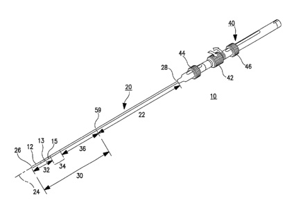
30 FIG. 1 schematically illustrates an anatomically-conforming, multi-curve
catheter
incorporating various features of the present invention for orienting a distal
tip
CA 02425640 2003-04-10
WO 02/030310 PCT/USO1/30578
-9-
electrode 12 (or electrodes) with respect to the heart wall for RF ablation
and/or EP
mapping. The mufti-curve catheter 10 can incorporate a porous tip and catheter
lumen for
emitting irrigating fluid around the distal tip electrode 12, but those
features are not
illustrated in FIG. 1 to simplify illustration. Moreover, the distal segment
32 is simplified
in FIG. 1 to show an elongated tubular shaped ablation electrode 12 and a pair
of mapping
electrodes 13 and 15 in the illustration of FIG 1, but the distal segment 32
may comprise a
plurality of ring-shaped electrodes, one or more coil electrode or the like
having other
shapes that are presently used or may come into use and including several
variations
described below in reference to other figures. It will be understood that the
catheter 10
1o also represents an ablation catheter construction delivering other forms of
ablation energy,
including visible or invisible light, infrared, and electrical energy from or
along the distal
tip.
The catheter 10 comprises a catheter shaft or body 20 and a handle 40. The
catheter shaft or body 20 has a shaft axis 24 and extends between a distal end
26 and a
proximal end 28 and is separated into a proximal section 22 and a distal
section 30.
Catheter body 20 may be of any suitable diameter and length and may be
straight or pre-
curved along its length, but preferably is straight when unrestrained. The
distal section 30
or the distal segment thereof can be tapered from the diameter of the proximal
section 22.
Preferably, the catheter body 20 has a uniform outside diameter of about 0.052
inch (1.32
mm) to 0.1040 inch (2.64 mm) and a length of about 50 cm to 110 cm.
The proximal section 22 has sufficient column strength and is capable of good
torque transmission to permit controlled placement of the distal section 30 at
a target site
in the heart including a selected cardiac valve or vessel in the manners
discussed below.
The distal section 30 is deflectable away from shaft axis 24 and includes a
distal segment
32, a curvable proximal segment 36 having a proximal segment length, and a
bendable
intermediate segment 34 having an intermediate segment length disposed between
the
distal segment 32 and the curvable proximal segment 36. The illustrative tip
electrode 12
is positioned along the distal segment 32, preferably extending proximally
from the
catheter body distal end 26 through all or part of the length of the distal
segment 32. The
3o distal segment 32 can include an elongated ablation electrode 12 that may
be solid or
irrigated and can include one or more proximal ring electrodes 13, 15 for use
in mapping
CA 02425640 2003-04-10
WO 02/030310 PCT/USO1/30578
-10-
that are either located proximally as shown or distally from ablation
electrode 12. Each
electrode is separately connected to insulated conductors extending proximally
through the
catheter body 20 to terminals of a cable connector in or on the handle 40 that
is connected
via a cable to the ablation energy source and/or mapping signal amplifiers. As
described
further below, a thermocouple is also typically included in the distal segment
32 of such
ablation catheters, and separately insulated thermocouple conductors extending
proximally
through the catheter body 20 to terminals of the cable connector in or on the
handle 40 that
are coupled via a cable to the temperature display and ablation energy control
apparatus
known in the art.
to The handle 40 can take any of the forms known in the art for making
electrical
connections with the conductors within the catheter body 20, for delivering
irrigation fluid
to an irrigation lumen (if present) of the catheter body 20. The handle 40
also comprises a
mechanism for deflecting the distal tip section 30 into the shapes provided by
the present
invention. The mechanism can take any form for pulling, pushing andlor
twisting the
deflection or push/pull wires within the catheter body 20 as described further
below. In
the illustrated embodiment, the handle 40 is attached to the catheter body
proximal end 28
and supports axially slidable manipulators comprising push-pull rings 44 and
46 and a
rotatable lateral deflection ring 42 that are coupled to the proximal ends of
a curve
deflection push-pull wire, a knuckle deflection push-pull wire, and a lateral
deflection wire
2o identified and described further below. The lateral deflection ring 42 can
be rotated to
impart a torque in a lateral deflection wire coupled thereto to laterally
rotate the distal
section 30 with respect to axis 24 within the proximal section 22. The details
of
construction of one embodiment of the components of the catheter body 20 are
set forth in
FIGS. 8-16 and these curve and rotation functions are described further below.
As shown in FIG. 1, when the push-pull wires are relaxed, the distal segment
32,
the bendable intermediate segment 34, and the curvable proximal segment 36 are
aligned
with the shaft axis 24 which is referenced as 0°. The knuckle
deflection push-pull wire
can be retracted or pulled by sliding ring 46 proximally to impart a small
radius bend from
substantially 0°, wherein the distal and proximal segments 32 and 36
are axially aligned, to
3o substantially 180°, whereby the distal and proximal segments 32 and
36 are substantially
in side-by-side alignment. The knuckle deflection push-pull wire can be
extended or
CA 02425640 2003-04-10
WO 02/030310 PCT/USO1/30578
-11-
pushed by sliding push-pull ring 46 distally to impart a small radius bend
from
substantially 0° to about -90°, that is in a bend direction
opposite to the bend direction
imparted when the knuckle deflection push-pull wire is retracted or pulled by
sliding ring
46 proximally. The intermediate segment 34 is bent in a bending radius of
between 2.0
mm and 7.0 mm, and preferably less than about 5.0 mm within the bending angle
range.
The abrupt knuckle bend angle range can be restricted further by positioning
of the slide
end stops for the push-pull ring 46 during assembly.
The manipulator push-pull ring 44 can be moved proximally or distally to move
the curve deflection push-pull wire coupled thereto proximally or distally to
form a curve
1o in the proximal segment 36 that is opposed to or in the same direction as
the bend
imparted in the intermediate segment 34. The bend or curve of the proximal
segment 36
that can be induced relative to the catheter body axis 24 as depicted in the
figures can be
between
-90° to +270° relative to the proximal section 22. The curvature
range of the proximal
segment 36 can be restricted further by position of the slide end stops for
the push-pull
ring 44 during assembly.
FIGS. 2 through 7 illustrate four of many possible co-planar curves induced in
the
segments of the distal section 30 in relation to the catheter body axis 24
accomplished by
selective movement of the axially slidable manipulator rings 46 and 44 coupled
to the
2o knuckle deflection push-pull wire S6 and the curve deflection push-pull
wire S4,
respectively. The distal end of the knuckle deflection push-pull wire 56
terminates at the
junction of the intermediate segment 34 with the distal segment 32, and the
curve
deflection push-pull wire 54 terminates at the junction of the intermediate
segment 34 with
the proximal segment 36. The knuckle deflection push-pull wire 56 and the
curve
deflection push-pull wire S4 extend in parallel with and are radially aligned
to the catheter
body axis 24 along a common radius extending from the catheter body axis 24
through the
proximal section 22 and the proximal segment 36. The knuckle deflection push-
pull wire
56 is spaced further away from the axis 24 than the curve deflection push-pull
wire 54
through the proximal section 22 and proximal segment 36. The distal section of
the
knuckle deflection push-pull wire 56 traversing the intermediate segment 34 is
axially
aligned with the axis of the curve deflection push-pull wire 54 in the
proximal segment 36.
CA 02425640 2003-04-10
WO 02/030310 PCT/USO1/30578
-12-
In FIG. 2, both the knuckle deflection push-pull wire 56 and the curve
deflection
push-pull wire 54 are pulled proximally to induce a short radius, 90°
knuckle bend in the
intermediate segment 34 and a long radius curve in the same plane and
direction in the
proximal segment 36. A 90° bend of the intermediate segment 34 with
respect to the
proximal shaft section 22 provides an optimum angular orientation of the
distal electrode
12 for pushing or pulling it against the heart wall.
FIG. 3 illustrates the same short radius, 90° knuckle bend formed
in the
intermediate segment 34 but without any curvature formed in the proximal
segment 36.
As set forth above, a knuckle bending radius between 2.0 mm and 7.0 mm and
preferably
less than about 5.0 mm is provided.
FIG. 4 illustrates the full substantially 180° knuckle bend formed
in the
intermediate segment 34 without any curvature formed in the proximal segment
36, so that
the distal and proximal segments 32 and 36 are substantially in side-by-side
orientation.
The curve deflection push-pull wire 54 can be both pulled proximally as shown
in
FIG. 2 to induce a curvature in the same direction as the knuckle bend in
intermediate
segment 34 and pushed distally as shown in FIG. 5 to induce a curvature in the
opposite
direction as the knuckle bend in intermediate segment 34. The curvature that
can be
induced in the proximal section ranges from -90° to +270°
relative to the proximal section
22 and with respect to catheter body straight axis 24, but smaller ranges can
be selected.
FIG. 6 illustrates a +270° curvature in the distal section 22 effected
by retraction of
both push-pull wires 54 and 56, and FIG. 7 illustrates a +270°
curvature in the distal
section 22 effected by retraction of only curve deflection push-pull wire 5.
In this way, the
distal electrode 12 is positioned at -90° to the proximal section 22,
which is a useful
orientation for ablating or mapping the heart wall at the caval-tricuspid
isthmus or sites
under in the ventricles the mitral or tricuspid valve flaps.
The lateral deflection that can also be induced to orient the distal tip
electrode 12
out of the plane of FIGS. 2-7 using the lateral deflection wire 52 and
manipulator ring 42 is
not shown in these figures since it would be out of the plane of the paper
that the drawings
are printed on. When the ring 42 is rotated clockwise or counterclockwise, the
lateral
3o deflection wire is twisted, causing the junction of the proximal and
intermediate segments
36 and 34 to rotate. It will be understood from the construction of the
lateral deflection
CA 02425640 2003-04-10
WO 02/030310 PCT/USO1/30578
-13-
wire described below that a lateral deflection of the tip segment 32 and the
intermediate
segment 34 in the range of -90° to +90° with respect to catheter
body straight axis 24 can
be achieved by such rotation.
The structure of the catheter body 20 that achieves these angular tip section
deflections and the lateral deflection is illustrated in FIGs. 8-16. FIGS. 9-
16 also show the
internal arrangement of the pull wires and wire lumens as well as the wires
that apply RF
energy to the tip electrode 12 and a thermocouple located in a cavity in the
tip electrode
12.
The proximal section 22 shown in FIGS. 8 and 15-16, is formed of an outer
shaft
to jacket or sheath 50, preferably made of high durometer (such as 72D) Pebax~
reinforced
by a braided wire tubing formed of flat, stainless steel wire embedded within
the sheath
wall that encloses a sheath lumen 58. Pebax~ polyamide polyether block
copolymer is
made by Elf Atochem, Inc. of Philadelphia, PA. The sheath lumen 58 encloses
the
knuckle deflection push-pull wire 56, the curve deflection push-pull wire 54,
and the
lateral deflection wire 52. The sheath lumen 58 also receives the distal tip
electrode
conductor 70 extending between the handle 40 and the distal tip electrode 12
and
thermocouple wires 72 and 74 that extend between a thermocouple 90 (depicted
in FIG. 9)
and temperature monitoring circuitry of the RF energy generator. The
thermocouple 90
provides temperature readings to modulate the delivered energy level or duty
cycle to
2o avoid undue heating of the distal tip electrode 12 during ablation. The
distal tip electrode
conductor 70 is used to convey electrical signals of the heart sensed through
the tip
electrode 12 to ECG display equipment coupled to a terminal of the handle 40
during EP
mapping or to deliver the RF energy from the RF energy generator to the distal
tip
electrode 12. These conductors 70, 72 and 74 would be separately electrically
insulated
from one another and the knuckle deflection push-pull wire 56, the curve
deflection push-
pull wire 54, and the lateral deflection wire 52. It will be understood that
the lumen 58 can
be configured with a fluid conduit to direct irrigation fluid to irngation
ports of the distal
tip electrode 12 and can be used to carry further wires coupled to additional,
more
proximally or more distally located, EP mapping andlor ablation electrodes
than electrode
12.
CA 02425640 2003-04-10
WO 02/030310 PCT/USO1/30578
-14-
The knuckle deflection push-pull wire 56 and the curve deflection push-pull
wire
54 are encased within incompressible spiral wire tubes 66 and 64,
respectively, that extend
from proximal tube ends abutting a stop plate within the distal end of handle
40 distally
through the proximal sheath lumen 58. A distal section of the incompressible
spiral wire
tube 66 and knuckle deflection push-pull wire 56 extends distally from
junction 59 of the
proximal section 22 and proximal segment 36 through a lumen 68 of proximal
segment
tube 60. The distal end of the incompressible spiral wire tube 66 is located
abutting the
proximal insulator 80 shown in FIGS. 8, 12 and 13 that the knuckle deflection
push-pull
wire 56 passes through, and it is adhered to the proximal insulator 80 when
the proximal
l0 insulator is thermally bonded between the tubes 60 and 82. The
incompressible spiral wire
tube 66 is not attached at its proximal end to the handle 40, and it therefore
"floats" over
the proximal portion of the knuckle deflection push-pull wire 56 that
traverses the catheter
body proximal section 22 and the proximal section 36 of the distal section 30.
This
floating feature advantageously prevents the stretching of the coil turns of
the
incompressible spiral wire tube 64 when the knuckle deflection push-pull wire
56 is
pushed or when the adjacent curve deflection push-pull wire 54 is pushed
distally or pulled
proximally, inducing a curve in the proximal segment 36.
The distal end of the incompressible spiral wire tube 64 is located at the
junction
59 of the distal end of proximal sheath 50 with the proximal end of the multi-
lumen tube
60 of the proximal segment 36 shown in FIGs. 8 and 15. The junction 59 is a
butt welded
junction of the distal end of proximal sheath 50 with the proximal end of the
mufti-lumen
tube 60, and so the distal end of the incompressible spiral wire tube 66 is
affixed to
junction 59 by the solidification of the melted material to it. But, the
proximal end of the
incompressible spiral wire tube 66 is not attached to the handle 40, so that
the coil turns of
the incompressible spiral wire tube 66 when the curve deflection push-pull
wire 54 is
pushed or when the adjacent knuckle deflection push-pull wire 56 is pushed
distally or
pulled proximally, inducing a curve in the intermediate segment 34.
The incompressible spiral wire tubes 64 and 66 are preferably formed of
stainless
steel flat wire wound so that the narrow wire edges abut one another in each
turn, but do
not overlap one another when the coils are compressed by pulling proximally on
the curve
deflection push-pull wire 54 and the knuckle bend push-pull wire 56. The coil
turns of
CA 02425640 2003-04-10
WO 02/030310 PCT/USO1/30578
-15-
coils formed of circular cross-section wire tends to ride over one another.
Preferably, the
incompressible spiral wire 64 is 0.017 inches thick by 0.023 inches wide, and
the
incompressible spiral wire 66 is 0.013 inches thick by O.OI9 inches wide. The
coil turns
are close wound so that the thinner wire sides of each coil turn abut or
nearly abut one
another.
The knuckle deflection push-pull wire 56 is formed of a nickel-titanium
superelastic metal that has a straight memory shape and does not readily kink,
enabling the
repeated formation of small radius knuckle bends in the intermediate segment
34 as
described further below. The curve deflection push-pull wire 54 and the
lateral deflection
wire 52 are formed of stainless steel, and their distal ends are both attached
to the
proximal insulator member 80. The lateral deflection wire 52 is tapered and is
reduced in
diameter distally when it traverses the proximal segment 36. Wires 52, 54 and
56 are
preferably coated with a lubricious material, e.g. PTFE or Parylene, to reduce
sliding
friction.
As shown in FIG. 8, the distal section 30 is formed of the distal electrode 12
and
distal insulator 84 together forming the distal segment 32. The intermediate
segment 34 is
formed of the two-lumen intermediate tube 82 and includes the distal section
of knuckle
deflection push-pull wire 54. The proximal segment 36 is formed of the mufti-
lumen tube
60 and proximal insulator 80 along with the wires passing through their
lumens. The
2o mufti-lumen tube 60 is preferably formed of intermediate durometer (such as
SSD)
PebaxO polyamide polyether block copolymer. The proximal insulator 80
illustrated in
cross-section in FIGS. 12 and 13 is formed of a relatively rigid PEED
(polyether-ether-
ketone) or other hard, temperature-resistant material with a number of lumens
81, 83, 85
and 86 extending through it aligned axially with the lumens 63, 65 and 68 of
mufti-lumen
tube 60 and lumens 88 and 93 of two-lumen intermediate tube 82.
The proximal end of the mufti-lumen tube 60 is butt welded to the distal end
of
proximal sheath 50 at the junction 59 as described above and the various
conductors and
wires are directed through the lumens 63, 65 and 68 as shown in FIG. 14. The
knuckle
deflection push-pull wire 56 and incompressible spiral wire 66 are directed
through
3o elliptical lumen 68 along with the conductors 70, 72 and 74. The
incompressible spiral
wire 66 terminates in abutment against the proximal insulator 80, but the
knuckle
CA 02425640 2003-04-10
WO 02/030310 PCT/USO1/30578
-I 6-
deflection push-pull wire extends distally through the proximal insulator 80.
The curve
deflection push-pull wire 54 is extended distally through the lumen 63 to an
attachment
with the proximal insulator 80, but the distal end of the incompressible
spiral wire 64 is
terminated at junction 59 as described above. The lateral deflection wire 52
extends
distally through lumen 65 to a connection with the proximal insulator 80.
Additional
lumens can also be provided in tube 60 that make the tube 60 more flexible and
easier to
bend.
The two-lumen intermediate tube 82 is preferably formed of relatively soft
durometer (such as 35D) Pebax~ polyamide polyether block copolymer. The
conductors
l0 70, 72 and 74 pass through the central lumen 86 and into a lumen 88 of the
two-lumen
intermediate tube 82. The knuckle deflection push-pull wire 56 extends
distally through
lumen 83 of the proximal insulator 80. The curve deflection push-pull wire 54
within
Iumen 68 extends distally through the Iumen 81 where its distal end is bent
over and
attached to the distal surface of the proximal insulator 80. Similarly, the
lateral deflection
15 wire 52 in lumen 65 extends distally through lumen 85 where its distal end
is bent over
and attached to the distal surface of the proximal insulator 80.
During manufacture, the lumens 93 and 88 of the two-lumen intermediate tube 82
are aligned with the lumens 83 and 86, respectively, of proximal insulator 80
as shown in
FIGS. 11 and 13 which are aligned with the central lumen 68 of the multi-lumen
tube 60
20 and the wires are passed through them as described above. The lumens 63 and
65 of the
multi-lumen tube 60 are aligned with the lumens 81 and 85 of proximal
insulator 80, and
the wires 54 and 52 are passed through the aligned lumens as described above.
Heat and
pressure are applied to the assembly to fuse the proximal insulator 80 between
the
proximal end of the two lumen intermediate tube 82 and the distal end of the
multi-lumen
25 proximal tube 60. The applied heat causes the tube material to flow over
scalloped
sections of the outer surface of proximal insulator 80 thereby fusing the
proximal end of
the two lumen intermediate tube 82 with the distal end of the multi-lumen
proximal tube
60.
The distal insulator 84 illustrated in cross-section in FIGS. 9 and 10 is
formed of a
3o relatively rigid PEEK or other hard, temperature-resistant material and is
attached between
tube 82 and distal tip electrode 12 preferably using a mechanical interlock
and/or adhesive.
CA 02425640 2003-04-10
WO 02/030310 PCT/USO1/30578
-17-
The distal end of two lumen intermediate tube 82 is shaped to fit over and be
adhered
through the use of appropriated adhesive, thermal bond, or other appropriate
methods to
the proximal end of the distal insulator 84 after aligning the lumen 93 with
the lumen 87 of
distal insulator 84. The conductors 70, 72 and 74 pass through the central
lumen 88 of the
two-lumen intermediate tube 82 and through a central lumen 89 of the distal
insulator 84
as shown in FIGs. 9-11. The distal end of the distal insulator 84 extending
through lumen
89 is attached to the distal tip electrode 12 as shown in FIG. 9, and the
conductor 70 is butt
welded to the distal tip electrode 12. The distal ends of the thermocouple
conductors 72
and 74 extend through lumen 89 and are attached to the thermocouple 90
positioned within
to a cavity of the distal tip electrode 12 as shown in FIG. 14. The knuckle
deflection push-
pull wire 56 extends distally through lumen 87 of the distal insulator 84. The
enlarged
diameter distal ball-tip end 57 of the knuckle bend pull wire 56 fits into a
bore 95 of the
distal insulator 84 so that the distal end of knuckle bend pull wire 56 is
fixed in place.
FIG. 17 is a partial perspective view of the distal end of an interior frame
member
41 and a coil wire stop plate 43 within the distal end of the handle 40 that
is joined with
the proximal end of the catheter body 20. FIG. 17 shows that the proximal ends
of the
incompressible coils 66 and 64 surrounding proximal portions of the knuckle
deflection
push-pull wire 56 and the curve deflection push-pull wire 54, respectively,
simply abut the
plate 43. The proximal portions of the knuckle deflection push-pull wire 56,
the curve
2o deflection push-pull wire 54, and the lateral deflection wire 52 pass
through holes in the
plate 43. The incompressible coils 66 and 64 are not otherwise restrained so
that the
incompressible coils 66 and 64 can move away from the plate 56 and not be
stretched if
the catheter body 20 is extended distally. In this way, the knuckle deflection
push-pull
wire 56 and the curve deflection push-pull wire 54 can be extended or pushed
distally to
impart the negative curvature in the intermediate and proximal segments 34 and
36
without stretching the incompressible coils 66 and 64.
Handle 40 may be of a conventional design, e.g. as shown in the above-
referenced,
commonly assigned '200 patent, except for the plate 56 and its above described
function.
Handle 40 also includes an electrical connector connected to electrical
conductors 70, 72
3o and 74 (and any additional conductors) for connection with a cable that is
attached to the
ECG and/or ablation equipment. Handle 40 may also be configured to be coupled
with a
CA 02425640 2003-04-10
WO 02/030310 PCT/USO1/30578
-18-
source of irngation fluid if the catheter body 20 and electrode 12 are
modified to provide
an irrigation fluid lumen and ports through the electrode 12.
Returning to the bendable intermediate segment 34, the relatively flexible
tube 82
is thus bounded on its proximal end by the proximal insulator 80 and on its
distal end by
the distal insulator 84. The length of the tube 82 and the distal section of
the knuckle
deflection push-pull wire 56 traversing lumen 93 forming the intermediate
segment 34 is
preferably on the order of about 4.0 mm to 15.0 mm. The length of the tube 60
of the
proximal segment 36 is preferably on the order of about 30.0 mrn to 120.0 mm.
The proximal segment 36 can be curved as shown in FIGS. 2, 5, 6 and 7 by
to retraction of the curve deflection push-pull wire 54 by retracting axially
slidable
manipulator ring 44. The proximal retraction of the knuckle bend pull wire 56
by
retracting axially slidable manipulator ring 46 induces a knuckle bend in the
tube 82 of the
intermediate section 34 as depicted in FIGS. 2-6. independently of the curve
induced in the
proximal segment 36. The knuckle bend that is induced has a bending radius of
less than
about 5.0 mm within a bend of substantially 180°.
The incompressible spiral coil wires 64 and 66 prevent the compression of the
tube
60 of proximal segment 36 or the sheath 50 of the proximal section 22. The
incompressible spixal wires 64 and 66 are not stretched or compressed by
retraction of one
or another of the push-pull wire 54 or the knuckle bend pull wire 56 or
twisting induced by
2o manipulation of the lateral deflection wire 52 because the proximal ends of
the
incompressible spixal wires 64 and 66 are not attached at the handle 40.
FIGS. 18 - 20 are schematic illustrations of the selective locations of the
distal
section 30 of the catheter body 20 of the catheter 10 described above for
cardiac mapping
and/or ablation of the heart 100. In the following discussion, it will be
assumed that the
distal tip electrode 12 is first applied to the location of interest, ECG
readings are made to
determine the existence and location of accessory pathways, and ablation is
selectively
performed.
FIGs. 18 - 20 illustrate, in simplified form, a sectioned heart 100 and the
major
vessels bringing venous blood into the right atrium RA, oxygenated blood into
the left
3o atrium LA and the aorta and aortic arch (FIG. 20) receiving oxygenated
blood from the left
ventricle LV. The venous blood is delivered to the RA through the superior
vena cava
CA 02425640 2003-04-10
WO 02/030310 PCT/USO1/30578
-19-
SVC, the inferior~vena cava IVC and the coronary sinus CS which all open into
the right
atrium RA superior to the annulus of the tricuspid valve leading into the
right ventricle.
Oxygenated blood from the two lungs is delivered into the Ieft atrium by the
Ieft and right,
inferior and superior, pulmonary veins LIPV, LSPV, RIPV and RSPV which are
superior
to the mitral valve. The right and left atria are separated by an inter-atrial
septum and the
right and left ventricles are separated by a ventricular septum. The tricuspid
valve TV and
mural valve MV are not shown completely to simplify the figures.
Accessory pathways develop in several parts of the RA and LA that are reached
by
the catheter 10 to be mapped and/or ablated in accordance with methods of use
thereof of
l0 the present invention depicted, for example, in FIGS. 18 and 19,
respectively. Certain
atrial tachycardias also employ left-sided accessory pathways in tight areas
under the cusps
of the mitral valve MV that can be reached in the manner depicted in FIG. 20.
In these
illustrations, it will be understood that the catheter body proximal section
is flexible
enough so that it curves to traverse the vascular system and is curved within
a heart
is chamber by the heart chamber wall by the catheter body
In FIG. 18, the distal section 30 of the catheter body 20 is introduced into
the RA
through the IVC, and the distal segment 32 is oriented to selected locations
of the RA
heart wall through selective manipulations of the manipulator rings 42, 44 and
46. The
RA is separated into a posterior, smooth walled portion that the SVC, IVC and
CS orifices
20 open through and a thin walled trabeculated portion separated by a ridge of
muscle which
is most prominent superior to the SVC ostium. Vestigial valve flaps can adjoin
the IVC
and CS orifices in some patient's hearts.
A thickened isthmus or Eustachian ridge extends between the IVC orifice and
the
medial cusp of the tricuspid valve. Certain atrial flutter tachyarrhythmias
are known to be
25 caused by accessory pathways situated in the myocardium at or along the
Eustachian ridge
toward the annulus of the tricuspid valve, and ablation to create a lesion
from the IVC
orifice over the Eustachian ridge can be used to sever the accessory pathways
therein. In
FIG. 18, the distal section of the catheter body is formed into a hook shape
within the IVC
to "hook" the distal tip electrode over the Eustachian ridge and draw it
against the tissue in
30 location 1A. A +150° to +180° knuckle bend is made in the
intermediate segment in the
manner of FIG. 4 to form this hook shape and access this location 1A.
CA 02425640 2003-04-10
WO 02/030310 PCT/USO1/30578
-20-
Alternatively, the distal section is advanced into the RA and a +150°
to +180°
curve is formed in the proximal segment 36 of the distal section along with
the +180°
knuckle bend made in the intermediate segment in the combined manner of FTGs.
2 and 4
to access the location 1B. The catheter body 20 is then retracted to apply the
distal tip
electrode 12 at the distal end of this compound hook shape against the tissue
location 1 B
adjacent or overlying location 1A at the Eustachian ridge.
The heart wall can be mapped and continuous lesions can be made along the
Eustachian ridge by successively moving the distal electrode 12 to an
adjoining location to
location 1A or 1B to sense the heart signals or apply RF ablation energy to
the new site.
The movement can be effected by twisting the distal segment 32 about the
catheter body
axis 24 by rotating the lateral deflection manipulator ring and wire and/or by
adjusting the
curvature in the proximal segment 36.
Other accessory pathways in the inter-atrial septum adjacent the AV node or
elsewhere along the RA wall or in the triangle of Koch can be accessed as
shown by the
exemplary location 1C of the distal tip electrode. In this illustrated
example, a +90°
knuckle bend is made in the intermediate segment in the manner of FIG. 3, and
a further
positive direction +90° bend is made in the proximal segment 36. Or, if
the entire distal
section 30 is within the RA, then the configuration of FIG. 2 can be employed
to locate
and hold the distal tip electrode against the atrial wall around the AV node
at the
2o exemplary location 1 C.
Premature activations occur frequently in the LA wall, particularly from
pulmonary
venous foci around the annular orifices of certain or all of the pulmonary
veins RIPV,
RSPV, LIPV, LSPV shown in FIG. 19 that cause atrial fibrillation. The LA can
be
accessed in a retrograde manner through the aorta. However, another convenient
approach
to the LA is via a puncture made through the inter-atrial septum from the RA
employing a
transseptal sheath 38 as depicted in FIG. 19. The distal section 30 can be
formed with
about a +90° knuckle bend is made in the intermediate segment in the
manner of FIG. 3
and slight positive, neutral or negative curvatures in the range of about -
45° to +45° in the
proximal segment 36 as in FIGS. 2, 3, and 4 to align the distal tip to
locations 2A, 2B or
2C. Continuous lesions can be made around the selected pulmonary valve orifice
by
successively moving the distal electrode to the next location and applying RF
ablation
CA 02425640 2003-04-10
WO 02/030310 PCT/USO1/30578
-21-
energy. The movement can be effected by twisting the distal segment about the
catheter
body axis using the deflection wire and manipulator.
The left-sided accessory pathways for atrial tachycardia in tight areas under
the
cusps of the mural valve MV are advantageously accessed by advancing the
distal section
30 of catheter body 20 in a retrograde manner through the aorta and into the
LV and then
angling and advancing the distal tip electrode under the cusps to exemplary
location 3 as
shown in FIG. 20. The distal segment 32 extends inwaxd in relation to the
plane of the
drawing of FIG. 20, and can be worked under the cusps around the MV to map
and/or
ablate a succession of adjoining sites.
to While the preferred embodiment only illustrates a single mapping/ablation
distal
tip electrode 12 particularly used in a unipolar ablation and/or mapping mode,
it will be
understood that it may be advantageous to locate one or more additional
mapping/ablation
electrodes in the distal segment 32 and/or proximally in the curvable proximal
segment 36
for selective operation either in a unipolar or bipolar mapping/ablation mode.
In the latter
case, bipolar mapping/ ablation across or through the Eustachian ridge can be
achieved in
the hook configuration depicted in FIG. 4 and at location 1A of FIG. 18.
In further embodiments of the present invention depicted in FIGS. 21 - 27,
particularly for ablating or mapping the Eustachian ridge, a plurality of
mapping and/or
ablation electrodes are located along extended distal segments 132 and 232
distal to
2o electrode 112 (which can be eliminated in variations to these embodiments).
The extended
distal segments 132 and 232 are formed to comply to the particular shape of
the caval-
tricuspid isthmus extending anteriorly from the orifice of the IVC and toward
the valve
flaps of the tricuspid valve to map and ablate that area as shown in FIG. 27.
The extended
distal segments 132 and 232 can have a pre-formed curved axis particularly
shaped to the
surface curvature of the caval-tricuspid isthmus. Or, the extended distal
segments 132 and
232 can have an elasticity and flexibility that conforms to the surface
curvature of the
caval-tricuspid isthmus effected by selection of a suitable low durometer
insulating tubular
member supporting the electrode(s). In either case, the hook shape is formed
in the
knuckle bend segment 34 in the manner illustrated in FIGS. 24, 26 and 27, and
a positive
3o curve can be induced in the proximal segment 36 as shown in FIGs. 24 or 26
as necessary.
A 0° or negative curvature can alternatively be induced in the proximal
segment 36 as
CA 02425640 2003-04-10
WO 02/030310 PCT/USO1/30578
_22_
shown in FIG. 5 if found necessary in a particular heart. From the handle 40
outside the
body, it is therefore possible to hook the intermediate segment 34 over the
Eustachian
ridge to orient the elongated, flexible electrode support body of the distal
segments 132,
232 against and in conformance with contours of the heart wall between the
Eustachian
ridge and the tricuspid valve cusps. A guide sheath or introducer may be
required to
straighten the pre-formed curvature or the highly flexible distal segment 132,
232 to
enable introduction through the vascular system and into the RA.
The extended distal segment 132 is formed of a plurality (e.g. six) of ring
electrodes 116, 118, 120, 122, 124, and 126 supported on a highly flexible or
pre-formed
l0 electrode support tube 114 as shown in FIGS. 21 - 23. The proximal end of
the extended
distal segment 132 including the distal insulator 84 is coupled to the
intermediate segment
34 of the catheter otherwise shown in FIGS. 1 and 8-17 and described above. A
further
insulator 130 separates electrode 112 from the electrode support tube I I4,
and the distal
tip 128 is fitted to the distal end of the electrode support tube 114. The
conductors to the
15 electrodes 116, 118, 120, 122, 124, and 126 that traverse the lumens of the
electrode
support tube 114, the insulator 130, the electrode 112, and the distal
insulator 84 are not
shown in FIG. 21 to simplify the drawing. Such conductors would be formed of
high
conductivity metals, e.g., copper, copper-silver alloys, and silver cored
wire.
In the alternative embodiment of FIGS. 24-26, the extended distal segment 232
is
20 formed of one or plurality (e.g. two) of spiral wound electrodes) 216
supported on a
highly flexible or pre-formed electrode support tube 214. The proximal end of
the
extended distal segment 132 including the distal insulator 84 is coupled to
the intermediate
segment 34 of the catheter otherwise shown in FIGS. 1 and 8-17 and described
above. A
further insulator 230 separates electrode 112 from the electrode support tube
214, and the
25 distal tip 228 is fitted to the distal end of the electrode support tube
214. The conductors)
to the electrodes) 216 that traverse the lumens of the electrode support tube
214, the
insulator 230, the electrode 112, and the distal insulator 84 are not shown in
FIG. 21 to
simplify the drawing. Such conductors) would be formed of high conductivity
metals,
e.g., copper, copper-silver alloys, and silver cored wire.
3o It is also contemplated that in the embodiments depicted in FIGS. 24-27 may
be
further modified by eliminating the proximal segment 36 and its associated
manipulator
CA 02425640 2003-04-10
WO 02/030310 PCT/USO1/30578
-23-
structure described above. In such an embodiment, the distal end of the
proximal section
22 would be coupled directly to the proximal end of the intermediate segment
34.
In the above-described preferred embodiments, the knuckle bend wire 56 and the
curve deflection wire 54 extend through the proximal section 22 and the
curvable proximal
segment 36, and the knuckle bend wire 56 extends further distally through the
bendable
intermediate segment 34 in a common radius extending from the catheter body
axis 24 as
shown in FIGS. 2-16. Therefore, the bend induced in the bendable intermediate
segment
34 upon retraction proximally of the knuckle bend wire 56 and the curve
induced in the
curvable proximal segment 36 upon retraction proximally of the curve
deflection wire 54
l0 are in a common plane with respect to the catheter body axis 24 and in a
common direction
as shown in FIG. 2. The bend induced in the bendable intermediate segment 34
upon
retraction proximally of the knuckle bend wire 56 and the curve induced in the
curvable
proximal segment 36 upon extension distally of the curve deflection wire 54
are in a
common plane with respect to the catheter body axis 24 but in a different
direction as
is shown in FIG. 5. The knuckle bend that can be induced in the intermediate
segment 34 is
very tight, falling within a xadius of about 2.0 mm to 7.0 mm through a range
of about
-90° to about +180° with respect to the catheter body axis 24 at
the intermediate segment
proximal end.
It will be understood that certain features of the present invention can be
2o advantageously employed in modifications of the preferred embodiment, e.g.,
by
displacing the knuckle bend wire 56 and its associated lumens 68, 83, 93 and
87, in a
radius that is not common with the curve deflection wire 54 and its associated
lumens. In
this regard, the knuckle bend wire 56 and its associated lumens 68, 83, 93 and
87, can be
arranged in a radius that is diametrically opposed to the radius that the
curve deflection
25 wire 54 and its associated lumens are aligned with, i.e., in a common
diametric line but on
either side of the catheter body axis 24. The lateral deflection wire 52 and
its associate
lumens illustrated in the FIGS. 12 - 16 occupy such a location, and they can
be displaced
either radially or to the other side of the axis 24. Then, the knuckle bend
induced in the
intermediate segment 34 would be in the opposite direction than is depicted in
FIGS. 2-7.
3o The catheter shaft or body and handle of the present invention allows
manipulation
with a high degree of sensitivity and controllability to provide the degree of
precision
CA 02425640 2003-04-10
WO 02/030310 PCT/USO1/30578
-24-
required for proper positioning of the tip electrode(s). The distal section of
the catheter
body is sufficiently resilient in order to position the distal tip electrodes)
against the
endocardium and to maintain the distal tip electrodes) in position during
mapping or
ablation without being displaced by movement of the beating heart, by
respiration, or by
blood flow. Along with steerability, flexibility, and resiliency, the catheter
body has a
sufficient degree of torsional stiffness to permit user imparted torque to be
transmitted to
the distal tip electrodes) from the handle. Moreover, the catheter body has
sufficient
column strength to convey axial loading to push the distal tip electrodes)
against the
tissue at target positions to be mapped or ablated.
to Other modification and variation can be made to the disclosed embodiments
without departing from the subject of the invention as defined in the
following claims. For
example, materials, diameters and lengths can be changed to suit the
particular needs or
desires of the user. A single mappinglablation electrode, or more than two
mapping/ablation electrodes could be present. A plurality of small sized
mapping
electrodes displaced apart along the distal section of the catheter body are
typically
provided and paired electrically to increase sensing resolution of the
electrical signals of
the heart traversing the adjoining heart wall site. Mapping electrodes could
also be located
between ablation electrodes. In some cases it may be desired to apply energy
to more than
one ablation electrode at the same time; for example, four ablation electrodes
could be
used and powered in pairs.
Although particular embodiments of the invention have been described herein in
some detail, this has been done for the purpose of providing a written
description of the
invention in an enabling manner and to form a basis for establishing
equivalents to
structure and method steps not specifically described or listed. It is
contemplated by the
inventors that the scope of the limitations of the following claims
encompasses the
described embodiments and equivalents thereto now known and coming into
existence
during the term of the patent. Thus, it is expected that various changes,
alterations, or
modifications may be made to the invention as described herein without
departing from
the spirit and scope of the invention as defined by the appended claims.