Note: Descriptions are shown in the official language in which they were submitted.
CA 02425796 2003-04-15
WO 02/32325 PCT/USO1/32320
NON-OVERLAPPING SPHERICAL THREE-DIMENSIONAL VASO-OCCLUSIVE COIL
FIELD OF THE INVENTION
This invention relates to the field of vaso-occlusive devices. More
particularly, it relates to a three-dimensional vaso-occlusive device made up
of a
plurality of non-overlapping loops.
BACKGROUND
Vaso-occlusion devices are surgical implements or implants that are placed
within the vasculature of the human body, typically via a catheter, either to
block the
flow of blood through a vessel malting up that portion of the vasculature
through the
formation of an embolus or to form such an embolus within an aneurysm stemming
from the vessel. One widely used vaso-occlusive device is a helical wire coil
having
windings which may be dimensioned to engage the walls of the vessels. Other
less
stiff helically coiled devices have been described, as well as those involving
woven
braids.
For instance, U.S. Pat. No. 4,994,069, to Ritchart et al., describes a vaso-
occlusive coil that assumes a linear helical configuration when stretched and
a folded,
convoluted configuration when relaxed. The stretched condition is used in
placing the
coil at the desired site (by its passage through the catheter) and the coil
assumes a
relaxed configuration--which is better suited to occlude the vessel--once the
device is
so placed. Ritchart et al. describes a variety of shapes. The secondary shapes
of the
disclosed coils include "flower" shapes and double vortices. A random shape is
described, as well.
Other three-dimensional vaso-occlusive devices have been described. U.S.
Patent No. 5,624,462 to Mariant describes a three-dimensional in-filling vaso-
occlusive coil. U.S. Patent No. 5,639,277 to Mariant et al. describes embolic
oils
having twisted helical shapes and U.S. Patent No. 5,649, 949 to Wallace et al.
describes variable cross-section conical vaso-occlusive coils.
U.S. Patent No. 5,334,210 to Gianturco, describes a vascular occlusion
assembly comprising a foldable material occlusion bag and a filled member, for
CA 02425796 2003-04-15
WO 02/32325 PCT/USO1/32320
example, a helical coil with a J-hook on the proximal end. The bag expands to
form a
diamond shape structure and the filler member inside the bag is forced into a
convoluted configuration as it is advanced into the cavity of the foldable
bag.
Implantable devices using variously shaped coils are shown in U.S. Patent No.
5,537,338 to Purdy. Purdy described a multi-element intravascular occlusion
device
in which shaped coils may be employed. U.S. Patent No. 5,536,274 to Neuss
shows a
spiral implant wluch may assume a variety of secondary shapes. Some complex
shapes can be formed by interconnecting two or more of the spiral-shaped
implants.
Spherical shaped occlusive devices are described in U.S. Patent No. 5,645,558
to Horton. Horton describes how one or more strands can be wound to form a
substantially hollow spherical or ovoid shape comprising overlapping strands
when
deployed in a vessel. Notably, the device as deployed must assume a
substantially
minimal energy configuration in which the loops making up the spherical shape
overlap with (n+1) circumference length at a minimum.
Vaso-occlusive coils having little or no inherent secondary shape have also
been described. For instance, co-owned U.S. Patent Numbers 5,690,666 and
5,826,587 by Berenstein et al., describes coils having little or no shape
after
introduction into the vascular space.
A variety of mechanically detachable devices are also known. For instance,
U.S. Pat. No. 5,234,437, to Sepetka, shows a method of unscrewing a helically
wound
coil from a pusher having interlocking surfaces. U.S. Pat. No. 5,250,071, to
Palermo,
shows an embolic coil assembly using interlocking clasps mounted both on the
pusher
and on the embolic coil. U.S. Pat. No. 5,261,916, to Engelson, shows a
detachable
pusher-vaso-occlusive coil assembly having an interlocking ball and keyway-
type
coupling. U.S. Pat. No. 5,304,195, to Twyford et al., shows apusher-vaso-
occlusive
coil assembly having an affixed, proximally extending wire carrying a ball on
its
proximal end and a pusher having a similar end. The two ends are interlocked
and
disengage when expelled from the distal tip of the catheter. U.S. Pat. No.
5,312,415,
to Palermo, also shows a method for discharging numerous coils from a single
pusher
by use of a guidewire which has a section capable of interconnecting with the
interior
of the helically wound coil. U.S. Pat. No. 5,350,397, to Palermo et al., shows
a pusher
2
CA 02425796 2003-04-15
WO 02/32325 PCT/USO1/32320
having a throat at its distal end and a pusher through its axis. The pusher
sheath will
hold onto the end of an embolic coil and will then be released upon pushing
the
axially placed pusher wire against the member found on the proximal end of the
vaso-
occlusive coil.
None of these documents disclose an anatomically shaped vaso-occlusive coil
where the loops making up the three-dimensional configuration are not non-
overlapping.
SUMMARY OF THE INVENTION
In one aspect, the invention includes a vaso-occlusive device comprising at
least one substantially linear strand of a vaso-occlusive member wound into a
stable,
three-dimensional relaxed configuration comprising a plurality of non-
overlapping
loops, wherein said relaxed configuration self forms upon release from a
restraining
member. In certain embodiments, the relaxed configuration of the vaso-
occlusive
device fills a body cavity or, for example, approximates the shape of sphere.
The
vaso-occlusive devices described herein can include any number of non-
overlapping
loops, for example in certain embodiments the device will have between about 6
and
loops while in other embodiments the device will have between about 6 and 12
loops. The vaso-occlusive devices described herein can be comprised of a
metal, for
20 example, platinum, palladium, rhodium, gold, tungsten and alloys thereof.
In other
embodiments, the vaso-occlusive devices described herein comprise a stainless
steel
or super-elastic metal alloy. In still other embodiments, the vaso-occlusive
member
comprises nitinol.
In other embodiments, any of the devices described herein further include
additional filamentary material attached to the vaso-occlusive member. In
still further
embodiments, the device comprises a deployment tip attached to at least one of
the
two ends of the vaso-occlusive member. The deployment tip can be, for example,
mechanically detachable or electrolytically detachable (e.g., by the
imposition of a
current on the pusher).
CA 02425796 2003-04-15
WO 02/32325 PCT/USO1/32320
In another aspect, the invention includes a method of occluding a body cavity
comprising introducing any of the vaso-occlusive devices described herein into
a body
cavity (e.g., an aneurysm).
hi yet another aspect, the invention includes a method of making a non-
overlapping three-dimensional vaso-occlusive device described herein, the
method
comprising (a) winding a substantially linear strand of a vaso-occlusive
member
around a winding mandrel, said winding comprising a winding pattern that
produces a
non-overlapping three-dimensional vaso-occlusive device described herein; and
(b)
heating the mandrel and vaso-occlusive member to produce said vaso-occlusive
device. In certain embodiments, the winding pattern approximates a Figure 8
shape or
an hourglass shape. In other embodiments, the winding mandrel is a three-
dimensional structure (e.g., approximate sphere, cube, cylinder, tetrahedron).
Further,
the mandrel may include grooves adapted to fit the substantially linear strand
and/or
pins on the surface thereof (e.g., a winding mandrel comprising 3 intersecting
posts
which form a 6 post structure and wherein each post is at approximately 90
relative to
the adjacent posts). One or more pins may have the same cross-section (e.g.,
shape
such as round or square, diameter, etc.) or, alternatively, each pin may have
a different
cross-section.
These and other embodiments of the subject invention will readily occur to
those of skill in the art in light of the disclosure herein.
BRIEF DESCRIPTION OF THE DRAWINGS
Figure 1 depicts an exemplary Figure 8 pattern for winding a device according
to the present invention. As shown, the loops making up each half of the
Figure 8 are
of approximately equivalent diameter.
Figure 2 depicts a wire wound around a cylindrical mandrel in a pattern that
will create a non-overlapping three-dimensional vaso-occlusive device.
Figure 3 depicts a wire wound around a three-dimensional mandrel. The
mandrel may include channels to guide the wire as it is wound around the
mandrel.
Figure 4 depicts a wire wound around a six post mandrel to form a non-
overlapping three-dimensional vaso-occlusive device.
4
CA 02425796 2003-04-15
WO 02/32325 PCT/USO1/32320
Figure 5 depicts a non-overlapping device according to the present invention
as deployed.
DESCRIPTION OF THE INVENTION
Vaso-occlusive devices, particularly coils, are described. Upon deployment
from a restraining member, the devices described herein self form into a
relaxed,
three-dimensional configuration approximating an anatomically cavity. The
three-
dimensional configuration is made up of a plurality of loops of a first
configuration.
However, unlike other three-dimensional vaso-occlusive devices, the loops of
the wire
making up the three-dimensional configuration of the device as deployed do not
overlap. Preferably, the loops do not overlap with each other or with
themselves.
Methods of making and using these devices also form an aspect of this
invention.
Advantages of the present invention include, but are not limited to, (i)
reducing or eliminating rotation upon deployment; (ii) reducing or eliminating
whipping upon deployment; (iii) providing vaso-occlusive devices that readily
and
substantially conform to fill a target vessel in a relaxed configuration; and
(iv)
providing methods and materials for making these non-overlapping vaso-
occlusive
devices.
All publications, patents and patent applications cited herein, whether supra
or
infra, are hereby incorporated by reference in their entirety.
It must be noted that, as used in this specification and the appended claims,
the
singular forms "a", "an", and "the" include plural referents unless the
content clearly
dictates otherwise. Thus, for example, reference to "a coil" includes a
mixture of two
or more such devices and the like.
The self forming, non-overlapping three-dimensional coil designs of the
present invention are particularly useful in treating aneurysms. The non-
overlapping
loop design described herein provides an improvement over known devices, for
example in terms of ease of deployment. Available three-dimensional coils are
made
up of a plurality of overlapping and interi:wined loops. Upon deployment from
a
substantially linear configuration these devices often rotate or whip
undesirably
during deployment. Whipping refers to the phenomena where a device stores
energy
CA 02425796 2003-04-15
WO 02/32325 PCT/USO1/32320
imparted by a user and then releases the energy very quickly. For example,
vaso-
occlusive devices are often deployed and manipulated at the target site using
a
guidewire controlled by the operator at a proximal location. Whipping occurs
when
the rotation imparted by the operator on the guidewire does not result in the
same 1:1
rotation of the distal end of the device. Rather, the device stores up the
rotational
energy and then may suddenly release the energy and rotate suddenly in a short
time.
Rotation, whipping and other problems associated with available vaso-occlusive
devices can impede formation of the three-dimensional relaxed configuration.
In
contrast, the non-overlapping configuration of the devices described minimizes
rotation and whipping upon deployment and promotes formation of a three-
dimensional configuration that substantially conforms to the target vessel.
Described herein are vaso-occlusive devices having a relaxed three-
dimensional configuration in the approximate shape of an anatomically cavity.
The
three-dimensional
configuration is made up of non-overlapping loops. Further, the approximately
diameter of the relaxed configuration preferably conforms to the target vessel
in
which is deployed. As used herein, the "first configuration" or "primary
configuration" refers to the structure obtained when a wire is shaped into a
coil, for
example, as a strand of a linear helically wound coil. The "secondary
configuration"
refers to the structures obtained when at least one strand of the first
configuration is
further shaped, for example, by winding around a mandrel. The relaxed
configuration
refers to the three-dimensional configuration assumed by the secondary
configuration
after is has been deployed from the restraining member (e.g., catheter). A
device may
have multiple relaxed configurations, for example depending on whether it is
deployed into a body cavity, the size of the body cavity, etc. The relaxed
configuration typically comprises a three-dimensional structure made up of non-
overlapping loops of the first configuration. The structure may be composed of
any
number of non-overlapping loops. In certain embodiments, the three-dimensional
configuration has between about 4 and 40 loops, more preferably between about
6 and
20 loops and even more preferably, between about 6 and 12 loops. The non-
6
CA 02425796 2003-04-15
WO 02/32325 PCT/USO1/32320
overlapping loops can form an open shape (e.g., a "C", "U", Figure 8 or
hourglass
shape) or a closed, non-overlapping shape (e.g., circle, oval, etc.).
The non-overlapping devices described herein promote formation of a three-
dimensional structure while minimizing rotation and whipping upon deployment.
Thus, depending on the winding pattern and mandrel, the coil will readily self
form
into its secondary, three-dimensional configuration and, accordingly, can be
more
easily deployed into a body cavity by the user. Determining the patterns, size
and
location to achieve the desired structures is within the purview of the
skilled artisan in
view of the teachings herein. The overall device is made up of a primary coil
made
from a wire. The primary coil is then wound into a secondary form, for example
on a
mandrel. The device is substantially straightened for deployment, for example,
into a
restraining member such as a deployment catheter. Upon release from the
restraining
member, the device self forms into the secondary, relaxed, three-dimensional
device.
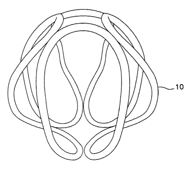
As shown, for example, in FIG. 5, a wire 10 is wound into a secondary
configuration
of non-overlapping turns. Further, as shown, the final shape of the secondary
configuration (as deployed) can, in certain embodiments, approximate a sphere.
The material used in constructing the vaso-occlusive member (e.g., the wire)
may be any of a wide variety of materials; preferably, the wire is a radio-
opaque
material such as a metal or a polymer. Suitable metals and alloys for the wire
making
up the primary coil include the Platinum Group metals, especially platinum,
rhodium,
palladium, rhenium, as well as tungsten, gold, silver, tantalum, and alloys of
these
metals. These metals have significant radiopacity and in their alloys may be
tailored
to accomplish an appropriate blend of flexibility and stiffness. They are also
largely
biologically inert. Highly preferred is a platinum/tungsten alloy.
The wire may also be of any of a wide variety of stainless steels if some
sacrifice of radiopacity may be tolerated. Very desirable materials of
construction,
from a mechanical point of view, are materials which maintain their shape
despite
being subjected to high stress. Certain "super-elastic alloys" include
nickel/titanium
alloys (48-58 atomic % niclcel and optionally containing modest amounts of
iron);
copper/zinc alloys (38-42 weight % zinc); copper/zinc alloys containing 1-10
weight
of beryllium, silicon, tin, aluminum, or gallium; or nickel/almninum alloys
(36-38
7
CA 02425796 2003-04-15
WO 02/32325 PCT/USO1/32320
atomic % aluminum). Particularly preferred are the alloys described in U.S.
Pat. Nos.
3,174,851; 3,351,463; and 3,753,700. Especially preferred is the
titanium/nickel alloy
lmown as "nitinol". These are very sturdy alloys which will tolerate
significant flexing
without deformation even when used as a very small diameter wire. If a
superelastic
alloy such as nitinol is used in the device, the diameter of the coil wire may
be
significantly smaller than that used when the relatively more ductile platinum
or
platinum/tungsten alloy is used as the material of construction.
The coils may be made of radiolucent fibers or polymers (or metallic threads
coated with radiolucent or radiopaque fibers) such as Dacron (polyester),
polyglycolic
acid, polylactic acid, fluoropolymers (polytetrafluoro-ethylene), Nylon
(polyamide),
or even sill. Should a polymer be used as the major component of the vaso-
occlusive
member, it is desirably filled with some amount of a lmown radiopaque material
such
as powdered tantalum, powdered tungsten, bismuth oxide, barium sulfate, and
the
like.
Generally speaking, when the device is formed of a metallic coil and that coil
is a platinum alloy or a superelastic alloy such as nitinol, the diameter of
the wire used
in the production of the coil will be in the range of 0.0005 and 0.006 inches.
The wire
of such diameter is typically then wound into a primary coil having a primary
diameter of between 0.005 and 0.035 inches. For most neurovascular
indications, the
preferable diameter is 0.010 to 0.018 inches. We have generally found that the
wire
may be of sufficient diameter to provide a hoop strength to the resulting
device
sufficient to hold the device in place within the chosen body cavity without
distending
the wall of the cavity and without moving from the cavity as a result of the
repetitive
fluid pulsing found in the vascular system.
The axial length of the primary coil will usually fall in the range of 0.5 to
100
cm, more usually 2.0 to 40 cm. Depending upon usage, the coil may well have
100-
400 turns per centimeter, preferably 200-300 turns per centimeter. All of the
dimensions here are provided only as guidelines and are not critical to the
invention.
However, only dimensions suitable for use in occluding sites within the human
body
are included in the scope of this invention.
CA 02425796 2003-04-15
WO 02/32325 PCT/USO1/32320
The overall diameter of the device as deployed is generally between 2 and 20
millimeters. Most aneurysms within the cranial vasculature can be treated by
one or
more devices having those diameters. Of course, such diameters are not a
critical
aspect of the invention.
Also contemplated in this invention is the attachment of various fibrous
materials to the inventive coil for the purpose of adding thrombogenicity to
the
resulting assembly. The fibrous materials may be attached in a variety of
ways. A
series of looping fibers may be looped through or tied to coil and continue
axially
down the coil. Another variation is by tying the tuft to the coil. Tufts may
be tied at
multiple sites through the coil to provide a vast area of embolus forming
sites. The
primary coil may be covered by a fibrous braid. The method for producing the
former
variation is described in U.S. Pat. Nos. 5,226,911 and 5,304,194 to Chee. The
method
of producing the fibrous braid is described in U.S. Pat. No. 5,382,259, issued
Jan. 17,
1995, to Phelps and Van.
The coils described herein can also include additional additives, for example,
any material that exhibits biological activity in vivo. Non-limiting examples
of
suitable bioactive materials are known to those of skill in the art.
The inventive compositions may be associated with other materials, such as
radioactive isotopes, bioactive coatings, polymers, fibers, etc., for example
by
winding, braiding or coating onto the device one or more of these materials,
typically
prior to introduction into the subject. Methods of associating polymeric
materials
with a solid substrate such as a coil are lenown to those of skill in the art,
for example
as described in U.S. Patent Nos. 5,522,822 and 5,935,145. In yet other
embodiments,
the solid substrate itself is made to be radioactive for example using
radioactive forms
of the substrate material (e.g., metal or polymer). Polymeric or metallic
substrates can
be made radioactive by known methods such as electrodeposition (see, e.g.,
Hafeli et
al. (1998) Biomate~ials 19:925-933); ion beam deposition (see, e.g.,
Fehsenfeld et al.
(1998) Semite IhteYV Ca~diol. 3:157-161), impregnation techniques or the like.
Thus,
the solid substrates can be made to be radioactive after formation by
deposition (e.g.,
coating, winding or braiding), impregnantion (e.g., ion-beam or
electrodeposition) or
other techniques of introducing or inducing radioactivity.
9
CA 02425796 2003-04-15
WO 02/32325 PCT/USO1/32320
The mechanical occlusive devices may include a wide variety of synthetic and
natural polymers, such as polyurethanes (including copolymers with soft
segments
containing esters, ethers and carbonates), ethers, acrylates (including
cyanoacrylates),
olefins (including polymers and copolymers of ethylene, propylene, butenes,
butadiene, styrene, and thermoplastic olefin elastomers), polydimethyl
siloxane-based
polymers, polyethyleneterephthalate, cross-linked polymers, non-cross linked
polymers, rayon, cellulose, cellulose derivatives such nitrocellulose, natural
rubbers,
polyesters such as lactides, glycolides, caprolactones and their copolymers
and acid
derivatives, hydroxybutyrate and polyhydroxyvalerate and their copolymers,
polyether esters such as polydioxinone, anhydrides such as polymers and
copolymers
of sebacic acid, hexadecandioic acid and other diacids, orthoesters may be
used. In a
preferred embodiment, the polymeric filament comprises the materials of the
present
invention or other suture materials that have already been approved for use in
wound
heating in humans.
Methods of Making Non-Overlapping Three-Dimensional Coils
Vaso-occlusive devices are typically formed by winding a wire (e.g., a
metallic wire) around a mandrel, for example winding the wire into a primary
configuration such as a helical coil. The primary coil can be wound into a
secondary
form which can be straightened for deployment and self form into a three-
dimensional
structure. Once wound onto a mandrel, the assembly of mandrel and coil is
typically
heat treated. The secondary form is one which, when ejected from a delivery
catheter,
forms a generally three-dimensional shape, conforming generally to the outer
periphery of the target vessel. Desirably, the vaso-occlusive device is of a
size and
shape suitable for fitting snugly within a vascular cavity (e.g., an aneurysm,
or
perhaps, a fistula).
Suitable winding mandrels may be a variety of shapes (e.g., cylindrical,
square, spherical, circular, rectangular, etc.) and may be solid or hollow.
Some
exemplary shapes of mandrels are shown in the FIGS and in co-owned U.S. Patent
No. 5,957,948 to Mariant et al. As noted above, the winding mandrel is
typically of
sufficient heat resistance to allow a moderate annealing step. The mandrel may
be
CA 02425796 2003-04-15
WO 02/32325 PCT/USO1/32320
made of a refractory material such as alumina or zirconia (for heat-treating
devices
made of purely metallic components) or may be made of a metallic material.
Composite mandrels (e.g., composites of conductive and non-conductive
materials)
described in co-owned U.S. Serial No. 09/637,470 may also be employed.
The winding mandrel should be of sufficient heat resistance to allow a
moderate annealing step. A typical annealing step for a platinum/tungsten
alloy coil
would involve a 1100 °F heating step in air for about between about 15-
20 minutes to
about 6 hours. The mandrel may be made of a refractory material such as glass,
alumina or zirconia (for heat-treating devices made of purely metallic
components) or
may be made of a metallic material (e.g., stainless steel). The pattern of
winding on
the mandrel provides both the three dimensional shape of the invention at
deployment
and also determines which areas of the coil are in contact with which areas of
the
mandrel.
Thus, the non-overlapping three-dimensional devices described herein are
typically made by winding a wire onto a mandrel into a configuration that can
be
substantially straightened for deployment and self forms into the three-
dimensional
configuration. FIG. 1 shows one exemplary pattern of winding to produce a non-
overlapping three-dimensional device in which a wire 15 is wound into a Figure
~
shape in which the diameter of each loop is essentially equivalent. The
mandrel is not
shown in this Figure and the pattern of winding is depicted in two dimensions.
Also
shown in FIG. 1 are potential areas of which may be made "softer" 20 to help
promote
coil formation in situ. Methods of malting selectively soft coils are
described, for
example, in co-pending U.S. Serial No. 09/637,470.
FIG. 2 depicts how a wire 25 is wound around a cylindrical mandrel 30 to
form the non-overlapping devices described herein. As shown in FIG. l, the
basic
pattern used is a Figure ~. FIG. 4 shows a wire 25 wound about a 6-post
mandrel 40
in a variation of the Figure ~ pattern.
FIG. 3 shows, yet another mandrel in which a primary coil 45 is wound around
a three-dimensional mandrel 50. Circumferentially continuous grooves (not
shown)
on the surface of the mandrel may be preferably provided to assist in
regularly
aligning the strand as it is being wound about the core.
11
CA 02425796 2003-04-15
WO 02/32325 PCT/USO1/32320
FIG. 4 shows a 6-post mandrel 40 suitable for forming a device described
herein. As will be apparent from the teachings herein, the winding mandrel can
include any number of posts and each post can be of virtually any shape. Shown
in
Figure 4 is a 6 post winding mandrel 40 with two square posts 41, 42 and four
round
posts 43, 44, 45, 46. Further, in certain embodiments, each post is positioned
at
approximately 90 ° relative to the other posts.
The winding mandrel is typically made of a refractory material, such as
alumina or zircoW a., primarily to form a support for winding that will not
pollute the
vaso-occlusive device during the heat-treatment step to be described below,
and will
provide a the three-dimensional form for the vaso-occlusive device during the
heat-
treatment step. Additionally, a small strand receptacle may be provided to
insert and
hold the end or ends of the strand in place when performing the heating step.
Other methods of winding a strand around a core will be apparent to one
skilled in the art. The continuous grooves are preferably provided to permit
the strand
to be wound about the core such that the resulting three-dimensional
configuration
contains non-overlapping loops, for example, by providing Figure 8 shaped
channels
in a spherical mandrel. The continuous grooves, reduce or eliminate the
90° plane
positions associated with whipping upon deployment of vaso-occlusive coils.
Alternatives to grooved mandrels, include, for example, using mandrels with
pins or
other protruding structures to provide guides for winding the primary
configuration.
Spherical mandrels with continuous grooves therein can be encapsulated in half
bricks with hollow half spheres cut out for annealing.
Methods of Use
The non-overlapping, three-dimensional devices described above are typically
loaded into a carrier for introduction into the delivery catheter and
introduced to the
chosen site using the procedure outlined below. This procedure may be used in
treating a variety of maladies. For instance, in treatment of an aneurysm, the
aneurysm itself may be filled with the mechanical devices prior to introducing
the
inventive composition. Shortly after the mechanical devices and the inventive
composition are placed within the aneurysm, an emboli begins to form and, at
some
12
CA 02425796 2003-04-15
WO 02/32325 PCT/USO1/32320
later time, is at least partially replaced by neovascularized collagenous
material
formed around the vaso-occlusive devices.
In using the occlusive devices of the present invention, a selected site is
reached through the vascular system using a collection of specifically chosen
catheters
and guide wires. It is clear that should the site be in a remote site, e.g.,
in the brain,
methods of reaching this site are somewhat limited. One widely accepted
procedure
is found in U.S. Patent No. 4,994,069 to Ritchart, et al. It utilizes a fine
endovascular
catheter such as is found in U.S. Patent No. 4,739,768, to Engelson. First of
all, a
large catheter is introduced through an entry site in the vasculature.
Typically, this
would be through a femoral artery in the groin. Other entry sites sometimes
chosen
axe found in the necl~ and are in general well l~nown by physicians who
practice this
type of medicine. Once the introducer is in place, a guiding catheter is then
used to
provide a safe passageway from the entry site to a region near the site to be
treated.
For instance, in treating a site in the human brain, a guiding~catheter would
be chosen
which would extend from the entry site at the femoral artery, up through the
large
arteries extending to the heart, around the heart through the aortic arch, and
downstream through one of the arteries extending from the upper side of the
aorta. A
guidewire and neurovascular catheter such as that described in the Engelson
patent are
then placed through the guiding catheter as a unit. Once the tip of the
guidewire
reaches the end of the guiding catheter, it is then extended using
fluoroscopy, by the
physician to the site to be treated using the vaso-occlusive devices of this
invention.
During the trip between the treatment site and~the guide catheter tip, the
guidewire is
advanced for a distance and the neurovasculax catheter follows. Once both the
distal
tip of the neurovascular catheter and the guidewire have reached the treatment
site,
and the distal tip of that catheter is appropriately situated, e.g., within
the mouth of an
aneurysm to be treated, the guidewire is then withdrawn. The neurovascular
catheter
then has an open lumen to the outside of the body. The devices of this
invention are
then pushed through the lumen to the treatment site. They are held in place
variously .
because of their shape, size, or volume. These concepts are described in the
Ritchart
et al patent as well as others. Once the vaso-occlusive devices are situated
in the
vascular site, the embolism forms.
13
CA 02425796 2003-04-15
WO 02/32325 PCT/USO1/32320
The mechanical or solid vaso-occlusion device may be used as a kit with the
inventive polymeric composition.
Modifications of the procedure and device described above, and the methods
of using them in keeping with this invention will be apparent to those having
skill in
this mechanical and surgical art. These variations are intended to be within
the scope
of the claims that follow.
14