Note: Descriptions are shown in the official language in which they were submitted.
CA 02425973 2003-04-15
WO 02/34169 PCT/USO1/32254
1
ANCHORING DEVICES AND IMPLANTS F'OR INTERVERTEBRAL DISC
AUGMENTATION
s
BACKGROUND OF THE INVENTION
The present invention relates to spinal implants, devices for
anchoring, and methods for implantation of, such implants in an
to intervertebral disc space.
The intervertebral disc functions to stabilize the spine and to
distribute forces between vertebral bodies. A normal disc includes a
gelatinous nucleus pulposus, an annulus fibrosis and two vertebral end
plates. The nucleus pulposus is surrounded and confined by the annulus
is fibrosis.
Intervertebral discs may be displaced or damaged due to trauma or
disease. Disruption of the annulus fibrosis allows the nucleus pulposus to
protrude into the spinal canal, a condition commonly referred to as a
herniated or ruptured disc. The extruded nucleus pulposus may press on
2o the spinal nerve, which may result in nerve damage, pain, numbness,,
muscle weakness and paralysis. Intervertebral discs may also deteriorate
due to the normal aging process. As a disc dehydrates and hardens, the
disc space height will be reduced, leading to instability of the spine,
decreased mobility and pain.
2s One way to relieve the symptoms of these conditions is by surgical
removal of a portion or all of the intervertebral disc. The removal of the
damaged or unhealthy disc may allow the disc space to collapse, which
could lead to instability of the spine, abnormal joint mechanics, nerve
damage, as well_ as severe pain. Therefore, after removal of the disc,
3o adjacent vertebrae are typically fused to preserve the disc space.
Several devices exist to fill an intervertebral space following removal
of all or part of the intervertebral disc in order to prevent disc space
collapse and to promote fusion of adjacent vertebrae surrounding the disc
CA 02425973 2003-04-15
WO 02/34169 PCT/USO1/32254
2
space. Even though a certain degree of success with these devices has
been achieved, full motion is typically never regained after such
intervertebral fusions. Attempts to overcome these problems has led to the
development of disc replacements. Many of these devices are
s complicated, bulky and made of a combination of metallic and elastomeric
components and thus never fully return the full range of motion desired.
More recently, efforts have been directed to replacing the nucleus pulposus
of the disc with a similar gelatinous material, such as a hydrogel. However,
once positioned in the disc space, many hydrogel implants may migrate in
to the disc space and/or may be expelled from the disc space through an
annular defect. Closure of the annular defect, or other opening, using
surgical sutures or staples following implantion is typically difficult and,
in
some cases, ineffective. Moreover, such hydrogel implants may be subject
to extensive deformation. Additionally, such hydrogel implants typically
is lack mechanical strength at high water content and are therefore more
prone to excessive deformation, creep, cracking, tearing or other damage
under fatigue loading conditions.
A need therefore exists for more durable nucleus pulposus or other
spinal implants, including implants that are less resistant to deformation, as
Zo well as devices and methods that anchor the implants so that the implants
are more resistant to migration andlor expulsion through an opening in the
annulus fibrosis. The present invention addresses these needs.
CA 02425973 2003-04-15
WO 02/34169 PCT/USO1/32254
3
SUMMARY OF THE INVENTION
Devices for anchoring a spinal implant in an intervertebral disc
space are provided. In one form of the invention, a device includes an
s elongated anchoring body, such as an anchoring rod, and at least one
securing member attached to the anchoring rod. The anchoring body or
rod is configured to anchor, hold, or otherwise retain a spinal implant. In
certain forms of the invention wherein more than one securing member is
included, the securing members are spaced apart along the length of the
to anchoring rod and may define a region for disposing an implant
therebetween. The anchoring rod has a first end and a second end,
wherein the first end is securable to an adjacent vertebra. The anchoring
deerices may be made from metallic materials, non-metallic materials and
combinations thereof.
is Spinal implant systems are also provided that include the anchoring
device described above and an elastic spinal implant. In certain forms of
the invention, the anchoring devices include an anchoring rod and at least
one securing member attached to the anchoring rod. The anchoring rod
includes a first end, a second end, a longitudinal axis and extends at least
2o partially through the implant. The anchoring component is securable to an
adjacent vertebra. Iri one form of the invention, the securing members may
be external to the implant, while in other forms of the invention the securing
members may be internal to the implant or may be both internal and
external to the implant.
2s Spinal implants are also provided that are resistant to lateral
deformation as they are restrained, or otherwise reinforced, by a flexible,
peripheral supporting band. in one form of the invention, the .implant
includes an elastic body sized for introduction into the intervertebral disc
space. The elastic body includes an upper surface and a lower surface for
3o contacting adjacent vertebral endplates. A flexible peripheral supporting
band is disposed circumferentially about the elastic body to reduce
CA 02425973 2003-04-15
WO 02/34169 PCT/USO1/32254
4
deformation of the body. At least a portion of the upper and lower surfaces
of the elastic body are free of the supporting band. The implant, including
the band, is sized to fit within an intervertebral disc space which is at
least
partially defined by an annulus fibrosis.
s Methods of anchoring a spinal implant are also provided. A
preferred method includes providing an elastic spinal implant and an
anchoring component that includes the anchoring devices described above,
extending the anchoring rod of the device at least partially through the
implant, and securing the anchoring component to an adjacent vertebra.
to Methods of reducing deformation of a spinal implant are also
provided. In one embodiment, a method includes disposing a flexible
peripheral supporting band circumferentially about the implants described
above.
One object of the present invention is to provide devices for
is anchoring spinal implants so they will be resistant to excessive migration
in,
and/or expulsion from, the intervertebral disc space.
Yet another object of the invention is to provide spinal implant
systems including an elastic spinal implant and an anchoring component
for anchoring the implant.
2o A further object of the invention is to provide spinal implants that are
more resistant to lateral deformation.
These and other objects and advantages of the present invention
will be apparent from the descriptions herein.
CA 02425973 2003-04-15
WO 02/34169 PCT/USO1/32254
s
BRIEF DESCRIPTION OF THE FIGURES
FIG. 1 is a side view of a device for anchoring a spinal implant in an
intervertebral disc space.
s FIG. 2 is an end view of the device of FIG. 1, taken along line 2-2.
FIG. 3 is a side view of an alternative embodiment of a device for
anchoring a spinal implant in an intervertebral disc space, having a ball-
and-socket joint.
FIG. 4 is a perspective view of the device of FIG. 3.
to FIG. 5 depicts a side view of an alternative embodiment of a device
for anchoring a spinal implant in an intervertebral disc space.
FIGS. 6 is an end view of the device of FIG. 5, taken along line
6-6.
FIGS. 7A-7T depict top views of alternative embodiments of
is securing members of the anchoring devices described herein. The
anchoring members are shown with a superimposed outline of how an
implant I may be disposed on the anchoring device.
FIGS. 8A-5H depict top views of further alternative embodiments of
securing members of the anchoring devices described herein. The
2o anchoring members are shown with a superimposed outline of how an
implant I may be disposed on the anchoring device.
FIG. 9 is a side view of a spinal implant system.
FIG. 10 depicts an end view of the system of FIG. 9, taken along line
10-10.
2s FIG. 11 depicts a side view of the spinal implant system of FIG. 9,
implanted in an intervertebral disc space, that includes an anchoring
component 10, an elastic body 100 and, optionally, a peripheral supporting
band 101.
FIG. 12 depicts a side view of an alternative embodiment of a spinal
3o implant system.
CA 02425973 2003-04-15
WO 02/34169 PCT/USO1/32254
6
FIG. 13 depicts an end view of the system of FIG. 12, taken along
line 13-13.
FIG. 14 depicts a side view of the system of FIG. 12 implanted in an
intervertebral disc space.
s FIG. 15A depicts a. perspective view of a spinal implant that may be
anchored with the anchoring devices described herein.
FIG. 15B depicts a side view of the implant of FIG. 15A.
FIG. 16 is a side view of a spinal implant reinforced with a flexible
peripheral supporting band.
to FIG. 17 depicts a top view of the implant of FIG. 16.
FIG. 18A shows the effect of imposing a load, represented by the
darkened arrows, on the deformation of a spinal implant reinforced with a
flexible supporting band. Top to bottom: no load; low load, moderate load;
high load.
is FIG. 18B is a graphical representation of the effect of imposing a
load on the deformation of a spinal implant of FIG. 18A.
FIGS. 19A-19D depict alternative embodiments of a flexible
peripheral supporting band of the present invention.
FIG. 20 depicts a side view of a spinal implant of the present
2o invention that is reinforced, and otherwise supported, by peripheral
supporting band 130' and straps 134 and 135.
FIG. 21 shows a top view of the implant of FiG. 20.
FIG. 22 depicts a side view of an alternative embodiment of a spinal
implant of the present invention, that includes a peripheral supporting band
2s 130" and securing straps 134', 135', 820, 830, 840 and 850.
FIG. 23 depicts a top view of the implant of FIG. 22.
FIG. 24 shows a cut-away view of an alternative embodiment of an
anchoring device implanted in an intervertebral disc space for anchoring
implant 100 with a tension band 700 extending between vertebrae 107 and
30 109.
FIG. 25 depicts a side view of the device of FIG. 24.
CA 02425973 2003-04-15
WO 02/34169 PCT/USO1/32254
7
FIG. 26 depicts a top, cut-away view of an alternative embodiment of
a device for anchoring a spinal implant that is implanted in an intervertebral
disc space.
FIG. 27 shows a top, cut-away view of an alternative embodiment of
s a device for anchoring a spinal implant that is implanted in an
intervertebral
disc space.
FIGS. 28-31 depicts cut-away, top views of anchoring devices, along
with anchored implants, inserted via posterior, lateral, oblique and anterior
approaches, respectively.
io FIG. 32 depicts a top, cut-away view of a device for anchoring a
spinal implant that is implanted in an intervertebral disc space, wherein two
implants are advantageously anchored.
FIG. 33 depicts a top, cut-away view of an alternative embodiment of
a device for anchoring a spinal implant, wherein two devices are used to
is anchor two spinal implants.
CA 02425973 2003-04-15
WO 02/34169 PCT/USO1/32254
8
DESCRIPTION OF THE PREFERRED EMBODIMENTS
For the purposes of promoting an understanding of the principles of
the invention, reference will now be made to preferred embodiments and
s specific language will be used to describe the same. It will nevertheless be
understood that no limitation of the scope of the invention is thereby
intended, such alterations and further modifications of the invention, and
such further applications of the principles of the invention as illustrated
herein, being contemplated as would normally occur to one skilled in the art
to to which the invention relates.
The present invention relates to devices for anchoring a spinal
implant in an intervertebral disc space to prevent excessive migration in
and/or expulsion from the disc space, as well as novel spinal implants.
Spinal implant systems are also described that include the anchoring
15 device as well as an anchored elastic spinal implant. The spinal implants
described herein include those that may be useful as nucleus pulposus
replacements, partial or complete disc replacements, and those that may
be useful in other disc reconstruction or augmentation procedures.
In other aspects of the invention, spinal implants are provided that
2o include an elastic body that is constrained and supported by a flexible
supporting member, such as a peripheral supporting band. The band may
advantageously have high resistance to hoop stress, and may thus function
in a similar manner as the annulus fibrosis. More particularly, the hoop
stress in the band preferably increases exponentially after some small,
2s allowable initial deformation. Such implants may advantageously be used
where the integrity of the annulus fibrosis has been negatively affected, or
in other circumstances wherein increased support of an implant is needed.
In one aspect of the invention, a device for anchoring a spinal
implant in an intervertebral disc space is provided. The device may include
3o an elongated anchoring body, such as an anchoring rod, having at least
one securing member attached thereto, or otherwise disposed thereon.
CA 02425973 2003-04-15
WO 02/34169 PCT/USO1/32254
9
Referring now to FIGS. 1 and 2, anchoring device 10 may include an
elongated anchoring body, or rod, 20, first securing member 30 and second
securing member 40. Securing members 30 and 40 may oppose each
other, may be spaced apart along the length of anchoring rod 20 and may
s define a region R for disposing a spinal implant therebetween. Moreover,
the longitudinal axes A of the securing members preferably extend
transverse with respect to the longitudinal axis X of the anchoring rod. The
device may advantageously be secured to an adjacent vertebra.
For example, in one form of the invention, anchoring device 10
to includes a first end 21 and a second end 22, wherein first end 21 is
securable to an adjacent vertebra. First end 21 may define a bracket 23, or
other similar structure, for securing first end 21 to an adjacent vertebra.
Bracket 23 includes a vertebra-contacting surface 24 and at least one
aperture 25 through which a bone screw, or other similar securing device,
is may be placed to secure the elongated body to an adjacent vertebra as
more fully described below. Moreover, a screw securing mechanism, such
as a lock screw or other known mechanism, may be used to further secure
the screw so it will not back out, or otherwise loosen. Bracket 23 is shown
as generally V-shaped in FIG. 2, although a wide variety of other shapes
2o are contemplated, as long as first end 21 is securable in some form to an
adjacent vertebra. As seen in FIG. 2, bracket 23 includes arm 23a and arm
23b. Arms 23a and 23b may be formed from one piece, or may be formed
of more than one piece that are attached, or otherwise connected, to each
other by methods known to the skilled artisan. Moreover, first end 21 may
2s define a bracket that extends along the length of two adjacent vertebrae,
so
that the bracket may be secured both to an upper adjacent vertebra and to
a lower adjacent vertebra in order to more stably secure anchoring rod 20,
and ultimately to more stably secure a spinal implant.
In another form of the invention, the bracket described herein may
3o be mounted on, or otherwise connected to, first end 21. For example, as
shown in FIGS. 3 and 4, first end 21' of anchoring rod 20' may define a ball
CA 02425973 2003-04-15
WO 02/34169 PCT/USO1/32254
or other spherical-shaped end that fits in a socket 26 on bracket 23' to form
a ball-and-socket joint, or ball joint. The ball joint advantageously allows
further movement of the attached elongated body of anchoring device 10',
which may reduce or eliminate stress that may otherwise exist near end E'
s of the elongated body.
Anchoring rod 20 may be formed from rigid, or otherwise non-flexible
materials, including carbon fiber reinforced composite, such as carbon
fiber/epoxy composites or carbon fiber/polyaryletherketone composites..
Anchoring rod 20 may further be formed from a wide variety of metallic
to materials, including, for example, shape memory materials, stainless steel,
titanium, titanium alloys, cobalt chrome alloys, and combinations thereof.
The shape memory materials may be made from, for example, the nickel-
titanium alloy known as Nitinol. The response of the shape memory
material to deformation generally has two triggers as known in the art to
is induce the material to partially or fully recover its memorized shape. The
first trigger is a thermal trigger where the deformed state is initially at a
temperature such that the deformed state is stable. Upon heating, the
temperature rises until the deformed state is no longer stable and begins to
change to the memorized state. The second trigger is a stress-actuated
2o trigger and may take advantage of superelasticity. The undeformed state is
at a temperature such that at least some of the material is in the austenitic
state. That is, the temperature may be such that the material is within the
hysterisis loop responsible for the superelastic phenomenon or behavior.
Under the influence of sufficient stress, the austenitic material will
2s transform into the martensitic state. Upon the release of some or all of
the
stress, the temperature is such that the martensitic state is unstable and
will automatically attempt to revert to the austenitic state with consequent
shape reformation. It should also be understood that the shape memory
material may attempt to recover the memorized shape by using some
3o combination of thermal and stress actuation. Preferred shape memory
materials will exhibit superelastic behavior. In devices formed from such
CA 02425973 2003-04-15
WO 02/34169 PCT/USO1/32254
11
rigid materials, anchoring rod 20 preferably includes an end E having an
arcuate shape, as seen in FIG. 1, so that elongated body 20 may be
secured to an adjacent vertebra.
The anchoring rod component of the device may also, in other forms
s of the invention, be formed of flexible materials so that the anchoring rod
acts as a tether, or other flexible anchor. Such a flexible, anchoring rod
component of an anchoring device 50 is shown in FIGS. 5 and 6. Flexible,
anchoring rod 60 also includes a first securing member 70 and a second
securing member 80. Anchoring rod 60 further includes a first end 61 and
to a second end 62, wherein the first end is securable to an adjacent
vertebra.
First end 61 may also define a bracket, such as bracket 23 as described
above. First end 61 of anchoring rod 60 may also be mounted, or
otherwise attached, to bracket 23' through a ball-and-socket joint as
described above by modifying first end 61 appropriately. In preferred forms
is of the invention, first end 61 may be secured to an adjacent vertebra with
an interference screw, especially when the device is implanted via a
posterior approach as discussed below. Securing members 70 and 80 also
define a region R' for disposing a spinal implant therebetween. Moreover,
although rod 20 is shown as being cylindrical herein, it is realized that the
2o rods described herein may assume a wide variety of shapes as known in
the art, including pyramidal, square and other polygonal shapes. The
shapes of the rods may be advantageously chosen so that the rods are
effective in anchoring the implants described herein.
A wide variety of materials may be used to form flexible anchoring
25 rod 60, including the same materials that may be used to form a rigid
anchoring rod described above, although the thickness or diameter of the
rod will be smaller than with the rigid rod so that the rod will be flexible.
The metallic materials may be in the form of a wire, cable, chain or have
some other appropriate configuration. Other suitable materials include
3o non-metallic, polymeric materials, such as polyaryletherketone,
polymethylmethacrylate, polycarbonate, polyurethane, silicone, polyolefins,
CA 02425973 2003-04-15
WO 02/34169 PCT/USO1/32254
12
including polytetrafluoroethylene, and combinations thereof; non-metallic,
fiber or fabric materials, including cellulose, polyester, polyvinyl alcohol,
polyacrylonitrile, polyamide, polytetrafluoroethylene, polyparaphenylene
terephthalamide, polyolefins such as polyethylene, or from combinations of
s these materials. The polymeric materials may be braided, in the form of a
cord, cable, or may have some other appropriate configuration, and
combinations thereof. The elongated anchoring bodies described herein,
as well as other portions of the anchoring component, may also be formed
from a combination of flexible and rigid components. For example, bracket
l0 23 or 23' of an elongated anchoring body may be formed from a non-
flexible material whereas the remainder of the body may be formed from a
flexible material. Other combinations are possible as one skilled in the art
would be aware after reviewing the description herein.
The securing members may be either integral with the anchoring rod
15 or may be otherwise attached thereto. Referring again to FIGS. 1 and 2,
securing members 30 and 40 are disposed on anchoring rod 20 and
include an inner surface 31 and 41, respectively, for contacting and
securing a spinal implant, as well as an outer surface 32 and 42,
respectively. As mentioned above, securing members 30 and 40 define a
2o region R along anchoring rod 20 wherein a spinal implant may be disposed
and secured. Thus, inner surfaces 31 and 41 of securing members 30 and
40, respectively, preferably abut the outer surface of an implant. The
securing members may be attached to anchoring rod 20 in a variety of
ways. For example, securing member 40 may include threads so that
2s securing member 40 may be screwed onto an end 22 of anchoring rod 20
that is threaded. Moreover, the securing members may be attached with
an adhesive, or other non-resorbable, biocompatible securing materials,
including cyanoacrylate adhesive and epoxy glue. Furthermore, securing
members may be secured by other means, including clamps, pins, knots,
3o by friction fit, mechanical interlocking or combinations thereof.
CA 02425973 2003-04-15
WO 02/34169 PCT/USO1/32254
13
Securing members 30, 40, 70 and 80 may, for example, be formed
from the same materials as described above for the elongated anchoring
body, or rod. In one preferred form of the invention, wherein the anchoring
rod is formed from a flexible, non-rigid material, such as a braided fabric,
s the securing members may also be formed from fabric. For example,
securing member 70 may be formed from a fabric that has been formed
into a knot and secured to the anchoring rod and end 62 may be formed
into, and otherwise define, a knot to form securing member 80.
As briefly mentioned above, the elongated body, or rod, of the
to anchoring device described herein may include at least one securing
member, and may include two, three, four or more securing members
disposed thereon or attached thereto. Furthermore, the securing members
may be variously-shaped and may be configured to internally secure,
externally secure, or both internally and externally secure an implant,
is including the implants described herein. Anchoring components that may
be used to internally secure implants are shown, for example, in FIGS. 7A-
7T.
Referring now to FIGS. 7A-7D, anchoring devices (200, 220, 240,
and 260) including elongated bodies, or anchoring rods (201, 221, 241, and
20 261, respectively) having a second end (203, 223, 243, and 263,
respectively) defining at least one securing member (210, 230, 250 and
270, respectively), shaped in the form of one or more hooks are shown.
FIG. 7E depicts an anchoring device 280 having a securing member 290
that includes at least one, preferably two or more, such as four, rod
2s extending radially from second end 293 of anchoring rod 291. A multiplicity
of such a set of four projecting rods, such as securing members 290' and
295', may be present, and may be spaced apart along the length of
elongated member 291' of anchoring device 280' as seen in FIG. 7F. In
alternative forms of the invention as seen in FIG. 7G, anchoring device 300
3o includes a single rod defining securing member 310 that has a longitudinal
axis aligned transverse, in this case perpendicular, to the longitudinal axis
CA 02425973 2003-04-15
WO 02/34169 PCT/USO1/32254
14
of anchoring rod 301, although two or more of these extending rods 310
and 310', preferably separated along the length of elongated body 301 from
each other, may be present as seen in FIGS. 7H and 71 (anchoring
components 500 and 520, respectively). In these, as well as other forms of
s the invention, an adhesive or other similar agent that bonds, or otherwise
secures the implant to the anchoring device may be disposed along the
length of the elongated body that will be in contact with the implant to
further secure the implant. The adhesive may further be used without any
other securing member being present and may thus act as a securing
to member itself. Suitable adhesives include, for example, cyanoacrylate
adhesives, epoxy adhesives and silicone adhesives.
In other embodiments of the invention, second end 323 or 323' of
elongated body 321 or 321' of anchoring component 320 or 320' may
further define a spherical-shaped body 324 or a rectangular-shaped body
is 324' as seen in FIGS. 7J and 7L, respectively. A single spherical-shaped
securing member may be present, or more than one member may be
present wherein each securing member is preferably spaced apart along
the length of the elongated body as seen, for example, in FIGS. 71C and 7M
(anchoring devices 340 and 360). These configurations of the securing
2o members may provide mechanical locking for increased fixation. Other
anchoring components having securing members that may provide for
mechanical locking include anchoring components 380 and 390 in FIGS.
7Q and 7R, respectively. In other forms of the invention, the second ends
of the securing members of the anchoring components may further define
2s sinusoidal or other wave shapes as seen in FIG. 7N (anchoring component
400) or may be a coiled, or spring element, (anchoring component 420) as
seen in FIG. 70. A multi-lobed securing member 430 is also encompassed
as seen with anchoring component 440 in FIG. 7S. Moreover, securing
member 470 may be defined by a tapered second end 463 of anchoring
3o rod 461 of anchoring device 460 as seen in FIG. 7P.
CA 02425973 2003-04-15
WO 02/34169 PCT/USO1/32254
An anchoring device, such as anchoring device 480, may include
securing members 490, such as fibers or other flexible elements, extending
radially from anchoring rod 481, preferably from second end 483 of the
anchoring rod as seen in FIG. 7T. It is realized that the anchoring devices
s described above having securing members thafi internally secure an
implant may, if the implant is appropriately positioned on the anchoring
device, act to externally secure, or both externally and internally secure,
the
implant.
For example, anchoring device 300 may externally secure an
to implant as shown in FIG. 8A. Anchoring device 500 may be used to both
internally and externally secure an implant as seen in F1G. 8B with
appropriate adjustment in the spacing of the securing members and/or the
size of the implant. Similarly, one skilled in the art would be aware that
repositioning the implant on many of the anchoring devices described
is herein with internal securing members may provide for both internal and
external securement of an implant.
In yet other embodiments shown in FIGS. 8C-8E, anchoring devices
with external securing members are shown, but may aid in internally
securing an implant due to their construction. Anchoring device 560
2o includes an anchoring rod 561 that is bent at end 562 and is attached, or
otherwise connected, to securing member 40, or other similar securing
member as described herein. In a further form of the invention shown in
F1G 8D, anchoring device 580 includes an elongated anchoring body, or
rod, 581 that connects, or otherwise attaches, to a connecting rod 585
2s preferably at a point equidistant from the ends 586 of the rod. Securing
members, such as securing members 40, may be attached, or otherwise
connected, to rod 585. Referring now to FIG. 8E, anchoring device 600
that includes an anchoring body 601 having opposing securing members,
such as securing members 30 and 40, spaced along the length of the
3o implant and defining a region R for disposing an implant therebetween is
depicted. A connecting member, or bar 605 is attached to the anchoring
CA 02425973 2003-04-15
WO 02/34169 PCT/USO1/32254
16
rod in region R, preferably at a point equidistant from ends 606 of the bar
and preferably extends radially from the anchoring body. Ends 606 of bar
605 are preferably connected to two other securing members, such as
securing members 40. FIG. 8F depicts a variation of anchoring device 500
s wherein securing members 630 and 640 of anchoring device 620 are wave-
shaped and are therefore configured to extend through the implant they will
secure. FIG. 8G depicts an anchoring device 640 that includes a
combination of the mechanical locking features 650 similar to those
previously described herein as well as an external securing element 651.
to In other forms of the invention, an anchoring device is provided that
helps to reinforce an implant to prevent the implant from undergoing
excessive creep under high load. Referring now to FIG. 8H, anchoring
device 660 includes internal securing member 670 that is rectangular-
shaped and is sized to prevent the implant from undergoing excessive
is creep under high load. It is noted in all of FIGS. 7 and 8 that implant I
is
shown in outline to denote how the anchoring bodies may be positioned
therein and it is realized that I may represent any of the implants described
herein.
The devices described herein are advantageously utilized with a
2o spinal implant, thus forming a spinal implant system. Referring now to
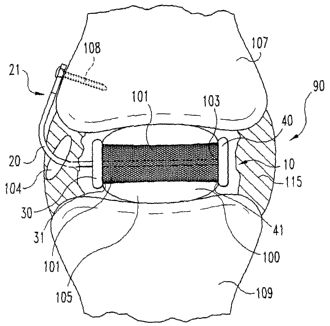
FIGS. 9-11, spinal implant system 90 includes a spinal implant 100 and a
spinal implant anchoring device 10 as described in reference to FIGS. 1
and 2. Inner surface 31 and 41 of securing members 30 and 40,
respectively, abut outer surface 105 of implant 100. As seen in FIG. 11,
25 anchoring rod 20 extends through aperture, or other defect, 104 in annulus
fibrosis 115 so that the first end 21 of anchoring device 10 may be
anchored to upper vertebra 107 with a bone screw 108. First end 21 may,
of course, be anchored to lower vertebra 109, or may be secured to both
vertebrae 107 and 109 if first end 21 is appropriately configured as
3o discussed above. The longitudinal axis X of the rod may extend parallel to
the longitudinal axis Y of the implant, but may extend through the implant in
CA 02425973 2003-04-15
WO 02/34169 PCT/USO1/32254
17
a wide variety of directions, as long as the rod functions to anchor the
implant in the disc space. Furthermore, the anchoring rod preferably
extends at least partially through the implant, but may extend completely
through the implant, entering one location, such as an end, and exiting
s another location, such as another end, including an opposing end. In
preferred forms of the invention, implant 100 may include a peripheral
supporting band 101 as further described below to provide further lateral
support for the implant, as well as to improve the strength of the implant. In
one form of the invention, band 101 may have apertures, or other openings
to therethrough, on opposing sides of the band which are in contact with the
securing member to allow the anchoring rod of the anchoring component,
or device, to be placed therethrough. Moreover, implant 100 further
includes a channel 103 extending therethrough through which the
anchoring rod may be disposed. The implant is preferably molded such that
is the channel is formed during the molding process. However, the channel
may be formed after formation of the implant in a variety of ways, including
drilling to form a channel having a desired shape with an appropriate drill
bit.
Referring now to FIGS. 12-14 in another form of the invention, a
2o spinal implant system 120 is shown which includes spinal implant 100 and
spinal implant anchoring device 50. Anchoring rod 60 extends through
aperture, or defect, 104 of annulus fibrosis 115. Furthermore, first end 61
of anchoring rod 60 of the anchoring device is secured to upper vertebra
107, but may be secured to lower vertebra 109, or both upper and lower
2s vertebrae, with an interference screw 110 as more fully described below
and as shown in FIG. 14. As seen in FIG. 14, one end of the anchoring rod
is wedged between the screw and the bone. Furthermore, first end 61 of
anchoring device 50 may be secured to both vertebra 107 and 109 for
added stability if first end 61 is appropriately configured as discussed
so above.
CA 02425973 2003-04-15
WO 02/34169 PCT/USO1/32254
18
The interference screws described herein can be non-resorbable,
resorbable and made form a wide variety of materials, including metals,
ceramics, polymers and combinations thereof. Non-resorbable metallic
materials include stainless steels, cobalt chrome alloys, titanium, titanium
s alloys, shape memory materials as described above, especially those
exhibiting superelastic behavior and including metals, and alloys thereof.
Resorbable materials include polylactide, polyglycolide, tyrosine-derived
polycarbonate, polyanhydride, polyorthoester, polyphosphazene, bioactive
glass, calcium phosphate, such as hydroxyapatite, and combinations
to thereof. The anchoring devices may also be anchored with other soft
tissue anchors known in the art, including suture anchors commonly used
in arthroscopy or sports medicine surgeries, for example. In the case of a
soft tissue or suture anchor, the end of the elongated body of the anchoring
device is attached to the end of the anchor, which is embedded and
is anchored in an adjacent vertebral body.
A wide variety of spinal implants for serving differing functions may
be anchored with the anchoring devices described herein, including
implants sized and configured for nucleus pulposus replacements, sized
and configured for partial or full disc replacements or other disc
2o reconstruction or augmentation purposes. Elastic, or otherwise resilient,
implants are most preferred. For example, implants may be formed from
hydrophilic materials, such as hydrogels, or may be formed from
biocompatible elastomeric materials known in the art, including silicone,
polyurethane, polyolefins such as polyisobutylene and polyisoprene,
2s copolymers of silicone and polyurethane, neoprene, nitrite, vulcanized
rubber and combinations thereof. In a preferred embodiment, the
vulcanized rubber is produced by a vulcanization process utilizing a
copolymer produced, for example, as in U.S. Patent No. 5,245,098 to
Summers et al., from 1-hexene and 5-methyl-1,4-hexadiene. Preferred
3o hydrophilic materials are hydrogels. Suitable hydrogels include natural
hydrogels, and those formed from polyvinyl alcohol, acrylamides such as
CA 02425973 2003-04-15
WO 02/34169 PCT/USO1/32254
19
polyacrylic acid and poly (acrylonitrile-acrylic acid), polyurethanes,
polyethylene glycol, poly(N-vinyl-2-pyrrolidone), acrylates such as poly(2-
hydroxy ethyl methacrylate) and copolymers of acrylates with N-vinyl
pyrolidone, N-vinyl lactams, acrylamide, polyurethanes and polyacrylonitrile
s or may be formed from other similar materials that form a hydrogel. The
hydrogel materials may further be cross-linked to provide further strength to
the implant. Examples of polyurethanes include thermoplastic
polyurethanes, aliphatic polyurethanes, segmented polyurethanes,
hydrophilic polyurethanes, polyetherurethane, polycarbonate-urethane and
to silicone polyether-urethane. Other suitable hydrophilic polymers include
naturally-occurring materials such as glucomannan gel, hyaluronic acid,
polysaccharides, such as cross-linked carboxyl-containing polysaccharides,
and combinations thereof. The nature of the materials employed to form
the elastic body should be selected so the formed implants have sufficient
is load bearing capacity. In preferred embodiments, a compressive strength
of at least about 0.1 MPa is desired, although compressive strengths in the
range of about 1 MPa to about 20 MPa are more preferred.
The implants can be shaped as desired. For example, the nucleus
pulposus implants may take the form of a cylinder, a rectangle, or other
2o polygonal shape or may be substantially oval. The implants may include
elastic bodies 750 that are tapered, such as at one end, as seen in FIGS.
15A and 15B, in order to create or maintain lordosis. Furthermore, in
certain forms of the invention, the implants generally conform to the shape
of the nuclear disc space. Additionally, implants can be sized to fit within
2s an intervertebral disc space, preferably surrounded by an annulus fibrosis,
or at least partially surrounded by an annulus fibrosis. That is, the implants
preferably are of a height and have a diameter that approximates the height
and diameter of an intervertebral disc space. In certain forms of the
invention, a spinal implant may be a nucleus pulposus implant and may
3o thus be sized to fit within the natural intervertebral disc space. In other
embodiments, the spinal implants may be disc replacements as described
CA 02425973 2003-04-15
WO 02/34169 PCT/USO1/32254
herein, and may be sized to fit within the intervertebral disc space that
includes the space resulting when the inner annulus fibrosis layer, or a
portion thereof, is removed. Such a spinal implant would therefore be sized
to fit within the larger intervertebral disc space that includes the space
s resulting from removal of a portion of the annulus fibrosis, and would thus
typically have a width or diameter that is substantially larger than the
natural nucleus pulposus.
As mentioned above, the implant to be anchored preferably is
reinforced for increased strength and to decrease lateral deformation of the
io implant. Accordingly, in yet another aspect of the invention, a reinforced
spinal implant is provided. Referring now to FIGS. 16 and 17, implant 120
includes a load bearing elastic body 121 with an upper surface 122 and a
lower surface 123. Implant 120 includes a preferably flexible, supporting
member, such as peripheral supporting band 130 disposed
is circumferentially about body 121. Band 130 is similar to band 100
discussed above, with the exception that band 130 does not have openings
therethrough on opposing sides of the band. As the implant, including the
elastic body and supporting band, advantageously may replace all or a
portion of the natural nucleus pulposus, while retaining the annulus fibrosis
20 or a portion thereof, the implant may be sized to fit within the
intervertebral
disc space defined by the annulus fibrosis or a portion thereof.
As seen in FIG. 16, elastic body 121 includes upper and lower
surfaces 122 and 123, respectively, portions of which are exposed to
directly contact adjacent vertebral endplates. This exposure allows the
2s lubricated upper and lower surfaces of elastic body 121 to articulate
against
the endplates to minimize abrasive wear of supporting band 130 and the
endplates. Although the amount of the upper and lower surfaces of elastic
body 121 that are exposed may vary, typically at least about 50%,
preferably at least about 70%, more preferably at least about 80% and
3o most preferably at least about 90% of the surfaces are exposed. In certain
forms of the invention, the elastic body core may function as a nucleus
CA 02425973 2003-04-15
WO 02/34169 PCT/USO1/32254
21
pulposus, and thus functions as a load bearing component with stress
transfer capabilities.
Peripheral supporting band 130 helps restrict excessive horizontal
deformation of elastic body 121 upon loading conditions, as seen
s progressively in FIG. 18A, thereby helping to restore and maintain disc
height. The hoop stress in the band increases exponentially after some
small, initial deformation as seen in FIG. 18B. Band 130 preferably
decreases lateral deformation, compared to deformation of an implant
without the circumferential reinforcing band, as desired. Band 130 may, for
to example, decrease lateral deformation by at least about 20%, preferably at
least about 40%, further preferably at least about 60%, more preferably at
least about 80% and most preferably at least about 90%. An implant, such
as one that includes an elastic body, having such a flexible supporting
band, will be flexible and otherwise resilient to allow the natural movements
is of the disc and provides shock absorption capability at low to moderate
applied stress, but will resist excessive deformation for disc height
maintenance under high loading conditions. As described herein in the
case of a lumbar disc, for example, low applied stress includes a force of
about 100 Newtons to about 250 Newtons, moderate stress includes a
2o force of about 250 Newtons to about 700 Newtons, and high loading
conditions, or high stress, includes a force of about above 700 Newtons.
Such a reinforced implant may be advantageously anchored with the
anchoring devices described herein. Moreover, other outer covers, or
jackets, as described in U.S. Patent No. 5,674,295 may be utilized to
2s reinforce implants to be anchored with the devices described herein. In
preferred forms of the invention, the bands, jackets, or other outer covers
or similar supporting members are flexible in that they may be folded or
otherwise deformed, but are substantially inelastic so that the implant is
more fully reinforced or otherwise supported.
so Peripheral supporting band 130, as well as other outer covers, or
jackets, may be made from a wide variety of biocompatible polymers,
CA 02425973 2003-04-15
WO 02/34169 PCT/USO1/32254
22
metallic materials, or combination of materials that form a strong but
flexible support to prevent excessive lateral (horizontal) deformation of the
core under increasing compressive loading. Suitable materials include
non-woven, woven, braided, or fabric materials made from polymeric fibers
s including cellulose, polyethylene, polyester, polyvinyl alcohol,
polyacrylonitrile, polyamide, polytetrafluoroethylene, polyparaphenylene
terephthalamide, and combinations thereof. Other suitable materials
include non-reinforced or fiber-reinforced elastomers such as silicone,
polyolefins such as polyisobutylene and polyisoprene, polyurethane,
io copolymers of silicone and polyurethane, neoprene, nitrite, vulcanized
rubber and combinations thereof. In a preferred form of the invention, a
combination, or blend, of silicone and polyurethane is used. Furthermore,
the vulcanized rubber is preferably produced as described above for the
spinal implants. Supporting band 130 is advantageously made from
Is materials described herein that allow it to be porous, which, in the case
of
an elastic body made from a hydrogel, or other hydrophilic material, allows
fluid circulation through the elastic core body to enhance pumping actions
of the intervertebral disc. Supporting members may further be formed from
carbon fiber ceramic, ceramic fibers, metallic fibers, or other similar fibers
2o described, for example, in U.S. Patent No. 5,674,295, or from metallic
materials that include shape memory materials as described above,
especially those exhibiting superelastic behavior, titanium, titanium alloys,
stainless steel, cobalt chrome alloys and combinations thereof. FIGS. 19A-
19D show supporting bands of various patterns, including braided patterns
2s (bands 140, 145 and 150) or porous patterns (band 155). It is realized that
the braided materials may also be porous.
In addition to reinforcing the implants described herein with an outer
cover, jacket or supporting band as described above, spinal implants 100,
such as those formed from a hydrogel material, that are advantageously
3o anchored with the anchoring devices described herein may be reinforced
by forming the implant by molding hydrogels of different stiffness together
WO 02/34169 PCT/USO1/32254
2
CA 02425973 2003-04-15
WO 02/34169 PCT/USO1/32254
23
and by annealing methods that include dipping the hydrogel in a hot oil
bath, as described in U.S. Patent No. 5,534,028. Other suitable reinforced
spinal implants, such as nucleus pulposus implants, that may
advantageously be used in the system of the present invention include
s those described in U.S. Patent Nos. 5,336,551, as well as the novel
implants described herein. As discussed above, the implant may be
advantageously shaped to conform to the intervertebral disc space, or
shaped as otherwise desired, as long as the implant has load bearing
capability. Although the amount of load the implant is required to bear may
to vary depending on several factors, including the particular location in
which
the implant will be positioned, as well as the general health of the
surrounding intervertebral discs, it is preferred that the implant be able to
bear a load of at least about 20 Newtons for cervical discs, at least about
50 Newtons for thoracic discs and at least about 100 Newtons for lumbar
is discs.
In yet other forms of the invention, an implant reinforced with a
peripheral supporting band as described above is provided that is further
reinforced with one or more straps. The straps may be advantageous in
preventing the peripheral supporting band described herein from slipping,
20 or otherwise sliding off the implant. Referring now to FIGS. 20 and 21, at
least one strap 134 extends along upper surface 122 and at least one strap
135 extends along lower surface 123 of elastic body 121 of implant 140.
Ends 136 of strap 134 and ends 137 of strap 135 are each preferably
connected, or otherwise attached, to peripheral supporting band 130'. The
2s point of attachment may be any location that will secure the strap,
including
at the upper margins 138 of the band, lower margins 139 of the band or
any region between the upper and lower margins. Although two straps
134 and 135 are shown extending along upper surface 122 and lower
surface 123, respectively, in FIGS. 20 and 21, one continuous strap may be
3o utilized that extends completely around the implant, or the strap utilized
may be in multiple pieces, as long as the combination of straps are
CA 02425973 2003-04-15
WO 02/34169 PCT/USO1/32254
24
sufficient to prevent excessive slipping and or sliding of the supporting
band. Furthermore, more than one strap may extend along upper surface
122 and more than one strap may extend along lower surface 123. For
example, as seen in FIGS. 22 and 23, straps 820, 830, 840 and 850 of
s implant 150 are attached to strap 130". Straps 820 and 830 are also
attached to strap 134' and straps 840 and 850 are also attached to strap
135'.
As mentioned above, the spinal implant with the flexible peripheral
supporting band may be anchored utilizing the anchoring devices described
to herein. In other forms of the invention, implants as described herein may
be anchored with an outer, preferably resorbable, shell as described in U.S.
Patent Application 09/650,525 to Trieu, filed August 30, 2000. In further
forms of the invention, the implant may further include various outer surface
features that may further restrain movement of the implant in the
is intervertebral disc space, with or without the outer shell. Such surface
features are also more fully described in U.S. patent application 09/650,525
to Trieu, filed August 30, 2000.
In yet other forms of the invention, a tension band 700 may be
secured to the anchoring device and to an adjacent vertebra to, for
2o example, provide further stabilization of the device, especially wherein
the
annulus and/or the ligament surrounding the annulus at the defect site are
compromised. Referring now to FIGS. 24 and 25, one end 701 of band
700 may be attached to an anchoring device, such as anchoring device 10"
(similar to anchoring device 10 except that bracket 123" is utilized), at, for
2s example, bracket 123", and the other end 702 may be secured to a plate
710, such as a metal plate, that is secured to the adjacent vertebra utilizing
screws 108 as described herein. Band 700 may be attached to the
anchoring device in a variety of ways, including crimping, tying, mechanical
locking or may be secured with the same screws used to secure the
so anchoring device to the vertebral bodies. If two anchoring devices are
utilized as described below, or if a single anchoring device is used that is
CA 02425973 2003-04-15
WO 02/34169 PCT/USO1/32254
secured to both adjacent vertebrae, one end 701 of tension band 700 may
be attached to one of the brackets, or other areas, of the first anchoring
device and the other end 702 of band 700 may be attached to the other
bracket, or other area, of the second anchoring device. The tension band
s is preferably flexible fio allow some degree of motion, but is substantially
inelastic to prevent excessive extension.
The tension band may be formed from a wide variety of natural or
synthetic tissue biocompatible materials. Natural materials include
autograft, allograft and xenograft tissues. Synthetic materials include
to metallic materials and polymers. The metallic materials can be formed
from shape memory alloy, including shape memory materials made from,
for example, the nickel-titanium alloy known as Nitinol as described above.
The shape memory materials may exhibit shape memory as described
above, but preferably exhibit superelastic behavior. Other metallic
is materials include titanium alloy, titanium, stainless steel, and cobalt
chrome
alloy. Suitable polymeric materials include, for example, polyethylene,
polyester, polyvinyl alcohol, polyacrylonitrile, polyamide,
polytetrafluoroethylene, poly-paraphenylene, terephthalamide and
combinations thereof. The materials used to form the tension band can be
2o in a variety of forms, including the form of a fiber, woven, or non-woven
fabric, braided, bulk solid and combinations thereof. The tension band may
further be treated, such as by coating and/or impregnating, with bioactive
materials that may enhance tissue ingrowth and/or attachment, including
hydroxyapatite, bioglass, and growth factors. Suitable growth factors
2s include transforming growth factors, insulin-like growth factors, platelet-
derived growth factors, fibroblast growth factors, bone morphogenetic
proteins as further described herein and combinations thereof.
In yet another aspect of the invention, methods of anchoring a spinal
implant are provided. In one form of the invention, a method includes
3o providing an elastic spinal implant and an anchoring component as
described herein. The elongated body, or anchoring rod, component of the
CA 02425973 2003-04-15
WO 02/34169 PCT/USO1/32254
26
anchoring component is at least partially extended, or otherwise disposed,
through the implant. The implant may include a pre-formed channel
therethrough, preferably formed during formation of the implant, through
which the anchoring rod may be extended. In alternative embodiments, the
s implant may be formed around internal securing members as discussed
above. The longitudinal axis of the anchoring rod may also extend parallel
to the longitudinal axis of the implant, or any other direction as mentioned
above that will allow the anchoring rod to anchor, secure, restrain or
otherwise hold the implant in the disc space. As an example, the anchoring
to rod, as well as the securing members, may take a tortuous path through
the implant, especially when the anchoring bodies have ends defining
variously-shaped securing members, as more fully described above, with
reference to, for example, FIGS. 7N, & 70 and 7T.
As further discussed above, in those forms of the invention wherein a
is securing member is at an end of the implant, the securing member may be
attached after the elongated body component is extended through the
implant. For example, with reference to FIGS. i and 7, securing member 40
may be attached to end 22 of elongated body 20 after anchoring rod 20 is
extended through channel 103 of implant 100. Moreover, the securing
2o member may also be formed after rod 20 is extended through channel 103, as
in the case where securing member 40 is defined by a knot structure. In other
forms of the invention, the channel may be formed after the implant is formed
by forming a channel with an appropriate tool, such as a drill with an
appropriately sized and shaped drill bit. ~ne of the ends of the anchoring
2s component are then secured to an adjacent vertebra.
In further aspects of the invention, methods of reducing deformation of
a spinal implant are provided. In one embodiment, a method includes
disposing a flexible peripheral supporting band as described above
circumferentially about the implant.
3o The implants formed from a hydrogel, or other similar hydrophilic
material described herein, including the supporting band of the reinforced
CA 02425973 2003-04-15
WO 02/34169 PCT/USO1/32254
27
implants, may advantageously deliver desired pharmacological agents.
The pharmacological agent may include a growth factor that may
advantageously repair a damaged annulus fibrosis, endplates or may have
some other beneficial effect. A wide variety of growth factors may
s advantageously be employed in the present invention. For example, the
growth factor may include a bone morphogenetic protein, transforming
growth factors, such as transforming growth factor-~3 (TGF-~3), insulin-like
growth factors, platelet-derived growth factors, fibroblast growth factors, or
other similar growth factor having the ability to repair the endplates,
io annulus fibrosis and/or nucleus pulposus of an intervertebral disc, or the
ability to have some other beneficial effect. The growth factors, or other
pharrriacological agents, are typically included in the implant in
therapeutically effective amounts. For example, the growth factors may be
included in the implants in amounts effective in repairing an intervertebral
is disc, including repairing the endplates, annulus fibrosis and nucleus .
pulposus. Although these amounts will depend on the specific case, the
implants may typically include no more than about five weight percent of
the growth factors, and preferably no more than about one weight percent
of the growth factors. In a preferred form of the invention, the growth factor
2o is a bone morphogenetic protein. Recombinant human bone
morphogenetic proteins (rhBMPs) are further preferred because they are
available in large quantities and do not transmit infectious diseases. Most
preferably, the bone morphogenetic protein is a rhBMP-2, rhBMP-4 or
heterodimers thereof. However, any bone morphogenetic protein is
2s contemplated, including bone morphogenetic proteins designated as BMP-
1 through BMP-18.
BMPs are available from Genetics Institute, Inc., Cambridge,
Massachusetts and may also be prepared by one skilled in the art as
described in U.S. Patent Nos. 5,187,076 to Wozney et al.; 5,366,875 to
3o Wozney et al.; 4,877,864 to Wang et al.; 5,108,922 to Wang et al.;
5,116,738
to Wang et al.; 5,013,649 to Wang et al.; 5,106,748 to Wozney et al.; and PCT
CA 02425973 2003-04-15
WO 02/34169 PCT/USO1/32254
28
Patent Nos. W093/00432 to Wozney et al.; W094/26893 to Celeste et al.;
and W094/26892 to Celeste et al. All bone morphogenic proteins are
contemplated whether obtained as above or isolated from bone. Methods for
isolating bone morphogenetic protein from bone are described, for example,
s in U.S. Patent No. 4,294,753 to Urist and Urist et al., 81 PNAS 371, 1984.
In other forms of the invention, the pharmacological agent may be one
that is used for treating various spinal conditions, including infected spinal
cords, cancerous spinal cords and osteoporosis. Such agents include
antibiotics, analgesics and anti-inflammatory drugs, including steroids. Other
io such agents are well know to the skilled artisan. These agents are also
used
in therapeutically effective amounts that will treat the various conditions
and
the symptoms they cause. Such amounts may be determined by the skilled
artisan depending on the specific case.
The~pharmacological agents are preferably dispersed within the
is hydrogel, or other hydrophilic, implant for in vivo release, and/or, with
respect
to implants with an elastomeric resorbable outer shell or those with a
flexible
supporting band, may be dispersed in either the band, the outer shell, or
both.
The hydrogel can be cross-linked chemically, physically, or by a combination
thereof, in order to achieve the appropriate level of porosity to release the
2o pharmacological agents at a desired rate. The agents may be released upon
cyclic loading, and, in the case of implants including a resorbable outer
shell,
upon resorption of the shell. The pharmacological agents may be dispersed
in the implants by adding the agents to the solution used to form the implant,
as long as the processing conditions will not adversely affect the agent.
2s Alternatively, the implants may be soaked in an appropriate solution
containing the agent, or by other appropriate methods known to the skilled
artisan.
While the invention has been illustrated and described in detail in the
drawings and foregoing description, the same is to be considered as
3o illustrative and not restrictive in character, it being understood that
only the
preferred embodiment has been shown and described and that all changes
CA 02425973 2003-04-15
WO 02/34169 PCT/USO1/32254
29
and modifications that come within the spirit of the invention are desired to
be protected. For example, in addition to being straight, the elongated
bodies of the anchoring device may exhibit other advantageous shapes as
shown in FIGS. 26 and 27. As seen in FIG. 26, anchoring rod 920' is
s arcuate. As seen in FIG. 27, anchoring rod 920" has a bend adjacent to
securing member 40. Other bent or angled anchoring components may be
understood by those of ordinary skill in the art, and such embodiments are
encompassed by this invention. Furthermore, the devices described herein
may be inserted and anchored via a wide variety of approaches, including
to posterior, lateral, oblique and anterior as shown in FIGS. 28-31,
respectively. Moreover, the nucleus pulposus implant systems may include
one or more implants disposed on the anchoring rods of the anchoring
devices described herein. As seen in FIG. 32, two implants 100' are
disposed on anchoring rod 20 of anchoring device 10. Thus, typically at
I5 least one implant is included in the implant systems described herein.
Additionally, in other forms of the invention, the spinal implant
systems may include one or more elastic bodies and one or more
anchoring devices. Referring now to FIG. 33, two anchoring devices are
included in the system along with two elastic bodies, each elastic body
2o disposed on a different anchoring device 950 or 960. Each anchoring
device may be independently anchored to an adjacent vertebra. In
alternative embodiments, first ends 951 and 961 of anchoring rods 953 and
963, respectively, may be connected, or otherwise attached to each other
to form a single extension, or end, of the anchoring rods, which may in turn
2s be attached to an adjacent vertebra or bracket as described herein. The
latter case is shown in FIG. 33, wherein first ends 951 and 961 of
elongated bodies 953 and 963, respectively, of anchoring devices 950 and
960 are integral with each other. Utilizing such a system with anterior and
posterior implants IA and IP, respectively, implants having different heights
3o may be used to create or maintain lordosis. For example, if a cylindrical
CA 02425973 2003-04-15
WO 02/34169 PCT/USO1/32254
implant is desired, anterior implant IA may have a larger diameter, and thus
a larger height, than posterior implant IP.
All references cited herein are indicative of the level of skill in the art
and are hereby incorporated by reference in their entirety.