Note: Descriptions are shown in the official language in which they were submitted.
CA 02426064 2003-04-22
BIOLOGICAL REPLACEMENT VALVE ASSEMBLY
Field of the Invention
This invention relates to a prosthesis that includes a biological valve
contained within a vein that is attached to a stmt for percutaneous
implantation into
a predetermined site within a human body.
Background of the Invention
There is an ongoing need in the medical field to be able to replace
malfunctioning heart valves and the like without the need for major surgery. A
number of advances have been made in procedures involving the percutaneous
implantation of biological valvular prosthesis taken from animals. One such
procedure is disclosed in 1.1.5. Patent No. 5,840,081 to Anderson in which an
animal
vein containing a valve is sutured to the inside of a stmt and delivered to a
valve site
by a balloon catheter.
The stmt employed by Andersen and others such as Bessler in U.S. Patent
170. 5;855,601 is fabricated from a relatively rigid metal, such as stainless
steel, that
is specifically. designed so that the elastic limit of the metal is exceeded
when the
stmt is expanded by the balloon. Accordingly, the expanded st:ent is unable to
totally conform to the shape and irregularities of the implantation site and
thus may
become dislodged over time. Furthermore if a need arises to further expand the
stmt
after the initial implantation, as may be the case in children who are
growing, the
only alternative is to resort to surgery.
Many scents in current usage are laser cut from a solid metal cylinder. This
in turn, can produce sharp edges along the cutting lines which valvular
prosthesis
can cut into a biological valve during the implantation procedure leading to
early
failure. Other stems are formed of wire strands that are welded together to
establish
a spring like structure. Here again the laser can produce rough or sharp edges
that
can damage tissue of a biological valvular prosthesis. In addition the welds
typically
are stronger than the wire strands of the stem and, as a result, the strands
will
normally break before welds causing the scent to fragment which in turn can
have
serious consequences.
CA 02426064 2003-04-22
-2-
Many stems that are in present day usage, contract axially as the stmt is
expanded radially. This, of course can cause problems where the stmt is
employed
to implant a biological valvular prosthesis. The shrinkage in length can
constrict the
vein or crimp portion of the biological valve structure as well as causing the
valve
structure from detaching itself from the stmt.
Many biological valves are harvested from animals such as cows wherein the
valve is located within a relatively thick vein such as the jugular vein.
Because of
the thick wall structure of the vein the delivery package; mounted upon the
balloon of
the catheter becomes rather bulky and thus difficult to percutaneously implant
in a
human patient, as for example, into the heart through the femoral artery.
Summary of the Invention
It is therefore and object of the present invention to improve a biological
valvular prosthesis used for percutaneous implantation into a human body site
for
1 j example, the heart region of a patient.
It is a further object of the present invention to reduce t:he thickness of a
biological valvular prosthesis that is used for percutaneous implantation into
a
patient.
A still further object of the invention is to provide an improved biological
valvular prosthesis that includes a stmt that exhibit minimal axial
contraction as the
stmt is expanded radially.
Another object to the invention is to provide a stmt for implanting a venous
valvular replacement for a human valve that will readily conform to the shape
of the
valve implantation site.
Yet another object of the present invention is to improve a scent mounted
biological valve that can be collapsed onto a balloon catheter to provide a
very low
profile replacement package for percutaneous implantation.
Still another object of the present invention is to more precisely fit a
venous
valvular replacement for a human valve to a stmt for percutane:ous implanting
of the
valve into a human patient.
CA 02426064 2003-04-22
-3-
These and other objects of the present invention. are attained by a prosthetic
device for implanting a biological valve into a patient. The prosthesis
includes a
stmt having a plurality of wire ribbon sections, each of which is fabricated
from a
strand of fine round wire. The ribbon sections are interconnected by welds to
form a
tubular member. Each ribbon section further contains a periodic series of
substantially sinusoidal bends along the length of the ribbon. Each bend
contains an
apex that is welded to an apex carried by an adjacent ribbon section. The
ribbon
sections are preferably fabricated from a fully annealed platinum alloy strand
of wire
having little or no shape memory. Initially the stmt is expanded to a desired
diameter related to the diameter of the body lumen at the implantation site.
The vein
wall that contains the biological valve is trimmed or pef;led back to a size
such that
the wall thickness of the vein is reduced to about between 50% and 90% of its
original size so that the outside diameter of the vein is about edual to the
inside
diameter of the expanded stmt. The vein is then sutured to the expanded stmt
so
that the vein is supported in a cylindrical fully opened configuration. The
welds
used to cojoin the stmt ribbons are formed so that they are weaker than the
tensile
strength of the ribbons wire strand. As a result a weld vwill break before the
wire
strand can be stressed to a point of fragmentation. The welds are all
contained inside
the boundaries described by the inside and outside diameters of the stmt when
the
stmt is expanded. Because the size of the vein that supports the biological
valve has
been considerably reduced, the stmt and valve prosthesis can be more compactly
compressed about the balloon of a catheter to enhance the ease of percutaneous
insertion of the package.
Brief Description of the Drawing
For a further understanding of these and other objects of the invention,
reference will be made to the following detailed description of t:he invention
which is
to be read in connection with the accompanying drawing, wherein:
CA 02426064 2003-04-22
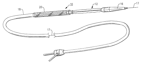
FIG. 1 is a schematic representation of a balloon catheter used to
percutaneously implant the prosthesis of the present invention within a
desired body
site;
FIG. 2 is a side elevation showing a stmt suitable for use in the present
j invention;
FIG. 3 is a perspective view further illustrating a prosthetic biological
valvular replacement for a human valve sutured to the expanded stmt;
FIG. 4 is a section taken along lines 4-4 in Fig. 2; and
FIG. 5 illustrates the vein section of a biological valvular replacement being
trimmed to reduce the wall thickness of the vein section of the replacement.
Detailed Description of the Invention
Turning now to the drawings, Fig. 1 illustrates a balloon catheter generally
referenced 10, that is suitable for percutaneous implanting a prosthetic
device 12
containing a biological replacement valve within a human patient. The catheter
includes an inflatable balloon upon which the prosthetic device 12 is mounted
in a
tightly crimped configuration. Although not shown, the balloon is connected to
a
lumen inside the catheter through which a radio-opaque fluid is provided to
the
balloon to inflate the balloon and thus expand the stmt in a radial direction
to
implant the prosthesis in a desired location. After implantation the fluid is
removed
through the lumen to deflate the balloon and the catheter is removed from the
implantation site. A pointed tip 16 is mounted at the distal end of the
catheter to
help direct the catheter through a body lumen into the implantation site. The
catheter contains a central lumen through which a guide wire I7 is slidably
contained. The guide wire is further arranged to pass through the balloon
section
and the tip section of the catheter. The guide wire is initially introduced
into the
desired implantation site through a suitable body lumen and the catheter is
then
guided along the wire into the site.
The catheter is covered by a sheath I 8 and a close running fit is provided
between the sheath and the catheter to allow for axial movement between the
sheath
CA 02426064 2003-04-22
-j-
and the catheter. A cylindrical shield 20 is attached at the distal end of the
sheath
and is arranged to protectively house a prosthetic device that has been
tightly
crimped upon the balloon section of the catheter.
As will be further explained below, with reference to Figs. 2-5, the
prosthetic
device 12 includes a collapsable stmt 28 and a biological venous valvular
replacement unit 29 which preferably has been harvested from the jugular vein
of an
animal, such as a cow, and is secured to the inside of the stmt. Initially,
the sheath
along with the attached shield is pulled back along the catheter to expose the
collapsed balloon and the prosthetic device is passed over the balloon and
crimped
tightly to the balloon to establish a compact low profile package. The sheath
is then
moved forward along the catheter to place the attached shield over the package
to
protect the prosthesis during percutaneous insertion. Once the package is
positioned
within the insertion site the shield again is moved back and the balloon
inflated to
implant the biological valve replacement unit within the site.
Turning more specifically to Figs. 2-5, there is illustrated a stmt 28 that is
particularly well suited for use in the present invention. A biological venous
valvular replacement 29 for a defective heart valve is carried inside of the
scent.
Although the present valve replacement 29 is for percutaneous implantation of
a
pulmonary valve within the heart of a patient, it should clear that the
present device
can be used in a number of similar applications without departing from the
teachings
of the invention. As illustrated in Fig. 3, the biological replacement unit
includes a
section of vein 32 that contain a valve 33. As will be explained below in
further
detail the venous valvular replacement is attached to the stmt by means of
sutures
34.
The present expandable stmt 28 includes a series of fine wire ribbon
sections, each designated 3S that are joined together to create a tubular or
cylindrical
member. The wire stand of each section is fabricated.o.f a soft,, highly
malleable
metal alloy that has been fully annealed to remove as much of its spring
memory as
possible. Preferably the wire material is fabricated of an alloy consisting of
about
90% platinum and 10% iridium that has a tensile strength of between 150,000
psi
CA 02426064 2003-04-22
and 175,000 psi. Although a platinum iridium wire is preferred for use in the
present
stmt, other alloys having similar properties such as a gold nickle alloy may
also be
employed. Prior to winding the wire ribbon sections into a cylindrical shape,
each
section is formed so that it contains a series of alternating sinusoidal bends
36. The
sections are formed by winding the strand of wire between rows of vertical
pins
projecting from the surface of a flat substrate. The strand is wound about the
pins in
alternate rows to create a sinusoidal shaped ribbon sections having a desired
number
of bends and a free length of wire at each end of the ribbon sections.
Each ribbon section is next wound into a cylinder and the cylinders
are then placed in axial alignment so that the apex of each bend section is
located in
close proximity with the apex of a bend section on an adjacent ribbon section.
The
adjacent bends are then welded together to cojoin the ribbon section in
assembly.
Although not shown, the free ends of the adjacent cylindrical ribbons, in
turn, are
bent into parallel overlapping alignment and are cojoined using similar
section
welds.
Referring to Fig. 4, there is illustrated a typical weld faint 37 used in the
practice of the present invention. Each weld is formed so that it lies inside
the
boundaries of the cylindrical scent as described by the inside diameter and
outside
diameter of the stmt. Accordingly, the weld does not protrude beyond the
boundaries of the wire cylinder into regions where rough edges of the welds
might
come in contact with the tissue of the biological valve replacement thereby
preventing rips or tears from forming in the tissue which might potentially
lead to
failure of the prosthesis.
A stmt of the construction and configurafion as herein describe has
extremely good flexibility, dimensional stability, very smooth surfaces, a low
profile
when collapsed and an immunity to fatigue and corrosion. As should be evident
the
length of the scent can be varied by varying the number of ribbon sections
that are
utilized. By the same token, the working range of the stem between its fully
collapsed condition and it fully expanded condition can also be varied by
varying the
number of bends in each of the ribbon sections. As can be seen each stmt can
be
CA 02426064 2003-04-22
tailored for insertion into a particular body site to provide for the most
effective
implantation of the biological valve which is attached to the stmt.
Because of the scent construction there is very little or no axial deformation
of the stmt as it is radially expanded or collapsed. Another feature of the
present
stmt is its ability to be reconfigured even after implantation without
adversely
effecting the stems performance. This feature is important in cases where a
valve
has been implanted in a growing child. Rather than replacing a valve
periodically
during the growth period, the supporting scent can be simply reconfigured to
accommodate for growth using a percutaneously introduced balloon catheter for
re-
engaging the stmt to reconfigure the stmt so that it will conform to the
changes in
the implantation site produced by growth.
As illustrated in Fig. ~, the stent is initially expanded to a desired
diameter
which generally conforms to the body vessel configuration at the implantation
site.
Next, as illustrated in Fig. 5, the vein section of the valve is trimmed to a
desired
length conforming to the length of the stmt with the valve 33 being located in
about
the mid-region of the stmt. In addition, the wall of the vein 32 is reduced in
thickness by 50% to 90% to considerably reduce the size of the valve package
when
the stmt is collapsed over the balloon prior to insertion. As illustrated in
Fig. 5, it
has been found that the jugular vein of a bovine animal is formed by layers of
tissue
that can be readily peeled back using a sharp instrument 40 to remove the
layers
without destroying the integrity of the vein structure or its ability to
function in a
replacement prosthesis. The wall of the vein is trimmed so that its outside
diameter
about matches the inside diameter of the expanded stmt. The vein is then
passed
into the expanded stmt and the vein sutured to the stmt as illustrated in Fig.
3. The
sutures are arranged to support the vein in a fully opened circular
configuration
within the expanded stmt.
Once the prosthesis has been sutured in place, it is passed over the balloon
section of the catheter and the scent is collapsed tightly agaialst the
balloon to provide
a more compact than normal package that can more easily be delivered through a
CA 02426064 2003-04-22
_g_
body lumen into an implantation site when compared to similar devices
employing
bovine or equine biological valves replacements.
While the present invention has been particularly shown and described with
reference to the preferred mode as illustrated in the drawing, it will be
understood by
one skilled in the art that various changes in detail may be/ effected therein
without
departing from the spirit and scope of the invention as defined by the claims.