Note: Descriptions are shown in the official language in which they were submitted.
CA 02426246 2003-04-16
WO 02/33380 PCT/USO1/32456
METHOD AND DEVICE FOR DILUTING A FLUID AND DETECTING
ANALYTES WITI-IIN A DILUTED FLUID
FIELD OF THE INVENTION
The present invention relates to a method and device for processing,
sampling, and diluting a fluid and to a method and device for detecting
analytes
within a processed, sampled, and diluted fluid.
BACKGROUND OF THE INVENTION
Within countries of the world with sophisticated and well-developed medical
care systems and facilities, there remain substantial portions of the
population that do
not access the medical care system. Those who fail to obtain medical diagnoses
may
represent up to 50°r'° of those at risk for solve medical
conditions, This failure to
access medical diagnosis and care may be due to fear, mistrust, restricted
availability,
or lack of information or finances. Undiagnosed and untreated individuals
serve as a
reservoir for increased spread of infection. In the past yeas, new AIDS eases
in San
I 5 Francisco have doubled to 900 (Investors Business Daily, page A2, July 3,
2000)
marking what many doctors fear is a breakdown of our current approach to
controlling the infection.
In countries with less developed medical care systems and sophisticated
diagnostic testing laboratories, most of the population may not receive prompt
diagnosis of potentially treatable conditions. Illnesses such as AIDS,
tuberculosis,
malaria, and other infectious diseases may drain the country's talent and
economic
resources to fine extreme, with an overall reduction in the standard of living
and gross
domestic product. (Confronting AIDS, Public Priorities in a Global Epidemic,
World
Bank Research Report, 1~~7j. In sub-Saharan Africa, more than 10°~0
of the
population aged 15-X19 carries HIV. In seven of the sixteen countries
20°~o are
infected and in one country, Botswana, one in every three adults carries HIV
(Investors Business Daily, page Al, June 28, 2p00, Global View of HIV
Infection).
This toll of medical illness stands as a barrier to becoming part of the
community of
twenty-first century planet earth with all of its beneFlts in education,
communication
j~ alld 1111'OI'I11at1Q11 exchange. These COlllltl'leS rlSk belng hOpeleSSly
Illlred 111 SICkIleSS,
CA 02426246 2003-04-16
WO 02/33380 PCT/USO1/32456
death, and economic instability, without the help of more developed countries
and
new methods of diagnosis, treatment and prevention of disease.
Inexpensive, widely available and easily performed diagnostic tests that could
be used by individuals anywhere and at any time, without the need of
instrumentation
or formal training, would contribute to earlier diagnosis of medical
conditions for
which the tests were available. These tests would also facilitate improved
education
regarding those medical conditions being detected by empowering individuals to
become involved in their early detection and treatment. Earlier detection and
improved education would be expected to result in reduced transmission of
those
infections for which tests were available to individuals, with the result of
benefiting
the entire society in terms of fewer infections, and increased health and
workforce
productivity. Researchers at the Centers for Disease Control in Atlanta, GA,
have
used mathematical models fio predict that availability of rapid tests for I-
IIV would
lead to testing of at least an additional 700,000 people, and detect more than
8,000
additional infected individuals (Los Angeles Times, page A10, June 14. 2000,
FDA
Blamed for Holding Up Rapid AIDS Tests).
The major technological impediment to development of diagnostic tests
suitable for use by individuals has been the lack of device formats that are
both
accurate and user-friendly for individuals. Accurate test methods have been
available
for many years but most have required instrumentation. A test that requires
instrumentation does not fulfill the needs of individuals who do not choose to
or
cannot access the medical system, and hence do not become tested. A user-
friendly
test must be capable of being quickly pei°formed by individuals wha
have no formal
training, and it should require few steps and allaw testing in any location at
anytime
?5 chasm by the user.
In recent years, user-friendly diagnostic tests have been developed that allow
individuals to detect analytes in undiluted fluid samples, Examples are
pregnancy
tests that individuals may purchase in any large supermarket and perform on
undiluted urine at any location and at any time that they choose. Patents by
Ullman
3p et al., L.I.S. Patent No. ~1,857,~153, issued August I 5, 1989; Nazareth et
al., LI,S. Patent
CA 02426246 2003-04-16
WO 02/33380 PCT/USO1/32456
No. 5,739,011, issued April 14, 1998, and Pawlalc et al., U.S. Patent No.
5,770,160
issued .lone 23, 199$, are examples of urine HCG tests for pregnancy. Widely
available tests for detecting serum glucose may be easily performed by
individuals,
but they still requii°e instrumentation. Tests requiring
instrumentation are more
expensive and do not fit our strict definition of being user-friendly.
Some analytes may be detected in undiluted whole blood, serum or plasma.
U.S. Patent No. 5,762,871, by Neyer, issued March 10,1998, and U.S. Patent
No. 6,027,692 by Galen et al., issued February 22, 2000, teach tests of
undiluted
blood serum or plasma for glucose and fructosamine. U.S. Patent No. 5,166,051
by
Killeen et al., issued Novembei° 2~, 1992, instructs regarding tests of
whole blood for
analysing serum cholesterol.
For other tests and assay formulations, detection is more accurate only after
dilution of the test liquid. An example is the test for antibodies to HIV.
Commercially available immunoassays, as well as rapid strip format tests for
HIV
antibody, routinely dilute the sample approximately 1:100 before testing, as
shown in
U.S. Patent No. 5,922,533 by Vallari et al., issued July 13, 1999. In these
tests a
uniform dilution of serum is prepared in a separate location, and the test is
then
conducted with the uniformly diluted serum.
Attempts to dilute plasma or serum within the test device have employed
washing the plasma or serum from a plasma separatorlcallector pad. An example
is
that taught by Bernstein et al., U,S. Patent No. 5,753,97. The resulting
dilutions are
variable depending upon the volumes of wash fluid added. In addition, the
dilutions
are not uniform and result in gradient concentrations of serum components
migrating
down the test strip. The initial eluents from the collector pad contain high
~> concentrations of plasma or serum relative to diluent, and later eluents
contain small
amounts since most of the plasma or serum has already been washed from the
collector pad. This may produce undesirable effects on test performance, such
as
inconsistent migration rates down the test strip, or inadequate completion oI~
reactivity between test labeling reagents and plasma or serum components that
are
3p present in high concentrations, such as immunoglobulin. This results in
variations in
_,_
CA 02426246 2003-04-16
WO 02/33380 PCT/USO1/32456
r
the time required to complete the test and in some instances adverse effects
on
sensitivity or speciCcity.
Individuals conducting a test at home, or stafF in a physician's office or
point
of care location, cannot easily separate plasma or serum from whole blood.
They
also cannot safely use pipettes to produce a reliable dilution for testing.
Persons
conducting the test also will not usually have available to them
instrumentation for
evaluating test strip results.
It would be useful to have a method and device that permits individuals to
separate plasma or serum from finger-stick whole blood and obtain a reliable
and
1Q relatively uniform dilution of that serum or plasma for testing. It would
further be
useful for the device design to allow migration of the diluted liquid sample
along
diagnostic test strips contained within the device, so that a diagnostic test
result is
rapidly produced. Optimally, the test device must provide a clear result that
is easily
interpreted by visual observation without instrumentation. Finally, to be
widely
accepted for testing anywhere and at anytime, fine device and method must
provide
these results with a minimum number of easily performed steps and provide the
diagnostic test result within approximately ten minutes.
A method and device that would permit reliable dilution of a sample liquid,
and rapid determination of the presence or absence of specific analytes within
that
diluted sample, without requirements of formal training or instruments, would
be
very useful worldwide. The method and device of this invention seek to fulfill
this
need,
SUMMARY OF THE INVENTION
The present invention relates to a method and device for processing,
'~5 sampling, and diluting a fluid and to a method and device for detecting
analytes
within the processed, sampled, and diluted fluid.
In one aspect, the invention provides a method for processing and sampling a
Fluid. In the method, at least a portion of a porous membrane is saturated
with a first
Cluid. A portion of the membrane saturated with the first fluid is then
isolated and a
3(~ second Cluid is applied to the isolated portion of the membrane releasing
the first
CA 02426246 2003-04-16
WO 02/33380 PCT/USO1/32456
fluid from the isolated portion of the membrane. The first fluid can be
biological
Fluid such as whole blood or urine. In one embodiment, the first fluid is
applied to
the membrane at position other than of the isolated portion of the membrane
and is
migi°ated to the isolated portion of the membrane. Depending on the
application, the
second fluid can be a gas or a liquid.
In another aspect of the invention, a method for diluting a liquid sample is
provided. In the method, at least a portion of a porous membrane is saturated
with a
processed liquid. The portion o~ the membrane saturated with the processed
liquid is
isolated and a diluent is applied to the isolated portion releasing the liquid
sample
From the isolated portion of the membrane to provide a diluted liquid sample.
The
liquid sample can be biological fi7uid such as whole blood or urine. In one
embodiment, the first fluid is applied to the membrane at position other than
at the
isolated portion of the membrane and is migrated to the isolated portion o~
the
membrane.
In a further aspect, the invention provides a method for detecting an analyte
in a liquid sample. 1n the method, at least a portion of a porous membrane is
saturated with a liquid sample containing an analyte. A portion of the
membrane
saturated with the liquid sample is then isolated and a diluent is applied to
the
isolated portion releasing the liquid sample from the isolated portion of the
membrane. The released and diluted liquid is then directed to receptacle in
fluid
communication with a test strip where the presence of the analyte is detected.
In one
embodiment, the receptacle can be in fluid communicatian with a second strip,
for
example, a control strip. In one embodiment, the method detects an antibody to
HIV.
In another embodiment, the method detects an antibody to H. pylori antigen. In
a
further embodiment, the method detects HCG antigen.
In another aspect of the invention, a device for processing and sampling a
l~Liid is provided. In one embodiment, the device includes a membrane for
receiving
a first fluid; first and second members adjacent opposing major surfaces of
the
membrane for isolating a portion of the membrane, and a receptacle in fluid
;Q communication with the isolated membrane for receiving fluid from the
isolated
CA 02426246 2003-04-16
WO 02/33380 PCT/USO1/32456
membrane. The Cirst and second members can be engaged with the membrane to
isolate a portion of the membrane. The isolated portion of the membrane
maintains a
void volume substantially the same as the void volume of the unengaged
membrane.
In another aspect, the invention provides a device for diluting a liquid
sample.
In one embodiment, the device includes a membrane For receiving a liquid,
first and
second members adjacent opposing major surfaces of the membrane for isolating
a
portion of the membrane, and a receptacle in fluid communication with the
isolated
membrane for receiving the sample of liquid from the isolated membrane. The
first
and second members can be engaged with the membrane to isolate a portion of
the
membrane. The isolated portion of the membrane maintains a void volume
substantially the same as the void volume of the unengaged membrane.
In another aspect of the invention, a device for detecting an analyte in a
liquid
sample is provided. In one embodiment, the device includes a membrane for
receiving a liquid, first and second members adjacent opposing major surfaces
of the
membrane for isolating a portion of the membrane, a receptacle in fluid
communication with the isolated membrane for receiving the sample of liquid
from
the isolated membranes and a test strip in fluid communication with the
receptacle.
The first and second members can be engaged with the membrane to isolate a
portion
of the membrane. The isolated portion of the membrane maintains a void volume
?0 substantially the same as the void volume of the unengaged membrane. The
test strip
detects the presence of analyte in the liquid sample. In one embodiment, the
device
Further includes a control strip in fluid communication with the receptacle.
The
device can be used to detect the presence of an I-IIV antibody, an antibody to
H.
pylori antigen, or HCG antigen in the liquid sample.
In one embodiment of the device, a porous membrane is used to process and
migrate a liquid to a dilution port zone. In the dilution port zone, the
liquid saturated
membrane is compressed circumFerenfially isolating a defined volume of sample
within the isolated membrane. A specified quantity of diluent is then forced
through
the isolated volume of the membrane perpendicular to the direction of membran
a
~(~ lateral flow. The result is removal of the deFmed volume of liquid sample
from the
-6-
CA 02426246 2003-04-16
WO 02/33380 PCT/USO1/32456
membrane and simultaneous dilution o~ the processed sample liquid. In another
embodiment, the extracted sample liquid is directed through a narrow orifice
that
causes mixing of the diluent and production of a diluted sample. The diluted
sample
collects in a receptacle reservoir within the device that is in Fluid
communication
with one or more membranes. These membranes can include diagnostic and control
test strips po5ltlolled such that the diluted sample wicks from the receptacle
well and
migrates along each strip. Using such a methodology, the present invention
provides
a rapid diagnostic test that can be readily visually interpreted without a
requirement
for instrumentation.
In other aspects, kits For detecting an HIV antibody, an antibody to H. pylori
antigen, or HCG antigen are provided. Each Icit includes a device as described
above
and a container comprising a suitable diluent.
The present invention provides a method and device that permits processing,
sampling, and reproducible dilution of processed and sampled fluids, and
subsequent
detection of analytes within the diluted fluid sample. In one embodiment,
processing, sampling, dilution and detection are accomplished with a minimum
number of user-friendly steps that produce a result within ten minutes. In one
embodiment, the diagnostic result are lines that are clearly visible on a
white
background and that do not Fade and are easily interpreted without
instrumentation.
BRIEF DESCRIPTION OF THE DRAWINGS
The Foregoing aspects and many of the attendant advantages of this invention
will become more readily appreciated by reference to the following detailed
description, when taken in conjunction with the accompanying drawings,
wherein:
FIGURE 1 is an exploded perspective view of a representative device of the
?5 invention including a dilution port (10), a yoke (1 1), a cover (12), an o-
ring (13), a
midpiece (l~l), and a base (15);
FIGURE 2A is a top plan view and FIGURE 2B is a bottom plan view of
dilution port (10);
_7_
CA 02426246 2003-04-16
WO 02/33380 PCT/USO1/32456
FIGURE 3 is a cross section view of dilution port (10) through its long axis
hook arms { 16) and channel { 17) {the plane of fine cross section and
direction of sigh t
are indicated in FIGURE 2A);
FIGURE ~lA is a top plan view and FIGURE 4B is a fronfi elevation view of
yoke (11);
FIGURE SA is a top plan view and FIGURE SB is a front elevation view of
cover {I2);
FIGURE 6A is a top plan view, FIGURE 6B is a bofitom plan view, and
FIGURE 6C is a front elevation view of midpiece (14) (fine direction of sight
for
FIGURE 6C is indicated by the arrow labeled 6C in FIGURE 6A);
FIGURE 7A is a top plan view and FIGURE 7B is a front elevation view of
base (I S);
FIGURES 8A, 8B, and $C are top plan views of base {15) of a representative
device of the invention, FIGURE 8A illustrates a representative base { 15),
FIGURE 8B illustrates a representative base (15) with midpiece (1~), and
FIGURE 8B illustrates a representative base {15) with midpiece {14) and sample
membrane (20);
FIGURE 9A is a top sectional view of the dilution port, sample membrane,
o-ring, midpiece, base, and diagnostic test strip, when the dilution port is
depressed
2p into the locked position compressing the sample membrane in a
representative device
ofthe invention;
FIGURE 9B is a cross sectional view of the dllutloll port, sample membrane,
o-ring, midpiece, base, and diagnostic test strip shown in FIGURE 9A {the
cross-
section location and direction of sighfi of FIGURE 9B is indicated in FIGURE
9A);
?5 FIGURhS 10A, 10B, l OC, and 1 pD are top views of a representative device
of the invention showing visual results obtained by the method and device of
this
inven fiion to deflect antibodies to I-IIV, FIGURE 10A shows a valid negative
result,
FIGURE I0B shows a valid positive resulfi for antibody to I-IIV-l, FIGURE 1pC
shows an invalid negative results due to an insul~iicient amounfi of blood
tested, and
_g_
CA 02426246 2003-04-16
WO 02/33380 PCT/USO1/32456
FIGURE 1QD shows an invalid negative result due to a problem with the I-IIV-1
antigen;
FIGURES 1 1 A and 1 1 B are graphs illustrating the reproducibility of
dilutions
obtained in two series of tests using a representative method and device of
the
111VeI1tlo11;
FIGURES 12A and 12B are graphs illustrating the effect of membrane
thickness and sample-holding capacity of the sampling membrane on the amount
of
sample obtained for analysis by a representative method and device of the
invention;
FIGURE 13 is a graph illustrating the effect on sample volume added to
receiving well on the amount of sample obtained for analysis by a
representative
method and device of the invention; and
FIGURE I~l is a graph illustrating the effect of time delay between adding
sample and adding diluent on the final amount of sample obtained for analysis
by a
representative method and device of the invention.
DETAILED DESCRIPTION OF THE PREFERRED EMBODIMENT
The present invention relates to a method and device for processing,
sampling, and diluting a Iluid and to a mefhod and device for detecting
analytes
within the processed, sampled, and diluted fluid.
In the method for processing, sampling, and diluting a fluid, an amount of a
fluid is accepted by a porous membrane having a substantially uniform porous
structure, and a portion of the membrane saturated with the fluid is isolated
therEby
defining a predetermined volume of fluid. The predetermined fluid volume is
then
released from the isolated membrane with a specified quantity of diluen t to
provide a
diluted fluid sample. The device tar diluting a fluid includes a membrane For
~~ accepting a fluid and a mechanism For isolating a portion of the membrane
that is
saturated with the fluid. When the diluted fluid includes an analyte, the
invention
provides a method and device for detecting one or more analytes in the fluid.
In one aspect, the present invention provides a method and device For
processing, sampling, and diluting a liquid sa111p1e. The method can be
carried out
using the device illustrated in FIGURE 1. Referring to FIGURE I,
representative
CA 02426246 2003-04-16
WO 02/33380 PCT/USO1/32456
device I includes dllLltloll port 10, yoke I l, cover 12, o-ring 13, midpiece
1~, and
base 15. FIGURES 2A and 2B illustrate dilution port 10. FIGURE 3 is a crass
section view of dilution port 10 through its long axis hook arms 16 and
channel 17
(the plane of the cross section and direction of sight are indicated in FIGURE
2A).
FIGURES ~A and 4B illustrate yoke 11. FIGURES SA and SB illustrate cover 12.
FIGURES 6A, 6B, and 6C midpiece 14 (the direction of sight for FIGURE 6C is
indicated by the ar>'ow labeled 6C in FIGURE 6A). FIGURES 7A and 7B illustrate
base 15.
With reference to the illustrated device, in one embodiment, the method
includes (a) adding a liquid sample to the sample collection well of the
device, where
the well is in fluid communication with a porous membrane; (b) saturating at
least a
portion the membrane with the liquid by waiting approximately two to three
minutes
for migration of the liquid sample along a membrane within the device, which
in the
case of whole blood separates serum or plasma at the leading edge of the
membrane
leaving behind Cellular components; (c) isolating a portion of the membrane
saturated
with the liquid by depressing the dilution port of the device until it locks
in place,
thereby isolating a defined volume of the liquid-containing membrane; (d)
releasing
the isolated volume of liquid from the membrane with a diluent by inserting
the tip of
a provided vial into the depressed and locked dilution port to form a leak-
proof seal,
and squeezing the vial to deliver liquid through the dilution port, causing
removal of
the isolated volume of liquid from the defined isolated volume of membrane and
forcing the liquid into a well located in the base of the device,
simultaneously mixing
the diluted sample; (e) migrating the diluted sample along one or more
membranes,
such as one or more diagnostic fest strips and control Pest strips (migration
time from
aboLlt flue t0 aboLlt Sevell n1111L1teS), alld alloWlllg a VlSLlally I'eadable
result t0
develop; and (fib interpreting the test and control results in the viewing
windows of
the test device.
The method and device of the invention permits detection of any analyte
wlthlll a dllLlted llqllld Salllple that 1S a Illelllbel' Of d SpeGIfIC
blndlllg pall'. A blndlIlg
- I 0-
CA 02426246 2003-04-16
WO 02/33380 PCT/USO1/32456
pair consists of two different molecules that through physical, chemical, or
other
means, specifically bind to each other.
The detection of antibodies to HIV by a representative method and device of
the invention is described in Examples 7 and $. Tl~e detection of antibodies
to H.
pylari antigen by a representative method and device of the invention is
described in
Example 9. The detection of HCG antigen by a representative method and device
of
the invention is described in Example 10.
The method of the invention can be further illustrated by reference to the
device depicted in FIGURES l-10. Referring to FIGURE SA, liquid sample to be
diluted and analyzed is placed into receiving well 27. The well includes
sloping
sides ?6 and a volume sufficient to collect more than liquid sample sufficient
to
allow completion of the test. The liquid may be placed into the receiving well
with a
disposable pipette, which results in a defined amount of liquid sample added
to the
device. Alternatively, an amount of liquid sample can be added in excess of
the
minimum amount required to complete the test. For example, the user may be
instructed to add a volume su ff dent to fully caat the membrane at the bottom
of the
receiving well and sufficient to coat the lower edges of the receiving well
adjacent
the membrane.
A membrane (see membrane 20 in FIGURE $C) is in fluid communication
with the liquid sample received by the receiving well (see 27 in FIGURE SA)
and
transports at least a portion of the liquid sample from the receiving well to
directly
beneath the dilution port (see 30 in FIGURE SA).
The membrane transports at least a portion of the liquid to be diluted and
tested from the first end of the membrane locafed beneath receiving well 27 to
the
?5 second end of the membrane located beneath the dilution port (see 30 in
FIGURE SA
and ?~l in FIGURE 3) and above o-ring 13 contained within the upper surface
the
midpiece l~l (See FIGURES 1 and 6A).
The membrane allows transport of the analyte of interest. Ideally, the analyte
does not bind to the membrane, such that analyte quantity is reduced. The
membrane
should not otherwise interfere with the: analyte's accurate detection.
CA 02426246 2003-04-16
WO 02/33380 PCT/USO1/32456
The membrane has a specified length, width, and thickness and a substantially
uniForm thickness, porous structure, and void volume. The membrane can be
capable
of being cut into precisely sized strips for use in the assay. Through its
substantially
uniform thickness and void volume, the membrane provides for a substantially
constant amount of liquid contained within a given surface area (volume) of
membrane.
In the practice of the invention, the membrane is compressible. The
membrane's compressibility permits isolation of a portion of the membrane and
collapse of the void volume in those portions of the membrane compressed
between
mating edges of dilution port 10 (see 24 in FIGURES 3 and 9B) and o-ring 13
(see
FIGURE 9B and FIGURES GA and GC). The noncompressed area of membrane 20
(see FIGURE 9B) within the circumference of the ring of compressed membrane is
thereby isolated and defines a specified volume of liquid sample.
The membrane allows diluent to flow through its thickness in the portion of
membrane 20 (see FIGURE 9B) isolated between the mating and compressing
surfaces of the device noted above, thereby allowing release from the membrane
and
dilution for analysis.
Suitable membrane for use in the method and device are known in the art.
Certain membranes may be more preferable for specific tests than others. For
example, to separate red blood cells from a whole blood sample to produce a
diluted
sample of serum or plasma, suitable plasma separating membranes include those
described by Baumgardner et al., U.S. Patent No. 5,18G,$~3. Alternatively, a
membrane with suitable physical characteristics may be treated with a lectin
or
chemical to produce a plasma enriched sample for the membrane area isolated by
the
?5 mating ridge and o-ring of dilution port and midpiece respectively.
Bernstein et al,
discuss this approach in U.S. Patent No. 5,7~3,~197. In other applications,
the liquid
sampling membrane tnay contain bufFers, reagents, and molecules to protect the
analyte of interest from loss on the membrane, or any other adaptations that
promote
the performance oCthe membrane and optimize the ultimate defection of the
analyte
j0 OF 111te1'eSt.
-12-
CA 02426246 2003-04-16
WO 02/33380 PCT/USO1/32456
Sample receiving or collection pads can be mated to the sample transporting
membrane. Sample receiving and collection pads are described by Pawlak et al"
in
U.S. Patent No. 5,770,160. The sample receiving or collection pad can include
reagents to optimize the test performance.
A representative device of the invention that facilitates compression of a
circumscribed area of membrane thereby isolating a membrane volume centripetal
to
the mating compressing edges of the device is illustrated in FIGURES I-10.
Referring to FIGURES 1, 7A, and 8B, representative device 1 includes channel
28
(FIGURE 8B) for receiving sample membrane 20 (FIGURE 8C) that aligns the
membrane between opposing surfaces of dilution port 10 and midpiece l~l.
Dilution
port 10 (FIGURE 1) is held in alignment within cover 12 by hook arms 16 (see
FIGURE 3) that fit within hook arm channels 29 (FIGURE SA). The rectangular
protrusions of fhe dilution port (FIGURE l ) can only fit in one orientation
to match
corresponding portions of cover (see 30 in FIGURE SA). This fit also helps to
maintain alignment and stability of the device. Midpiece 1~1 (FIGURE l,
FIGURE ~A, FIGURE 6B and FIGURE 8B) is held in alignment with base 15 by
guide pegs and matching surfaces. Guide pegs 32 (FIGURE 7A) within receiving
well 31 of base 15 and guide peg 33 (FIGURE 7A) protrudes upward from the
junction of the channels in the base, which hold the diagnostic membrane 18
(FIGURE 8A) and control membrane 19 (FIGURE 8A). Guide pegs 32 mate with
receptacles 3~ (FIGURE 6B) on the undersurface of midpiece 14. Guide peg 33
(FIGURE 7A) passes through aperture 35 (FIGURES 6A and 6B) in midpiece l~l and
inserts into a mating receptacle on the undersurface of cover 12 (not shown).
0-ring
13 (FIGURE 1) is received within channel 36 (FIGURES 6A and 6C) in midpiece
1~1, which holds approximately 70°,~0 of the o-ring volume within the
channel.
Channel 36 provides a friction f t that prevents loss of the o-ring from the
midpiece
during assembly, and allows approximately 30°~'0 of the o-ring to
protrude above the
upper lip of the channel (see FIGURE dC). As noted above, 0-ring 13 is
compressible and serves to maintain compressive force on the sample membrane
between it and surface 2~1 of dilution port 10 (see FIGURES 3 and 9B). The
-13-
CA 02426246 2003-04-16
WO 02/33380 PCT/USO1/32456
alignment and stability of the dilution port and midpiece with o-ring in its
upper
surface facilitate delivery of a compressive force to a circular perimeter of
sample
membrane.
Yolce 11 (FLGURE 1) prevents dilution port 10 from being accidentally
depressed and locked into the bottom piece and thei°eby preventing flow
of the
sample along the sample membrane for testing. Yolce arms 37 {FIGURE ~lA) fit
into
matching slits 70 (FIGURE 3) of dilution port 10, Yolce arms 37 and yoke side
arm
38 {FIGURES ~lA and 4B), which fits beneath the long arm of the dilution port,
prevent the dilution port from being depressed into the device until the yoke
is
removed.
The invention provides a device that provides a user-friendly means to
efectively initiate and maintain the compressive farce on a circumscribed area
of
membrane. A representative device is shown in FIGURE 1. To operate the device,
yoke 11 (FIGURE 1 ) is grasped by handle 39 (FIGURES ~A and 4B) and pulled to
slide arms 37 away from mating slits 70 of dilution port 10. With the yoke in
this
position, dilution port 10 can be pressed downwardly into the device. The
downward
force compresses membrane 20 between the undersurface ridge 24 of dilution
port 10
and o-ring 13 {see FIGURE 9B). With further downward force, o-ring 13 is
compressed allowing hooks 21 (FIGURE 3) at the ends of hook arms 16 (FIGURE 3)
of dilution port 10 to lock into place with base 15 through receptacles 22
(FIGURE
7A). Ridges X10 (FIGURE 7A) adjacent the hook arm receptacles 22 lock the
hooks
of the dilution port hook arms in place. The hook arms maintain the alignment
and
stability of dilution port 10, and the alignment o~ midpiece 14 is maintained
by
matching guide pegs and receptacles and matching surfaces between the midpiece
and base. The compressive force on the membrane is maintained by the depressed
and locked-in-place dilution port, and by the resistance to compression of the
o-ring.
This effectively isolates the portion of membrane centripetal to these mating
surfaces
with minimal effort or complexity for the user. Fluid added to the membrane
within
this isolated area tends to move vertically through the membrane thickness and
does
not easily pass through the compressed areas to escape laterally along the
membrane.
-1 ~l-
CA 02426246 2003-04-16
WO 02/33380 PCT/USO1/32456
~lher methods that introduce and maintain compressive force to isolate a
circumscribed volume of the membrane are also be within the scope of this
invention. Any two surfaces that effectively mate and compress a perimeter
area of
membrane to isalate a portion of membrane within that perimeter are included
within
the scope of this invention. The compressing surfaces are not limited to the o-
ring
and plastic surfaces illustrated in the representative device depicted in the
drawings.
The invention provides a device that facilitates introduction of a defined
amount of diluent to an isolated area of membrane to be sampled. This can be
accomplished with minimal effort and complexity by the user when using the
device
illustrated in FIGURES 3 and 9B. Dilution port 10 includes channel 17 that
traverses
vertically through the port (see FIGURES 2A, 2B, and 9B). Channel entrance 42
can
be adapted to mate with a diluent vessel. Entrance X12 can be mated with the
neck of
a commercially available vial having a twist-off top. The vial can be
economically
manufactured in bulk and prefilled with defined amounts of sterile buffered
diluent.
1 S These vials can be compressible and their necks designed so that liquid
does not
escape from the vial when the cap has been twisted off and the vial is
inverted. The
neck of the inverted vial can mate to form a leak-proof seal with the tapering
sides dl
of the entrance to dilution port channel. The device described above allows
for
delivery of a defined amount of liquid diluent under pressure as follows: (1)
the
dilution port is pressed down into its locked position as described above to
isolate an
area of membrane containing a sample to be diluted; (2) the cap of the
compressible
vial is removed, the vial inverted, and its neck placed into the mating
entrance
channel of the dilution port to form a leak-proof seal; (3) the vial is
compressed
delivering under pressure a defined amount of diluent through the channel 17
in
dilutian port 10 to the isolated area of sample membrane 20s (FIGURE 9B).
The invention provides a device that provides for a Ilow of diluent though the
isolated area of membrane to be sampled, simultaneously removing the sample
from
the membrane and diluting the sample, and directing the diluted sample away
From
the membrane. Midpiece 1~ includes channel 23 (FIGURES 6A, 6B, and 9B) that
:0 passes vertically through the midpiece having entrance ~l~l (FIGURE 6A).
-I5-
CA 02426246 2003-04-16
WO 02/33380 PCT/USO1/32456
Channel ?3 directs diluen t fluid that passes through the isolated membrane.
When
diluent fluid is delivered under pressure to the isolated membrane as
described above,
diluent passes through the isolated area of membrane and away from the
membrane
through the vertical channel in the midpiece. The result is simultaneous
removal and
dilution of the sample contained within the isolated volume of sample membrane
and
exit of this diluted sample from the undersurface of the midpiece.
The device of the invention provides for mixing of sample and diluent to
deliver a relatively uniform dilution of sample that is collected in a well
within the
device. The wicking uptake ends of diagnostic and control test strips, which
allow
for the evaluation of the presence or absence of analytes within the diluted
sample,
terminate in the well. Referring to FIGURES 1, 7A, and 9A, when diluent is
delivered to the isolated membrane, the sample within the isolated membrane is
washed out and passes through channel 23 in the midpiece and exits to
reservoir well
31 in base 15. Reservoir well 31 holds all of the diluent introduced into the
device.
The amount of diluent introduced is sufficient to effect complete wicking to
the end
of both strips (diagnostic and control) in the device. The delivery of diluent
through
the isolated membrane, its passage through the narrow channel of the midpiece,
and
rapid flow to the reservoir well results in mixing action that produces a
relatively
uniform dilution of sample for analysis. The diluent may be pressurized and
?p delivered under pressure.
As noted above, the device of the invention includes a reservoir well that
collects the diluted sample and facilitates capillary wicking of the diluted
sample into
diagnostic and control test strips. Passage of the diluted sample along the
diagnostic
and control strips permits evaluation or the presence oc absence of analyte.
Referring
?5 to FIGURES l, 6C, 7A, 8A, 8B, and 9B, channels 56 and 58 (FIGURE 7A) and
guide pegs 57 and 59 (FIGURE 7A) hold diagnostic strip 18 and control strip 19
(FIGURE 8A). Guide pins 32 for the midpiece project upward from reservoir well
31 and serve to locate the wick end of the test strips. During assembly of the
device,
the wicking ends of the diagnostic and control test strips are depressed to
the bottom
3Q of the reservoir well in the base by projections 47 and 48 on the
undersurface of the
-1 (~-
CA 02426246 2003-04-16
WO 02/33380 PCT/USO1/32456
midpiece (FIGURES GB and GC). Some of these projections hold the wick portion
of
the diagnostic and control strips at the deepest portion of the reservoir to
ensure
access to the entire diluted sample. Other guides on the undersurface of the
midpiece
as well as on the undersurface of the top piece (not shown) hold the strips in
place
within the channels.
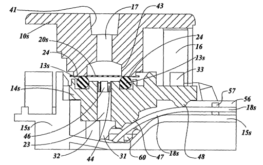
A cross-sectional view of a the assembled device is illustrated in FIGURE
9B. The plane and direction of sight of the cross-section are indicated in
FIGURE 9A. The cross-sectional surface of each part is indicated by its part
number
followed by the letter "s". Specifically, the sectional surface of the
dilution part is
l Os, and the sectional surfaces of the sample membrane, a-ring, midpiece,
base, and
diagnostic test strip are 20s, 13s, leis, 15s and 18s, respectively. With the
dilution
port depressed and locked into position, the sample membrane is compressed
between undersurface 2~ of the dilution port and the uppersurface of the o-
ring 13
contain ed within the midpiece. This results in isolation of the sample
membrane
contained within the perimeter of the o-ring. Liquid applied through the
dilution part
channel 17 exits onto a diamoter of membrane equivalent to the diameter of the
dilution port exit 43. This liquid does not escape along the sample membrane
20, but
instead passes perpendicularly through the sample membrane (i.e., through its
thickness) due to lower resistance. This flow of liquid through the isolated
portion of
the sample membrane removes sample contained mth m the void volume of the
sample membrane. The removed sample and
associated diluont
fluid passes to
channel 23. Sampleand diluent also passes four collection
into the areas of the
midpiece ~l~ that to the central through-channel. These collection
empty in areas are
located between fourmembrane support at the top of the
the pasts 4G midpiece
2s immediately the area of membrane The removed sample
beneath sampled. and
diluent mix as they pass by the collection areas iota channel 23 and collect
in
receptacle well 31 of th a brio. This diluted sample is then wiclcod from the
receptaclo wall along the diagnostic strip 18 and the control strip 19 (not
shown) to
ovaluate For the presence of analyte,
-17-
CA 02426246 2003-04-16
WO 02/33380 PCT/USO1/32456
The device of the invention can be made by automated assembly. Assembly
can be made as follows: base 15 lays flat on a pallet and is ready to receive
diagnostic and control strips, and optionally a desiccant tablet. These strips
are held
in place by the channel guides SG and 58 and pegs 57, 59, and 32 (FIGURE 7A).
Midpiece 1 ~ with o-ring 13 in place is added to the assembly guided by pegs
32 and
33 that project upward fi°om base I5. With the addition of the midpiece
to the
assembly, the diagnostic and control test strips are held in place within the
base, and
channel 28 (FIGURE 8B) for receiving sample membrane 20 (FIGURE 8C) is
formed by the base and adjacent midpiece. Sample membrane 20 is then added to
the assembly, and held in piece by channel guides ~I9 and 51 (FIGURE 7A) and
stop
peg 52 (FIGURE 8C). Cover 12 is then added and held onto the bottom piece by
mating pegs and tapered receptacles 50 (FIGURE 7A) that allow a secure press-
fit
between the two parts. Yolce 11 is connected to dilution port IO by sliding
yoke arms
37 (FIGURE ~A) into matching slots 70 of dilution port I0. The combined
dilution
port and yoke is then Fitted to the assembly by placing hook arms 16 of the
dilution
port into hook arm receptacles 29 (FIGURE 5A). The dilution port is held in
place
against removal from the top piece by hooks 21 (FIGURE 3) on the hook arms
which
rest against the undersurface of ridges 40 (FIGURE SA). The dilution port is
prevented fi°om depression into a locked position in the bottom piece
until the user
2p removes the yoke prior to conducting the test.
The method and device of the invention can be used for testing multiple
analytes and allows for internal quality controls on the reagents used in each
test. As
shown in FIGURE 8A, two strips enter the reservoir well permitting the diluted
sample to flow over the diagnostic test and control strips. In other
embodiments, the
device can include more than two strips contacting the same reservoir well.
FIGURES I OA, I 013, I 0C, and 1 pD illustrate the use of the method and
device of the invention in a rapid test for antibodies to HIV. An internal
control is
used to assess whether a sufficient amount of serum immunoglobulin has been
added
to the test (C3), In addition, control wells C 1 and C2 monitor the integrity
of two
s0 separate synthetic peptide-protein conjugates that are use to detect
antibodies to
-18-
CA 02426246 2003-04-16
WO 02/33380 PCT/USO1/32456
HIV-1 (C1) and hIIV-2 (C2), Without all oCthese controls, one is less certain
that a
negative result For antibodies to HIV-1 and HIV-? reps°esents a true
negative. One
could not conclude that a negative result is due to the absence of antibodies
to HIV in
the test sample rather than loss of integrity of HIV antigens, unless these
internal
controls for HIV-1 antigen (C1) and HIV-2 antigen (C2) are included and
demonstrate that these antigens are intact. A negative result is also not
definitive
without knowing that sufficient immunoglobulin was tested. If an individual
adds
diluent prematurely to the dilution port and initiates the wicking through the
diagnostic test strip before any sample has reached the area beneath the
dilution port,
a potentially false negative result would not be detected without control (C3)
that
confirms sufficient immunoglobulin has been evaluated. The diagnostic test
strip
used in this example also permits simultaneous determination of whether a
sample
positive for antibodies to HIV contains antibodies to HIV-1, HIV-2 or both.
Currently available diagnostic tests using lateral flow technology usually
include
1 ~ controls to confirm sufficient immunoglobulin has been tested, but no
currently
available tests include internal controls of antigen infiegrity.
In summary, in one embodiment, the invention provides a method and a
device, as exemplified in FIGURES I-10, that provides for testing for specific
analytes within a collected and diluted sample, both without need of
instrumentation
or formal training.
A representative device of the invention includes five components: (1)
dilution port; yoke; cover; midpiece, and base.
The device's cover includes a sample receiving well for collecting a sample
liquid to be tested and allowing the sample to confiact and fill the voids of
a sample
?5 membrane (sample membrane). The device's base includes a channel to contain
the
sample membrane and provide Iluid communication between the receiving well and
the dilution port. The sample membrane transports liquid From the receiving
well to
the dilu tion port. Depending on the application. the sample membrane can
include
components that prepare or modiFy the sample For subsequent testing. The
sample
s0 membrane is porous and contains the sample within its voids and allows the
sample
-19-
CA 02426246 2003-04-16
WO 02/33380 PCT/USO1/32456
to be subsequently removed from a defined volume of membrane isolated by the
device.
The dilution port, which fits into prescribed portions of the cover and base,
is
held in a non-active position on the top piece by a yoke, which slides into
grooves of
the dilution port thereby preventing it from being depressed down into the
device,
thus allowing free passage of the test liquid in the sample membrane without
obstruction fi°om any sLirfaces of the dilution port. When the yoke is
removed, the
dilution port may be depressed into the active position and catch hooks on the
tension
arms of the dilution port lock into place against catch receptacles causing
the flat
circular undersurface of the dilution port to compress the area of the sample
membrane between the dilution port and a matching circular upper-surface of an
o-ring contained within the midpiece. The compressed membrane effectively
prevents any substantial flow of liquid along the membrane either from outside
or
inside the area circumscribed by the corresponding mating surfaces of midpiece
o-ring and dilution port.
The midpiece includes a top surface completes the channel that confines the
sample membrane and contains a groove to hold the o-ring that forms one mating
surface with the dilution port to define the volume of sample membrane for
testing.
The midpiece includes a through-channel to allow passage of diluent and sample
out
of the sample membrane and down into the reservoir located in the bottom
piece.
The undersurface of the midpiece includes protrusions that guide the
diagnostic and
control membranes into the reservoir to allow analysis of the diluted sample,
The device's base includes components for alignment with the midpiece and
cover and catch receptacles for the hook arms of~ the dilution port to lock
the port and
base into the active position. The base includes one or more channels that
contain
test and/or control strips and a ohannel for the sample membrane. The test and
control strip channels permit the test and control strips to contact the
diluted test
sample in the reservoir well. and further align the strips to pass by viewing
windows
for test result analysis.
_~0_
CA 02426246 2003-04-16
WO 02/33380 PCT/USO1/32456
The test and control strips contact the diluted sample in the reservoir well.
The sample migrates out of the reservoir along the strip and
encounter°s a
microparticle pad that contains visible particles that bind to and label the
analyte of
interest. The migrating labeled analytes of the diluted sample then bind to
defined
areas of the strip, located beneath view windows (see FIGURE IO circular and
rectangular viewing windows). Any migrating fluid not bound to one of the
membranes o~ the test or control strips is then absorbed by blotters bringing
the test
reaction to completion and ready for reading (see FIGURE 10 example);
Control strips or reagent lines confirm reactive potency of the reagents used
in the test (see, for example, FIGURE 10, C1 and C2) and confirm that the
diluted
sample is adequate for testing (see, far example, FIGURE 10, C3).
In another aspect, the invention provides a system that includes, in addition
fo
the device, a vial that contains the precise volume of dilution reagent
required for a
given test. In one embodiment, the vial is compressible and further includes a
volume of gas equal or greater than the contained liquid. The vial includes a
neck
having a sLaFCciently small diameter to prevent leakage of the solution when
the
bottle is inverted and having a tip of appropriate size and malleability to
form a tight
seal when placed into dilution port vial tip receptacle. With the cap removed
and the
neck of the vial placed firmly into the vial tip receptacle of the dilution
port,
squeezing the vial forces out all of the liquid from the vial and down through
the
dilution port. A channel in the dilution port connects fhe dilution port vial
tip
receptacle with the undersurface of the dilution port and the area of isolated
sample
membrane. The mating surfaces of the pol't and midpiece define a circumscribed
volume of sample membrane. When a known volume of dilution reagent is passed
?5 through this volume of membrane, the sample liquid in the membl°ane
is removed
and diluted in a reproducible fashion.
In another' embodiment, the invention provides a method for easily and
reliably sampling a test liquid, and in some instances simultaneously diluting
that test
llqLlld. The IIlethod 111C1L1deS fllllllg the VOlds Of a pol'oLIS Inembl'alle
wltll a llqllld
a0 sample; isolating a defined amount of the liquid sample within the voids of
the
-? I -
CA 02426246 2003-04-16
WO 02/33380 PCT/USO1/32456
sample membrane; and releasing the defined amount of sample liquid from the
Illenlbl'alle.
The liquid sample is isolated by opposing two complementary surfaces on
opposing I11a~01' surfaces (i.e., on either side) of the sample membrane.
These
complementary surfaces form a perimeter around a defined volume of the
membrane.
The isolated liquid sample can be removed from the membrane by delivering a
second liquid or gas to the portion of the membrane containing the isolated
liquid
sample. When a second liquid is used, the liquid sample is simultaneously
diluted.
The liquid sample released (and diluted) from the membrane is collected into
a reservoir where it can be further analyzed to detect specific components or
analytes.
The representative device of the invention illustrated in FIGURE 1-10 was
designed using modeling, computer-aided design. The 3-D modeling and computer
aided design program Rhinoceros, copyrigh t 1993-199$, Robert McNeel and
Associates, Seattle, Washington, USA, was first used as a beta version, and
later as a
I 5 commercially available software application. Once a design was completed,
an STL
file was created and provided to several different rapid prototypes were
produced
using stereolithography. In some instances, critical portions of the parts
were
machine-tooled to achieve desired tolerances, Test results are presented in
the
following examples.
The follawing examples are intended to illustrate but not limit the scope of
this invention.
EXAMPLE 1
Reproducibility o~ Dilutions of Test Sample
CytoSep 1661 membrane was obtained from Ahlstrom Filtration Inc.,
?5 Chattanooga, TI~1 (see U.S. Patent No.5,186,$~13, Baumgardner etal., Blood
Separation Media and Method for Separating Plasma From Whole Blood). Single
sided adhesive coated polyester from Adhesives Research Inc., Glen Roclc, PA
(ARCARE 7$1 ~) was laminated to portions of both sides of the CytoSep 1661
membrane prior to sizing the strips to fit channel 51 shown in FIGURE 7A of
the
3p medlCal deVlCC_ ThIS lmpel'llleable pOlyeStel' Sheet 5e1'Ved t0 COIIfIIle
the llqllld flOW
-7?-
CA 02426246 2003-04-16
WO 02/33380 PCT/USO1/32456
to within the membrane, and not over surfaces of the device in those areas
where it
was applied. After lamination with polyester, strips 20 (FIGURE 8C) were cut
to
size to fit channel 51 shown in FIGURE 7A. Each sized strip of CytoSep 1661
contained on its undersurface laminated polyester, This polyester extended
from the
end of the strip that rests beneath the sample receiving well in the fully
assembled
device along the under surface up to approximately two millimeters short of
the outer
margin of the o-ring contained within the midpiece (see FIGURE 8B). The upper
surface of each sized CytoSep 1661 strip contained laminated polyester
beginning
approximately 2 mm beyond the downstream edge of the undersurface of the
device
sample receiving well 27 (FIGURE 5A) to 2 mm short of the outer margin of the
o-ring 13 contained within midpiece 1 ~ (FIGURE 8B). For purposes of this
example
size 008 o-ring and a midpiece designed to tightly hold size 008 o-ring were
used.
The sized and laminated CytoSep membrane strip was placed into the device and
all
five plastic components of the device were assembled before use.
SchillingTM Red food coloring (manufactured by McCormick & Co, Inc.,
Hunt Valley, MD, and containing FD&C Reds 40 and 3) was obtained from a local
supermarket. A stock 1:10 dilution of the food coloring was prepared in PBSAA
buffer consisting of 50 mM phosphate, 10 mM NaCI, pH 7.~, and 0.05°lo
sodium
azide and 0.1°,% bovine serum albumin in deionized water. Dilutions of
this stock
solution were scanned with a Gilford spectrophotometer (Gilford Systems, Ciba
Corning, Oberlin, OH). Peak absorbance was noted at a wavelength of
X195 nanometer s,
Experiments to test the reliability of the device to produce a consistent
dilution of the stock solution of red food coloring were conducted as follows.
Approximately two drops (100 to 120 microliters) of the stock solution were
added to
the fully assembled device containing the sized and laminated CytoSep 1661
membrane. AFter a wait of two minutes, the yoke was removed from the dilution
port, and the dilution port was pressed down into the locked position. A twist-
off
capped plastic 0.$ ml vial, obtained froth Automatic Liquid Packaging,
Woodstock,
IL, and prefulled with X50 microliters of PBSAA buffer diluent, was inverted
and its
-23-
CA 02426246 2003-04-16
WO 02/33380 PCT/USO1/32456
neck was pressed into the entrance to the channel in the dilution port to form
a leak-
proof seal. The vial was squeezed to express all of its contents, held for 3
seconds,
and then removed while keeping the vial squeezed.
Buffer diluent passed through the portion of CytoSep 1661 membrane
isolated for testing by the device. This isolated membrane consisted of the
noncompressed circular area of membrane located within the circumference of a
ring
of membrane compressed by the device. The ring of membrane compression was
produced by the undersurface of the dilution port in tile locked and active
position
and the o-ring held in the upper surface of the midpiece. Buffer diluent
introduced
1 p under pressure into the dilution port channel passed through this channel
and through
the isolated ring of membrane removing the red food coloring. The extracted
red
faod coloring and diluent passed down through the channel in the midpiece
resulting
in a mixed dilution of sample that collected in the reservoir in the bottom
piece.
The dilution of the red food coloring achieved by the device was assessed as
15 follows.
Immediately after expressing the diluent from the twist-off cap vial and
removing it from the device, the dilution part was unlocked, and 300
microliters of
diluted sample was removed from the reservoir and placed into a test tube. To
each
300 microliters of diluted sample was added an additional 300 microliters of
PBSAA
20 diluent for purposes of reading the result in the spectrophotometer
cuvette. This
resulted in a 1:2 dilution of the dilution produced by the device. The device
was used
repeatedly to evaluate dilutions that it produced of the red food coloring
stock
solLltloll Ll11de1' dlfferellt CondltlonS. The dilutions achieved were
eValLlated by
reading the 1:2 diluted samples at a wavelength of X195 manometers.
25 FLGURE 11 A presents the mean absorbance at X195 mm wavelength of two
separate sets of ten dilutions each performed on separate days using the
medical
device with CytoSep 1661 membrane and a size 008 o-ring in the midpiece. The
mean absorbance was 0.51 S and the median absorbanee was 0.51 ~. The range of
two
standard deviations from the mean was 0.125-0.605, When compared to a standard
30 CLII'Ve developed fro111 1110w11 d1111t1011S O1' the Stock SOllltlon I'ead
L111der the Sa111e
CA 02426246 2003-04-16
WO 02/33380 PCT/USO1/32456
conditions, the two standard deviations range of dilutions produced by the
device was
1/75 to 1/105 (FIGURE 11B).
EXAMPLE 2
Role of Sample Membrane Thickness on Dilution
FIGURE 12A presents the mean absorbance at 495 nm wavelength of two
separate sets of ten dilutions each performed on separate days using the
medical
device and a size 008 o-ring in the midpiece. For one set of ten dilutions
CytoSep
1661 with a thickness of 0.18 mm was used, and far the other set CytoSep 1660
with
a thickness of 0.33 mm was used. Three drops of stock solution (approximately
165 microliters) was used with the GytoSep 1660 membrane because of its
capacity
to hold a greater volume of sample. Two drops (100 to 120 microliters) of the
stock
solution were added to the sample receiving well for the series using CytoSep
1661.
In both series the volume added fully saturated the void volume of the
membranes
used. The average dilution produced with the CytoSep 1 G61 membrane was 1 /90
with a two standard deviation range of 1/75-1/105. The average dilution
produced
with the CytoSep 1660 membrane was 1150 wi h a two standard deviation range of
1/44 to 1/65 (FIGI~RE 12B). This illustrates that a range ofdilutions can be
obtained
using the method and device of this invention by selecting membranes of
different
thickness. Thase membranes that are thicker will contain more sample per unit
area,
and hence result in a lower dilution produced by a given quantity of diluent.
EXAMPLE 3
The Effect of Membrane Surface Area Sampled on Dilution
Tests were performed as in Example 1 using CytoSep 1661 membrane. One
hundred microliters of red dye solution were added to the receiving well. Four
?5 hundred fifty microliters of PBSAA diluent were added to remove sample from
the
circumscribed isolated CytoSep 1661 membrane, after a two minute delay between
adding red dye stock solution to the receiving well and adding diluent through
the
dilution port. Triplicate dilutions were performed with a size 008 o-ring with
an
internal diameter of x.55 mm, and compared with triplicate dilutions
pei°formed using
3p a dilution port and midpiece designed for use with a size 007 o-ring which
has an
-? 5-
CA 02426246 2003-04-16
WO 02/33380 PCT/USO1/32456
internal diameter of 2.55 mm. Using the formula err= it can be seen that the
comparison of surface areas sampled by the two o-rings is surface area
008lsurface
area 007 = (2.275)'-/(1,275)= ~ 5.18/1.63 = 3.18. One therefore expects that
the
device and method would remove 3,18 times more sample using the 008 o-ring.
Triplicate tests using the 008 membrane produced A495 readings of 0,447,
0.423,
and 0.445, for a mean A495 of 0.438. The triplicate tests using the 007 o-ring
produced A495 readings of 0.226, 0.184, and 0.212, fox a mean A495 of 0,207.
The
0.438 mean A495 reading corresponds to a 11120 dilution, and the mean A495 of
0.207 corresponds to a dilution of 1:380, when applied to the standard curve
of
dilutions obtained known dilutions of the stock red dye solution (FIGURES 11B
and
12B). The ratio of these two dilutions is 3801120 = 3.17, close to the
predicted result
based upon su dace area sampled.
EXAMPLE 4
Effect of Volume of Sample Placed into Receiving Well on Dilution
FIGURE 13 presents the results of an experiment to examine the effect of
volume of sample added to the receiving well of the device. As in Example 2,
CytoSep 1661 membrane was used and dilutions were produced with 450
microliters
of PBSAA diluent. Each dilution was produced 2 minutes after adding the sample
to
the receiving well. Each volume was tested in duplicate and the volumes tested
were
25, 50, 75, 100, 150, 200 and 250 microliters, corresponding to approximately
'/~, 1,
I !~~, 2, 3, 4 and 5 drops.
The results indicate that it is necessary to have enough volume to saturate
the
void volumes of the membrane. Twenty-five and fifty microliter amounts,
corresponding to '/ and I drop, were insufficient to saturate the CytoSep 1661
sampling membrane, resulting in low A495 readings of 0.168 and below
(FIGURE 13). I-Iowever, volumes of 75 microliters and larger, corrES~onding to
1 !~~
to 5 drops, all saturated the sampling membrane. The samples obtained by the
device
with these saturating volumes were essentially equivalent based upon their
A495
absorbances,
26_
CA 02426246 2003-04-16
WO 02/33380 PCT/USO1/32456
EXAMPLE 5
Effect of Time Delay Between Addin-g SanZple and Adding Diluent on Dilutiol7
PIGU'RE 1~1 presents the results of an experiment to examine the effect of
time between sample addition and dilution on the Final dilution produced by
the
method and design of this invention. The experiment was conducted with CytoSep
1661 membrane as in Example 1 using 100 mieroliters of stock red dye solution.
Time periods were tested in duplicate and were 5 seconds, 10 seconds, 15
seconds,
30 seconds and 1 minute. In addition, single dilutions were made after two
minutes
(120 seconds), five minutes (300 seconds) and fifteen minutes (900 seconds).
The
results through five minutes are graphed on the Egure. At least one full
minute was
required for the sample to migrate from receiving well end to the area beneath
the
dilution port and reach a steady state filling the void volume of the membrane
at its
end opposite the point of application. However, the absorbance at X95
nanometers
(A495) remained essentially unchanged from 2 minutes to 15 minutes after
adding
the sample to the I°eceiving well.
EXAMPLE 6
Preparation of Test Strips for Detecting Antibodies to
HIV within Diluted Serum Sam lies
Each test strip was prepared as four separate components. These components
include a wick, a micro-particle pad, a white nits°ocellulose membrane,
and a blotter.
The wick serves to draw the diluted sample up into the test strip from the
reservoir in
the base. The micro-particle pad for these experiments was coated with
recombinant
protein A (rPA) labeled with colloidal gold, a visually observable micro-
particle
reagent. As the diluted serum sample migrates through the test strip most of
the
antibodies within the sample are labeled with the micro-particle reagent and
their
subsequen t migration over the test strip may be tracked. HIV antigen was
coated to
the 171t1'OCeIILlIOSe II1e117bI'ane 111 a Mlle perpendlCLllar t0 tile
Inlgrat1011 flow. 1VIICrO-
particle labeled antibodies migrate down the test strip. Those directed at I-
LIV antigen
bind to it on the white nitrocellulose, and the remaining labeled antibodies
continue
Illlgl'atlOll Ollt OI' the 171t1'OCellLllOSe lntO the blOttel'. The pl'eSel7Ce
Of a pink t0 purple
_77_
CA 02426246 2003-04-16
WO 02/33380 PCT/USO1/32456
line in the same location as the bound HIV antigen on the nitrocellulose
identiFies the
presence of antibodies directed at that HIV antigen within the diluted serum
sample_
The blotter paper serves to absorb most of the liquid and reagents that
migrate along
the test strip, providing a white nitrocellulose background and facilitating
recognition
of any labeled antibodies bound to the HIV antigen on the white
nitrocellulose.
LoProSorbT~' from PALL Corporation, Port Washington, NY, was used for
both the wick and micro-particle application pad. Immunopore~"' nitrocellulose
paper
from Costar Scientific Corporation, Cambridge, MA, was used for the reading
zone
of the test, and paper 939 from Ahlstrom Filtration Inc., Chattanooga, TN, was
used
as the blotter.
The micro-particle pad component of the test strips was prepared separately
prior to assembly into the final test strips. The micro-particle pad was
coated with a
solution of colloidal gold-labeled recombinant protein A. The recombinant
protein A
(rPA), lot RC1041, was abtained from Repligen Corporation, Cambridge, MA. The
colloidal gold-rPA conjugate was prepared as described by Lea et al., J.
Histochemistry & Cytochemistry, 40(6):757-758 (1992), with the following
modifications. Gold chloride (tetrachlorauric acid trihydrate, ACS, Sigma
Chemical
Company, St. Louis. MO) was dissolved in HPLC pure water at 100 mg gold
chloride per liter of HPLC pure water. One hundred ml of this 0.1 mg/ml gold
solution was brought to a boil in a Pyrex flash with stirring and precautions
to
prevent evaporation. A volume of 3.2 ml of 1 °,~o sodium citrate was
added to the
boiling gold solution and stirring continued. The solution initially turned
blue-gray,
and then with continued stirring and heat the solution became orange-red after
two
minutes. Heat and stirring were continued another 6 minutes and the solution
was
then cooled.
The final colloid had an absorbance at 520 nm wavelength of 1.072. A
minimal protective test against N~aCI was performed with tile Lot RC 1041 rPA,
and
found to be 5 micrograms of rPA per ml of gold colloid. A 40 ml volume of the
gold
colloid was adjusted to pI-I 6.0 with K~CO; and I-I3P0~. Two hundred
microliters of
rPA at a 1 mglml concentration were added with mixing to the pI-I-adjusted
gold
-~ g_
CA 02426246 2003-04-16
WO 02/33380 PCT/USO1/32456
COIIOId SOlL1t1011. The SOILIt1011 WdS Inlxed fOr two 111111uteS alld then let
Stand for fOLlr
minutes. Two ml of 1 °~o PGG (polyethylene glycol) and 4.6 ml of 10%
bovine serum
albumin (BSA) were then added with mixing. Twelve aliquots of 1.~ ml each were
centrifuged in a MicroFuge'~"' (Beckman Instruments, Fullerton, CA), and the
solution
was centrifuged at maximum speed for 45 minutes. The supernatant above each
pellet of gold colloid-rPA was aspirated, and each pellet was resuspended in
50 microliters of a buffer of SO mM Tris pH 8.0, 100 mM NaCI, 0.02°~o
sodium azide,
0.02% PEG and 1 % BSA. All resuspended pellets were pooled and had an
absorbance at 520 nanometers of 3.15. This preparation was tested for
detection of
human IgG coated to nitrocellulose membrane in a lateral flow assay (see
below) and
showed easily visible strong staining of the IgG that had been coated to the
nitrocellulose at concentrations of 1 mg/ml and 10 mg/ml.
The stock colloidal gold rPA conjugate with an absorbance of 3.15 was tested
for its ability to bind to antibodies directed at HIV antigens, while
preserving the
1 > capacity of those antibodies to recognize the HIV antigens. A synthetic
peptide
representing an immunodominant region of gp~ll of HIV-I was conjugated through
its C-terminus to bovine serum albumin as described in Formos0 et al. (U.S.
Patent
N0.5,260,189). This peptide-protein conjugate was coated to nitrocellulose
membrane strips in a line perpendicular to the length of the strip,
approximately one-
fourth the distance from the downstream terminal end of the strip, at a
concentration
of 1 mghnl, and the strips' excess binding sites were then bl0clced, as
described
below. Serum containing antibodies to HIV was diluted 1:100 in a buffer
consisting
of 50 mM Tris HCI, pI-I 8, 100 mM NaCI, 0.025°r'o sodium azide and I
°J° BSA. Ten
microliters of HIV positive diluted serum was mixed with ten microliters of
the stock
colloidal gold rPA conjugate, and the colloidal gold rPA conjugate bound to
antibodies in test serum. This 20 microliter combination was added to the
upstream
end o1' the nitrocellulose strip and allowed to migrate along the
nitrocellulose strip
past the area of bound I-IIV peptide-protein conjugate, and off the downstream
end of
the strip onto a blotter. Most of the visible colloidal gold rPA conjugate
migrated
a0 thl'oLlgh the IlltroCellLIloSe papel' alld OIltO the blottel', bLlt a
1'eddlSh-plnk llne, agalnSt
-? 9-
CA 02426246 2003-04-16
WO 02/33380 PCT/USO1/32456
a white background, remained at the site on the nitrocellulose where the I-IIV
peptide-pi°otein conjugate had previously been bound. This experiment
was repeated
using 1:100 diluted serum known to not contain antibodies to HIV, and no
visible
line remained on the nitrocellulose membrane where HIV peptide-protein
conjugate
had previously been bound. Taken together, these experiments indicated that
the
stock colloidal gold rPA conjugate was capable of labeling antibodies to HIV,
which
then retained their ability to i°ecognize the HIV peptide-protein
conjugate bound to
the nitrocellulose membrane.
The stock colloidal gold rPA solution was used to prepare microparticle pads
for test strips as follows. LoProSorb~'"' membranes from PALL Corporation,
Port
Washington, NY, that had been backed with polyester (ARCARE 7815, Adhesives
Research Ins., Glen Rock, PA) were precoated with a solution of nonfat skim
milk.
The nonfat skim mills blocking buffer consisted of 0.5°r'o nonfat
skim milk
(Carnation) in deionized water with 50 mM Tris, pH 7.7, 0.03°~'o sodium
azide, and
0.45°lo PVP-~0 that had been filter sterilized. After saturating the
>aoProSorbl~~
membranes with the blocking solution, they were fully dried and then coated
with the
stock colloidal gold rPA solution diluted I:6 in deionized water containing
1 °~''o PVP-40, 0.02°,~'o sodium azide, 0.1 °r'o PEG, 1
°r'o BSA, 2.5°r'o sucrose.
These pads containing the colloidal gold microparticles were allowed to air
dry prior to assembly into the final test strips.
The HIV antigen coated nitrocellulose membrane component of the test strips
was prepared separately prior to assembly into the final test strips. The HIV
antigen
utilized was peptide SS76 described by fonnoso et al., LI.S. Patent No.
5,260,189.
This peptide was conjugated through its C-terminus to bovine serum albumin,
and
the peptide-protein conjugate was coated to the nitrocellulose. The
nitrocellulose
membranes employed were Immunopore~'"~ from Corning Costar, Cambridge, MA, or
5 micron backed nitrocellulose from Schleicher & Schuell (Keene, NH). The HIV
synthetic peptide-protein conjugate was used in concentrations ranging from
2.5 to
12.5 mg/ml in coating buffer consisting of 50 mM phosphate, 100 mM NaCI,
0.02°.r'o
sodium azide and 0.05°,~o PVP-X10. 1'he HIV peptide-protein conjugate
solution was
-30-
CA 02426246 2003-04-16
WO 02/33380 PCT/USO1/32456
applied to the nitrocellulose in a line perpendicular to the migratory flow of
the test
strip, and allowed to bind at room temperature for ten minufes. As a control,
recombinant protein A in a concentrations ranging from 0.2 mglml to 5 mg/ml in
the
same coating buffer was coated to the same nitrocellulose in a line
paralleling the
HIV peptide-protein conjugate antigen, separated by I cm distance. This was
allowed to bind to the nitrocellulose under the same conditions as HIV antigen
binding. Remaining active sites on the nitrocellulose were then blocked by
gentle
rocking of the nitrocellulose immersed in a filter-sterilized solution of
blocking
buffer consisting of 0.5°r'° nonfat skim mills, 0.45% PV P-40,
0.03% sodium azide in
50 mM Tris, pH 7.7 at room temperature for one hour, followed by air drying.
The final composite test strips were formed by lamination together using
single-sided adhesive coated polyester from Adhesives Research Ins., Glen
Rock, PA
(ARCARE 7$15 or ARCARE 8160. The first two components to be laminated to
the polyester were the antigen coated and blocked nitrocellulose membrane and
the
wick. The space between wick and nitrocellulose was adjusted so that the
subsequent lamination of micro-particle pad coated with colloidal gold labeled
rPA
produced an overlap region of 2 mm shared on the upstream side with the wick,
and
on the downstream side with the nitrocellulose membrane. This overlap region
allowed capillary flow from one membrane to the other during migration of
liquid
along the test strip. Subsequently, the blotter paper was laminated to the
composite
on the downstream side of the nitrocellulose with a 2 mm area of overlap
between the
blotter and nitracellulose. The width of the role of laminated four part
camposite
was cut to fit the length dimension of the test strip channel of the medical
device
(FIGURE G, 6-5). Prior to use, a single test strip was cut from the composite
with a
width to fit the width of the test strip channel of the medical device.
INXAMPLE 7
Detection of Antibodies to NIV Usini? Test Stri>as
Serum known to contain antibodies to HIV was compared to serum that did
nOt COlltaln aIltIbOdleS to I-IIV, In d11L1t10nS ranglllg II'om I :I0 through
1:000, Llslng
the test strips of Example G. The serum samples were diluted in a PBSAA
diluent
-J I -
CA 02426246 2003-04-16
WO 02/33380 PCT/USO1/32456
described in Example 1. With the samples known to contain antibodies to HIV, a
visible line of colored micro-particles appeared on the test strips in the
location of
bound HIV antigen, in serum dilutions of 1:10 through 1:2,000. This indicated
sensitive detection of antibodies to HIV in the known positive samples. No
such
visible line of micro-particles developed for the samples that did not contain
antibodies to HIV. This indicated specificity of the test strips configured as
in
Example 6. The control line of nitrocellulose bound rPA produced a visible
line of
micro-particles at serum dilutions of 1;10 through 1;300, independent of the
presence
of antibodies to HIV. This indicates that rPA coated to the nitrocellulose
membrane
under the conditions of Example 6 can be adapted for use as a control to
confirm that
an adequate amount of serum immunoglobulin was evaluated to validate a
negative
test result. This use of rPA as a control to confirm adequate serum added is
illustrated in FIGURE 10, well C3, regarding interpretation of test results.
EXAMPLE 8
Use of Device and Method to Detect Antibodies to
HIV to Free ~p41 HIV-1 Peptide Coated to Test Strips.
Peptide PVIR 126, an antigenic peptide from gp~ll of HIV-l, was obtained
from Bachem California (Torrance, CA). Peptide PVIR has the amino acid
sequence
HzN-Arg-Ile-Leu-Ala-Val-Glu-Arg-Tyr-Leu-Lys-Asp-Gln-Gln-Taeu-Leu-Gly-Ile-
Trp-Gly-Cys-Ser-Gly-Lys-Leu-Ile-Cys-Thr-Thr-Ala-Val-Pro-Trp-Asn-Ala-Ser-OH
(SEQ ID NO:I) with some cyclization of the peptide by an S-S creating an
antigenic
loop in the underlined portion. This peptide was dissolved in 1
°,~° acetic acid at
2 mglml, and then diluted fo 0.5 mg/ml using a nitrocellulose coating buffer
consisting of 50 mM phosphate pH 7.4, 150 mM NaCI, 0.05°,% PVP-~10, and
0.025°J°
?5 sodium azide. The peptide in nitrocellulose coating buffer was applied to
nitrocellulose strips taken from the nitrocellulose contained within the
Schleicher &
Schuell (Keene, NH) AccuSep~,~'~ membrane as 3 drops of 1.5 microliters each
in a
line and allowed to air dry. Upstream from the nitrocellulose on each strip
was first a
microparticle application pad connecting to the nitrocellulose by at leasf 3
mm and
Further upstream a wick cone ected to the microparticle pad by at least 3 nnn.
_;?_
CA 02426246 2003-04-16
WO 02/33380 PCT/USO1/32456
Downstream from the nitrocellulose was a blotter that connected to the
nitrocellulose
by at least 3 nnn of overlap. The wick was comprised of LoProSorb membrane
from
Pall-Gelman (Port Washington, NY). The microparticle pad was prepared by
pretreating the LoProSorb membrane with 0.1 °r'o IgG free BSA and
drying, followed
by application of protein A colloidal gold (20 nm size, $ O.D. in
5°~'° trehalose)
obtained from British Biocell International, Cardiff, UK. The colloidal gold
was
added to pad saturation and allowed to dry, and the pads used with each strip
were
approximately 5 mm wide by 10 mm long. The blotter was comprised of S&S
paper 300 and served to wick all fluid moving along the test strip from the
wick,
through the microparticle/colloidal gold pad and onto the nitrocellulose strip
coated
with PVIR gp~l peptide and out the downstream end of the test strip into the
S&S
300 paper blotter.
The test strips were first tested with a 1:100 dilution of a pool of 9 HIV
positive sera, and compared with a 1:100 dilution of an HIV negative test
serum.
The colloidal gold particles with attached HIV antibodies bound to the test
strip in
the area of applied peptide for the HIV positive serum sample, but not for the
HIV
negative serum sample.
The test strips were then placed into the test device of this patent
application.
Whole blood negative for HIV antibody was then used as the negative control,
and
compared to this same blood mixed two parts whole blood to 1 part HIV positive
serum pool. The test device was run in the normal fashion with separation of
the red
blood cells from serum over CytoSep 1661 (Pall-Gehnan), followed by creation
of a
dilution of approximately 1:90 through the sampling port by the addition of
serum
dilution buf-Fer consisting of 50 mM phosphate, 150 mM NaCI, 0. I
°~'° BSA (IgG free)
and 0.025°~"o sodium azide. The prepared and diluted whole blood
samples wicked up
onto the test strips from the sampling port well, and ran along the test
strips through
the microparticle pads picking up the colloidal gold protein A, and migrating
along
the nitrocellulose membranes past the bound PVIR gp41 peptide and into the
blotter.
The 1-IIV positive whole blood-serum mixture caused the colloidal gold to bind
to the
CA 02426246 2003-04-16
WO 02/33380 PCT/USO1/32456
peptide i°egion of the nitrocellulose membrane strips, and this binding
did not occLir
with the HIV negative whole blood.
EXAMPLE 9
Use of Device and Method to Detect Antibodies to
H. pylori Antigen Coated to Test Strips
Test strips were prepared identically to those in Example 8 except that the
nitrocellulose membrane used were test strips from the Beclanan-Coulter {Palo
Alto,
CA) FIexSure HPT"' tests that contain an H. pylori antigen line. These test
strips
were placed into the test device of this patent and reacted against whole
blood from a
person with antibody to H. pylori and another person without antibody to H.
pylori,
as in Example 9. The prepared and diluted whole blood samples wicked onto the
test
strips within the device and the sample with antibodies to H. pylori caused
the
colloidal gold particles to collect at the I-I. pylori antigen line, whereas
the sample
without H. pylori antibodies was unable to cause the colloidal gold to collect
at the
H. pylori line on the test strips.
EXAMPLE 10
Llse of Device and Method to Detect HCG Antigen in Whole Blood.
Dipsticks used to detect human chorionic gonadotrophin {HCG) in urine or
serum were obtained from Vancouver Biotech Ltd., Vancouver, British Columbia,
Canada, These strips were placed into the test device of this patent and run
and
tested against three whole blood samples. One sample was from a pregnant
female,
and two samples were from males whose blood did not contain HCG. Sixty-six
microliters of whole blood was placed into the blood collection well of the
device,
and allowed to migrate along the CytoSep 1661 membrane within it separating
red
2S blood cells from serum, over a three minute period. After three minutes,
the
sampling port was depressed and locked into place and 250 microliters of serum
dilution buffer consisting of 50 mM phosphate, pl-I 7,4, 150 mM NaCI, 0.025%
sodium aide, and 0.1 % BSA {IgG ii-~e~ was added through the port to collect
and
dilute the serum samples from each person. These prepared and diluted samples
then
wicked over the dipstick test strips from Vancouver Biotech Ltd. All three
strips
_~r1_
CA 02426246 2003-04-16
WO 02/33380 PCT/USO1/32456
Shoaled the COlltl'ol Mlle, bLlt only the pl"e~llallt felllale ShOWed a Mlle
In the area where
monoclonal antibody to I-ICG had been coated.
While the preferred embodiment of the invention has been illustrated and
described, it will be appreciated that various changes can be made therein
without
S departing from the spirit and scope of the invention.
-3 S-
CA 02426246 2003-04-16
WO 02/33380 PCT/USO1/32456
1
SEQUENCE LTSTING
<21a> Clarz~y Technologies Incorporated
Buchanan, Thomas
Buchanan, Mark
<12D> METHGD AND DEVTGE FOR DTLUT2NG A FLUTD AND DETEC'ETNG ANALYTES
WTTHIN THE DTLU'TED FLUTD SAMPLE
<13U> IMMT-1-28120
<140> PCT/US02/_
<141> 20~?1-10--1'7
<15p> US 60/241,4Q9
<151> 200Q-10-18
<160> 1
<170> Fa~entTn version 3.0
<210> 1
<211> 35
<212> FRT
<213> homo sapien
<400> 1
Arg Tle Leu Ala Val Glu Arg Tyr Le~z Lys Asp Gln G1n Lei Leu Gly
1 5 10 15
Tls Trp Gly Cys Ser Gly Lys Leu Tle Cys Thr '~hr Ala Val Pro Trp
2~ 25 30
Asn Ala Ser