Note: Descriptions are shown in the official language in which they were submitted.
CA 02426517 2003-03-12
WO 02/23197 PCT/USO1/28862
METHODS AND COMPOSITIONS FOR
GLYCOSIDASE ASSAYS
Related Application
This application claims priority to U.S. Provisional
Application No. 60/233,075 filed September 15, 2000.
Technical Field
This application relates to compositions and methods for
measurement of glycosidase activity. Additionally, the present
invention provides for compositions and methods for determining
metastatic and inflammatory states. The present invention further
provides compositions and methods for high throughput assays for
compounds that affect glycosidase activity.
Background of the Invention
Proteoglycans (PG) are complex macromolecules present on
the cell surfaces and in the extracellular matrices of a wide range of
cells (1-3). They are thought to play a major part in chemical
signaling between cells. They bind secreted signaling molecules,
which can enhance or inhibit the activity of the signaling molecule.
Proteoglycans can also bind and regulate the activity of secreted
proteins by immobilizing the protein, sterically blocking the activity
of the protein, providing a reservoir for delayed release, protecting the
CA 02426517 2003-03-12
WO 02/23197 PCT/USO1/28862
2
protein from proteolytic degradation, or altering the protein for more
effective presentation to cell surface receptors. .
Proteoglycans are polyanionic substances of high molecular
weight and contain many different types of heteropolysaccharide side
chains covalently linked to a polypeptide backbone. PG consists of a
protein core to which long carbohydrate chains termed
glycosaminoglycans (GAG) are covalently attached. GAGS are
linear, highly charged polysaccharides composed of a repeating pair
of sugars, one of which is always an amino sugar. Formerly, these
carbohydrate groups were called mucopolysaccharides, but they are
now termed glycosaminoglycans because they can contain derivatives
of glucosamine or galactosamine. In principle, proteoglycans have
the potential for almost limitless heterogeneity. The underlying
repeating pattern of disaccharides in each GAG can be modified by
patterns of sulfate groups.
The three major types of GAG found in PG are: 1) hyaluronan
(HA), 2) glucosaminoglycans (heparan sulfate (HS), heparin, and
keratan sulfate (KS)), and 3) galactosaminoglycans (chondroitin
sulfate (CS) and dermatan sulfate (DS)). Approximately 25% of
heparan sulfate linear polysaccharides consist of alternating N-
acetylated disaccharide units [-~4) a,-D-GlcNpAc-(1-~4)-13-D-
GlcAp(l~] and N-sulfated disaccharides [-~4)a-D-GlcNpS-(1-~4)-
13-D-GlcAp or oc-L-IdoAp(1-~]. These polymers are formed by the
attachment of a repeating ~[4)a-D-GlcNpAc(1~4)-l3-D-GlcAp(1~]
2S disaccharide sequence to a serine residue of a core protein through a
tetrasaccharide, glucuronosyl-galactosyl-galactosyl-xylosyl, linkage
CA 02426517 2003-03-12
WO 02/23197 PCT/USO1/28862
3
region. This molecule then undergoes partial N-deacetylation
followed by N-sulfation of the newly exposed amino groups,
followed by partial C-5 epimerization of D-GlcAp to L-IdoAp, and
finally O-sulfation. O-sulfates are always found in proximity to N-
sulfates, which enhances the clustering of the sulfate residues and the
heterogeneity in chemical composition and charge density of heparan
sulfate. A typical HS chain consists of a repeating disaccharide unit
of hexuronic acid and D-glucosamine. Heparan sulfate proteoglycans
are involved in many biological events such as angiogenesis, blood
coagulation, cell adhesion, lipid metabolism, tissue morphogenesis,
cell differentiation, and regulation of various growth factors and
cytokine activities.
Heparan sulfate proteoglycans are important components of the
subendothelial extracellular matrix and the basement membrane of
blood vessels (2). Basement membranes are continuous sheets of
extracellular matrix composed of collagenous and noncollagenous
proteins and proteoglycans that separate parenchymal cells from
underlying interstitial connective tissue. They have characteristic
permeabilities and play a role in maintaining tissue architecture.
In addition to heparan sulfate proteoglycan (HSPG), the basal
lamina consists predominantly of a complex network of adhesion
proteins, fibronectin, laminin, collagen and vitronectin (6). Heparan
sulfate (HS) is an important structural component of the basal lamina.
Each of the adhesion proteins interacts with HS side chains of HSPG
within the matrix. Thus, HSPG functions as a barrier to the
extravasation of metastatic and inflammatory cells. Cleavage of HS
by the endoglycosidase heparanase produced by metastatic tumor
CA 02426517 2003-03-12
WO 02/23197 PCT/USO1/28862
4
cells and inflammatory cells destroys the filtering properties of the
lamina. In addition, the degradation of the HS may assist in the
disassembly of the extracellular matrix and thereby facilitate cell
migration (5) by allowing blood borne cells to escape into the
bloodstream.
Heparanase activity has been described in a number of tissues
and cell types including liver, placenta, platelets, fibroblasts,
neutrophils, activated T and B-lymphocytes, monocytes, and
endothelial cells (7-16).
No sensitive non-radioactive method is currently available for
determination of heparanase activity in tissue or biological fluids.
There is currently a need for the development of compositions and
methods for simple, rapid, and non-radioactive quantitative assays for
the detection of glycosidase activity, particularly heparanase activity.
There is also a need for treatments and therapeutic compositions for
diseases associated with heparanse activities.
Summary of the Invention
The present invention is directed to methods for the
measurement of cellular activities. Additionally, the present
invention comprises compositions and methods for diagnosing
diseases, preferably the presence of metastases or neoplastic growth,
and for determining the metastatic potential for tumors. The present
invention furthers comprises compositions and methods for the
diagnosis of inflammatory states ih vitro and i~c vivo.
An aspect of the present invention comprises quantitative
measurements of glycosidase activity. A preferred method of the
CA 02426517 2003-03-12
WO 02/23197 PCT/USO1/28862
present invention comprises assays for glycosidase activity, more
preferably endoglycosidase activity, most preferably determination of
heparanase activity. The present invention provides for assays which
comprise biotinylated HS bound on streptavidin-coated wells binding
5 additional streptavidin molecules provided in a solution, and this
binding is inversely proportional to the extent of digestion of the HS
(see Figure 1). Thus, after digestion by heparanase, HS retains its
ability to bind the streptavidin coated in the wells, but HS loses its
ability to bind additional streptavidin molecules in solution. By using
enzyme-coupled streptavidin, the amount of streptavidin binding
biotin-HS following heparanase digestion can be effectively
determined, preferably by a color reaction.
The present invention also comprises compositions and
methods for screening for compounds that are capable of inhibiting
glycosidase activity, preferably heparanase activity. Additionally, the
compositions and methods of the present invention may be used in
high throughput assays for the identification of compounds capable of
inhibiting such enzymatic activity. The present invention also
comprises compositions and methods for determining compounds that
are capable of inhibiting the metastatic potential of tumors or altered
cells and compounds that are capable of inhibiting inflammatory
states.
Accordingly, it is an object of the present invention to provide
compositions and methods for assays of glycosidase activity that are
rapid, simple, and non-radioactive.
CA 02426517 2003-03-12
WO 02/23197 PCT/USO1/28862
6
Another object of the present invention is to provide
compositions and methods for the quantitative measurement of
glycosidase activity.
It is another object of the present invention to provide
compositions and methods for the measurement of heparanase
activity.
Yet another object of the present invention is to provide
compositions and methods for screening compounds capable of
inhibiting glycosidase activity, particularly heparanase activity.
It is a further object of the present invention to provide
compositions and methods for determining the effect of certain
compounds on various cellular and enzymatic activities.
It is another object of the present invention to provide
compositions and methods for diagnosing the presence of metastases.
Still another object of the present invention is to provide
compositions and methods that are useful in determining the
metastatic potential of tumors.
Yet another object of the present invention is to provide
compositions and methods for determining inflammatory states.
A further object of the present invention is to provide
compositions and methods for identifying compounds that are capable
of inhibiting the metastases of tumors and compounds that are
capable of inhibiting or resolving inflammatory states.
These and other objects, features and advantages of the present
invention will become apparent after a review of the following
detailed description and claims.
CA 02426517 2003-03-12
WO 02/23197 PCT/USO1/28862
7
Brief Description of the Figures
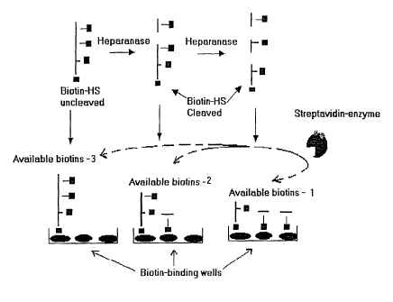
Fig. 1 is a diagram illustrating an assay of heparinase activity.
Fig. 2 is a diagram illustrating the linking of biotin to heparan
sulfate.
Fig. 3 is a graph showing the measurement of heparanase
activity using various amounts of Biotin-HS.
Fig. 4 is a graph showing the digestion of HS by heparanase.
Fig. 5 is a graph showing the substrate specificity of
heparanase.
Fig. 6 is a graph showing the specificity of assay in the
presence of proteases.
Fig. 7 is a graph showing the measurement of heparanase
activity in cell culture media and in serum.
Detailed Description of the Invention
The present invention may be understood more readily by
reference to the following detailed description included herein.
Although the present invention herein is described with reference to
specific details of certain embodiments thereof, it is not intended that
such details should be regarded as limitations upon the scope of the
invention. The entire text of the references mentioned herein is
hereby incorporated in their entireties by reference.
The present invention is directed to compositions and methods
for measurement of cellular and enzymatic activity. The present
invention also comprises compositions and methods for the diagnosis
of metastases, determination of the metastatic potential of tumors and
CA 02426517 2003-03-12
WO 02/23197 PCT/USO1/28862
g
the determination of the presence of inflammatory states.
Additionally, the present invention is directed to compositions and
methods for screening for compounds that can alter, preferably inhibit
glycosidase activity, particularly heparanase activity, compounds that
can alter, preferably inhibit metastases; and compounds that can alter,
preferably inhibit, inflammatory reactions and states.
There is increasing interest in heparan sulfate compounds and
their related enzymes due to a possible relationship between changes
in normal activity and tumor invasiveness and tumor metastatic
activity. An important process in tissue invasion by blood-borne
tumor cells and white cells involves their passage through the
vascular endothelial cell layer and subsequent degradation of the
underlying basal lamina or basement membranes and extracellular
matrix with a battery of secreted proteases and glycosidases (4,5).
Heparanase activity was shown to correlate with the metastatic
potential of animal and human tumor cell lines (7, 17-20). It is also
known to regulate growth factor activity. Many growth factors
remain bound to heparan sulfate in storage form and are disassociated
by heparanase during angiogenesis, improving the survival rate of
cancer cells.
Serum heparanase levels in rats were higher by more than an
order of magnitude after injection of the rats with highly metastatic
mammary adenocarcinoma cells. In addition, heparanase activity in
the sera of rats bearing MTLn3 tumors correlated well with the extent
of the metastases. Moreover, serum/urine heparanase activity in
cancer patients was shown to be 2-4 fold increased in particular where
tissue metastases were present. Because the cleavage of HS appears
CA 02426517 2003-03-12
WO 02/23197 PCT/USO1/28862
9
to be essential for the passage of metastatic tumor cells and
leukocytes through basement membranes, studies of heparanase
inhibitors provides the potential of developing a novel and highly
selective class of anti-metastatic and anti-inflammatory drugs.
Preferred embodiments of the present invention comprise
compositions and methods for the measurement of cellular and
enzymatic activities. Such assays can be used to measure such
activities, both qualitatively and quantitatively. The assays described
herein for determining the presence of such activities may be used in
methods for diagnosing metastases, metastatic potential and
inflammatory states. The assays of the present invention can also be
used to screen for compounds that alter, either stimulate or inhibit,
such cellular and enzymatic activities.
A preferred assay is herein described for heparanase activity.
Though this assay particularly describes measurement of heparanase
activity, it is not intended as a limitation of the invention. The
present invention contemplates compositions and methods for assays
measuring any glycosidase activity, including, but not limited to, any
enzymes with glycosaminoglycan-degrading activity, chondroitinase,
heparan sulfate endoglycosidase, heparan sulfate exoglycosidase,
polysaccharide lyases, keratanase, hyaluronidase, glucanase, amylase,
and other glycosidases and enzymes.
Despite the clinically significant role of heparanase in disease
processes, no sensitive high throughput assay to detect heparanase
activity is currently available. Existing heparanase assays are
cumbersome and time-consuming and require preparation of the
radiolabeled substrate and separation of degraded products from the
CA 02426517 2003-03-12
WO 02/23197 PCT/USO1/28862
uncleaved substrate (7, 12). Other heparanase assays require the
biosynthetic radiolabeling of matrix-associated HSPG and the
detection of HS chain degradation by gel-filtration analysis of
radiolabeled material released from the matrix (5). These assays
5 unfortunately do not discriminate between protease and heparanase
activities. Most heparanase assays also require extensive degradation
of the radiolabeled HS (or matrix-derived HSPG) substrate to allow
separation of the degraded product from the substrate by gel filtration.
Solid-phase heparanase assays have also been developed
10 where chemically and biosynthetically radiolabeled heparin and HS
chains were attached to a solid support, with release of radiolabel
from the solid support being a measure of enzyme activity. Such
assays, however, suffer from the disadvantage that the immobilized
substrate may be less accessible to the heparanase enzyme, and the
coupling of the radiolabeled substrate to the solid support, via the
substrate's reducing terminus, involves complex protocols. Assays
using such procedures are taught in U.S. Patent No. 4,859,581, which
is herein incorporated in its entirety.
Previous studies have also radiolabeled both heparin and
HS by iodination at naturally occurring glucosamine residues or by
N-acetylation of the partially de-N-sulfated substrate. Such
procedures require the use of radioactive iodine, which is a powerful y
emitter and therefore extremely hazardous. A sensitive radioactive
assay for heparanase has recently been reported (18). Although the
sensitivity of this method (nano units) is comparable to the present
method, it requires affinity chromatography of the heparanase-
CA 02426517 2003-03-12
WO 02/23197 PCT/USO1/28862
11
cleaved products on columns of histidine-rich glycoprotein-
Sepharose.
There are also some non-radioactive assays available for
heparanase. The most used assay for heparanase involves measuring
the optical density (at 230 nm) of unsaturated uronic acids formed
during degradation of heparin. Apart from that assay's having high
sensitivity (~mols of hexuronic acid), this assay suffers from the
disadvantage of interference from certain biological molecules
(proteins and nucleic acids), which show strong UV absorption.
Another color-based assay for measuring heparanase activity utilizes
heparin's ability to interfere with color development during the
interaction of protein with the dye Coomassie brilliant blue (25). This
assay is relatively specific for heparin, but requires large quantities
(up to 600 ~,g) of substrate.
The present invention, comprising compositions and
methods for assays for enzymatic activity, has advantages over the
previously described assays. Such advantages include the relative
ease of preparing the substrate, the rapid and simultaneous
determination of the presence of enzymatic activity in a large number
of samples, and the high sensitivity of such determination of activity.
Thus, the methods and compositions of the present invention can be
used for diagnostic purposes as well as in screening for compounds
that inhibit such activities.
A preferred method of the present invention comprises
the following. A composition comprising biotin-HS is mixed with a
sample, such as a tumor sample, bodily fluid, or other fluid suspected
of having heparinase activity, to form a reaction mixture. This
CA 02426517 2003-03-12
WO 02/23197 PCT/USO1/28862
12
sample may be pretreated to remove contaminating or reactive
substances such as endogenous biotin. After incubation, an aliquot or
portion of the reaction mixture is removed and placed in a biotin-
binding plate. After washing with buffers, a Streptavidin-enzyme
conjugate is added to the biotin-binding plate. Reagents for the
enzyme are added to form a detectable color product. For example, a
decrease in color formation, from a l~nown standard, indicates there
was heparinase activity in the sample. The biotin-binding plate
comprises any means for binding biotin, preferably to a solid surface.
In general, a preferred method comprises attaching one
of a binding partner to a substrate for the enzyme to be measured.
Incubation with a sample comprising the enzyme to be measured
allows for activity by the enzyme to be measured in a reaction
mixture. A portion or the whole reaction mixture, depending on the
amount needed, is then mixed with the complementary binding
partner, so that the binding partners are bound together. This is the
first binding reaction. After incubating to allow for binding,
washings are performed. A complementary binding partner,
complementary to the first binding partner attached to the substrate, is
added. This complementary binding partner may or may not be the
same as the first complementary binding partner. This is the second
binding reaction. The complementary binding partner in the second
binding reaction is labeled in a manner that is detectable. For
example, the complementary binding partner is labeled with an
enzyme that causes a detectable color change when the appropriate
reaction conditions exist.
CA 02426517 2003-03-12
WO 02/23197 PCT/USO1/28862
13
Preferred methods comprise use of binding partners,
including but not limited to, biotin and Streptavidin. Other ways of
binding one of the binding partners, such as biotin, can be used at
either biotin-binding step, either binding biotin to the plate or in
detection of the available biotins. The number of biotins, or other
binding partner, that are available for the second binding, is the
quantitative result of the assay. "Complementary binding partner"
means one of the pair of the binding partners, such as biotin and
Streptavidin or an antibody and its antigen. The biotin is the
complementary binding partner of Streptavidin, Streptavidin is the
complementary binding partner of biotin. An antibody that
specifically binds biotin is also a complementary binding partner of
biotin.
The enzyme of the sample, for which the activity or
presence is being detected, can be any of the enzymes, including but
not limited to, any enzymes with glycosaminoglycan-degrading
activity, chondroitinase, heparan sulfate endoglycosidase, heparan
sulfate exoglycosidase, polysaccharide lyases, keratanase,
hyaluronidase, glucanase, amylase, and other glycosidases and
enzymes.
The labeled binding partner, in the above method, the
enzyme labeled-streptavidin, can be labeled with any detectable
marker, including but not limited to, enzymes, dyes,
chemiluminescence, and other methods known in the art. A preferred
method comprises labeling with an enzyme that produces a color
change in its substrate that is detectable. This method is safe, easy,
CA 02426517 2003-03-12
WO 02/23197 PCT/USO1/28862
14
effective and can be used in both qualitative and quantitative
methods.
Using the above methods, the amount of enzyme activity
in a sample can be determined. Also, the above methods can be used
to determine compounds that can inhibit enzyme activity. For
example, a composition comprising the compound of interest is added
to a l~nown amount of heparinase either before or during the
incubation of the heparinase and its substrate-binding partner. If the
compound alters the activity of the heparinase, the assay methods of
the present invention will show a change in the amount of detectable
label. Such assays are used for high throughput determination of the
activity of compounds.
The compositions and methods of the present invention
can be used to diagnose the presence of metastases, which includes
1 S cancer, neoplastic growth, either initial or return metastatic growth.
A preferred embodiment of the present invention comprises the
following methods. Patients suspected of having one or several
tumors, either in an initial finding or in a return of tumor growth,
provide a biological sample for testing. This biological sample can be
any bodily fluid, tissue, or cellular sample. The biological sample
may be pretreated to remove endogenous biotin. The sample is used
in the assays of the present invention. An increase in the glycosidase
activity, particularly heparanase activity, or a high level of
glycosidase activity, is indicative of tumor or metastases presence.
The present invention can be used to measure the
metastatic potential of tumors. Tumor tissue or fluid samples are
assayed for the presence of glycosidase activity, particularly
CA 02426517 2003-03-12
WO 02/23197 PCT/USO1/28862
heparanase activity. Samples are taken once or in sequential biopsies
for testing. The transformed cells, such as cancerous or tumor cells,
may be found in vivo in living beings, or ih vitro, derived from cell
lines. A high level of glycosidase activity, or an increase in the
5 amount of glycosidase activity from a baseline determination
indicates that the metastatic potential of the tumor or cells is greater
than that of normal cells. Other tests known to those skilled in the art
can also be used in combination with the assays of the present
invention.
10 The present invention may also be used in determining
the presence of inflammatory reactions. An increase in the amount of
glycosidase activity, particularly heparanase activity, in a biological
sample, is indicative of the presence of an inflammatory reaction.
Other tests known to those skilled in the art can also be used in
15 combination with the assays of the present invention.
Another use of the present invention is for determining
compounds that influence the glycosidase activity in cells, tissues or
whole body responses. Because the present invention comprises
assays for quantitatively measuring glycosidase activity, compounds
that inhibit or enhance that activity can be determined easily using
such assays. For example, once a known amount of heparanase
activity is determined from the assays of the present invention,
compounds can be added to the assay and the amount of inhibition
can be determined. The present invention comprises high throughput
assays which can measure the effects on enzyme activity levels by
many different compounds. For example, the effect of compounds on
CA 02426517 2003-03-12
WO 02/23197 PCT/USO1/28862
16
the inhibition of glycosidase activity can be measured in vitro or in
vivo, using any type of sample known to those skilled in the art.
The present invention is further illustrated by the
following examples, which are not to be construed in any way as
imposing limitations upon the scope thereof. It will be clear to one of
skill in the art that various other modifications, embodiments, and
equivalents thereof exist which do not depart from the spirit of the
present invention and/or the scope of the appended claims.
EXAMPLES
Example 1
P~epa~ation of biotihylated HS
HS was biotinylated using biotin with extended spacer
arms using succinimidyl-6-(biotinamido) hexanoate (NHS-LC-Biotin)
obtained from Pierce. The chemistry of the reaction between HS and
biotin is shown in Figure 2 and other long chain analogs reduce steric
hindrance associated with binding biotinylated molecules to avidin.
0.5 ml HS solution (2 mg/ml in NaHC03, pH 8.5) was mixed with
0.05 ml of a freshly prepared solution of NHS-LC-Biotin in dimethyl
sulfoxide. The mixture was incubated at room temperature for 1
hour. Unconjugated biotin was removed by centrifugation (10,000
RPM) through Microcon-3 filter (Millipore) followed by dilution with
phosphate buffered saline (PBS). This procedure was repeated five
times to ensure complete removal of free biotin. Unwanted aldehydes
in the reaction were then quenched by incubation with one milliliter
of Tris-glycine buffer (25 mM-183 mM, pH 8.3) at room temperature
for 20 minutes. The mixture was subjected to three rounds of
microfiltration as described above. Biotinylated HS (5 mg/ml in PBS)
CA 02426517 2003-03-12
WO 02/23197 PCT/USO1/28862
17
was aliquoted and stored at -20°C. To obtain maximum
biotinylation, a 25 fold molar excess biotin was used. Using HABA
reagent, it was determined that the ratio of HS to biotin was 1:2.
Example 2
Assessment of biotihylation
The extent of biotinylation of HS was determined using
Avidin-HABA (Pierce Chemical Co) (22). The HABA assay can be
used over a wide range of pH and salt concentrations. HABA (4-
hydroxyazobenzene-2'-carboxylic acid) is a dye that binds to avidin
and can serve as an indicator of unoccupied binding sites. Avidin
combines stoichiometrically with biotin, making it possible to use any
physiochemical differences between avidin and the avidin-biotin
complex as the basis of a qualitative and quantitative assay method
for either component.
When HABA binds to avidin, there is a large spectral change in
the HABA dye. A new absorption band appears at 500 nm, which is
characteristic of the quinoid form of the dye. The avidin-biotin
complex does not bind HABA and because the dissociation constant
of the complex is so low, the dye is stoichiometrically displaced by
biotin. Consequently, the HABA assay can be the basis of both
colorimetric and titrimetric assays. The amount of avidin can be
calculated directly from the increased absorbance at 500 nm, or the
dye may be used as an indicator in a spectrophotometric titration with
biotin.
The absorption band that results from the avidin-HABA
complex decreases proportionately when biotin is added. Since biotin
CA 02426517 2003-03-12
WO 02/23197 PCT/USO1/28862
l~
has such a high affinity for avidin, it displaces the HABA dye. The
unknown amount of biotin can be determined by preparing a standard
curve using known amounts of biotin to displace the HABA which
bound to avidin, and plotting against the absorbance at 500 nm.
HABA solution was prepared by adding 24.2 mg of
HABA (Pierce) to 9.9 mI HaO, and then adding 0.1 ml 1 M NaOH.
Avidin-HABA reagent was prepared by adding 10 mg of avidin and
600 ~ul of HABA solution to 19.4 ml of phosphate buffered saline. To
1 ml of Avidin-HABA reagent in a cuvette, 100 ~1 of biotinylated HS
was added, and the optical density was measured at 500 nm in a
spectrophotometer. A standard curve was determined using known
amounts of HABA. The decrease in optical density of the HABA
following the addition of biotinylated HS was determined.
Example 3
Heparanase Assay
Biotin-labeled HS from Example 1 was digested with
heparanase, and the reaction containing undegraded and degraded HS
was bound to in a biotin-binding plate (Figure 1). Streptavidin,
conjugated with an enzyme, was added to the binding plate.
Quantitation of the color reaction measured the amount of available
biotin binding sites. A decrease in color from a known amount
reflects HS digestion by heparanase.
A lyophilized powder of heparanase (Heparanase III obtained
from Seikagaku) containing 0.1 units of enzymatic activity was
hydrated in 100 ~,1 of Reaction Buffer (3.33 mM calcium acetate pH
7.0, containing 0.1 mg/ml B~SA). This solution was then diluted to a
CA 02426517 2003-03-12
WO 02/23197 PCT/USO1/28862
19
working concentration of heparanase solution (O.Olmicro-units to 1
milk-unit) in Reaction Buffer. Enzyme activity was defined by the
manufacturer of the heparanase (Seikagaku) as follows: one unit of
enzyme activity is defined as amount required to generate 1 mole of
hexuronic acid per minute. Biotin-HS was diluted to a desired
concentration in Reaction Buffer.
To determine heparanase activity, 10 ~,1 of heparanase solution
was mixed with 200 ~.l of the biotin-HS substrate in a 96 well plate.
The reaction was incubated at 43°C for 1 hour. One hundred
microliters of the reaction mixture was added to a hydrated biotin-
binding plate (Chemicon) and incubated at 37°C for 30 minutes. The
biotin-binding plates were hydrated with 200 ~,1 of lx Assay Buffer
(Chemicon). Wells were washed five times with lx Assay Buffer and
incubated with 100 ~,l of 1:3000 diluted Streptavidin-Enzyme
Conjugate (Chemicon) for 30 minutes at 37°C. The wells were
washed five times with lx Assay Buffer and incubated for 20 minutes
with 100 ~,l of Substrate Solution (Chemicon). Color development in
the wells was assessed by measuring the optical density at 450 nm in
a microplate reader (Labsystems, Muliskan Ascent model).
CA 02426517 2003-03-12
WO 02/23197 PCT/USO1/28862
Example 4
Assay of hepa~ahase activity
The ability of bacterial heparanase (Seikagaku) to degrade
biotin-HS was determined. To determine the optimal amount of HS,
5 different concentrations of biotin-HS were used. These data are
presented in Figure 3. One-half milliunit of heparanase was sufficient
to completely digest up to 1 ~,g of biotin-HS. Biotin-HS was
incubated with the indicated amounts of heparitinase for 30 minutes
at 37° C. The extent of digestion was determined using streptavidin-
10 enzyme conjugate.
To determine the minimum amount of heparanase required for
digestion, 100 ng of biotin HS was digested with various amounts of
heparanase. One hundred nanograms of biotin-HS was incubated
with the indicated concentrations of heparitinase and the extent of
15 digestion at each concentration was determined using the
streptavidin-enzyme conjugate. At this concentration of HS (100 ng),
1 ,Unit of heparitinase digested approximately 40% of the initial
biotin HS (~40 ng), whereas complete digestion was achieved at 10
Units of heparitinase. These data are shown in Figure 4. At lower
20 substrate concentrations (5-10 ng of HS), heparanase activity in the
range of 10-100 nano units could be assayed. This sensitivity is
superior to previously described non-radioactive assays and equal or
superior to previous radioactivity-based heparanase assays.
Example 5
Specificity of a hepa~anase assay
CA 02426517 2003-03-12
WO 02/23197 PCT/USO1/28862
21
The next step was to determine whether the degradation of HS
was specific for HS and heparanase. Biotin-HS digestion was carried
out with heparanase in the presence or absence of excess unlabeled
HS to determine the specificity of heparanase towards HS. These
data are presented in Figure 5. Heparanase activity toward biotin-HS
was inhibited by approximately 60% in the presence of 50 fold (5 ~,g)
excess of unlabeled HS. The biotin-HS degrading activity was
specifically due to heparanase.
Example 6
Specificity of hepa~anase assay in the presence of proteases
The potential effect of proteases on the heparanase assay is of
particular concern because HS contains both a protein core and
attached polysaccharide chains. Since biological samples generally
contain other degradative enzymes such as proteases, the ability of
two different proteases (matrix metalloprotease (MMP-9) and trypsin)
to digest biotin-HS was determined. These data are presented in
Figure 6. Neither protease tested (MMP or trypsin) demonstrated any
activity that lead to a decrease in the amount of biotin-HS detected by
the assay. These data show that the activity that degrades the
polysaccharide portion of HS, i.e. the heparanase activity, can be
specifically measured in samples such as biological fluids that may
contain proteases.
Example 7
Assay of mafsamaliara heparayaase
CA 02426517 2003-03-12
WO 02/23197 PCT/USO1/28862
22
The present invention was also used to measure HS degrading
activity in mammalian cells. Previously, it was shown that
endothelial cells, stimulated with lysolecithin, produce an HS
degrading heparanase activity. This was demonstrated using
radiolabeled HS.
Confluent monolayers of endothelial cells were
incubated with 50 ~,M lysolecithin for 24 hours, and conditioned
medium and cell lysate were assayed for heparanase activity.
Measurement of the level of heparanase activity using the assays of
the present invention was as sensitive as using previously known
radioactive assays.
Example 8
Hepa~ahase assay ih serum
This assay is useful in high-throughput screening that may
involve samples containing culture medium or serum. Therefore, the
ability to measure heparanase activity in culture medium or serum
was determined. Certain culture media and serum may contain
endogenous biotin. In order to remove endogenous biotin, serum or
culture media was first pre-adsorbed on a streptavidin coated plate.
Alternatively, serum or culture media was centrifuge filtered to
remove any endogenous biotin. Heparanase was diluted either in
Reaction Buffer (3.33 mM calcium acetate pH 7.0, containing 0.1
mg/m1 BSA) as a control, culture medium, or serum, and enzyme
activity was determined. These data are presented in Figure 7.
Heparanase activity was not affected by the components of culture
CA 02426517 2003-03-12
WO 02/23197 PCT/USO1/28862
23
medium or proteins in serum that remained after the pre-treatment to
remove endogenous biotin.
Those skilled in the art will now see that certain modifications
can be made to the invention herein disclosed with respect to the
illustrated embodiments, without departing from the spirit of the
instant invention. And while the invention has been described above
with respect to the preferred embodiments, it will be understood that
the invention is adapted to numerous rearrangements, modifications,
and alterations, all such arrangements, modifications, and alterations
are intended to be within the scope of the appended claims.
The following references are hereby incorporated by reference
in their entirety.
References:
1. Kjellen, L. and Lindahl, U. (1991) Proteoglycans: structure and
interactions. Ann. Rev. Biochem. fi0, 443-475
2. Rosenberg, R. D., Shworak N. W., Liu J. Schwartz J. J., and
Zhang L. (1997) Heparan sulfate proteoglycans of the cardiovascular
system. J. Clin. Invest. 99:2062-70
3. Lindahl, U., Kusche-Gulberg, M., and Kjellen, L. (1998)
Regulated diversity of heparan sulfate. J. Biol. Chem. 273, 24979-82
4. Nakajima, M., Irimura, T., Di Ferrante, D., Di Ferrante, N. and
Nicolson, G. L. (1983) Heparan sulfate degradation: relation to tumor
invasive and metastatic properties of mouse B 16 melanoma sublines.
Science 220, 611-613
5. Vlodavsky, L, Eldor, A., Haimovitz-Friedman, A., Matzner, Y.,
Ishai-Michaeli, R., Lider, O., Naparstek, Y., Cohen, I. R. and Fuks, Z.
(1992) Expression of heparanase by platelets and circulating cells of
CA 02426517 2003-03-12
WO 02/23197 PCT/USO1/28862
24
the immune system: possible involvement in diapedesis and
extravasation. Invasion Metastasis 12, 112-127
6. Wight T. N. (1995) The extracellular matrix and
atherosclerosis. Curr Opin Lipidol. 1995 6, 326-34
7. Nakajima, M., Irimura, T. and Nicolson, G. L. (1986) Tumor
metastasis-associated heparanase (heparan sulfate endoglycosidase)
activity in human melanoma cells. Cancer Lett. 31, 277-283
8. Nakajima, M., Irimura, T. and Nicolson, G. L. (1988)
Heparanases and tumor metastasis. J. Cell. Biochem. 36, 157-167
9. Ricoveri, W. and Cappelletti, R. (1986) Heparan sulfate
endoglycosidase and metastatic potential in murine fibrosarcoma and
melanoma. Cancer Res. 46, 3855-3861
10. Gallagher, J. T., Walker, A., Lyon, M. and Evans, W. H. (1988)
Heparan sulphate-degrading endoglycosidase in liver plasma
membranes. Biochem. J. 250, 719-726
11. Dempsey LA, Plummer TB, Coombes SL, Platt JL. (2000)
Heparanase expression in invasive trophoblasts and acute vascular
damage. Glycobiology 10, 467-75.
12. Goshen R, Hochberg AA, Korner G, Levy E, Ishai-Michaeli R,
Elkin M, de Groot N, Vlodavsky I. (1996) Purification and
characterization of placental heparanase and its expression by
cultured cytotrophoblasts. Mol. Hum. Reprod. 2, 679-84.
13. Parish CR, Hindmarsh EJ, Bartlett MR, Staykova MA, Cowden
WB, Willenborg D. O. (1998) Treatment of central nervous system
inflammation with inhibitors of basement membrane degradation.
Immunol Cell Biol. 76, 104-13
CA 02426517 2003-03-12
WO 02/23197 PCT/USO1/28862
14. Gilat, D, Hershkoviz, R, Goldkorn, I, Cahalon, L, Korner, G,
Vlodavsky, I, Lider, O (1995) Molecular behavior adapts to context:
heparanase functions as an extracellular matrix-degrading enzyme or
as a T cell adhesion molecule, depending on the local pH. J. Exp.
5 Med. 181, 1929-34
15. Graham, L. D., Underwood, P.A. (1996) Comparison of the
heparanase enzymes from mouse melanoma cells, mouse
macrophages, and human platelets. Biochem. Mol. Biol. Int. 39, 563-
71.
10 16. Pillarisetti, S., Obunike, J. C. and Goldberg, I. J. (1995)
Lysolecithin induced alterations of subendothelial heparan sulfate
proteoglycans increases monocyte binding to matrix J.Biol.Chem.
270, 29760-29765
17. Nakajima. M, Welch. D. R, Irimura. T, Nicolson. G. L. (1986)
15 Basement membrane degradative enzymes as possible markers of
tumor metastasis. Prog Clin Biol Res. 212, 113-22
18. Freeman, C and Parish, C. R. (1997) A rapid quantitative assay
for the detection of mammalian heparanase activity Biochem. J. 325,
229-237
20 19. Vlodavsky, I, Friedmann, Y., Elkin, M., Aingorn, H., Atzmon,
R., Ishai-Michaeli, R., Bitan, M., Pappo, O., Peretz, T., Michal, L,
Spector, L., Pecker, I. (1999) Mammalian heparanase: gene cloning,
expression and function in tumor progression and metastasis. Nat.
Med. 5, 793-802
25 20. Hulett, M.D., Freeman, C., Hamdorf, B.J., Baker, R.T., Harris,
M.J. and Parish, C.R. (1999) Cloning of mammalian heparanase, an
CA 02426517 2003-03-12
WO 02/23197 PCT/USO1/28862
26
important enzyme in tumor invasion and metastasis. Nat Med. 5,
803-9
21. Foxall C, Holme KR, Liang W, Wei Z. (1995) An enzyme-
linked immunosorbent assay using biotinylated heparan sulfate to
evaluate the interactions of heparin-like molecules and basic
fibroblast growth factor. Anal. Biochem. 231, 366-73
22. Green, N.M. (1975). Avidin. In: Adv. in Protein Chemistry,
Academic Press, New York, 29, 85-133
23. Oosta, G. M., Favreau, L. V., Beeler, D. L. and Rosenberg, R.
D. (1982) Purification and properties of human platelet heparitinase J.
Biol. Chem. 257, 11249-11255
24. Bartlett, M. R., Underwood, P. A. and Parish, C. R. (1995)
Comparative analysis of the ability of leucocytes, endothelial cells
and platelets to degrade the subendothelial basement membrane:
evidence for cytokine dependence and detection of a novel sulfatase.
Immunol. Cell Biol. 73, 113-124
25. Khan, M.Y, Newman, S. A. (1991) A rapid colorimetric assay
for heparanase activity Anal. Biochem. 196, 373-6