Note: Descriptions are shown in the official language in which they were submitted.
CA 02426871 2003-04-24
WO 02/37938 PCT/USO1/50646
METHOD AND DEVICE FOR SEhECTIVEhY TARGETING CEhhS WITHIN
A THREE-DIMENSIONAh SPECIMEN
Background of the Invention
This invention relates to methods and devices
for selectively identifying and individually manipulating
particles in mixtures and more specifically to methods
and devices for selectively removing unwanted target
cells from a tissue with an energy beam.
Previous methods of removing target cells from
tissues have been developed based upon the ability to
effectively separate target cells from the tissue or
availability of toxic chemical agents that are delivered
specifically to the target cells. Effective separation
of target cells is difficult to achieve due to the
difficult balance to be struck between providing
conditions that are rigorous enough to remove target
cells from their native tissues and gentle enough not to
damage other cells desired to be maintained in the
tissue. Furthermore, many of the separation methods
require extensive disruption of the tissue thereby
precluding or rendering difficult reconstitution of
viable tissue with the remaining cells. . Although, toxic
agents can be delivered to the target cells while still
in the tissue, the degree of specificity required to
prevent collateral damage to surrounding tissue is often
difficult to achieve.
CA 02426871 2003-04-24
WO 02/37938 PCT/USO1/50646
2
Thus, there is a need for apparatus and methods
for rapidly and efficiently identifying and targeting
particular cells within complex populations found in
biological tissues. The present invention satisfies this
need and provides related advantages as well.
SUMMARY OF THE INVENTION
The invention provides an apparatus for
electromagnetically affecting a particle of interest in a
specimen. The apparatus includes (a) a stage capable of
20 supporting the specimen; (b) a detector including at
least one camera, wherein the detector is capable of
resolving a particle of interest within the specimen; (c)
a means for locating the particle of interest in three
dimensions; (d) a means for focusing electromagnetic
radiation to a focal volume within the specimen; and (e)
a means for adjusting the relative positions of the stage
and electromagnetic radiation focusing means, thereby
positioning the particle of interest within the focal
volume.
The invention further provides a method for
electromagnetically affecting a particle of interest in a
specimen. The method includes the steps of (a) obtaining
a plurality of nonidentical two-dimensional sectional
representations of the specimen in which the particle of
interest is discernable in at least one of the sectional
representations; (b) combining the plurality of sectional
two-dimensional representations to produce a three-
dimensional representation of the specimen; (c) locating
the particle of interest in three dimensions based on the
CA 02426871 2003-04-24
WO 02/37938 PCT/USO1/50646
3
three-dimensional representation; and (d) focusing
electromagnetic radiation to a focal volume, the focal
volume intersecting a portion of the specimen containing
the particle of interest, wherein the radiation within
the focal volume substantially affects only the portion
of the specimen within the focal volume, the portion of
the specimen within the focal volume being surrounded by
an envelope of substantially unaffected specimen.
Further provided is a method for
electromagnetically affecting a particle of interest in a
specimen. The method includes the steps of (a) focusing
a plurality of detectors on a plurality of focal planar
regions in a specimen; (b) obtaining a plurality of two-
dimensional sectional representations each corresponding
to one of the focal planar regions, wherein a particle of
interest can be discerned in at least one of the two
dimensional sectional representations; (c) storing the
plurality of two-dimensional sectional representations in
a computer memory; (d) combining the plurality of two-
dimensional sectional representations to produce a three-
dimensional representation of at least a portion of the
specimen; (e) locating the particle of interest in the
specimen based on the three-dimensional representation of
at least a portion of the specimen; and (f) focusing
electromagnetic radiation to a focal volume, the focal
volume intersecting a portion of the specimen containing
the particle of interest, wherein the radiation within
the focal volume substantially affects only the portion
of the specimen within the focal volume, the portion of
the specimen within the focal volume being surrounded by
an envelope of substantially unaffected specimen.
CA 02426871 2003-04-24
WO 02/37938 PCT/USO1/50646
4
The invention also provides a method for
electromagnetically affecting a particle of interest in a
specimen. The method includes the steps of: (a)
obtaining a plurality of two-dimensional sectional
representations of the specimen at different Z-levels,
wherein the particle of interest is discernable in at
least one of the sectional representations; (b) selecting
a two-dimensional sectional representation from the
plurality of two-dimensional sectional representations
wherein the particle of interest is in focus; (c)
locating the particle of interest in the specimen with
reference to X and Y coordinates of the particle in the
selected two-dimensional sectional representation and the
Z-level of the selected two-dimensional sectional
representation; and (d) focusing electromagnetic
radiation to a focal volume, the focal volume
intersecting a portion of the specimen containing the
particle of interest, wherein the radiation within the
focal volume substantially affects only the portion of
the specimen within the focal volume, the portion of the
specimen within the focal volume being surrounded by an
envelope of substantially unaffected specimen.
BRIEF DESCRIPTION OF DRAWINGS
FIGURE 1 is a perspective view of one
embodiment of a cell treatment apparatus and illustrates
the outer design of the housing and display.
FIGURE 2 is a perspective view of one
embodiment of a cell treatment apparatus with the outer
housing removed and the inner components illustrated.
CA 02426871 2003-04-24
WO 02/37938 PCT/USO1/50646
FIGURE 3 is a block diagram of the optical
subassembly design for one embodiment of a cell treatment
apparatus.
FIGURE 4 is a front view of the relative focal
5 planar regions achieved at stepped Z-levels by the CCD
array.
FIGURE 5 is a perspective view of a specimen
showing how the three-dimensional image processor module
assembles the images captured by the CCD array at stepped
Z-levels.
FIGURE 6 is a bottom view of a specimen of
cells illustrating the quadrants as seen by the CCD
array. Each rectangular quadrant represents an image
captured by a single camera focused at its respective
Z-level.
FIGURE 7 is a block diagram of the optical
subassembly that illustrates the interrelation of the CCD
array with the cell treatment apparatus.
FIGURE 8 is a perspective view of one
embodiment of an optical subassembly within one
embodiment of a cell treatment apparatus.
FIGURE 9 is a side view of one embodiment of
an optical subassembly that illustrates the arrangement
of the scanning lens and the movable stage.
FIGURE 10 is a bottom perspective view of one
embodiment of an optical subassembly.
CA 02426871 2003-04-24
WO 02/37938 PCT/USO1/50646
6
FIGURE 11 is a top perspective view of the
movable stage of the cell treatment apparatus.
DETAILED DESCRIPTION
This invention provides methods and apparatus
for selectively targeting and electromagnetically
irradiating specific particles in a specimen, for the
purpose of inducing a response in the targeted particles.
In one embodiment, the invention can be used to target a
particular cell or subset of cells in a biological
specimen. A cell can be targeted by microscopically
imaging or otherwise detecting a distinguishing feature
relative to other particles of the specimen. A cell once
targeted can be irradiated with electromagnetic radiation
to induce any of a variety of responses including
necrosis, activation or inhibition of a stage in the cell
cycle or development, change in gene expression or a
change in cellular composition such as by uptake of an
exogenous molecule or release of a caged or subcellular
compartmentalized ion or molecule. An advantage of the
apparatus and methods of the invention is that particular
cells within a mixed population found in a typical
biological specimen can be individually targeted and
electromagnetically affected, without substantially
affecting non-targeted cells within the specimen.
Furthermore, the invention provides for automation of the
apparatus or methods thereby allowing high-throughput
processing of a specimen.
As used herein the term "electromagnetically
affecting," when used in reference to a particle, is
intended to mean changing at least one property of the
CA 02426871 2003-04-24
WO 02/37938 PCT/USO1/50646
7
particle by intersecting the particle with
electromagnetic radiation energy. The term can include a
property that is changed transiently such as electronic
excitation state or permanently such as chemical
composition or structure. When used in reference to a
cell the changed property can include viability, membrane
integrity, cell cycle stage, gene expression level,
intracellular pH, intracellular composition, subcellular
or cytosolic ion concentration or developmental stage and
the like.
As used herein the term "discerning," when used
in reference to a particle in a specimen, is intended to
mean distinguishing the particle from at least one other
component of the specimen according to a detectable
property of the particle. The term can include
distinguishing based on shape, size, optical properties,
chemical composition, density, mass, presence or absence
of a natural or synthetic label, affinity for a label or
presence or absence of an associated chemical or
biological activity.
As used herein the term "focal volume" is
intended to mean a three-dimensional portion of an
irradiated region in space, or in a specimen, that
receives a higher amount of electromagnetic energy than
received by any other portion of the region. The term
can include a three-dimensional portion defined by a
focal point to which rays of electromagnetic radiation
converge or from which they appear to diverge after
passing through an optical system, where the rays
contacting the optical system can be collimated or non-
collimated. The term can also include a portion defined
CA 02426871 2003-04-24
WO 02/37938 PCT/USO1/50646
8
by the intersection of two or more beams of
electromagnetic radiation such that the portion receives
higher energy than those regions through which each
individual beam passes. The three-dimensional portion
can define a portion of a specimen that receives an
amount of electromagnetic radiation that substantially
affects only the portion while an envelope of specimen
surrounding the portion is substantially unaffected by
the radiation.
As used herein, the term "specimen" is intended
to mean any type of composition having particles within a
three-dimensional environment. The term can include a
biological specimen such as a tissue having cells or
other particles. The specimen may be enclosed by, or
associated with, a container. The container can be
constructed to maintain the sterility and viability of
the cells. Further, the specimen may incorporate, or be
associated with, a cooling or heating apparatus to keep
it above or below ambient temperature during operation of
the apparatus or methods described herein. The specimen
container, if one is used, can be made of a material that
is compatible with the use of the illumination laser,
back-light illuminator, and treatment laser, such that it
transmits adequate energy without being substantially
damaged itself.
A particle of the specimen can be a microscopic
particle such as a cell, cell aggregate, virus,
subcellular compartment such as an organelle, or large
macromolecule such as a ribosome or chromosome. The
"cells" used in the apparatus or method can be any
biological cells, including procaryotic and eucaryotic
CA 02426871 2003-04-24
WO 02/37938 PCT/USO1/50646
9
cells, such as animal cells, plant cells, yeast cells,
bacteria cells, human cells and non-human primate cells.
The cells can be taken from organisms or harvested from
cell cultures.
As used herein, the term "focal planar region"
is intended to mean a viewed region in three-dimensional
space that is elongated in two dimensions and
substantially confined between two parallel planes that
are orthogonal to the direction of view. The viewed
region can be a slice or section of a specimen or portion
of such a slice or section. Thus, a focal planar region
of a specimen can be used to produce a sectional image of
the specimen. The midplane of a focal planar region is
intended to mean the plane that is parallel to and midway
between the two parallel planes that confine the focal
planar region.
The invention provides a method for
electromagnetically affecting a particle of interest in a
specimen. The method includes the steps of (a) obtaining
a plurality of nonidentical two-dimensional sectional
representations of the specimen in which the particle of
interest is discernable in at least one of the sectional
representations; (b) combining the plurality of sectional
two-dimensional representations to produce a three-
dimensional representation of the specimen; (c) locating
the particle of interest in three dimensions based on the
three-dimensional representation; and (d) focusing
electromagnetic radiation to a focal volume, the focal
volume intersecting a portion of the specimen containing
the particle of interest, wherein the radiation within
the focal volume substantially affects only the portion
CA 02426871 2003-04-24
WO 02/37938 PCT/USO1/50646
'10
of the specimen within the focal volume, the portion of
the specimen within the focal volume being surrounded by
an envelope of substantially unaffected specimen.
The invention further provides a method for
electromagnetically affecting a particle of interest in a
specimen. The method includes the steps of (a) focusing
a plurality of detectors on a plurality of focal planar
regions in a specimen; (b) obtaining a plurality of two-
dimensional sectional representations each corresponding
to one of the focal planar regions, wherein a particle of
interest can be discerned in at least one of the two
dimensional sectional representations; (c) storing the
plurality of two-dimensional sectional representations in
a computer memory; (d) combining the plurality of two-
25 dimensional sectional representations to produce a three-
dimensional representation of at least a portion of the
specimen; (e) locating the particle of interest in the
specimen based on the three-dimensional representation of
at least a portion of the specimen; and (f) focusing
electromagnetic radiation to a focal volume, the focal
volume intersecting a portion of the specimen containing
the particle of interest, wherein the radiation within
the focal volume substantially affects only the portion
of the specimen within the focal volume, the portion of
the specimen within the focal volume being surrounded by
an envelope of substantially unaffected specimen.
Further provided by the invention is a method
for electromagnetically affecting a particle of interest
in a specimen. The method includes the steps of: (a)
obtaining a plurality of two-dimensional sectional
representations of the specimen at different Z-levels,
CA 02426871 2003-04-24
WO 02/37938 PCT/USO1/50646
11
wherein the particle of interest is discernable in at
least one of the sectional representations; (b) selecting
a two-dimensional sectional representation from the
plurality of two-dimensional sectional representations
wherein the particle of interest is in focus; (c)
locating the particle of interest in the specimen~with
reference to X and Y coordinates of the particle in the
selected two-dimensional sectional representation and the
Z-level of the selected two-dimensional sectional
representation; and (d) focusing electromagnetic
radiation to a focal volume, the focal volume
intersecting a portion of the specimen containing the
particle of interest, wherein the radiation within the
focal volume substantially affects only the portion of
the specimen within the focal volume, the portion of the
specimen within the focal volume being surrounded by an
envelope of substantially unaffected specimen.
The methods of the invention can be used to
selectively identify, and individually target with an
electromagnetic radiation beam, specific cells within a
cell population for the purpose of electromagnetically
affecting and thereby inducing a response in the targeted
cells. The population of cells can be a mixed population
in a biological specimen including a tissue such as from
an organ or biological fluid such as blood. A specimen
used in a method of the invention can also be homogenous
in origin such as in a cell culture. A cell within a
specimen that is targeted and electromagnetically
affected can include a tumor cell, non-tumor cell,
fibroblast, T-cell or teratoma-forming cell, to name a
few.
CA 02426871 2003-04-24
WO 02/37938 PCT/USO1/50646
12
The cells can be targeted according to a
variety of properties that distinguish the cells from
others in the specimen. A property useful in
distinguishing a cell can be its morphological
characteristics such as shape or size and physiological
characteristics such as the location or the presence or
absence of one or more detectable marker. For example, a
label having specificity for a target cell can be used in
a mixed population to distinguish the target cell. The
cells targeted by the apparatus and methods herein are
those that are selectively labeled, or otherwise
distinguished from others in the specimen such that a
focal volume produced by a treatment electromagnetic
radiation source can be specifically directed to the
targeted cells.
The chosen label can be any that substantially
identifies and distinguishes the first population of
cells from the second population of cells. The marker
need not be destructive nor permanently associated with
the cell. For example, monoclonal antibodies that are
directly or indirectly tagged with a fluorochrome can be
used as specific labels. Other examples of cell surface
binding labels include non-antibody proteins, lectins,
carbohydrates, or short peptides with selective cell
binding capacity. Membrane intercalating dyes, such as
PKH-2 and PKH-26, could also serve as a useful
distinguishing label indicating mitotic history of a
cell. Many membrane-permeable reagents are also
available to distinguish living cells from one another
based upon selected criteria. For example, phalloidin
indicates membrane integrity, tetramethyl rhodamine
methyl ester (TMRM) indicates mitochondrial transmembrane
CA 02426871 2003-04-24
WO 02/37938 PCT/USO1/50646
13
potential, monochlorobimane indicates glutathione
reductive stage, carboxymethyl fluorescein diacetate
(CMFDA) indicates thiol activity, carboxyfluorescein
diacetate indicates intracellular pH, fura-2 indicates
intracellular Ca2~ level, and 5,5',6,6'-tetrachloro-
1,1',3,3'-tetraethylbenzimidazolo carbocyanine iodide
(JC-1) indicates membrane potential. Cell viability can
be assessed by the use of fluorescent SYTO 13 or YO PRO
reagents. Similarly, a fluorescently-tagged genetic
probe (DNA or RNA) could be used to label cells which
carry a gene of interest, or express a gene of interest.
Further, cell cycle status could be assessed through the
use of Hoechst 33342 dye to label existing DNA combined
with bromodeoxyuridine (BrdU) to label newly synthesized
DN.A .
To provide even greater flexibility in the
ability to distinguish target cells from non-target
cells, combinations of two or more labels, each with a
different fluorochrome, can be used. For example, one
antibody labeled with phycoerythrin (PE) and another
antibody labeled with Texas Red~ could be used to
identify target cells that express one, both, or neither
of the antigens recognized by the antibodies. A cell can
also be identified according to a combination of present
and absent labels compared to other cells in the specimen
being processed. One skilled in the art could use a
variety of multi-color labeling approaches to identify
specific cell subpopulations within a complex mixture of
cells.
A cell can also be targeted based on expression
of an endogenous or exogenous reporter gene. Reporter
CA 02426871 2003-04-24
WO 02/37938 PCT/USO1/50646
14
genes useful for labeling a cell include the green
fluorescent protein (GFP) and derivatives thereof, beta-
galactosidase, and luciferase. Additionally, cells can
be detected according to the presence or absence of a
recombinantly fused reporter polypeptide such as a
polyhistidine tag (Qiagen; Chatsworth, CA), antibody
e~itope such as the flag peptide (Sigma; St Louis, MO),
glutathione-S-transferase (Amersham Pharmacia;
Piscataway, NJ), cellulose binding domain (Novagen;
Madison, WI), calmodulin (Stratagene; San Diego, CA),
staphylococcus protein A (Pharmacia; Uppsala, Sweden),
maltose binding protein (New England BioLabs; Beverley,
MA) or strap-tag (Genosys; Woodlands, TX). Reporter
genes such as those described above can be constructed
and detected using known methods as described in.Sambrook
et al., Molecular Cloning A Laboratory Manual, 2nd ed.,
Cold Spring Harbor Press, Plainview, New York (1989);
Sambrook et al., Molecular ClonincL A Laboratory Manual,
3rd ed., Cold Spring Harbor Press, Plainview, New York
(2001); Ausubel et al. (Current Protocols in Molecular
Bioloery (Supplement 47), John Wiley & Sons, New York
(1999) ) .
It should be noted that if no specific label is
available for cells of the first population, the method
can be implemented in an inverse fashion by utilizing a
specific label for cells of the non-target population.
For example, in hematopoietic cell populations, the CD34
or ACC-133 cell markers can be used to label only the
primitive hematopoietic cells, but not the other cells
within the mixture. In this embodiment, cells of the
first population are identified by the absence of the
label, and are thereby targeted by the energy beam.
CA 02426871 2003-04-24
WO 02/37938 PCT/USO1/50646
A specimen can be imaged and the three-
dimensional coordinates of a target cell in the specimen
determined as follows. Focal planar regions of the
specimen occurring at different depths within the
5 specimen can be observed and used to generate sectional
images of the specimen. A particle can be identified and
located in a selected in-focus sectional image as
described in more detail below. Alternatively, a three
dimensional image of the specimen can be reconstructed by
10 arranging neighboring sectional images. A target cell
can be identified in the three dimensional image
according to an above-described distinguishing feature
and its relative location in the specimen identified by a
set of coordinates in three-dimensions. The coordinates
15 of the target cell can then be used to aim a treatment
electromagnetic radiation beam at the target cell.
After cells of the first population are
identified, a treatment electromagnetic radiation beam,
such as from a laser, collimated or focused non-laser
light, RF energy, accelerated particle, focused
ultrasonic energy, electron beam, ar other radiation
beam, is used to deliver a targeted dose of energy that
induces the pre-determined response in at least one of
the cells of the first population, without substantially
affecting cells of the second population. The response
can be lethal or non-lethal. Examples of responses that
can be electromagnetically induced in the methods of the
invention include changes in viability, membrane
integrity, cell cycle stage, gene expression level,
intracellular pH, intracellular composition, subcellular
or cytosolic ion concentration or developmental stage and
the like as will be described in further detail below.
CA 02426871 2003-04-24
WO 02/37938 PCT/USO1/50646
16
Another response that can be
electromagnetically induced is photobleaching. In
photobleaching, a label in the form of ~a dye, such as
rhodamine 123, GFP, fluorescein isothiocyanate (FITC), or
phycoerythrin, is added to the specimen before the
instant methods are commenced. After the population of
cells has time to interact with the dye, the energy beam
is used to bleach a region of individual cells in the
population. Such photobleaching studies can be used to
study the motility, replenishment, dynamics and the like
of cellular components and processes.
Another response is internal molecular
uncaging. In such a process, the specimen is combined
with a caged molecule prior to the commencement of the
instant methods. Such caged molecules include the ~i-2,6-
dinitrobenzyl ester of L-aspartic acid or the 1-(2-
nitrophenyl)ethyl ether of 8-hydroxylpyrene-1,3,6-tris-
sulfonic acid. Similarly, caging groups including
alphacarboxyl-2-nitrobenzyl (CNB) and 5-carboxylmethoxy-
2-nitrobenzyl (CMNB) can be linked to biologically active
molecules as ethers, thioethers, esters, amines, or
similar functional groups. The term "internal molecular
uncaging" refers to the fact that the molecular uncaging
takes place on the surface or within the cell. Such
uncaging experiments study rapid molecular processes such
as cell membrane permeability and cellular signaling.
Yet another response is external molecular
uncaging. This uses approximately the same process as
internal molecular caging. However, in external
molecular uncaging, the uncaged molecule is not attached
to or incorporated into the targeted cells. Instead, the
CA 02426871 2003-04-24
WO 02/37938 PCT/USO1/50646
17
responses of the surrounding targeted cells to the caged
and uncaged variants of the molecule are imaged by the
instant apparatus and methods.
As discussed above, multiple cell
subpopulations can be identified with the appropriate use
of specific labels and illumination sources. Further,
multiple cellular responses can be induced with the
appropriate use of treatment lasers and treatment
substances added to the biological specimen. By
extension, the simultaneous identification and
processing of different cell subpopulations (i.e. to
induce different responses) in parallel is possible. For
example, one cell subpopulation can be identified and
targeted for induction of necrosis, while another cell
subpopulation in the same specimen can be targeted for
optoporation. As the process is carried out, both cell
subpopulations are treated in the appropriate manner,
under control of the computer.
In addition to the use of colorometric dyes,
30 embodiments of the invention also include a camera that
images a plurality of colors. For example, in one
embodiment, a color CCD camera is used to capture a color
image of a population of cells. Because different cells
are targeted by different colored compounds, images can
be gathered that distinguish, on the basis of color,
various cell types in a mixed population. For example,
the identity of a cancer cell can be confirmed by its
binding to two different colored compounds. A camera
that detects these two colors is then able to confirm the
identity of the cancer cell more reliably then if the
system only relied on a single identifying compound.
CA 02426871 2003-04-24
WO 02/37938 PCT/USO1/50646
18
The invention further provides an apparatus for
electromagnetically affecting a particle of interest in a
specimen. The apparatus includes (a) a stage capable of
supporting the specimen; (b) a detector including at
least one camera, wherein the detector is capable of
resolving a particle of interest within the specimen; (c)
a means for locating the particle of interest in three
dimensions; (d) a means for focusing electromagnetic
radiation to a focal volume within the specimen; and (e)
a means for adjusting the relative positions of the stage
and electromagnetic radiation focusing means, thereby
positioning the particle of interest within the focal
volume.
Figure 1 is an illustration of one embodiment
of a cell treatment apparatus 10. The cell treatment
apparatus 10 includes a housing 15 that stores the inner
components of the apparatus. The housing includes laser
safety interlocks to ensure safety of the user, and also
limits interference by external influences (e. g., ambient
light, dust, etc.). Located on the upper portion of the
housing 15 is a display unit 20 for displaying captured
images of cell populations in a three-dimensional
environment during treatment. These images are captured
by a camera array, as will be discussed more specifically
below. A keyboard 25 and mouse 30 are used to input data
and control the apparatus. An access door 35 provides
access to a movable stage that holds a specimen container
of cells undergoing treatment.
An interior view of the apparatus 10 is
provided in Figure 2. As illustrated, the apparatus 10
provides an upper tray 200 and lower tray 210 that hold
CA 02426871 2003-04-24
WO 02/37938 PCT/USO1/50646
19
the interior components of the apparatus. The upper tray
200 includes a pair of intake filters 215A and B that
filter ambient air being drawn into the interior of the
apparatus 10. Below the access door 35 is the optical
subassembly which is mounted to the upper tray 200 and is
discussed in greater detail below with regard to Figures
3 through 10.
On the lower tray 210 is a computer 225 which
stores the software programs, commands and instructions
that run the apparatus 10. In addition, the computer 225
provides control signals to the treatment apparatus
through electrical signal connections for steering the
laser to the appropriate spot on the specimen in order to
treat the cells.
As illustrated, a series of power supplies
230A,B and C provide power to the various electrical
components within the apparatus 10. In addition, an
uninterruptable power supply 235 can be incorporated to
allow the apparatus to continue functioning through short
external power interruptions.
Figure 3 provides a layout of one embodiment of
an optical subassembly design 300 for an embodiment of a
cell treatment apparatus 10. As illustrated, an
illumination laser 305 provides a directed laser output
that is used to excite a particular label that is
attached to targeted cells within the specimen. In this
embodiment, the illumination laser emits light at a
wavelength of 532 nm in order to optically excite
specific labels. Of course lasers that illuminate light
of other wave lengths could also be used within the
CA 02426871 2003-04-24
WO 02/37938 PCT/USO1/50646
system. Once the illumination laser has generated a
light beam, the light passes into a shutter 310 which
controls the pulse length of the laser light.
After the illumination laser light passes
5 through the shutter 310, it enters a ball lens 315 where
it is focused into an SMA fiber optic connector 320.
After the illumination laser beam has entered the fiber
optic connector 320, it is transmitted through a fiber
optic cable 325 to an outlet 330. By passing the
10 illumination beam through the fiber optic cable 325, the
illumination laser 305 can be positioned anywhere within
the treatment apparatus and thus is not limited to only
being positioned within a direct light pathway to the
optical components. In one embodiment, the fiber optic
15 cable 325 is connected to a vibrating motor 327 for the
purpose of mode scrambling and generating a more uniform
illumination spot.
After the light passes through the outlet 330,
it is directed into a series of condensing lenses in
20 order to focus the beam to the proper diameter for
illuminating one frame of cells. As used herein, one
frame of cells is defined as the portion of the
biological specimen that is captured within one image
captured by a single camera. This is described more
specifically below.
Accordingly, the illumination laser beam passes
through a first condenser lens 335. In one embodiment,
this first lens has a focal length of 4.6 mm. The light
beam then passes through a second condenser lens 340
which, in one embodiment, provides a 100 mm focal length.
CA 02426871 2003-04-24
WO 02/37938 PCT/USO1/50646
21
Finally, the light beam passes into a third condenser
lens 345, which provides a 200 mm focal length. While
the present invention has been described using specific
condenser lenses, it should be apparent that other
similar lens configurations that focus the illumination
laser beam to an advantageous diameter would function
similarly. Thus, this invention is not limited to the
specific implementation of any particular condenser lens
system.
Once the illumination laser beam passes through
the third condenser lens 345, it enters a cube
beamsplitter 350 that transmits the 532 nm wavelength of
light emanating from the illumination laser. Preferably,
the cube beamsplitter 350 is a 25.4 mm square cube
(Melles-Griot, Irvine, CA). However, other sizes are
anticipated to function similarly. In addition, a number
of plate beamsplitters or pellicle beamsplitters could be
used in place of the cube beamsplitter 350 to suit other
embodiments. Those skilled in the art will be able to
use beamsplitters having a variety of different
transmission wavelengths according to the particular
labels used, and wavelengths of the illumination laser
and transmission laser.
Once the illumination laser light has been
transmitted through the cube beamsplitter 350, it reaches
a long wave pass mirror 355 that reflects the 532 nm
illumination laser light to a set of galvanometer mirrors
360 that steer the illumination laser light, under
computer control, through a scanning lens (Special
Optics, Wharton, NJ) 365 to a specimen. The galvanometer
mirrors are controlled so that the illumination laser
CA 02426871 2003-04-24
WO 02/37938 PCT/USO1/50646
22
light is directed at the proper portion of the
three-dimensional cell population in the frame of cells
to be imaged. The scanning lens described in this
embodiment of the invention includes a refractive lens.
It should be noted that the term "scanning lens" as used
in the present invention includes, but is not limited to,
a system of one or more refractive or reflective optical
elements used alone or in combination. Further, the
scanning lens may include a system of one or more
diffractive elements used in combination with one or more
refractive and/or reflective optical elements. One
skilled in the art will know how to design a scanning
lens system in order to illuminate the proper cell
population.
The light from the illumination laser is of a
wavelength that is useful for illuminating the specimen.
In this embodiment, energy from a continuous wave 532 nm
Nd:YAG frequency-doubled laser (B&V~1 Tek, Newark, DE)
reflects off the long wave pass mirror (Custom
Scientific, Phoenix, AZ) 355 and excites fluorescent
labels in the specimen. In one embodiment, the
fluorescent tag is phycoerythrin (PE). Alternatively,
Alexa 532 (Molecular Probes, Eugene, 0R) can be used as a
fluorescent tag in this embodiment of the invention.
Phycoerythrin and Alexa 532 have emission spectra with
peaks near 580 nm, so that the emitted fluorescent light
from the specimen is transmitted via the long wave pass
mirror into the camera array. The use of the filter in
front of the camera array blocks light that is not within
the wavelength range of interest, thereby reducing the
amount of background light entering the camera array.
Those skilled in the art will be able to select
CA 02426871 2003-04-24
WO 02/37938 PCT/USO1/50646
23
appropriate filters based on the excitation wavelength,
excitation and emission spectra of the label used and the
optical properties of the long pass filter 355.
The 532 nm illumination laser is further
capable of exciting multiple fluorochromes that will emit
energy at different wavelengths. For example, PE, Texas
Red~, and CyChromeTM can all be efficiently excited by a
532 nm laser. However, they emit energy with spectra that
peak at 576 nm, 620 nm, and 670 nm, respectively. This
difference in transmitted wavelengths allows the signal
from each fluorochrome to be distinguished from the
others. In this case, the range of wavelengths
transmitted by the filter 460 is expanded. In addition,
the camera array is used to capture the emitted light, so
that the different signals are distinguished by the
computer. Alternatively, the emitted light can be
directed to three monochromatic cameras, each having a
filter for selective observation of one of the specific
fluorochrome's emission wavelengths. Fluorochromes
having a variety of differing excitation and emission
spectra can be used with appropriate filters and
illumination sources to allow detection and
differentiation of multiple signals from a single
specimen. Those skilled in the art will be able to
select fluorochromes that can be differentiated by a
particular set of optical components by comparison of the
excitation and emission spectra for the fluorochromes
with consideration for the known illumination and
detection wavelengths for the optical components.
Yet another embodiment involves replacing the
single fixed filter 460 with a movable filter cassette or
CA 02426871 2003-04-24
WO 02/37938 PCT/USO1/50646
24
wheel that provides different filters that are moved in
and out of the optical pathway. In such an embodiment,
fluorescent images of different wavelengths of light are
captured at different times during cell processing. The
images are then analyzed and correlated by the computer,
providing multicolor information about each cell target
or the cell population as a whole.
It is generally known that many other devices
could be used in this manner to illuminate the specimen,
including, but not limited to, a lamp such as an arc lamp
or quartz halogen lamp. Examples of arc lamps useful in
the invention include mercury arc lamps or xenon arc
lamps. One skilled in the art will know that an
appropriate lamp can be chosen based on a variety of
factors including average radiance across the spectrum,
radiance in specific regions of the spectrum, presence of
spectral lines, radiance at spectral lines, or arc size.
A light-emitting diode (LED) or laser other than the
Nd:YAG frequency-doubled laser described above can also
be used in the invention. Thus, the invention can
include an ion laser such as argon ion or krypton ion
laser, Helium neon laser, Helium cadmium laser, dye laser
such as a rhodamine 6G laser, YAG laser or diode laser.
One skilled in the art can choose an appropriate laser or
lamp according to desired properties such as those
described above or in Shapiro, Practical flow cytometry,
3rd Ed. Wiley-Liss, New York (1995).
Advantages of the Nd:YAG frequency-doubled
laser described above include high intensity, relatively
efficient use of energy, compact size, and low generation
of heat. It is also generally known that other
CA 02426871 2003-04-24
WO 02/37938 PCT/USO1/50646
fluorochromes with different excitation and emission
spectra could be used in such an apparatus with the
appropriate selection of illumination source, filters,
and long and/or short wave pass mirrors. For example,
5 Texas Red~, allophycocyanin (APC), and PharRedTM could all
be excited with a 633 nm HeNe illumination laser, whereas
fluoroisothiocyanate (FITC), PE, and CyChromeTM could all
be excited with a 488 nm Argon illumination laser. One
skilled in the art could utilize many other optical
10 layouts with various components in the invention in order
to illuminate cells so that they return fluorescent
energy in multiple wavelengths. The illumination sources
described above can be used alone or in combination with
other sources to provide a wide variety of illumination
15 wavelengths within a single instrument, thereby allowing
the use of many distinguishable labels simultaneously.
The invention can be configured to illuminate
the specimen in any wavelength or wavelength range
between 100 nanometers and 30 micrometers including ultra
20 violet (UV) which occurs in the range of about 200 to 390
nm, visible (VIS) occurring in the range of about 390 to
770 nm, and infrared (IR) in the range of about .77 to 25
micrometers. A particular wavelength or wavelength range
can be produced from a radiation source having a
25 specified output range as described above. As also
exemplified above, appropriate optical filters can be
chosen to selectively pass, reflect or block radiation
based on wavelength. Optical filters useful in the
invention include interference filters in which multiple
layers of dielectric materials pass or reflect radiation
according to constructive or destructive interference
between reflections from the various layers.
CA 02426871 2003-04-24
WO 02/37938 PCT/USO1/50646
26
Interference filters are also referred to in the art as
dichroic filters, or dielectric filters. Also useful in
the invention are absorptive filters which prevent
passage of radiation having a selective wavelength or
wavelength range by absorption. Absorptive filters
include colored glass or liquid.
A filter used in the invention can have one or
more particular filter transmission characteristics
including, bandpass, short pass and long pass. A band
pass filter selectively passes radiation in a wavelength
range defined by a center wavelength of maximum radiation
transmission (Tn,ax) and a bandwidth and blocks passage of
radiation outside of this range. TmaX defines the
percentage of radiation transmitted at the center
wavelength. The bandwidth is typically described as the
full width at half maximum (FWHM) which is the range of
wavelengths passed by the filter at a transmission value
that is half of TmaX. A band pass filter useful in the
invention can have a FWHM of 10 nanometers (nm), 20 nm,
30 nm, 40 nm or 50 nm. A long pass filter selectively
passes higher wavelength radiation as defined by a TmaX
and a cut on wavelength. The cut on wavelength is the
wavelength at which radiation transmission is half of
Tmaxi and as wavelength increases above the cut on
wavelength transmission percentage increases and as
wavelength decreases below the cut on wavelength
transmission percentage decreases. A short pass filter
selectively passes lower wavelength radiation as defined
by a TmaX and a cut off wavelength. The cut off
wavelength is the wavelength at which radiation
transmission is half of TmaX, and as wavelength increases
above the cut off wavelength transmission percentage
CA 02426871 2003-04-24
WO 02/37938 PCT/USO1/50646
27
decreases and as wavelength decreases below the cut off
wavelength transmission percentage increases. A filter
of the invention can have a Tmax of 50-100%, 60-900 or 70-
30a.
In addition to the illumination laser 305, a
treatment laser 400 is present to irradiate the targeted
cells once they have been identified by the detector.
In one embodiment, the radiation beam from the treatment
laser induces necrosis of targeted cells within the cell
population. As shown, the treatment laser 400 outputs an
energy beam of 523 nm that passes through a shutter 410.
Although in the embodiment described here the exemplary
laser outputs an energy beam having a 523 nm wavelength,
other electromagnetic radiation sources, such as those
described above with respect to the illumination source,
are also within the scope of the present invention and
can be selected according to the particular response
desired in the target particle.
Once the treatment laser energy beam passes
through the shutter 410, it enters a beam expander
(Special Optics, Wharton, NJ) 415 which adjusts the
diameter of the energy beam to an appropriate size at the
plane of the specimen. Following the beam expander 415
is a half-wave plate 420 which controls the polarization
of the beam. The treatment laser energy beam is then
reflected off a fold mirror 425 and enters the cube
beamsplitter 350. The treatment laser energy beam is
reflected by 90° in the cube beamsplitter 350, such that
it is aligned with the exit pathway of the illumination
laser light beam. Thus, the treatment laser energy beam
and the illumination laser light beam both exit the cube
CA 02426871 2003-04-24
WO 02/37938 PCT/USO1/50646
28
beamsplitter 350 along the same light path. From the
cube beamsplitter 350, the treatment laser beam reflects
off the long wave pass mirror 355, is steered by the
galvanometers 360, thereafter enters the scanning lens
365 which focuses the treatment electromagnetic radiation
beam to a focal volume within the three-dimensional
specimen. The focal volume receives a sufficient amount
of electromagnetic radiation energy to kill a cell within
the focal volume. However, cells in the envelope
surrounding the focal volume are not substantially
affected by the radiation from the treatment laser.
Thus, cells in the envelope surrounding the focal volume
are not killed by the treatment laser. However, a focal
volume need not entirely encompass a particle of interest
such that a particle of interest having at least a
portion within the focal volume can be substantially
electromagnetically affected in the methods and apparatus
of the invention.
It should be noted that a small fraction of the
illumination laser light beam passes through the long
wave pass mirror 355 and enters a power meter sensor
(Gentec, Palo Alto, CA) 445. The fraction of the beam
entering the power sensor 445 is used to calculate the
level of power emanating from the illumination laser 305.
In an analogous fashion, a small fraction of the
treatment laser energy beam passes through the cube
beamsplitter 350 and enters a second power meter sensor
(Gentec, Palo Alto, CA) 446. The fraction of the beam
entering the power sensor 446 is used to calculate the
level of power emanating from the treatment laser 400.
The power meter sensors are electrically linked to the
computer system so that instructions/commands within the
CA 02426871 2003-04-24
WO 02/37938 PCT/USO1/50646
29
computer system capture the power measurement and
determine the amount of energy that was emitted from the
treatment laser. Thus, the system provides feedback
control for altering the power of each laser to suit a
particular application.
The energy beam from the treatment laser is of
a wavelength that is useful for achieving a response in
the cells. In the presently described embodiment, the
radiation source produces a focal volume having
sufficient energy to kill a cell. More specifically, a
pulsed 523 nm Nd:YLF frequency-doubled laser is used to
heat a localised volume of fluid containing the targeted
cell, such that it is induced to die. The rate and
efficiency of cell death is dependent upon the actual
temperature achieved in the cell, as described by Niemz,
Laser-tissue interactions: Fundamentals and applications,
Springer-Verlag, Berlin 1996. Briefly, induction of
necrosis occurs at about 60 °C or higher due to
denaturation and coagulation of proteins. Further
heating to about 00 °C or higher causes a drastic
increase in cell membrane permeability, further damaging
the cell. The temperature required for reliable necrosis
induction changes with exposure duration, rising about
3.5 °C for each 10-fold decrease in exposure duration.
For example, necrosis can be induced following an
exposure at about 72 °C for about 1 msec, whereas
exposure at about 94 °C will induce necrosis after an
exposure time of about 1 nsec. Thus, the treatment
electromagnetic radiation beam wavelength and power can
be selected to kill a cell within a few milliseconds,
seconds or even minutes.
CA 02426871 2003-04-24
WO 02/37938 PCT/USO1/50646
A Nd:YZF frequency-doubled, solid-state laser
(Spectra-Physics, Mountain View, CA) is used because of
its stability, high repetition rate of firing, and long
time of maintenance-free service. However, most cell
5 culture fluids and cells are relatively transparent to
light in this green wavelength, and therefore relatively
high fluences of energy are used to achieve cell death.
A reduced amount of energy can be used for inducing cell
death in methods where a dye is added to or present in
10 the cell culture such that energy from the treatment
laser is efficiently absorbed thereby heating the
specimen. Specificity can also be achieved by
selectively labeling target particles in a specimen, such
as cells, with a dye and irradiating the specimen with a
15 low fluence of radiation sufficient to affect the labeled
particle but too weak to substantially affect non-labeled
particles. In the example shown, the non-toxic dye FD&C
red #40 (allura red) is used to absorb the 523 nm energy
from the treatment laser. However, one skilled in the
20 art could identify other laser/dye combinations that
would result in efficient absorption of energy by the
specimen. For example, a 633 nm HeNe laser's energy
would be efficiently absorbed by FD&C green #3 (fast
green FCF). Alternatively, a 488 nm Argon laser's energy
25 would be efficiently absorbed by FD&C yellow #5 (sunset
yellow FCF), and a 1064 run Nd:YAG laser's energy would
be efficiently absorbed by Filtron (Gentex, Zeeland, MI)
infrared absorbing dye. Through the use of an energy
absorbing dye, the amount of energy required to kill a
30 targeted within a cell population can be reduced since
more of the treatment laser energy is absorbed in the
presence of such a dye.
CA 02426871 2003-04-24
WO 02/37938 PCT/USO1/50646
31
Another method of killing cells is to adjust
the wavelength of radiation emitted from the treatment
laser. The wavelength of the radiation emitted from the
treatment laser can be any of a variety of wavelengths or
wavelength ranges including those described above with
respect to the illumination laser. Those skilled in the
art will be able to use a variety of available lasers as
a treatment laser in the apparatus of the invention such
as those described above with respect to the illumination
laser. For example, killing can be achieved by the use
of an ultraviolet laser. Energy from a 355 nm Nd:YAG
frequency-tripled laser will be absorbed by nucleic acids
and proteins within the cell, resulting in cell death.
In another embodiment, killing of cells can be achieved
with the use of a near-infrared laser. Energy from a
2100 nm Ho:YAG laser or a 2940 run Er:YAG laser will be
absorbed by water within the cell, resulting in cell
death. Other electromagnetic radiation sources can be
used alone or in combination with optical filters such as
those described above to generate a treatment
electromagnetic radiation beam for electromagnetically
affecting a particle or cell in a focal volume.
Although this embodiment describes the killing
of cells by the energy beam, one skilled in the art would
recognize that other responses can also be induced in the
cells by an energy beam, including photomechanical
disruption, photodissociation, photoablation, and
photochemical reactions, as reviewed by Niemz, supra.
For example, a sufficient amount of energy can be
supplied to the focal volume to specifically activate a
photosensitive substance photodynamic therapy agent such
as a hematoporphyrin derivative, tin-etiopurpurin or
CA 02426871 2003-04-24
WO 02/37938 PCT/USO1/50646
32
lutetium texaphyrin as described in Oleinick and Evans,
The photobiology of photodynamic therapy: Cellular
targets and mechanisms, Rad. Res. 150: 5146-5156 (1998).
Additionally, a small, transient pore could be made in
the cell membrane (Palumbo et al., Targeted gene transfer
in eukaryotic cells by dye-assisted laser optoporation,
J. Photochem. Photobiol. 36:41-46 (1996)) to allow the
entry of genetic or other material. This cell
permeabilization response can be achieved through at
least two potential mechanisms. The first is
optoporation, using a laser directed toward cells and the
surrounding media to induce shock waves, thereby causing
small holes to form temporarily in the surface of nearby
cells, allowing materials to non-specifically enter cells
in the area. The second is optoinjection, using a laser
directed at specific cells to selectively form a pore in
the membranes of those cells targeted. Further, specific
molecules in or on the cell, such as proteins or genetic
material, could be inactivated by the directed energy
beam (Grate and Wilson, Laser-mediated, site-specific
inactivation of RNA transcripts, PNAS 96:6131-6136
(1999); Jay, D. G., Selective destruction of protein
function by chromophore-assisted laser inactivation, PNAS
85:5454-5458 (1988)). Also, photobleaching can be
utilized to measure intracellular movements such as the
diffusion of proteins in membranes and the movements of
microtubules during mitosis (Ladha et al., J. Cell
Sci.,110(9):1041 (1997) Centonze and Borisy, J. Cell
Sci. 100 (part 1):205 (1991); White and Stelzer, Trends
Cell Biol. 9(2):61-5 (1999); Meyvis, et al., Pharm. Res.
16(8):1153-62 (1999). Further, photolysis or uncaging,
including multiphoton uncaging, of caged compounds can be
utilized to control the release, with temporal and
CA 02426871 2003-04-24
WO 02/37938 PCT/USO1/50646
33
spatial resolution, of biologically active products or
other products of interest (Theriot and Mitchison, J.
Cell Biol. 119:367 (1992); Denk, PNAS 91(14):6629
(1994)). These mechanisms of inducing a response in a
targeted cell via the use of electromagnetic radiation
directed at specific targeted cells are also intended to
be incorporated into the present invention.
An apparatus of the invention can include one
or more treatment electromagnetic radiation beam as
described above. For example, a plurality of
electromagnetic radiation beams can originate from one or
more electromagnetic radiation source such as a lamp or
laser that is divided and redirected in a plurality of
paths. The paths can end in a sigle focal volume or a
plurality of focal volumes. Each path can pass through
different optical components to produce treatment
electromagnetic radiation beams of differing wavelength
or intensity if desired. Alternatively or additionally,
a plurality of treatment lasers can be used in an
apparatus of the invention.
More than one laser can be directed to a
specimen such that the electromagnetic radiation beams
intersect at a focal volume within the specimen. The
focal volume at which the electromagnetic radiation beams
intersect will experience a higher intensity of radiation
than other regions within the envelope surrounding the
focal volume. The intensity and number of
electromagnetic radiation beams intersecting the specimen
can be selected to produce sufficient combined energy to
electromagnetically affect a particle within the focal
volume while individually producing an amount of energy
CA 02426871 2003-04-24
WO 02/37938 PCT/USO1/50646
34
that is not capable of substantially electromagnetically
affecting particles outside of the focal volume. Two or
more treatment electromagnetic radiation beams that
intersect a focal volume of a specimen can have differing
wavelengths and can irradiate a focal volume
simultaneously or sequentially as desired to induce a
particular electromagnetic effect or combination of
electromagnetic effects. The wavelengths and intensities
of the electromagnetic radiation beams can be selected
from within the ranges described previously.
In addition to the illumination laser 305 and
treatment laser 400, the apparatus includes a detector
having an array of cameras 450A, 450B, 450C and 450D that
capture images, or frames of the cell populations at
stepped Z-levels (Z-levels are also referred to herein as
depths of field). The camera array contains a plurality
of cameras having views offset vertically with respect to
each other. which allows the array to capture cell images
at various 2-levels, or depths, within the specimen. As
illustrated in Figure 3, each camera 450A, 450B, 450C,
450D and 450E is focused through a lens 455A, 455B, 455C,
455D and 455E, respectively to capture light reflected by
a beamsplitter 457A, 457B, 457C and 457D, respectively.
Prior to reaching the beamsplitters 457A, 457B, 457C and
457D the light from the specimen passes through a filter
460 to allow accurate imaging of the cells at the desired
wavelengths without capturing stray background light
occurring at other wavelengths. A stop 462 is positioned
between the filter 460 and mirror 355 in order to prevent
unwanted light from entering the camera array from angles
not associated with the image from the specimen. The
filter 460 is chosen to selectively pass light within a
CA 02426871 2003-04-24
WO 02/37938 PCT/USO1/50646
certain wavelength range. The wavelength range of
transmitted light includes wavelengths emitted from the
targeted cells upon excitation by the illumination laser
305, as well as those from a back-light source 475. The
5 filter 460 selectively prevents passage of light in the
wavelength region of the illumination laser which would
otherwise saturate the detector or render the
fluorescence signal undetectable.
The back-light source 475 is located above the
10 specimen 600 to provide back-illumination of the specimen
at a wavelength different from that provided by the
illumination laser 305. In the embodiment described
here, the back light source is an ZED that emits light at
590 nm, such that it can be transmitted through the long
15 wave pass mirror to be directed into the camera array.
This back-illumination is, useful for imaging cells
whether or not there are fluorescent targets within the
frame being imaged. The back-light can be used in
attaining proper focus of the system, even when there are
20 only unstained, non-fluorescent cells in the frame. In
one embodiment, the back-light is mounted on the
underside of the access door 35 (Figure 2). Thus, the
apparatus can be configured with an appropriate back
light, illumination laser and optical filters to
25 selectively pass illumination of a desired wavelength to
the camera array. Other wavelengths of light are
prevented from passing through the filter 460, and being
recorded by the camera array 450.
It should be noted that in the presently
30 described embodiment, the detector includes a camera
array having a plurality of charge-coupled devices (CCD).
CA 02426871 2003-04-24
WO 02/37938 PCT/USO1/50646
36
The cameras can be placed to view different focal planar
regions, each of the viewed focal planar regions being a
different sectional image of the specimen. The detector
can transmit the sectional images back to the computer
system for processing. As will be described below, the
computer system determines the coordinates of the
targeted cells in the specimen by reference to one or
more sectional images captured by the CCD camera array.
Referring generally to Figures 4 through 6, the
use of the CCD camera array is illustrated. As
illustrated, the views of the CCD cameras are
substantially parallel and each CCD camera views a
different focal length. Different focal planes can be
viewed by the cameras by vertically offsetting each
camera within the array or by placing focusing optics
between the camera and specimen. Using such an
arrangement it becomes possible to capture focused images
of cells within focal planar regions observed at
different depths of field within the specimen. As
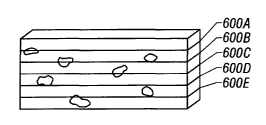
illustrated in Figure 5, the focal planar regions
observed at each depth of field, as indicated by sections
600A through 600E, can be captured as sectional images
and then assembled by a three-dimensional image processor
225A of Figure 7 to produce a three-dimensional volume
image of the specimen. This image is then used to
determine three coordinates for aiming the treatment
laser to the appropriate location within the volume of
the specimen.
The apparatus can produce sectional images at a
variety of depths of field according to the configuration
of optical devices. Those skilled in the art will be
CA 02426871 2003-04-24
WO 02/37938 PCT/USO1/50646
37
able to configure the detector to image at a shallow
depth of field which includes a depth of less than 100
microns. Depending upon the size of the specimen the
depth of field can be selected from 1, 2, 3, 4, 5, 6, 7,
8, 9, 10, 11, 12, 15, 20, 25, 30 , 35, 40, 45, 50, 60, 70,
80 90 or 100 microns. For larger specimens even greater
depths of field can be employed for deeper imaging.
The apparatus of the invention can be
configured to capture images of the specimen at different
resolutions or magnifications. This can be achieved by
altering the property of the lens 455 in front of one or
more cameras. A turret, cassette or wheel containing
different lenses can be placed between the camera and
specimen such that the magnification or resolution can be
rapidly changed. The turret, cassette or wheel can be
functionally attached to a positioning device for manual
or automated changes in resolution or magnification
during the course of or between specimen processing
procedures.
A detector used in the apparatus can also
include two or more cameras capable of imaging the
specimen from different directions of view. Imaging from
different directions of view, also referred to as stereo-
imaging, can be used to reconstruct an image of the
specimen. Two or more cameras can stereo-image a
specimen when their different directions of view are
separated by an angle selected from 1 to 180 degrees.
The invention can include cameras having different
directions of view separated by less than 1 degree, 2
degrees, 5 degrees, 10 degrees, 15 degrees, 20 degrees,
25 degrees, 30 degrees, 35 degrees, 40 degrees, 45
CA 02426871 2003-04-24
WO 02/37938 PCT/USO1/50646
38
degrees, 50 degrees, 90 degrees or 180 degrees, wherein a
degree is intended to be used consistent with
mathematical usage wherein it is an angle subtending
1/360 of the circumference of a circle.
A detector used in the invention can include
one or more cameras viewing a relatively shallow focal
planar region, wherein the focal planar region can be
refocused on different sections of the specimen. A
particular camera view can be refocused to observe a
0 specimen at different depths of field thereby obtaining
different sectional images of the specimen. Such
refocusing can be achieved by moving the camera. Other
lower inertia components of the detector are preferably
moved in order to achieve refocus and include lenses or
.5 mirrors placed in between the optical path of the camera
and specimen. The component to be adjusted can be
operably attached to a positioning device for manual or
automated refocusing. Automated focusing can be achieved
by incorporation of an automated positioning device that
:0 is capable of communicating with imaging processing
devices such as those described below.
Any detector capable of converting radiation
directed from a specimen or particle therein into a
signal that can be subsequently manipulated or stored to
5 determine the presence or quantity of a particle in a
specimen can be used in the apparatus or methods of the
invention. A detector can include a photodiode,
photomultiplier tube or charge-coupled device. A
detector can also include an imaging device that converts
0 radiation directed from a specimen or particle therein to
a set of signals that can be converted into a 3-
CA 02426871 2003-04-24
WO 02/37938 PCT/USO1/50646
39
dimensional representation of a specimen. Such an
imaging device can include a camera such as a CCD camera,
digital camera, film camera or photographic camera and
the like. One skilled in the art will be able to choose
a detector based on a variety of well known factors
including, for example, compatibility with the radiation
source used, sensitivity, spectral range of detection and
compatibility with data processing devices.
Referring now to Figure 8, a perspective view
of an embodiment of an optical subassembly is
illustrated. As illustrated, the illumination laser 305
sends a light beam through the shutter 310 and ball lens
315 to the SMA fiber optic connector 320. The light
passes through the fiber optic cable 325 and through the
output 330 into the condenser lenses 335, 340 and 345.
The light then enters the cube beamsplitter 350 and is
transmitted to the long wave pass mirror 355. From the
long wave pass mirror 355, the light beam enters the
computer-controlled galvanometers 360 and is then steered
to the proper frame of cells in the specimen through the
scanning lens 365.
As also illustrated in the perspective drawing
of Figure 8, the treatment laser 400 transmits energy
through the shutter 410 and into the beam expander 415.
Energy from the treatment laser 400 passes through the
beam expander 415 and passes through the half-wave plate
420 before hitting the fold mirror 425 and subsequently
entering the cube beamsplitter 350 where it is reflected
90° to the long wave pass mirror 355, from which it is
reflected into the computer controlled galvanometer
mirrors 360. The galvanometer mirrors 360 can be
CA 02426871 2003-04-24
WO 02/37938 PCT/USO1/50646
adjusted to steer the treatment laser beam through the
scanning lens 365 such that the beam strikes the portion
of a specimen where a particular target cell is located.
Accordingly, a desired response can be selectively
5 induced in the target cell using the apparatus of the
invention.
In order to accommodate a very large surface
area of specimen to treat, the apparatus includes a
movable stage that mechanically moves the specimen
10 container with respect to the scanning lens. Thus, once
a specific sub-population of cells within the scanning
lens field-of-view has been treated, the movable stage
brings another sub-population of cells within the
scanning lens field-of-view. As illustrated in Figure
15 11, a computer-controlled movable stage 500 holds a
specimen container 505 which contains a specimen 600 to
be processed. The movable stage 500 is moved by
computer-controlled servo motors along two axes so that
the specimen container can be moved relative to the
20 optical components of the instrument. The stage movement
along a defined path is coordinated with other operations
of the apparatus. In addition, specific coordinates can
be saved and recalled to allow, return of the movable
stage to positions of interest. Encoders on the x and y
25 movement provide closed-loop feedback control of stage
position.
A flat-field (F-theta) scanning lens 365 can be
mounted below the movable stage. The scanning lens
field-of-view comprises the portion of the specimen that
30 is presently positioned above the scanning lens by the
movable stage 500. The lens 365 can be mounted to a
CA 02426871 2003-04-24
WO 02/37938 PCT/USO1/50646
41
stepper motor that allows the lens 365 to be
automatically raised and lowered (along the z-axis) for
the purpose of focusing the system.
As illustrated in Figures 8-10, below the
scanning lens 365 are the galvanometer-controlled
steering mirrors 360 that deflect electromagnetic energy
along two perpendicular axes. Behind the steering mirrors
is the long wave pass mirror 355 that reflects
electromagnetic energy of a wavelength shorter than 545
nm. Wavelengths longer than 545 nm are passed through
the long wave pass mirror, directed through the filter
460, coupling lens 455, and into the CCD camera array,
thereby producing an image of the appropriate size on the
CCD sensor of the camera array 450 (See Figures 3 and 4).
The magnification defined by the combination of the
scanning lens 365 and coupling lens 455 can be chosen to
reliably detect single cells while maximizing the area
viewed in one frame by each camera. Although a CCD
camera array (DVC, Austin, TX) is illustrated in this
embodiment, the camera can be any type of detector or
image gathering equipment known to those skilled in the
art, as described above. The optical subassembly of the
apparatus is preferably mounted on a vibration-damping
platform to provide stability during operation as
illustrated in Figures 2 and 9.
Referring now to Figure 11, a top view of the
movable stage 500 is illustrated. As shown, a specimen
container can be detachably mounted in the movable stage
500. The specimen container 505 rests on an upper axis
nest plate 510 that is designed to move in the forward
and backward direction with respect to the movable stage
CA 02426871 2003-04-24
WO 02/37938 PCT/USO1/50646
42
500. A stepper motor can be connected to the upper axis
nest plate 510 and computer system so that commands from
the computer direct forward or backward movement of the
specimen container 505.
The movable stage 500 is also connected to a
timing belt 515 that provides side-to-side movement of
the movable stage 500 along a pair of bearing tracks 525A
and B. The timing belt 515 attaches to a pulley housed
under a pulley cover 530. The pulley is connected to a
stepper motor 535 that drives the timing belt 515 to
result in side-to-side movement of the movable stage 500.
The stepper motor 535 is electrically connected to the
computer system so that commands within the computer
system control side-to-side movement of the movable stage
500. A travel limit sensor 540 connects to the computer
system and causes an alert if the movable stage travels
beyond a predetermined lateral distance.
A pair of accelerometers 545A and B is
preferably incorporated on this platform to register any
excessive bumps or vibrations that may interfere with the
apparatus operation. In addition, a two-axis
inclinometer 550 is preferably incorporated on the
movable stage to ensure that the specimen container is
level, thereby reducing the possibility of
gravity-induced motion in the specimen container.
The specimen chamber has a fan with ductwork to
eliminate condensation on the specimen container, and a
thermocouple to determine whether the specimen chamber is
within an acceptable temperature range. Additional fans
are provided to expel the heat generated by the
CA 02426871 2003-04-24
WO 02/37938 PCT/USO1/50646
43
electronic components, and appropriate filters are used
on the air intakes 215A and B (see Figure 2).
The computer system 225 controls the operation
and synchronization of the various components of
electronic hardware described above. The computer system
can be any commercially available computer that can
interface with the hardware. One example of such a
computer system is an Intel Pentium~ IV-based computer
running the Microsoft Windows~ 2000 operating system.
Software is used to communicate with the various devices,
and control the operation in the manner that is described
below.
When the apparatus is first initialized, the
computer loads files from the hard drive into RAM for
proper initialization of the apparatus. A number of
built-in tests are automatically performed to ensure the
apparatus is operating properly, and calibration routines
are executed to calibrate the cell treatment apparatus.
Upon successful completion of these routines, the user is
prompted to enter information via the keyboard and mouse
regarding the procedure that is to be performed (e. g.
patient name, ID number, etc.). Once the required
information is entered, the user is prompted to open the
access door 35 and load a specimen onto the movable
stage.
Once a specimen is in place on the movable
stage and the door is closed, the computer passes a
signal to the stage to move into a home position. The
fan is initialized to begin warming and defogging of the
specimen. During this time, cells within the specimen
CA 02426871 2003-04-24
WO 02/37938 PCT/USO1/50646
44
are allowed to settle to the bottom surface. In
addition, during this time, the apparatus may run
commands that ensure that the specimen is properly
loaded, and is within the focal range of the system
optics. For example, specific markings on the specimen
container can be located and focused on by the system to
ensure that the scanning lens has been properly focused
on the bottom of the specimen container. After a
suitable time, the computer turns off the fan to prevent
excess vibrations during treatment, and cell treatment
processing begins.
First, the computer instructs the movable stage
to be positioned over the scanning lens so that the first
area of the specimen to be treated is directly in the
scanning lens field-of-view. The
galvanometer mirrors are instructed to move such that the
center frame within the field-of-view is imaged in the
camera. As discussed below, the field imaged by the
scanning lens is separated into a plurality of frames.
Each frame is the proper size so that the cells within
the frame are effectively imaged by the camera array.
The back-light 475 is then activated in order
to illuminate the field-of-view so that it can be brought
into focus by the scanning lens. Once the scanning lens
has been properly focused upon the specimen, the computer
system divides the field-of-view into a plurality of
frames so that each frame is analyzed separately by the
camera array. This methodology allows the apparatus to
process a plurality of frames within a large
field-of-view without moving the mechanical stage.
Because the galvanometers can move from one frame to the
CA 02426871 2003-04-24
WO 02/37938 PCT/USO1/50646
next very rapidly compared to the mechanical steps
involved in moving the stage, this method results in an
extremely fast and efficient apparatus.
The apparatus of the invention can further
5 include an image processing device 225A for combining one
or more two-dimensional representations of a specimen and
producing a three-dimensional representation. A two-
dimensional or three-dimensional representation refers to
an image or any characterization of a specimen, or
10 portion thereof, that specifies the coordinates of at
least one particle of interest therein such as a
graphical or tabular list of coordinates or a set of
computer commands that can be used to produce an image.
Initially, one or more two-dimensional
15 representations such as two-dimensional sectional images
can be captured by the camera array and stored to a
memory in the computer. Although, a single two-
dimensional image can contains sufficient information to
produce a three dimensional representation of a specimen,
20 it may be desirable to process two or more or a plurality
of two-dimensional images to produce a three-dimensional
image. Instructions in the computer can produce or
calculate a three dimensional representation such as a
three-dimensional image. A three-dimensional image
25 calculated as such can be analyzed with respect to the
size, shape, number, or other object features in the
image at each stepped Z-level. If necessary, the
computer instructs the z-axis motor attached to the
scanning lens to raise or lower in order to improve focus
30 on the frame of interest. The galvanometer-controlled
mirrors are then instructed to image a first frame,
CA 02426871 2003-04-24
WO 02/37938 PCT/USO1/50646
46
within the field-of-view, in the camera array. Once the
galvanometer mirrors are pointed to the first frame in
the field-of-view, the shutter in front of the
illumination laser is opened to illuminate the first
frame through the galvanometer mirrors and scanning lens.
The camera array captures an image of any fluorescent
emission from the specimen in the first frame of cells.
Once the image has been acquired, the shutter in front of
the illumination laser is closed and a software program
(Epic, Buffalo Grove, IZ) within the computer processes
the image.
The image processing device 225A can include
the capability of virtual autofocusing by searching
sectional images of a specimen and identifying a
25 sectional image that is in-focus. Virtual autofocusing
does not require production of a three-dimensional
representation of any part of the specimen and can,
therefore, be performed prior to or absent formation.of a
three-dimensional representation. A plurality of
sectional representations such as sectional images can be
obtained as described above using one or more cameras
viewing different focal planar regions. Virtual
autofocusing can be achieved by analysing multiple
sectional images and selecting an in-focus image.
Subsequent image processing can then be selectively
carried out for the in-focus sectional image in order to
efficiently identify a desired target particle. Thus, a
particle of interest can be identified or located in a
specimen based on its X and Y coordinates in the in-focus
sectional image and the Z-level of the sectional image.
The X and Y coordinates as used herein refer to
coordinates in two dimensions forming a plane orthogonal
CA 02426871 2003-04-24
WO 02/37938 PCT/USO1/50646
47
to the direction of view. A plurality of in-focus
sectional images selected by virtual autofocusing can be
used to calculate a three-dimensional image as described
above.
Although real-time autofocusing can be used in
the invention, virtual autofocusing provides the
advantage of more rapid throughput. Specifically, real-
time autofocusing often requires multiple adjustments of
optical components and re-imaging until an in-focus
sectional image is obtained. In contrast, when a
plurality of fixed cameras are placed to view non-
overlapping focal planar regions, at least one camera
will have a focused view without the need to move any
component of the detector. Subsequently, the images can
be analyzed using algorithms similar to those used in
real-time autofocusing methods without the requirement
for time-consuming movement of optical components and
reacquisition of images.
Known autofocusing algorithms such as those
used in microscopy or autofocus cameras can be used to
analyze sectional images and identify an in-focus
sectional image in the apparatus and methods of the
invention. An example of an autofocus method that can be
used in the apparatus or methods of the invention is
binary search autofocus. Binary search autofocus can be
performed virtually by preselecting two sectional images
between which an in-focus sectional image is thought to
exist and iteratively reducing the number of intervening
sectional images until one having a desired focus is
identified. The iterations include the steps of
selecting a sectional image that is halfway between the
CA 02426871 2003-04-24
WO 02/37938 PCT/USO1/50646
48
boundary sectional images, evaluating the selected
sectional image for a predetermined focus value and
further reducing the boundary distance until a sectional
image having the desired focus value is identified.
Alternatively, a sequential autofocus method can be used
in which sectional images are analyzed in a stepwise
fashion starting from a preselected initial sectional
image.
In another embodiment, the detector can capture
an image of a two-dimensional specimen. Virtual
autofocusing will work if the depth of the specimen is
less than the depth of field of the detector view.
Virtual autofocusing can be carried out as described
above to identify or locate the X and Y coordinates for a
particle of interest located in the two-dimensional
specimen. The particle identified as such can be
targeted and electromagnetically affected using an
apparatus or method of the invention.
The power sensor 445, discussed above, detects
the level of light intensity emitted by the illumination
laser. Based on the measured intensity, the computer can
determine if an appropriate amount of light has
illuminated the frame of cells for the particular
application. In the event a particular threshold has not
been obtained or the signal surpasses a desired maximum
the laser intensity can be adjusted and another
illumination and image capture sequence performed. Such
iteration can be carried out until the appropriate
conditions are achieved or after a preselected number of
iterations the system can pause or indicate in an error
condition that is communicated to the operator.
CA 02426871 2003-04-24
WO 02/37938 PCT/USO1/50646
49
The threshold or maximum energy levels will
depend upon the particular application of the apparatus
or methods of the invention. The term "threshold" refers
to the amount of energy sufficient to change a particular
property. For example, a threshold amount of
electromagnetic energy can be an amount sufficient to
kill a cell, to transiently permeabilize a membrane or
cell, to induce a cell cycle stage, to activate or
repress gene expression, to increase or decrease
intracellular pH within a defined range, or to alter a
morphological, metabolic or developmental stage of a
cell. Other cells in the same specimen that do not
receive radiation at or beyond the threshold will not
undergo the particular change. A range of
electromagnetic energy used in the apparatus or methods
of the invention can be defined by a threshold and a
ceiling. A "ceiling" is intended to mean an amount of
energy that is greater than a threshold amount and
sufficient to induce an unwanted change in a particular
property. The ceiling can be defined by any detectable
change including those described above in relation to a
threshold energy. Thus, a range of electromagnetic
energy used in the invention can include an amount of
energy sufficient to transiently permeabilize a membrane
or cell without causing permanent permeabilization or
cell death.
Shuttering of illumination light can be used to
reduce undesirable heating and photobleaching of the
specimen and to provide a fluorescent signal in a desired
range of detection. An image analysis algorithm is run
to locate the x-y-z centroid coordinates of all targeted
cells in the frame by reference to features in the
CA 02426871 2003-04-24
WO 02/37938 PCT/USO1/50646
captured image. If there are targets in the image, the
computer calculates the three-dimensional coordinates of
the target locations in relation to the movable stage
position and field-of-view, and then positions the
5 galvanometer-controlled mirrors to point the treatment
electromagnetic radiation beam to the location of the
first target in the first frame of cells. It should be
noted that the z-coordinate may be calculated by the
algorithm based in part upon the focal length of the
10 camera that captured the image. It should further be
noted that only a single frame of cells within the
field-of-view has been captured and analyzed at this
point. Thus, there should be a relatively small number
of identified targets within this sub-population of the
15 specimen. Moreover, because the camera array is pointed
to a smaller population of cells, a higher magnification
is used so that each target is imaged by many pixels
within the CCD camera.
Once the computer system has positioned the
20 galvanometer controlled mirrors to point to the location
of the first targeted cell within the first frame of
cells, the treatment laser is fired for a brief interval
so that the first targeted cell is given an appropriate
dose of energy. The power sensor 446 discussed above
25 detects the level of energy that was emitted by the
treatment laser, thereby allowing the computer to
calculate if it was within a desired range to induce a
response in the targeted cell. The power of the
treatment laser can be adjusted and the treatment laser
30 fired at the same target again. The iterative targeting,
firing, and sensing steps can be repeated until
appropriate conditions axe achieved or up to a
CA 02426871 2003-04-24
WO 02/37938 PCT/USO1/50646
51
predetermined number of rounds after which the iteration
is paused or an error message communicated to the
operator. In another embodiment, the treatment laser can
be fired once at more than one, a group or all of the
target cells within a frame, and subsequently the
computer can direct the target laser to return to any
cells that did not receive a sufficient level of energy
to induce a response.
Once all of the targets have been irradiated
with the treatment laser in the first frame of cells, the
mirrors can be positioned to the second frame of cells in
the field-of-view, and the processing repeated at the
point of frame illumination and camera imaging. This
processing can be continued for all frames within the
field-of-view above the scanning lens. When all of these
frames have been processed, the computer instructs the
movable stage to move to the next field-of-view in the
specimen, and the process repeated from the back-light
illumination and auto-focus steps. Frames and
fields-of-view can be overlapped to reduce the
possibility of inadvertently missing areas of the
specimen. Once the specimen has been fully processed,
the operator is signaled to remove the specimen, and the
apparatus is immediately ready for the next specimen.
Although the text above describes the analysis of
fluorescent images for locating targets, those skilled in
the art will understand that the non-fluorescent
back-light LED illumination images can be useful fox
locating target cells based on other properties such as
those viewable in a standard microscope based on
absorbance, transmittance or refraction of light.
CA 02426871 2003-04-24
WO 02/37938 PCT/USO1/50646
52
The galvanometer mirrors provide the advantage
of controlling the imaging of successive frames and the
irradiation of successive targets. One brand of
galvanometer is the Cambridge Technology, Inc. model
number 6860 (Cambridge, MA). This galvanometer can
reposition very accurately within a fraction of a
millisecond, making the processing of large areas and
many targets possible within a reasonable amount of time.
In addition, the movable stage can be used to move
specified areas of the specimen into the scanning lens
field-of-view. This combination of movements can be
automated providing increased throughput of the apparatus
or methods.
It should be understood that other embodiments
of the invention are also possible. For example, a
movable stage, similar to a conveyer belt, could be
included to continuously move a specimen of cells through
the above-described process. Error signals continuously
generated by the galvanometer control boards are
monitored by the computer to ensure that the mirrors are
in position and stable before an image is captured, or
before a target is fired upon, in a closed-loop fashion.
In one embodiment, the apparatus described
herein is capable of electromagnetically affecting
particles in at least 1, 2, 3, 4, 5, 6, 7, 8, 9, or 10
square centimeters of a biological specimen per minute.
In another embodiment, the apparatus described herein
processes at least 0.~5, 0.5, 1, 2, 3 or 4 million cells
of a biological specimen per minute. The rate at which
the apparatus operates can be measured as the number of
particle containing focal volumes that are
CA 02426871 2003-04-24
WO 02/37938 PCT/USO1/50646
53
electromagnetically affected per minute. The apparatus
can electromagnetically affect at least 1, 2, 3, 4, 5, 6,
8, 10, 15, 20, 30, 60, 100, 300, 500, 1000, 3000, 5000,
10000 , 30000, 50000, 100000, 300000, or 500000 separate
focal volumes in the specimen per minute. Furthermore,
the rate of imaging can be at the rate of at least 1 Hz,
2 Hz, 3 Hz, 4 Hz, 5 Hz, 6 Hz, 8 Hz, 10 Hz, 15 Hz, 30 Hz,
50 Hz, 100 Hz, 150 Hz, 300 Hz, 500 Hz, or 1000 Hz.
One embodiment allows in vitro maintenance of
the biological specimen in the apparatus, such that it
can be monitored and processed periodically in situ.
This is accomplished by maintaining the specimen under a
standard tissue culture environment of 37°C. In this
manner, specific cells in the specimen are followed over
time, periodically assessing their state, and/or
periodically rendering treatment to those specific cells.
The system includes software and computer memory storage
for providing the apparatus with the ability to record
cell locations and thereafter direct the movable stage to
return to those recorded locations such that large
specimens can be repeatedly processed in a scanning mode.
Of course, many variations of the
above-described embodiments are possible, including
alternative methods for illuminating, imaging, and
targeting the cells. For example, movement of the
specimen relative to the scanning lens could be achieved
by keeping the specimen substantially stationary while
the scanning lens is moved. Steering of the illumination
beam, images, and energy beam could be achieved through
any controllable reflective or diffractive device,
including prisms, piezo-electric tilt platforms, or
CA 02426871 2003-04-24
WO 02/37938 PCT/USO1/50646
54
acousto-optic deflectors. Additionally, the apparatus
can image/process from either below or above the
specimen. Because the apparatus is focused through a
movable scanning lens, the illumination and energy beams
are directed to different focal planes along the z-axis.
Thus, portions of the specimen that are located at
different vertical heights are specifically imaged and
processed by the apparatus in a three-dimensional manner.
The sequence of the steps could also be altered without
changing the process. For example, one might locate and
store the coordinates of all targets in the specimen, and
then return to the targets to irradiate them with energy
one or more times over a period of time.
To optimally process the specimen, it should be
placed on a substantially flat surface so that a Large
portion of the specimen appears within a narrow range of
focus. The density of cells on this surface can, in
principle, be at any value. However, increasing the cell
density can minimize the total surface area required to
be scanned or detected in the apparatus or methods of the
invention.
The following examples illustrate the use of
the described method and apparatus in different
applications.
Example 1: Autologous HSC Transplantation
A patient with a B cell-derived metastatic
tumor in need of an autologous HSC transplant is
identified by a physician. As a first step in the
treatment, the patient undergoes a standard HSC harvest
CA 02426871 2003-04-24
WO 02/37938 PCT/USO1/50646
procedure, resulting in collection of approximately 1 x
101° hematopoietic cells with an unknown number of
contaminating tumor cells. The harvested cells are
enriched for HSC by a commercial immunoaffinity column
5 (Isolex~ 300, Nexell Therapeutics, Irvine, CA) that
selects for cells bearing the CD34 surface antigen,
resulting in a population of approximately 3 x 10⁸
hematopoietic cells, with an unknown number of tumor
cells. The mixed population is thereafter contacted with
10 anti-B cell antibodies (directed against CD20 and CD23)
that are conjugated to phycoerythrin, and anti-CD34
antibodies that are conjugated to CyChromeTM.
The mixed cell population is then placed in a
sterile specimen container on a substantially flat
15 surface near confluence, at approximately 500,000 cells
per square centimeter. The specimen is placed on the
movable stage of the apparatus described above, and all
detectable tumor cells are identified by the presence of
the phycoerythrin tag and absence of the CyChromeTM tag,
20 and are then targeted with a lethal dose of energy from a
treatment laser. The design of the apparatus allows the
processing of a clinical-scale transplant specimen in
under 4 hours. The cells are recovered from the specimen
container, washed, and then cryopreserved. Before the
25 cells are reinfused, the patient is given high-dose
chemotherapy to destroy the tumor cells in the patient's
body. Following this treatment, the processed cells are
thawed at 37°C and are given to the patient
intraveneously. The patient subsequently recovers with
30 no remission of the original cancer.
CA 02426871 2003-04-24
WO 02/37938 PCT/USO1/50646
56
Example 2: Allogeneic HSC Transplantation
In another embodiment, the significant risk and
severity of graft-versus-host disease in the allogeneic
HSC transplant setting can be combated. A patient is
selected for an allogeneic transplant once a suitable
donor is found. Cells are harvested from the selected
donor as described in the above example. In this case,
the cell mixture is contacted with phycoerythrin-labeled
anti-CD3 T-cell antibodies. Alternatively, specific
allo-reactive T-cell subsets could be labeled using an
activated T-cell marker (e.g. CD69) in the presence of
allo-antigen. The cell population is processed by the
apparatus described herein, thereby precisely defining
and controlling the number of T-cells given to the
patient. This type of control is advantageous, because
administration of too many T-cells increases the risk of
graft-versus-host disease, whereas too few T- cells
increases the risk of graft failure and the risk of
losing of the known beneficial graft-versus-leukemia
effect. The present invention and methods are capable of
precisely controlling the number of T-cells in an
allogeneic transplant.
Example 3: Tissue Engineerincr
In another application, the present apparatus
is used to remove contaminating cells in inocula for
tissue engineering applications. Cell contamination
problems exist in the establishment of primary cell
cultures required for implementation of tissue
engineering applications, as described by Langer and
Vacanti, Sci. Amer. 280:86-89 (2999). In particular,
CA 02426871 2003-04-24
WO 02/37938 PCT/USO1/50646
57
chondrocyte therapies for cartilage defects are hampered
by impurities in the cell populations derived from
cartilage biopsies. Accordingly, the present invention
is used to specifically remove these types of cells from
the inocula. For example, a cartilage biopsy is taken
from a patient in need of cartilage replacement. The
specimen is then grown under conventional conditions as
described in Brittberg et al., N.E.J. Med. 331:889-895
(1994). The culture is then stained with a specific
label for any contaminating cells, such as fast-growing
fibroblasts. The cell mixture is then placed within the
apparatus described and the labeled, contaminating cells
are targeted by the treatment laser, thereby allowing the
slower growing chondrocytes to fully develop in culture.
Exam»le 4: Stem Cell Thera~y
Yet another embodiment involves the use of
embryonic stem cells to treat a wide variety of diseases.
Since embryonic stem cells are undifferentiated, they can
be used to generate many types of tissue that would find
use in transplantation, such as cardiomyocytes and
neurons. However, undifferentiated embryonic stem cells
that are implanted can also lead to a jumble of cell
types which form a type of tumor known as a teratoma as
described in Pedersen, Sci. Amer. 280:68-73 (1999).
Therefore, therapeutic use of tissues derived from
embryonic stem cells must include rigorous purification
of cells to ensure that only sufficiently differentiated
cells are implanted. The apparatus described herein is
used to eliminate undifferentiated stem cells prior to
implantation of embryonic stem cell-derived tissue in the
patient.
CA 02426871 2003-04-24
WO 02/37938 PCT/USO1/50646
58
Example 5: Generation of Human Tumor Cell Cultures
In another embodiment, a tumor biopsy is
removed from a cancer patient for the purpose of
initiating a culture of human tumor cells. However, the
in vitro establishment of primary human tumor cell
cultures from many tumor types is complicated by the
presence of contaminating primary cell populations that
have superior in vitro growth characteristics over tumor
cells, and the limited number of tumor cells obtainable
from such a specimen. For example, contaminating
fibroblasts represent a major challenge in establishing
many cancer cell cultures. The disclosed apparatus is
used to particularly label and destroy the contaminating
cells, while leaving the biopsied tumor cells intact.
Accordingly, the more aggressive primary cells will not
overtake and destroy the cancer cell line. The apparatus
described within allows purification of cells with
relatively high yield, which is particularly important in
application where the starting cell number is limited.
The high yield of purified cells from the disclosed
apparatus provides a significant advantage over other
cell purification methods.
Example 6: Generation of a Specific mRNA Expression
Library
The specific expression pattern of genes within
different cell populations is of great interest to many
researchers, and many studies have been performed to
isolate and create libraries of expressed genes for
different cell types. For example, knowing which genes
are expressed in tumor cells versus normal cells is of
CA 02426871 2003-04-24
WO 02/37938 PCT/USO1/50646
59
great potential value as described in Cossman et al.,
Blood 94:411-416 (1999). Due to the amplification
methods used to generate such libraries (e. g. PCR), even
a small number of contaminating cells can result in an
inaccurate expression library. One approach to overcome
this problem is the use of laser capture microdissection
(LCM), in which a single cell is used to provide the
starting genetic material for amplification as described
in Schutze et al., Nat. Biotech. 16:737-742 (1998}.
Accurate purification of a significant cell number prior
to extraction of mRNA would enable the generation of a
highly accurate expression library, one that is
representative of the cell population being studied,
without biases due to single cell expression or
25 expression by contaminating cells. The methods and
apparatus described in this invention can be used to
purify cell populations with high yield so that no
contaminating cells are present during an RNA extraction
procedure.
Within a human prostate tumor, there are
multiple cell types, each of which lead to metastatic
disease. To understand the basis of prostate cancer and
its progression, the tools of genomics such as DNA
sequencing of normal prostate and cancer prostate cDNAs
(expressed sequence tags, or ESTs) and DNA array
comparison of normal and cancerous tissues have been used
to investigate the distinct patterns of gene expression
in normal prostate and in different prostate tumors. It
has been shown that different prostate cell
subpopulations with different surface antigens have
different mRNA expression profiles. Using the apparatus
disclosed herein, primary human prostate tumors are
CA 02426871 2003-04-24
WO 02/37938 PCT/USO1/50646
purified for specific cell subpopulations to investigate
their gene expression. For example, four populations of
primary prostate cells are purified for mRNA analysis by
binding antibodies against specific cell markers to new
5 cell surfaces. The purified populations are: CD44'CD13'
normal basal, CD44'CD13' basal cell-like cancer,
CD57'CD13' normal luminal, and CD57'CD13 luminal
cell-like cancer epithelial cells. Understanding the
nature of each subpopulation, and its relation to disease
10 onset and progression, could lead to new diagnostic and
therapeutic approaches in the treatment of
the disease.
Example 7: Transfection, Monitorincr,, and Purification of
a Specific Cell Population
15 Many research and clinical gene therapy
applications are hampered by the inability to transfect
an adequate number of a desired cell type without
transfecting other cells that are present. The method of
the present invention would allow selective targeting of
20 cells to be transfected within a mixture of cells. By
generating a photomechanical shock wave at or near a cell
membrane with a targeted energy source, a transient pore
can be formed, through which genetic or other material
can enter the cell. This method of gene transfer has
25 been called optoporation. The apparatus described above
can selectively induce optoporation in the cells of
interest in a rapid, automated, targeted manner.
For example, bone marrow cells are plated in a
specimen container having a solution containing plasmid
30 DNA to be transfected. The plasmid DNA encodes a
CA 02426871 2003-04-24
WO 02/37938 PCT/USO1/50646
61
therapeutic gene (e. g. MDR), as well as a marker gene
(e. g. green fluorescent protein). PE-labeled antibodies
(anti-CD34) having specificity for stem cells are added
into the medium and bind to the stem cells. The specimen
container is placed within the cell processing apparatus
and a first treatment laser is targeted to any cells that
become fluorescent under the illumination laser light,
thereby facilitating transfection of DNA specifically
into the targeted cells. The cells are maintained in
situ at 37°C, all targeted cells being periodically
analyzed for expression of green fluorescent protein to
indicate successful transfection with the plasmid DNA.
After 48 hours, all cells not expressing green
fluorescent protein are eliminated with a second
treatment laser, thereby yielding a pure population of
stem cells expressing the transfected genes.
Example 8: Selection of Desirable Clones in a
Biotechnology Application
In many biotechnology processes where cell
lines are used to generate a valuable product, it is
desirable to derive clones that are very efficient in
producing the product. This selection of clones is often
carried out manually, by inspecting a large number of
clones that have been isolated in some manner. The
present invention would allow rapid, automated inspection
and selection of desirable clones for production of a
particular product. For example, hybridoma cells that
are producing the greatest amounts of antibody can be
identified by a fluorescent label directed against the F~
region. Cells with no or dim fluorescent labeling are
targeted by the treatment laser for killing, leaving
CA 02426871 2003-04-24
WO 02/37938 PCT/USO1/50646
62
behind the best producing clones for use in antibody
production.
Example 9: Automated Monitoring of Cellular Responses
Automated monitoring of cellular responses to
specific stimuli is of great interest in high-throughput
drug screening. Often, a cell population in one well of
a well-plate is exposed to a stimulus, and a fluorescent
signal is then captured over time from the cell
population as a whole. Using the methods and apparatus
described herein, more detailed monitoring could be done
at the single cell level. For example, a Cell population
can be labeled to identify a characteristic of a
subpopulation of cells that are of interest. This label
is then excited by the illumination laser to identify
those cells. Thereafter, the treatment laser is targeted
at the individual cells identified by the first
label, for the purpose of exciting a second label,
thereby providing information about each cell's response.
Since the cells are substantially stationary on a
surface, each cell could be evaluated or treated multiple
times, thereby providing temporal information
about the kinetics of each cell's response. Also,
through the use of the large area scanning Lens and
galvanometer mirrors, a relatively large number of wells
~5 could be quickly monitored over a short period of time.
As a specific example, consider the case of
alloreactive T-cells as presented in Example 2 above. In
the presence of allo-antigen, activated donor T-cells
could be identified by CD69. Instead of using the
treatment laser to target and kill these cells, the
CA 02426871 2003-04-24
WO 02/37938 PCT/USO1/50646
63
treatment laser could be used to examine the
intracellular pH of every activated T-cell through the
excitation and emitted fluorescence of carboxyfluorescein
diacetate. The targeted laser allows the examination of
only cells that are activated, whereas most screening
methods evaluate the response of an entire cell
population. If a series of such wells are being
monitored in parallel, various agents could be added to
individual wells, and the specific activated T-cell
response to each agent could be monitored over time.
Such an apparatus would provide a high-throughput
screening method for agents that ameliorate the
alloreactive T-cell response in graft-versus-host disease
Based on this example, one skilled in the art could
I5 imagine many other examples in which a cellular
response to a stimulus is monitored on an individual cell
basis, focusing only on cells of interest identified by
the first label.
Throughout this application various
publications have been referenced. The disclosures of
these publications in their entireties are hereby
incorporated by reference in this application in order to
more fully describe the state of the art to which this
invention pertains.
Although the invention has been described with
reference to the examples provided above, it should be
understood that various modifications can be made without
departing from the spirit of the invention. Accordingly,
the invention is limited only by the claims.