Note: Descriptions are shown in the official language in which they were submitted.
CA 02427163 2003-04-28
WO 02/36790 PCT/EPO1/12532
Influenza Virus Vector for Infection of Dendritic Cells
The present invention relates to a method for preparing patient-derived immune
cells, such as dendritic cells, capable of expressing antigens, such as tumor-
associated antigens or virus associated antigens, patient derived immune cells
obtainable by said method, their use for the treatment of tumors or virus
infections and to a method for the expression of tumor-associated antigens or
virus associated antigens by utilizing such patient-derived immune cells. The
method utilizes attenuated, recombinant influenza virus vectors allowing
efficient
transduction and a high expression rate of genes encoding one or more tumor-
or virus-associated antigens in patient-derived immune cells. The patient-
derived
immune cells obtained by said method can be used for ex-vivo therapy of cancer
or diseases caused by viral microbial or parasitic infections..
Introduction
Anti-tumor immune responses can be efficiently generated in vivo and in vitro
by
2o patient derived immune cells such as dendritic cells (hereinafter shortly
referred
to as "DC"), which have been genetically modified to express tumor-associated
antigens (hereinafter shortly referred to as "TAA"). Several viral expression
vectors derived from adenoviruses, retroviruses, and poxviruses have been used
to transduce foreign genes into DC. These viruses contain DNA or form a DNA
intermediate, which could integrate into the host genome with the risk of
adverse mutations, and have limited host-cell range. In contrast, influenza
viruses are sensitive to antiviral pharmaceuticals, fail to establish
persistent
infections, do not integrate their RNA genome into the chromosome, have a
broad host-cell range, and may even be genetically designed entirely from
3o cloned cDNA thus providing a favourable alternative. Here we show that self-
attenuating recombinant avian influenza virus vectors very efficiently deliver
TAA
genes and other genes, which can include genes of virus associated antigens
(hereinafter "VAA") into patient derived immune cells, in particular into
human
monocyte-derived DC. The modified DC retained their characteristic phenotypic
and immunity-stimulating properties. Thus the influenza virus vector
represents
a valuable and safe tool in the development of immune cell- (DC-)based
immunotherapy in man.
CONFIRMATION COPY
CA 02427163 2003-04-28
WO 02/36790 PCT/EPO1/12532
DCs are highly potent initiators of immune responses and are widely
distributed
throughout lymphoid and non-lymphoid tissues (R.M. Steinman, Fundamental
Immunology, fourth Ed. Ed W.E. Paul. Philadelphia: Lippincott-Raven. pp. 547
(1999)). To launch immune responses, DC have to capture and to process
antigens in the periphery and to present them to rare antigen-specific T
cells,
which they encounter after migration to lymphoid organs. DC are therefore
considered as "Natures adjuvant" (J.W. Young, K. Inaba, J. Exp. Med. 183, 7-11
(1996); G. Schuler, R.M. Steinman, J. Exp. Med. 186, 1183-1187 (1997); J.
1o Banchereau, R.M. Steinman, Nature 392, 245-252 (1998), M. Rescigno et al.,
Immunol. Today 20, 200-203 (1999)). Antigen uptake is accomplished by
immature DC. They mature into potent T cell stimulators in response to
environmental danger signals like pro-inflammatory cytokines, dsRNA or LPS. DC
which are activated in this way, migrate to the lymphoid organs, develop
cytoplasmic protruding veils and up-regulate a set of characteristic surface
proteins. These are required for the interaction and activation of antigen-
specific
T cells. Among them are CD25, CD40, CD54, CD80, CD83, CD86 as well as high
levels of MHC class I and class II molecules.
2o The ability to present antigens to naive or quiescent T cells qualifies DC
as a
potent biological tool for cell-based immuno-therapeutical strategies directed
against tumors and infectious diseases (G. Schuler, R.M. Steinman, J. Exp.
Med.
186, 1183-1187 (1997); J. Banchereau, R.M. Steinman, Nature 392, 245-252
(1998)). Several studies reported beneficial anti-tumor and anti-viral effects
due
to DC mediated vaccinations in mice (J.I. Mayordomo et al., Nat. Med. I., 1297-
1302 (1995); R.C. Fields et al., Proc. Natl. Acad. Sci., 95, 9482-9487 (1998);
B.
Ludewig et al., J. Virol. 72, 3812-3818 (1998)). Recently immunization with
peptide- and/or tumor lysate-pulsed DC has also been reported in humans (F.J.
Hsu et al., Nat. Med. 2, 52-58 (1996); G. Murphy et al., Protstate 29, 371-380
3o (1996); F.O. Nestle et al., Nat. Med. 4, 328-332 (1998); M.V. Dhodapkar et
al.,
J. Clin. Invest. 104, 173-180 (1999); M. Marchand ~et al., Int. J. Cancer 80,
219
230 (1999); B. Thurner et al., J. Exp. Med. 190, 1669-1678 (1999)). Although
these data are quite promising, the peptide-loading strategy has several
disadvantages, including HLA restrictions and the fact that only a limited
number
of tumor-specific peptides are known.
2
CA 02427163 2003-04-28
WO 02/36790 PCT/EPO1/12532
On the other hand it is known that the strategy to genetically modify DC by
genes coding for full-size tumor antigens has several advantages over pulsing
DC with whole tumor lysates or tumor peptides: The heterologous proteins
expressed from the introduced genes are endogenously processed, loaded onto
MHC class I molecules (Y. Yang et al., J. Virol. 69, 2004-2015 (1995); C.A.
Mack
et al., Hum. Gene Ther. 8, 99-109, (1997)) and presented to T lymphocytes.
Thus genetically modified DC have the potential to present previously unknown
epitopes in association with different, individually divergent, MHC molecules.
Therefore, new strategies for effective genetic modifications are needed in
order
to generate new DC based anti-tumor vaccines. DC can be generated in vitro
from cultured human monocytes (N. Romani et al., J. Exp. Med. 180, 83-93
(1994); F. Sallusto, A. Lanzavecchia, J. Exp. Med. ,179, 1109-1118 (1994); A.
Bender et al., J. Immunol. Methods 196, 121-135 (1996); N. Romani et al., J.
Immunol. Methods 196, 137-151 (1996)) even in large numbers from
leukapheresis products for clinical use (B. Thurner et al., J. Exp. Med. 190,
1669-
1678 (1999)). Transduction of these DC with tumor-associated or viral antigens
leads to the presentation of these antigens via MHC class I molecules.
Viral vectors represent a very powerful method to deliver heterologous genes
2o into primary cells. Several viral vector systems have been described that
are
capable to introduce foreign genes into DC. Adenoviral vectors have been
reported to efficiently transduce human CD34+-derived (M. Bregni et al., Gene
Ther. 5, 465-472 (1998)) as well as monocyte-derived DC (A.B. Dietz, P.S. Vuk,
Blood 91, 392-398 (1998); L. Zhong et al., Eur. J. Immunol. 29, 964-972
(1999)). Retrovirus vectors have been shown to transduce proliferating human
DC progenitors (J.F. Arthur et al., Cancer Gene Ther. 4, 17-25 (1997); P.
Szabolcs et al., Blood 90, 2160-2167 (1997); B. Verhasselt et al., Blood 91,
431-
440 (1998)). However, there are several disadvantages associated with
retroviral vectors. They have relatively low transduction rates (P. Szabolcs
et al.,
Blood 90, 2160-2167 (1997)) and the genomes of these, vectors integrate into
the host genome. Furthermore, non-dividing cells, like terminally
differentiated,
mature DC cannot be transduced. Vaccinia viruses have also been used to
efficiently transfer genes into human DC. However this vector employs multiple
mechanisms to evade the immune system including down modulation of the
costimulatory molecule CD80 and induces apoptotic cell death in both stages of
3
CA 02427163 2003-04-28
WO 02/36790 PCT/EPO1/12532
DC (J. Engelmayer et al., J. Immunol 163, 6762-6768 (1999), R. Drillien et
al.,
Virology 268, 471-481 (2000)).
Again, viral expression vectors derived from adenoviruses, retroviruses, and
poxviruses have been reported to efficiently transduce foreign genes into DC
(M.E. Reeves et al., Cancer Res. 56, 5672-5677 (1996); W. Song et al., J. Exp.
Med. 186, 1247-1256 (1997); M. Di Nicola et al., Cancer Gene Ther. 5, 350-356
(1998); M. Subklewe et al, Blood 94, 1372-1381 (1999); L. Zhong et al., Eur.
J.
Immunol. 29, 964-972 (1999); S. Yang et al., J. Immunol. 164, 4204-4211
(2000)). These viruses contain DNA or have an DNA replication intermediate,
which could integrate into the host chromosomal sequences thus posing the risk
of adverse mutations. This is impossible with negative-strand RNA viruses as
influenza viruses. Interestingly, DC infected by influenza viruses are strong
stimulators of human CD8+ T cells (N. Bhardwaj et al., J. Clin. Invest. 94,
797-
807 (1994); A. Bender et al., J. Exp. Med. 182, 1663-1671 (1995) and
Immunology 198, S52-567 (1998)). Furthermore, infection by influenza virus
results in the maturation of immature DC and to increased antigen presentation
and T cell stimulatory capacity (M. Cella et al., J. Exp. Med. 189, 821-829
(1999)). Therefore, gene transfer into DC by influenza virus vectors as
disclosed
2o in Strobel et al., J. of Dermatological Science 16(1), 5133 (1988) and J.
of
Investigative Dermatology 110(4), 605 (1998) was a promising approach to
induce anti-tumor or anti-viral immune response.
The generation of recombinant influenza viruses was hampered for a long time
by the fact that the virus has a segmented RNA genome. The development of the
RNA polymerase I technique allows the generation of recombinant viruses with
additional genomic segments capable of expressing complete heterologous genes
(G. Neumann et al., Virology 202, 477-479 (1994)), which was built around in
vivo synthesis of recombinant vRNA molecules by cellular RNA polymerase I
3o transcription of the respective template cDNA constructs. Modified terminal
viral
RNA sequences (hereinafter "promoter-up mutations" or promoter-up variants")
were designed by nucleotide substitutions (Neumann and Hobom, Mutational
analysis of influenza virus promoter elements in vivo, J. Gen. Virol. 76, 1709-
1717 (1995); WO 96/10641). The above promoter-up variants carry up to five
nucleotide substitutions (in promoter-up variant 1920; see Flick and Hobom, J.
Gen. Virol. 80, 2565-2572 (1999)). When these promoter-up variants are
4
CA 02427163 2003-04-28
WO 02/36790 PCT/EPO1/12532
_ a
attached to a recombinant ninth vRNA segment its increased transcription and
amplification rate will not only compensate for the losses suffered
spontaneously,
but even cause accumulation of the foreign vRNA segment during simple viral
passaging, in the absence of any selection.
However, due to its over-replication relative to all of the regular influenza
vRNA
segments (which of course .are connected to wild-type promoter sequences)
after
catching up with the others the foreign segment will become over-abundant.
This
increasingly will result in viral particles that have incorporated several
copies of
1o recombinant vRNA, but no longer have a full set of all eight standard
segments
incorporated among an average of about 15 vRNA molecules present within a
virion. Such particles are defective and will not result in plaque formation,
hence
. after an initial increase of recombinant viral particles. during the first
steps of
propagation a dramatic decrease is observed, usually at the third or fourth
step
Of viral passaging, depending on the size of the recombinant vRNA and the
level
of the promoter-up mutation attached. A balanced situation with regard to the
insert length and the level of promoter activity can be achieved, and has been
propagated in a particular case over 11 passages, with essentially stable
levels
of recombinant viruses among a majority of helper viruses (around 80%) during
2o these steps.
WO 00/53786 then discloses a recombinant influenza virus for high-yield
expression of incorporated foreign gene(s), which is genetically stable in the
absence of any helper virus and which comprises at least one viral RNA segment
being an ambisense RNA molecule (ambisense RNA segment). Said ambisense
RNA segment contains one of the standard viral genes in sense orientation and
a
foreign, recombinant gene in anti-sense orientation, or vice versa, in overall
convergent arrangement. The ambisense RNA segment in the recombinant
influenza virus of WO 00/53786 is - if stably integrated - one of the eight
3o segments of the virus, and is a so-called "bicistronic" segment containing
two
different genes in covalent junction which are to be expressed by the
recombinant virus. A further stable recombinant influenza virus with a
bicistronic
segment is described in PCT/EP01/08124. Here, the influenza virus comprises at
least one viral RNA segment being a bicistronic RNA molecule coding for two
genes in tandem arrangement (tandem RNA segment). In said tandem RNA
segment one of the standard viral genes is in covalent junction with a
foreign,
s
CA 02427163 2003-04-28
WO 02/36790 PCT/EPO1/12532
recombinant gene and said tandem RNA segment has an upstream splice donor
and a downstream splice acceptor signal surrounding the proximal coding
region.
Both applications mention that such stable recombinant influenza viruses
containing a foreign gene in a bicistronic segment are suitable as viral
vectors for
infection, transfection or transduction of patient-derived immune cells such
as
dendritic cells. These stable recombinant viruses are suitable for DC-based
therapy when very pure, mature DC preparations are used, which afford abortive
infections, but may be less desirable when DC preparations still contain some
immature DC or monocytes. In the latter instance the use of attenuated
recombinant viruses or self-attenuating recombinant viruses is preferred. The
limited replication of self-attenuating viruses may in addition strengthen the
immune response.
Summary of the Invention
It was found that a recombinant influenza virus as disclosed in WO 96/10641
having a ninth , i.e. an additional, segment carrying a TAA or VAA gene under
the control of a high-capacity promoter is a suitable vector for transduction
of
TAA and VAA genes. These viruses still abortively infect DC without
interfering
with their antigen presenting capacity but - due to their "instability" - are
self-
2o attenuating whilst retaining the high and persistent antigen expression
controlled
by the high-capacity promoter. The attenuated recombinant influenza viruses
retain the immunostimulatory capacity of mature dendritic cells while
expressing
the antigens. In contrast to other viral vectors used for DC transduction -
which
often decrease DC function and have a limited host-cell range - influenza
viruses
can be efficiently controlled by antiviral pharmaceuticals, cannot integrate
into
host chromosomes and fail to establish persistent infections. The invention
thus
provides
(1). a method for preparing patient derived immune cells capable of expression
of
one or more tumor-associated antigens (TAA) or virus-associated antigens
(VAA), said method comprising ~ .
(a) preparing a recombinant influenza virus having at least one additional
segment containing
(i) a nucleotide sequence coding for the one or more TAA or VAA, and
(ii) terminal viral RNA sequences which have been modified by nucleotide
substitutions in up to five positions, resulting in improved transcription
6
CA 02427163 2003-04-28
WO 02/36790 PCT/EPO1/12532
rates of both the vRNA promoter as well as the cRNA promoter as present
in the complementary sequences; and
(b) infecting patient derived immune cells with the recombinant influenza
virus
obtained in step (a);
(2) a method for preparing patient derived immune cells capable of expression
of
one or more tumor-associated antigens (TAA) or virus-associated antigens
(VAA), said method comprising
(a) preparing an attenuated recombinant influenza virus containing a
nucleotide
sequence coding for the TAA or VAA, wherein said nucleotide sequence is
incorporated into the influenza virus genome resulting in an attenuated
phenotype; and
(b) infecting patient derived immune cells with the recombinant influenza
virus
obtained in step (a);
(3) patient derived immune cells, preferably dendritic cells, expressing one
or
more TAA or VAA which are obtainable according to the method of (1) or (2)
above;
(4) a pharmaceutical composition comprising patient derived immune cells of
(2)
above or a mixture thereof;
(5) the use of patient derived immune cells of (3) above for preparing a
2o medicament for tumor therapy or antiviral therapy;
(6) a method for the expression of tumor-associated antigens or virus
associated antigens in
patient-derived immune cells which comprises utilizing (culturing) patient-
derived immune
cells of (3) above; and
(7) a method for treating tumors or virus infections in a mammal comprising
administering
25 the mammal patient derived immune cells of (3) above.
In the recombinant influenza virus used for infecting the patient derived
immune
cells of the method of embodiment (1), the additional gene incorporated into
the
influenza virus genome results in an attenuated phenotype. This is for example
3o achieved by having the gene on an additional segment. However, other or
additional mechanisms of viral attenuation may also be used and may lead to an
attenuated or self-attenuating recombinant influenza virus vector (embodiment
35 Vaccination with dendritic cells presenting tumour antigens will induce a
potent
primary immune response or amplify existing cytotoxic antitumor T cell
7
CA 02427163 2003-04-28
WO 02/36790 PCT/EPO1/12532
responses. Therefore, tumor antigens most suitable for immunotherapy are,
ideally, strictly tumor specific, or at least the immune response should have
tumoral specificity. Many tumor antigens, however, are shared with normal
cells
and are overexpressed in the tumor. This implies that an immune response could
potentially be harmful, if an immune response to self-antigens occurs causing
autoimmunity. The technology for DC vaccines shall thus result in an immune
response of sufficient quality and magnitude of tumor-specific T cell
responses.
Tumor-specific antigens are rare. However, a growing family of testicular
1o antigens has been identified that are aberrantly expressed in a significant
proportion of tumors of various histological types and -in addition- only in
testis
cells. These antigens, called cancer/testis antigens, should ensure strict
tumor ~l
specificity of the immune responses as the germ line cells do not express MHC-
I
molecules.
These antigens, for example the MAGE-, GAGE- and BALE-families, NY-ESO-I or
HOM-MEL-40 (a.k.a. SSX-2) are thus prime candidates for the DC-based anti-
tumor vaccines when part of a potent dendritic cell vaccine based on influenza
virus-mediated gene transfer. These antigens have the required selectivity for
a
2o flu-vector based DC vaccine, can most likely be readily incorporated into
the
recombinant virus, and are able to induce cellular immune responses. Peptides
derived from MAGE-A3 have been "pulsed unto" dendritic cells, by Schuler and
coworkers and the vaccine was found to induce specific CTL. This study could
serve as an historical reference.
Furthermore, the number of tumor antigens suitable for potent therapeutic
vaccines is still limited and a search for novel turi~or antigens, as well as
viral
antigens, seems warranted. The influenza virus vector system is suitable for
antigen discovery. Co-expression of additional antigens with known
cancer/testis
3o antigens in an attenuated recombinant virus is an important option for
widening
the vaccine spectrum.
In experiments it was shown that genetically modified DC, which express tumor-
associated antigens can efficiently induce anti-tumor immunity and thus have a
high potential as tools in cancer therapy. The gene delivery is most
efficiently
achieved by viral vectors. Genes encoding a melanoma derived TAA, such as
s
CA 02427163 2003-04-28
WO 02/36790 PCT/EPO1/12532
MAGE-3 or the green fluorescence protein (GFP) were introduced into a high-
expression self-attenuating avian influenza virus vector. Monocyte-derived
mature DC infected by these recombinants efficiently produced GFP or MACE-3.
More than 90 % of the infected DC express a transduced gene. Importantly,
these transduced DC retained their characteristic phenotype, their potent
allogeneic T cell stimulatory capacity and were able to stimulate MAGE-3
specific
CD8+ cytotoxic T cells. Thus influenza virus vectors provide a highly
efficient
gene delivery system to transduce human DC with TAA, which consequently
stimulate TAA specific T cells, also when attenuated, for example, but not
limited
to, by expressing the TAA from a ninth, additional segment under the control
of
a high-capacity promoter.
Description of the Figures
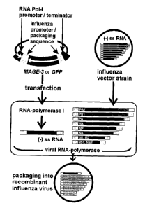
Fia -1' Insertion of the MACE-3 gene into recombinant influenza viruses: A
plasmid containing the MAGE-3 (or GFP) open reading frame flanked by influenza
virus promoter-up variant signals and more externally RNA-polymerise I control
sequences was transfected into 293T cells. These cells were infected with
influenza viruses. The heterologous cDNA sequences are transcribed by the
cellular RNA polymerise I in virus RNA- (vRNA-) like molecules, which are
2o subsequently amplified by the viral polymerise. The heterologous vRNA and
the
eight homologous influenza virus genomic RNAs are all co-packaged into virus
particles.
Fia 2' High level expression of heterologous proteins from recombinant
influenza
viruses: MDCK cells were infected with recombinant influenza virus stocks
expressing MAGE3-AU1 (FPV-MAGE3-AU1) or GFP (FPV-GFP) under control of
promoter-up variant 1104.
(A) Heterologous RNA produced by the recombinant viruses was detected by RT
PCR with primers specific for MACE-3 and GFP, respectively. The fi-actin RT-
PCR
served as an internal control. The PCR products were detected in ethidium
3o bromide stained agarose gels.
(B) Proteins extracted from infected cells were analyzed by immunoblot using
specific antibodies directed against the AU-1 sequence tag of MAGE3-AU1(left
panel). The corresponding bands were visualized by a secondary antibody
conjugated with horseradish peroxidase and an enhanced chemoluminescence
(ECL) assay. The same blot was reprobed using antibodies against GFP (right
panel).
9
CA 02427163 2003-04-28
WO 02/36790 PCT/EPO1/12532
FicL 3: Efficient gene transfer by recombinant influenza virus into human DC~
Mature human dendritic cells were infected with the recombinant influenza
virus
FpV-GFP at a MOI = 1.
(A) After 8 hours cells expressing GFP were detected by UV-fluorescence
microscopy (right panel). The left panel shows the same detail in phase
contrast
microscopy.
(B) Quantification of the transduction rate by FACS analysis: More than 90% of
the cells were found to express GFP. Uninfected and FPV wildtype infected DC
were used as controls.
1o Figi. 4: Expression of heteroiogous MAGE-3 protein from recombinant
influenza
virus transduced human DC: Mature human DC were infected (MoI = 1) with the
influenza virus recombinants FPV-MAGE3-AUI or, FPV-GFP. After 24 hours,
proteins were extracted, separated and analyzed by the immunoblot technique.
(A) To detect the MAGE3-AU1 fusion protein the blot was incubated with
specific
antibodies to the AU-1 sequence tag. The corresponding band was visualized by
a secondary antibody conjugated with horseradish peroxidase and an enhanced
chemiluminescence (ECL) assay.
(B) To detect GFP the same blot was reprobed using specific antibodies.
Fia. 5: Unchanged DC surface marker expression after giene transfer ~i
2o recombinant influenza virus: Mature DC were infected (MoI = 1) with FPV-GFP
or
left uninfected. After 24h, cells were counter-stained using antibodies
directed
CD86, CD80, HLA class I and class II and secondary PE-conjugated goat anti-
mouse IgG F(ab')2 fragments. The expression of cell surface markers and GFP
was quantified by flow cytometry. Percentages of cells in the quadrants are
indicated.
Figi. 6: Influenza virus transduced DC are potent T cell stimulators The allo-
stimulatory capacity of DC infected with FPV-GFP(~), with FPV-MAGE3-AUI(~) or
with the FPV wild type strain(. ) was tested in a MLR assay. As indicated,
graded
DC (6000-220) were co-cultivated with 2x10⁵ T cells. After four days, cells
were
3o pulsed with [3H]-thymidine for 16 hours and incorporation of radioactivity
was
determined. Shown are the mean counts ~ SEM of triplicates. Counts for T cells
alone or DC alone were always less than 1000 cpm.
to
CA 02427163 2003-04-28
WO 02/36790 PCT/EPO1/12532
Fig.7: Detection of MHC-I/IMP complexes on the transduced DC by a specific CTL
clone: Uninfected or FPV-MAGE3-AU1 infected DC (7.5 x 10⁴) were co-cultured
with an IMP-specific CTL clone ( 5 x 10³) for 18 hours. The capacity of the
CTL
clone to release IFN-y was immunochemically visualized by the ELISPOT-
technique. Results are shown as mean spot numbers ~ SEM of triplicates.
Fig_ 8' Expansion of CD8+ memor~r T cells in the absence of exogenous
cytokines
b~~ transduced DC: To demonstrate that DC infected with recombinant influenza
virus are able to stimulate influenza virus-specific CTL, autologous T cells
(4 x
106 ) were co-cultured with 0.2 x 10⁶ DC for seven days without addition of
1o exogenous cytokines.
(A) Subsequently the IMP-specific CTLs were stained with tetrameric MHC-I/IMP
complexes at 37°C and counterstained with labelled CD8 mAb at
4°C. The IMP-
specific CD8+ T cells were identified by FAGS analyses (upper right sector).
The
dot plots show T cells co-cultured with uninfected DC (control), IMP peptide-
pulsed DC (IMP-peptide), FPV-GFP infected DC or with FPV-MAGE3-AU1 infected
DC.
(B) Demonstration of IFN-y release from IMP-specific CTLs. The CTL activity
upon stimulation with IMP peptide (1pM) was assessed by ELISPOT-assay with
0.5 x 10⁶ T cells per well. Cells were cultured for 18 hours and IFN-y release
from
2o individual T cells was immunochemically visualized. Shown are means ~ SEM
of
triplicates
Fig. 9: Stimulation of MAGE-3 specific CTL b~~transduced DC: Uninfected or FPV-
MAGE3-AU1 infected DC (7.5 x100, were co-cultured with MAGE-3 specific CTL
clone (5 x 10³) for 18 hours. The capacity of transduced DC to present antigen
was evaluated by IFN-y secretion of individual CTL using the ELISPOT-
technique.
Results are the mean of three experiments ~ SEM.
Detailed Description of the Invention
"Patient derived immune cells" according to the present invention comprises
3o antigen presenting cells including, but not limited to, dendritic cells.
The patient
derived immune cells are derived from mammals, preferably humans.
The provision of patient derived immune cells utilized in the method of
embodiments (1) and (2) is hereinafter described in more detail with reference
to DC (again, the present invention is not limited to the use of DC). The
(human)
DC can be derived from various sources including peripheral blood, bone marrow
11
CA 02427163 2003-04-28
WO 02/36790 PCT/EPO1/12532
etc. The DC generation starting peripheral blood mononuclear cells (CPBMC) is
preferred (see WO 93/20185, WO 97/29182, EP-A-922 759, incorporated
herewith in its entirety). PBMC can be isolated from buffy coats by Ficoll-
Hypaque density gradient centrifugation. CD14 cells are then selected (e.g. by
using electric microbeads) from the PBMC, cultured and differentiated in a
suitable medium (e.g. RPMI supplemented with plasma and IL-4) for about 1 to
6 days, and matured by adding a suitable maturation cocktail (e:g. containing
IL-
1f3, IL-6, TNF-a and PGEZ to the culture medium and further culturing for
about 1
to 4 days. After washing with serum-free medium (e.g. RPMI) the mature DC are
ready for infection).
Infection of the DC according to step (b) of embodiment (1) and (2) of the
invention is achieved by adding recombinant virus vector (at a MoI of about
0.1
to 5, preferably about 1) to a DC culture in serum-free medium (e.g. RPMI or
the
like) for about O.S to 5 h (preferably at 37 °C/5% COZ). Thereafter the
infected
DC are washed with medium (RPMI or the like) and are cultured in medium
supplemented with plasma (e.g. RPMI + 1% AB-plasma).
The recombinant influenza viruses used in embodiment (1) preferably contain a
2o ninth segment which is a so-called "monocystronic" segment (viz, it
contains one
TAA or VAA gene flanked by the terminal viral RNA sequences).
Moreover it is preferred in embodiments (1) and (2) that the terminal viral
RNA
sequences, which are active as promoter signal, are modified by nucleotide
substitution in up to 5 positions, resulting in improved transcription rates
(of
both the vRNA promoter and the cRNA promoter as present in the
complementary sequence) as well as enhanced replication and/or expression
rates relative to the wild-type sequence. Said modified terminal viral RNA
sequences preferably differ from the wild-type sequence in that in said ninth
3o vRNA segment the 12 nucleotide conserved influenza 3' terminal sequence has
been modified by replacement of one to three nucleotides occurring in said
sequence at positions 3, 5 and 8 relative to the 3' end by other nucleotides
provided that the nucleotides introduced in positions 3 and 8 are forming a
base
pair (i.e., if the nucleotide position 3 is G, than that in position 8 is C;
if the
nucleotide in position 3 is C, than that in position 8 is G; etc.).
12
CA 02427163 2003-04-28
WO 02/36790 PCT/EPO1/12532
The 3' conserved regions of the wild-type influenza virus have the following
sequences:
Influenza A: (5')-CCUGCUUUUGCU-3'
Influenza B: (5')-NN(C/U)GCUUCUGCU-3'
Influenza C: (5')-CCUGCUUCUGCU-3'.
Moreover, the 13 nucleotide conserved influenza 5'-terminal sequence may be
modified by replacement of one or two nucleotides occurring in said sequence
as
positions 3 and 8 by other nucleotides, again provided that the introduced
nucleotides are forming a base pair. The 5' conserved regions of the wild-type
influenza virus have the following sequences:
Influenza A: 5'-AGUAGAAACAAGG
Influenza B: 5'-AGUAG(A/U)AACA(A/G)NN
Influenza C: 5'-AGCAGUAGCAAG(G/A):
Preferred influenza viruses of the invention are those wherein in the 3'
conserved
region the replacements G3A and C8U have been performed, more preferred are
those where also the replacement U5C has been performed (the above
mutations are annotated relative to the 3' end; such counting from the 3' end
is
also indicated by a line on top of the digit, e.g., G 3A). Another preferred
2o influenza virus mutant comprises the 3'-terminal nucleotide sequence G3C,
U5C
and C8G (relative to the 3' end) resulting in the following 3' terminal
nucleotide
sequence (5')-CCUGGUUCUCCU-3'. Among the influenza viruses defined
hereinbefore those having a 3'-terminal nucleotide sequence of (5')-
CCUGUUUCUACU-3' are most preferred. In case of an influenza A virus the
segment may further have the modifications U3A and A8U in its 5' terminal
sequence, in case of influenza C it may have the modifications C3U and G8A in
its 5' terminal sequence. The most preferred influenza viruses of the present
_
invention comprise the following general structures:
Influenza A (mutant pHL1102)'
5'-AGUAGAAACAAGGNNNUS_6..(880-2300 ntds)..N'N'N'CCUGUUUUUACU-3'
Influenza A (mutant pHL1104~
5'-AGUAGAAACAAGGNNNUS_6..(880-2300 ntds)..N'N'N'CCUGUUUCUACU-3'
Influenza A (mutant~HL1920O
5'-AGAAGAAUCAAGGNNNUS_6..(880-2300 ntds)..N'N'N'CCUGUUUCUACU-3'
Influenza A (mutant pHL1948O
5'-AGUAGAAACAAGGNNNUS_6..(880-2300 ntds)..N'N'N'CCUGGUUCUCCU-3'
13
CA 02427163 2003-04-28
WO 02/36790 PCT/EPO1/12532
Influenza B:
5'-AGUAG(A/U)AACA(A/G)NNNNNUS_6..(880-2300 ntds)..N'N'N'N'N'(C/U)GUUUCUACU-3'
Influenza C:
5'-AGUAGUAACAAG(G/A)GUS_6..(880-2300 ntds)..CCCCUGUUUCUACU-3'
In the above structures the variables are defined as follows:
(1) Underlined and enlarged letters show the required mutations relative to
the
wild-type sequence for preparing a promoter mutant with enhanced properties;
(2) enlarged A in position 10 in the 5'-part of the sequence: unpaired A
residue,
bulge-forming;
(3) (A/G) in one position: different isolates or single segments with-
variable
sequence at the respective position, which are functionally interchangeable;
(4) N and N': positions undefined, but base-paired relative to each other
because
of complementarity between the 5' and 3' termini, different among the 8
segments, but constant for each segment throughout all viral isolates;
(5) (880-2300 ntds): the lengths of the viral RNA segments, in case of
segments
with foreign genes increased up to 4,000 nucleotides.
Here is described a new transduction system for DC based on a recombinant
avian influenza virus vector. Using this vector more than 90 % of the DC
2o expressed the marker green fluorescent protein (GFP) and similarly the
tumor-
associated antigen MAGE-3 was efficiently expressed. These transduced DC
retained their characteristic phenotype and their potent T cell stimulatory
capacity. Furthermore, they were also able to stimulate MAGE-3 specific CD8+
memory T cells as well as CTL clones. As this recombinant virus is an
attenuated
virus, other attenuated recombinant influenza viruses that show limited or no
replication in vivo, but do allow expression of foreign genes will be equally
efficacious. Such recombinant viruses can be obtained be deleting genes not
required for replication in vitro, or by deleting genes and supplementing
these
genes in trans in producer cell lines, as known to those skilled in the art.
Expression of heterologous genes in dendritic cells (DC) is a powerful
strategy to
elicit strong immune reactions against tumor antigens. Here we report that
influenza viruses are capable to efficiently introduce and express foreign
proteins
such as tumor associated antigens (TAA) like MACE-3 or virus associated
antigens (VAA) in functional monocyte-derived human DC, thus providing a
potential tool for immune therapy of malignant melanoma and other tumors.
14
CA 02427163 2003-04-28
WO 02/36790 PCT/EPO1/12532
Suitable TAAs in accordance with the present invention are those known in the
art, including, but not limited to, TAAs from the class of cancer/testis
antigens.
Preferably the TAA is MAGE-3, NY-ESO-1 or HOM-MEL-40.
Suitable VAAs in accordance with the present invention are those known in the
art including, but not limited to, VAAs selected from a Herpes simplex virus
immediate early or early, protein, a human papilloma virus-antigen, preferably
Lz, E5 or E6, a Human Immunodeficiency Virus protein, preferably tat, rev, nef
or
gag, or a Hepatitis C virus protein known to elicit T cell response in a
mammal.
1o The TAA or VAA may also be a fusion protein, preferably a fusion protein
comprising entirely or partially the TAA or VAA proteins listed above.
In experiments it was shown that more than 90 % of mature DC expressed the
GFP marker after infection with recombinant influenza virus without undergoing
~5 lysis. This is comparable with the very high transduction rate reported for
recombinant GFP adenoviral vectors (L. Zhong et al., Eur. J. Immunol. 29, 964-
972 (1999)). However adenovirus has no receptor on the DC and very high
multiplicities of infection are needed to transduce genes into DC, which can
lead
to immunosuppression. The DC transfected with the specific recombinant
2o influenza viral vector did not reveal changes in their typical surface
marker
expression with the exception of a moderate down-modulation of CD83. This is
most probably a strain specific feature of FPV, since infection with another
influenza virus strain (PR-8) had no impact on the typical surface expression
of
this protein (M. Cella et al., J. Exp. Med. 189, 821-829 (1999)). The DC
25 transfected with the recombinant influenza virus vector express viral and
heterologous protein and generate MHC-I peptide complexes as shown by using
CTL clones in conjunction with interferon-y ELISPOT. The immunogenicity of the
transfected DC was also shown. They were able to expand influenza virus matrix
protein-specific memory T cells in the absence of exogenous cytokines. This is
a
3o unique feature of mature potent immunostimulatory DC. Furthermore, infected
DC were still very potent allogeneic T cell stimulators and were also able to
stimulate MAGE-3 specific CTL clones. Therefore, mature DC which have been
subjected to influenza vector-mediated heterologous gene transfer are potent
antigen presenting cells.
15
CA 02427163 2003-04-28
WO 02/36790 PCT/EPO1/12532
The features of this influenza virus vector suggest that it can be developed
into a
safe vector system useful also for the application in humans, e.g. for a ex
vivo
tumor or antiviral therapy. In contrast to other viral vector systems,
influenza
virus does not contain DNA nor does its replication depend on a DNA
intermediate. Thus influenza virus sequences cannot integrate into genomic
host
sequences and. cause mutations or cancer. There is no clinical indication that
influenza virus infection can become persistent or chronic in humans, as has
been known for retroviruses and adenoviruses (H. Hayder A. Mullbacher,
Tmmunol. Cell. Biol. 74, 504-512 (1996)).
Recombinant influenza viruses utilized in embodiment (1) of the current
invention have a limited capacity to replicate in culture, thus can be
considered
as self-attenuating. Usually after four passages the infectivity of viral
stock is
drastically decreased. The self-attenuation is due to the superior capacity of
the
promoter variant driving expression of the genes) on the heterologous
segment= Thereby it prevents the incorporation of essential genomic segments
into viral particles after exceeding a certain threshold. Finally, several
antiviral
substances including neuraminidase inhibitors are known to interfere with any
unwanted influenza virus, propagation providing a backup in case of an
2o accidentally induced infection (R. Lambkin, J.S. Oxford, Textbook on
Influenza,
p. 487 (1998); C.R. Penn, Textbook on Influenza, pp. 477-487 (1998)).
Like other viral vector systems used for gene transfer into DC, the influenza
virus vector results in the presentation of viral vector antigens in addition
to the
TAA. Simultaneous presentation of several antigens could result in unequal
immune reactions with the activation of CTL against one or few dominant
antigens. Such immunodominance in the CTL response probably results from
interference between T cell responses and not from insufficient presentation
of
peptide epitopes (E.Z. Wolpert et al., J. Immunol. 161, 4449-4505 (1998)).
so Tmmunodominance of viral vector antigens over heterologous tumor associated
antigens cannot be excluded for all MHC-I haplotypes. However, the activation
of
a MAGE-3-specific CTL clone by influenza virus-transduced DC strongly argues
for effective presentation of the TAA. Furthermore, since dominant T cell
epitopes for influenza viruses are well characterized and influenza viruses
can be
generated entirely from cloned DNA (G. Neumann et al., Proc. Natl. Acad. Sci.,
96, 9345-9350 (1999)), competing viral epitopes in the vector may be removed
16
CA 02427163 2003-04-28
WO 02/36790 PCT/EPO1/12532
by mutagenesis, if necessary. For long-term genetic modifications, the
immunogenicity of the used vector might be a major obstacle, since the
cellular
immunity directed against influenza virus could lead to the destruction of
genetically modified DC. This has been reported for instance for adenoviral
vectors upon application of these vectors to elicit anti-tumor immune
responses
in gene therapy studies (G.P. Gao et al., J. Virol. 70, 8934-8943 (1996) and
Immunology 91, 135-144 (1997); M. Christ et al., Immunol. Lett. 57, 19-25
(1997)). On the other hand, for immunotherapy studies, using short-lived
genetically modified DC this cellular immunity problem might not be as severe.
The presence or development of neutralizing antibodies against the viral
vector
might not be of major concern in an ex vivo gene transfer approach with no
production of viral progeny. Influenza viruses are internalized upon DC
infection,
which also becomes abortive during late gene expression (A. Bender et al.,
Exp.
Med. 182, 1663-1671 (1995) and Immunobiology 198, 552-567 (1998)). This
suggests that the chances of direct exposure of complete viral surface
antigens
that can be recognized by host antibodies are reduced. Additional techniques
to
attenuate the virus are available leading to further reduction in replication
(Hobom, unpublished). Finally, since it is now possible to efficiently
generate
influenza A viruses entirely from cloned cDNAs (Neumann et al., 1999) it is
2o feasible to directly introduce new haemagglutinin genes into the viral
vector
which are not recognized by the immune system. In addition, to using influenza
virus vectors based on hemagglutinins from different influenza serotypes,
these
vectors might also be applied in combination with other viral vectors in a
sequential approach to boost the immune reaction against the heterologous
antigens and to minimize the reaction to the viral proteins.
The pharmaceutical composition according to embodiment (4) above and the
medicament according to (5) above are suitable in ex vivo and in vivo
application
schemes contain the recombinant influenza virus in a pharmaceutically
effective
3o amount. Besides said recombinant influenza virus, the pharmaceutical
composition and the medicament may contain further pharmaceutically
acceptable carrier substances well-known to a person skilled in the art, such
as
binders, desintegrants, diluents, buffers, preservatives, etc. The
pharmaceutical
composition and medicaments are solid or liquid preparations and are suitable
to'
be administered orally, intravenously or subcutaneously.
17
CA 02427163 2003-04-28
WO 02/36790 PCT/EPO1/12532
The pharmaceutical composition and medicament are suitable as (ex vivo)
vaccines. The method of embodiment (7) of the invention includes the
administration of an effective amount to the mammal or the administration of a
sufficient infective dose of the recombinant virus to the cell system that is
used
for ex vivo therapy or for in vitro investigations, whereby the amount and
dose
will be determined by a person skilled in the respective arts or knowledgeable
of
the desired treatments. Said method is an antologous immunotherapy wherein
the patient derived cells which are ex vivo infected, ant the transduced cells
are
reintroduced into the patient.
The method according to embodiment (6) of the invention pertains to the
expression of the VAA or TAA by the patient derived immune cells obtained in
step (b) of embodiments (1) and (2).
In summary, the influenza virus expression vector system offers a unique
combination of features which are advantageous for an ex vivo transduction of
DC. It combines high efficiency with genomic simplicity, lack of genomic
integration, and the availability of a chemotherapeutic backup safety measure.
Thus influenza virus vectors may be a valuable alternative in the development
of
zo genetically modified DC. To minimize the anti-vector immunization and
booster
the therapeutic anti-tumor immune responses, different influenza viral vectors
based on different strains can be used in sequential immunization protocols.
The invention is further explained by the following non-limitative
experiments.
Experiments
1. Materials and Methods Cell lines and viruses: The human embryonic kidney
cell line 293T and the Madin-Darby canine ~ kidney (MDCK) cell line were
cultivated in Dulbecco's modified Eagle Medium (DMEM) supplemented with
10% fetal calf serum (FCS), glutamine (0.35 mg/ml), penicillin (0.12 mg/ml)
and
streptomycin sulfate (0.12 mg/ml). The medium for 293T cells was additionally
supplemented with 6418 (500pg/ml) in order to maintain high T-Ag expression.
The human melanoma cell line MZ2-MEL 43, the EBV-transformed B cell line LG-
2-EBV and the HLA-A1 restricted MADE-3-specific CTL clone LB 705 CTL 434/1
were kindly provided by P. v.d. Bruggen (Ludwig Institute for Cancer Research;
is
CA 02427163 2003-04-28
WO 02/36790 PCT/EPO1/12532
Brussels, Belgium). MZ2-MEL 43 and LG-2-EBV cells were cultured in DMEM or
Iscove's medium (Life Technologies, Karlsruhe, Germany) supplemented with
10% FCS. The MACE-3.A1 CTL clone was cultured in Iscove's medium
supplemented with 10 % pooled human serum, L-arginine (116 pg/ml), L-
asparagine (36 pg/ml) and gentamycin (10 pg/ml). This CTL clone was
restimulated weekly with irradiated (100 Gy) MAGE-3.A1 peptide loaded MZ2-
MEL 43 cells. Irradiated (100 Gy) LG-2-EBV cells were used as feeder cells.
The
CTL clone C9, which specifically recognizes the HLA-A2.1 restricted epitope of
influenza virus matrix protein 1 (IMP) was cultured in the same medium as the
1o MAGE-3.A1 CTL clone. The CTL clone C9 was re-stimulated with PHA-treated
irradiated (30 Gy) PBMC as stimulator cells. Again, irradiated (100 Gy) LG-2-
EBV
were used as feeder cells. All cell lines and clones were maintained at
37°C in an
8 % COZ atmosphere.
The avian influenza A virus (fowl plaque virus, FPV H7N7) was propagated in
MDCK cells as previously described (G. Neumann, G. Hobom, J. Gen. Virol. 76,
1709-1717 (1995)). Infection at a multiplicity of infection (MoI) of 1x10⁻⁵ to
1x10⁻⁶ resulted in virus stocks containing approximately 1x10⁹ plaque forming
units (pfu) per ml after two days of incubation. As a measure of the virus
particle
2o content, titers of virus hemagglutination were determined using chicken
erythrocytes. For the quantification of infectious particles, plaque assays
and
limiting dilution assays were performed on MDCK cells as described previously
(A. Bender et al., J. Exp. Med. 182, 1663-1671 (1995)). To determine the
fraction of viruses expressing the heterologous green fluorescent protein
(GFP),
limiting dilution assays were examined using a fluorescence microscope (Leica
DM IRB; Leica, Bensheim, Germany).
2. Preparation of recombinant influenza virus' Heterologous genes were
introduced into influenza viruses by the application of the RNA polymerise I
3o technique (A. Zobel et al., Nucleic Acid Res. 21, 3607-3614 (1993)). This
strategy leads to the incorporation of a heterologous gene into the influenza
virus particle as an additional, ninth genomic segment (pseudo-vRNA). The
required RNA is transcribed from transfected plasmid DNA, which encodes the
recombinant gene in combination with modified influenza virus 3' plus 5'
terminal
sequences (Fig.1). These constitute an influenza virus promoter-up variant and
contain the viral promoter and packaging signals. For expression of the
influenza
19
CA 02427163 2003-04-28
WO 02/36790 PCT/EPO1/12532
virus pseudo-vRNA the plasmid contains RNA polymerise I transcription control
elements, that allow the generation of uncapped, non-pofyadenylated RNA with
the flanking influenza virus control sequences. Influenza virus recombinants
were essentially prepared as described before (G. Neumann et al., Virology
202,
477-479 (1994)). , Briefly, plasmids (3 pg) encoding the heterologous viral
genomic segment with a promoter-up mutation (G. Neumann, G. Hobom, .7. Gen.
Virol. 76, 1709-1717 (1995)) were transfected into 293T cells cultured in 6cm
plates using Lipofectamin Plus (Life Technologies, Karlsruhe, Germany). After
24
hours, medium was removed from the culture and cells were inoculated with 2
1o ml of virus suspension (FPV Bratislava) in phosphate buffered saline (PBS)
supplemented with CaCl2 (0.91mM) and MgCl2 (0.49mM) (=PBS++) at a MoI of
2. After 45 min, the cells were washed once with PBS++ before 3 ml of medium
were added. Eight hours later, after one replication cycle, the supernatant
containing the recombinant virus was harvested. To enrich for recombinant
viruses, stocks were serially passaged on MDCK cells at a MoI of 2 or 3. After
three passages most viruses contained the heterologous sequence as was
monitored by GFP expression in limiting dilution assays. The enrichment is due
to the higher replication rate of the heterologous fragment compared to the
wild
type segments originating from the helper virus.
3. Plasmid construction: The plasmid pHHlO contains all cis-acting sequences
required for the synthesis of an influenza virus genomic segment. These
include
the influenza virus vRNA 5' and 3' terminal sequences with promoter and
packaging signal activity which are flanked by an RNA polymerise I (pol I)
promoter and an RNA polymerise I terminator element. Heterologous sequences
were cloned between the two parts of the influenza virus promoter in negative
orientation relative to the pol I promoter, using the pBSK multiple cloning
site
present in this location. The coding region for MADE-3 was generated by PCR
amplification of a 983 by fragment using plasmid pcDNA1-MAGE3 (B. Gaugler et
3o al., J. Exp. Med. 179, 921-930 (1994)) as template (kindly provided by P.
v. d.
Bruggen, Brussels, Belgium). The sense primer adds a Bgl II site to the 5' end
5 ' - GGA GAT CTC ATC ATG CCT CTT GAG CAG -3 ' ) and the reverse pri mer adds
an AU1 tag (P.S. Lim et al., J. Infect. Dis. 162, 1263-1269 (1990)) as well as
an
Xba I site to the 3 ' end (5 ' - CCT CTA GAT TAT ATA TAG CGA TAG GTG TCC TCT
TCC CCC TCT CTC -3 ' ). The PCR fragment was cleaved using Bgi II and Xba I,
purified from agarose~ gel (QIAEX II gel extraction kit, Qiagen, Hilden,
Germany)
CA 02427163 2003-04-28
WO 02/36790 PCT/EPO1/12532
and cloned into pHHlO via Bam HI and Xba I. The resulting plasmid was
designated pM4HH. The plasmid pHL2251 is a derivative of pHHlO containing a
variant of the Aequorea victoria GFP gene.
4. RT-PCR: Total ,RNA was extracted from infected MDCK cells 8 hours post
infection using the High Pure RNA Isolation Kit (Roche Molecular Biochemicals,
Mannheim, Germany). Subsequently, RT-PCR was performed using the Titan One
Tube RT-PCR System (Roche Molecular Biochemicals, Mannheim, Germany)
according to the manufacturers instructions. The PCR amplification was
1o performed using the following conditions: 95°C for 2 min (first
cycle only), 95°C
for 20 s, 57°C for 20 s, 72°C for 45 s (21 cycles), 95°C
for 20 s, 57°C for 20 s,
72°C for 60 s (19 cycles), 72°C for 4 min. Each RT-PCR reaction
was performed
in a final volume of 25 p1, with 1 p1 of total RNA as template. The nucleotide
sequences of the PCR primers used, were as follov~is: sense 5 °-CGG GAA
ATC
GTG CGT GAC AT -3' and reverse 5'-GAA CTT TGG GGG ATG CTC GC -3 °
yielding a 712 by beta-actin fragment; sense 5 °- TAA TAC GAC TCA CTA
TAG
GGC -3' and reverse 5'- CAG CAC GTG TCT TGT AGT TCC -3' yielding a 397 by
GFP fragment; sense 5 °- CAG CAC GTG TCT TGT AGT TCC -3' and
reverse 5 °-
ATA TAG CGA TAG GTG TCC TC -3 ° yielding a 747 by MAGE-3-AU1
fragment.
5. Immunoblot: Total cellular protein from infected MDCK cells and DC was
prepared according to a freeze-thaw lysis protocol (I. Schmitt et al., J.
Virol. 72,
633-640 (1998)). The lysis buffer contained 1 mM PMSF, O.OZmg/ml Aprotinin,
0.02 mg/ml Leupeptin and 2 mM DTT in 1 x THE (Tris, NP-40, EDTA). After
electrophoresis on a 12% polyacrylamide gel, proteins were transferred onto
Immobilon P membrane (Millipore, Eschborn, Germany), and the blots were
incubated with an anti-AU1 mouse monoclonal antibody (1:5000) (HISS
Diagnostics, Freiburg 1m Breisgau, Germany), washed three times and then
incubated with a secondary anti-mouse antibody (1:2000) conjugated with
3o horseradish peroxidase (Amersham Buchler, Braunschweig, Germany). GFP on
immunoblots was detected with an anti-GFP rabbit pofyclonal antibody (1:1000)
(Clontech, Heidelberg, Germany) and a secondary anti-rabbit antibody (1:2000)
also conjugated with horseradish peroxidase (Amersham Buchler, Braunschweig,
Germany). Proteins recognized by these antibodies were detected using an
enhanced chemoluminescence assay (Amersham Buchler, Braunschweig,
Germany).
21
CA 02427163 2003-04-28
WO 02/36790 PCT/EPO1/12532
6. Generation and infection of human DC: Dendritic cells were generated from
CD14+ human monocytes. PBMC were isolated from huffy coats of healthy HLA-
typed donors by Ficoll-Hypaque density gradient centrifugation (460 x g, 25
min,
20°C). To remove ,platelets, PBMC were repeatedly centrifuged with
decreasing g
numbers. CD14+ cells were positively selected using magnetic microbeads
(Miltenyi, Bergisch Gladbach, Germany). As assessed by FACS-analyses, the
CD14+ cells were on average over 95% pure. Immunophenotypically these
CD14+ cells were HLA-class I++, HLA-DR++, CD86++, CD40+, CD83-, CD80-, CD25-
. The isolated CD14+ cells were cultured at a density of 7 x 10⁶ in a volume
of 12
ml in 10 cm dishes (Falcon 3003, Becton Dickinson Heidelberg, Germany) in
RPMI (BioWhittaker, Walkersville, MD, USA) supplemented with 1 % heat-
inactivated AB-plasma. To start the differentiation to DC, 800 U/ml rhGM-CSF
(Leukomax, Novartis, Basel, Switzerland) and 1000 U/ml rhIL-4 (Novartis
Research Institute, Vienna, Austria) were added (day zero). On day three one
third of the culture medium was replaced with fresh culture medium
supplemented with 400 U/ml rhGM-CSF and 500 U/ml rhIL-4. On day six the
final maturation of DC was induced by adding IL-i-(3 (2 ng/ml) (Sigma,
Deisenhofen, Germany), IL-6 (1000 U/ml) (Novartis, Basel, Switzerland), PGEZ
(1 pg/ml) (Sigma) and TNF-a (10 ng/ml) (Boehringer Ingelheim Austria, Vienna,
Austria) according to H. Jonuleit et al., Eur. J. Immunol. 27, 3135-3142
(1997).
On day eight or nine FACS-analyses revealed the typical cell surface marker
expression of mature. DC: CD25++, CD40++, CD80++, CD83++, CD86++, HLA-DR++,
HLA-class I++, CD14-. On day eight or nine mature DC (1 x 10⁶) were infected
in
1 ml serum-free medium for 1 hour at 37°C/5% C02 at a MoI of 1.
Thereafter,
cells were washed extensively with RPMI and cultured in 6-well-plates in RPMI
supplemented with 1 % AB-plasma.
7. Immunophenotyping with antibodies and tetramers~ Cell surface antigens
3o were detected by incubating cells (1 x 10⁶/ ml) for 30 min at 4°C
with
monoclonal antibodies specific for CD25 (clone 2A3) from Becton Dickinson
(Heidelberg, Germany), CD14 (UCHM-1), CD40 (B-B20), CD86 (BU-63), HLA
class I (W6/32), HLA-DR (DDII), all from Cymbus Biotechnology (Chandlers Ford
Hants, U.K.), CD80 (MAB104), and CD83 (HBl5a) both from Immunotech
(Hamburg, Germany). After washing, cells were incubated with PE-conjugated
goat anti-mouse IgG F(ab')Z fragments (Jackson Immuno Research, West Grove,
22
CA 02427163 2003-04-28
WO 02/36790 PCT/EPO1/12532
PA, USA) for 30 min at 4°C. FACS-analyses were performed on a
FACScan
(Becton Dickinson, Heidelberg, Germany) using the CeIIQuest software. For the
detection of HLA-A2.1/IMP-specific CTL, cells were incubated with PE-labelled
tetrameric MHC-I/IMP peptide complexes (tetramers) (Dunbar et al., 1998) for
15 min at 37°C. Subsequently cells were counterstained with Tricolor-
labelled
CD8 specii=tc mAb (Caltag, Burlingame, CA, USA) for 15 min on ice. After
several
washes 0.5 x 10⁶ cells were analyzed by flow cytometry.
8. T cell stimulation assays: For mixed lymphocyte reaction (alto-MLR),
1o allogeneic CD4+ or CD8+ T cells were isolated by positive magnetic cell
sorting
according to the manufactures instructions (Miltenyi, Bergisch Gladbach,
Germany). The T cells (2 x10⁵/well) were cocultured_ with various numbers
(220,
660, 2000, 6000) of DC in 96-well flat bottomed plates. After 4 days, cells
were
pulsed with 1 pCi/well [3H]methyl-thymidine (Amersham Buchler,
~5 Braunschweig, Germany) and harvested 16 hours later onto glassfiber
filters.
Incorporation of [3H]-thymidine was measured with a microplate counter
(Wallac, Turku, Finland).
To assess the capability of infected DC to expand specific autologous
cytotoxic T
20 lymphocytes, DC were co-cultured with T cells (DC : T ratio of 1:20) in 24-
well
plates in RPMI supplemented with 10 % heat-inactivated human serum. After
seven days of culture without addition of exogenous cytokines the specificity
and
functionality of CTL was evaluated by staining with tetramers (P.R. Dunbar et
al.,
Curr. Biol. 8, 413-416 (1998)) and by ELISPOT-assay.
9. Enzyme-linked immunospot assay(ELISPOTy: The ability of the transduced
DC to stimulate antigen-specific CTL clones (i.e. IFN-y production) and the
specificity of expanded autologous T cells for defined peptides was measured
ori~
a single cell basis using the ELISPOT technique. Highly protein-binding
3o nitrocellulose membrane-bottomed 96-well plates (MAHA S4510, Millipore,
Eschborn, Germany) were precoated with 0.1 pg/ml primary anti-IFN-~
monoclonal antibody (1-D1k Mabtech, Stockholm, Sweden). After blocking with
5 % human serum 0.5-1x10⁴ antigen-specific T cells were added per well.
Subsequently 7.5x10⁴ DC were carefully added. After 18 hours of culture at
37°C the plates were washed six times with 0.05 % Tween~ in PBS and
then
incubated with biotinylated secondary anti-IFN-y monoclonal antibody (7-B6-1,
23
CA 02427163 2003-04-28
WO 02/36790 PCT/EPO1/12532
Mabtech, Stockholm, Sweden). Plates were washed again six times and stained
with streptavidin/peroxidase (Vectastain Elite kit, Vector Laboratories,
Berlingame, CA; USA) and 3-amino-9-ethylcarbazole (AEC, Sigma, Deisenhofen,
Germany) as substrate. Spots were counted using a special computer assisted
video imaging analysis system (Carl Zeiss Vision, Eching, Germany), using the
KS ELISPOT software version 4.1.143.
10. Expression of the MACE-3 tumor antigen usingi recombinant influenza
viruses: Since influenza viruses can infect dendritic cells (DC) in an
abortive way
1o and at the same time enhance their T cell stimulatory potential (M. Cells
et al., J.
Exp. Med. 189, 821-829 (1999)), influenza virus derived vectors should be well
suited to express antigens in these cells, which subsequently will be
efficiently
presented to the immune system. To introduce the tumor-associated antigen
(TAA) MAGE-3 or, as a control, the green fluorescent protein (GFP) into
influenza
viruses, the RNA polymerise I (pol I) technique was applied (A. Zobel et al.,
Nucleic Acid Res. 21, 3607-3614 (1993)). The foreign gene is under the control
of a high-capacity promoter (promoter 1104 having 3' modifications G3A, U5C
and C8U and no 5' modification). To facilitate the detection of the protein,
the
MAGE-3 open reading frame was fused with the coding sequence for the AU-1
2o epitope tag. The coding sequences were inserted into the vector with the
pol I
promoter and viral packaging/promoter sequences (Fig.1). Due to the viral
control sequences including the packaging signal, the pol I transcripts were
incorporated into influenza virus particles when the transfected culture was
inoculated with the avian influenza virus strain. Four to eight hours after
infection, the cells started to stain green in UV light due to the expression
of GFP
(data not shown). The fraction of recombinants within the viral stock. was
determined by measuring their capacity to generate plaques and to express GFP
in limiting dilution assays. Using the GFP recombinant virus .as a model for
the'
enrichment of the additional segment, we found that the heterologous segment
3o is enriched from passage to passage. Starting from about 1-3 %, the content
of
recombinants in the viral stocks increased up to 30fold after two to three
passages on MDCK cells (data not shown). This enrichment is due ~to the
optimized viral promoter and packaging signal sequences present in the
heterologous segment, which allow for a more efficient replication in
comparison
with the homologous influenza virus RNA segments (G. Neumann, G. Hobom, J.
Gen. Virol. 76, 1709-1717 (1995)).
24
CA 02427163 2003-04-28
WO 02/36790 PCT/EPO1/12532
To check for the presence of MAGE-3 recombinants in viral stocks, MDCK cells
were inoculated with virus and MACE-3 transcripts were detected from total
cellular RNA by RT-PCR (Fig. 2A). To verify the MACE-3 expression on the
protein level, total protein extracts from infected MDCK cells were prepared.
The
heterologous protein was detected in immunoblots. As shown in Fig.2B high
amounts of heterologous MAGE-3 protein could be detected. The protein
expression of GFP was high resulting in a similar band intensity as MAGE-3
(Fig.2B). The recombinant viruses expressing GFP or MAGE-3 were termed FPV
1o GFP and FPV-MAGE3-AU1, respectively.
11. Efficient transfer of MAGE-3 into mature human DC using recombinant
influenza virus; To investigate whether heterologous genes can be transferred
into human DC using recombinant influenza viruses, mature DC were generated.
For this purpose CD14+ monocytes were isolated from peripheral blood and
treated with GM-CSF and IL-4 for six days; subsequently the cells were
maturated by the addition of IL-1~3, IL-6, TNF-a and PGE2. These cells showed
the typical phenotype of fully mature DC (see 6. above). To determine whether
human monocyte-derived DC are accessible to gene transfer by recombinant
2o influenza virus, the mature DC were infected with GFP encoding viruses at a
MoI
of 1. Microscopic examination revealed that almost all cells expressed the
marker
protein GFP (Fig. 3A). As expected the infection of these mature DC was
abortive
and did not result in cell lysis or production of progeny virus (data not
shown).
Quantitative analyses of green fluorescence using FACS showed that more than
90% of these DC were infected and expressed the heterologous protein (Fig.
3B). Maximal levels of GFP expression were detected within eight hours after
infection. The protein was expressed for more than five days as was
determined by daily examination of GFP fluorescence by microscopy. To verify
the expression of the MACE-3 gene, DC cultures were infected with the
3o appropriate recombinant viral vector and in ~ parallel with the GFP
expressing
virus. Protein extracts were subjected to immunoblot analyses. Both, the MAGE-
3 gene and GFP gene were found to be expressed at high levels. For both
proteins prominent specific bands of similar intensity could be demonstrated
by
using either an antibody binding the AU-1 tag of MAGE-3 or a GFP specific
antiserum, respectively. (Fig. 4). Immature DC could be transduced with a
2s
CA 02427163 2003-04-28
WO 02/36790 PCT/EPO1/12532
similar efficiency (data not shown). However in the immature cell population
infection with a MoI of 1 did result in increased cytopathic effects.
12. Phenotypic characterization of influenza virus infected DC: To investigate
whether infection with recombinant influenza virus affects the phenotype of
DC,
typical cell surface markers were analysed by FACS 24 hours after infection
with
the GFP-coding recombinants. As shown in Fig. 5, the transduced DC produced
high levels of the typical surface markers. About 100% of the GFP-positive
cells
expressed CD86, CD 80, HLA class II and more than 80% of those cells HLA
1o class I molecules. Other characteristic cell surface markers such as CD25,
CD40
and CDIa were also detected on GFP+ DC. Comparison with uninfected DC did
not reveal any major differences in any of these markers. A modest down-
modulation of CD83 surface expression was the only deviation observed (data
not shown). The same expression patterns were obtained for DC infected with
FPV-MAGE3-AU1 or wild-type virus. These data indicate that the infection with
recombinant influenza virus and the high level expression of the heterologous
antigens does not cause major phenotypic alterations in the infected DC .
13. T cell stimulation bk influenza virus transduced DC: The crucial point for
the
2o application of DC in immunotherapy is their capacity to stimulate both CD4+
helper as well as CD8+ cytotoxic T cells (CTL). In order to address this
issue, we
firstly tested the ability of the transduced DC to induce T cell proliferation
in
allogeneic MLR. Therefore, DC were infected with one of the recombinant
influenza virus strains or with FPV wild-type and co-cultured for four days
with
allogeneic CD4+ T cells. After a 16 hours pulse, the incorporation of [3H]-
thymidine was measured. As five individual experiments showed, all co-cultures
resulted in a significant increase in [3H]-thymidine incorporation, even
though
300 and more T cells per DC were present. This indicated that DC infected with
FPV-GFP or with FPV-MAGE3-AU1 or with FPV wild-type have an unimpaired high
3o T cell stimulatory capacity (Fig.6). Similar results were obtained with
allogeneic
CD8+ T cells (data not shown). Secondly, in order to evaluate the capacity of
influenza virus transduced DC to present viral antigen to CD8+ CTL, transduced
DC were co-cultured with an influenza virus matrix protein (IMP) specific CTL
clone and the activation was determined by IFN-y ELISPOT (T. Hanke et al.,
vaccine 16, 426-435 (1998)). As shown in three individual experiments,
influenza virus transduced DC were potent stimulators of this T cell clone,
26
CA 02427163 2003-04-28
WO 02/36790 PCT/EPO1/12532
indicating the presence of MHC-I/IMP complexes on its surface (Fig. 7). In
contrast, uninfected DC with CTL or the CTL clone alone did not show any
activation. Thirdly, we proved that these DC were capable to expand CD8+
memory T cells in the absence of exogenously added cytokines; this is an
unique
property of DC. To this end, DC were infected with FPV-GFP or FPV-MAGE3-AU1.
Uninfected DC and IMP peptide pulsed DC were prepared as negative and
positive control, respectively. The DC were co-cultured with autologous CD8+ T
cells at a responder/stimulator ratio of 20:1. Specifically activated T cells
were
allowed to proliferate during a seven days culture period. Subsequently the
1o number of IMP-specific CD8~ cells were identified by double staining using
monoclonal antibodies and tetrameric MHC-I/IMP complexes binding to specific T
cell receptors (P.R. Dunbar et al., Curr. Biol. 8, 413-416 (1998)). As shown
in
Fig. 8A the infection with influenza virus recombinants resulted in an 11 to
18-
fold increase of IMP specific CD8+ positive T cells (for FPV-GFP: 0.49%, for
FPV-
MAGE: 0.32%; uninfected: 0.03%). Comparable numbers of specific CD8+T cells
(0.57%) were obtained with IMP peptide pulsed DC . To assess whether the
effector T cells expanded by the co-cultivation can be specifically activated,
they
were incubated with IMP peptide and analyzed for IFN-y production. As shown by
ELISPOT assays, 'the number of individual IFN-y -producing T cells after co-
2o culture, with infected DC was about 30 times higher than that of the
negative
control (Fig. 8B). Next we investigated the ability of the FPV-MAGE3-AU1
transduced DC to generate MHC-IMAGE-3 peptide complexes using a MAGE-3
specific CTL clone and ELISPOT as test system. As shown in Fig. 9, the
transduced DC were able to stimulate IFN-y production from this CTL clone
whereas the clone by itself did not spontaneously secrete IFN-y. Hence, TAA
gene transfer by influenza virus vectors into DC yields antigen presenting
cells,
which present the corresponding tumor antigen apart from vector antigen and
Which specifically stimulate T cells.
27
CA 02427163 2003-04-28
WO 02/36790 PCT/EPO1/12532
1/7
SEQUENCE LISTING
<110> Artemis Pharmaceuticals GmbH
<120> Influenza Virus Vector forInfection of Dentritic Cells
<130> 012734wo/JH/ml
<140>
<141>
<160> 34
<170> PatentIn Ver. 2.1
<210> 1
<211> l2
<212> RNA
<213> Influenza A virus
zo
<400> 1
ccugcuuuug cu ~ 12
<2l0> 2
<211> 12
<212> RNA
<213> Influenza B virus
<400> 2
nnygcuucug cu 12
<210> 3
<211> 12
<212> RNA
<213> Influenza C virus
<400> 3
ccugcuucug cu . 12
<21'0> 4
<211> 12
<212> RNA
<213> Artificial Sequence
<220>
<223> Description of Artificial Sequence: Modified
influenza A 5'-sequence
<400> 4
ccuguuucua cu 12
<210> 5
<211> 12
<212> RNA
<213> Artificial Sequence
<400> 5
ccugguucuc cu 12
CA 02427163 2003-04-28
WO 02/36790 PCT/EPO1/12532
2/7
<210> 6
<211> 13
<212> RNA
<213> Influenza A virus
<900> G
aguagaaaca agg 13
<210> 7
<211> 13
<212> RNA
<213> Influenza B virus
<400> 7
aguagwaaca rnn 13
<210> 8
<211> 13
<212> RNA
<213> Influenza C virus
<400> 8
agcaguagca agr 13
<210> 9
<211> 13
<212> RNA
. <213> Artificial Sequence
<220>
<223> Description of Artificial Sequence: Modified
influenza A 5'-sequence
<400> 9
agaagaauca agg 13
<210> 10
<211> 21
<212> RNA
<213> Influenza A virus
<400> 10
aguagaaaca aggnnnuuuu a 21
<210> 11
<211> 21
<212> RNA
<213> Artificial Sequence
.
<220>
<223> Description of Artificial Sequence: Modified
influenza A 5'-sequence (pHL1920)
<400> 11
agaagaauca aggnnnuuuu a 21
CA 02427163 2003-04-28
WO 02/36790 PCT/EPO1/12532
3/7
<210> 12
<211> 21
<212> RNA
<213> Influenza B virus
<400> 12
aguagwaaca rnnnnnuuuu a 21
<210> 13
<2l1> 19
<212> RNA
<213> Artificial Sequence
<220>
<223> Description of Artificial Sequence: Modified
influenza C 5'-sequence
<400> 13
aguaguaaca agrguuuuu 19
<210> 14
<211> 15
<212> RNA
<213> Artificial Sequence
<220>
<223> Description of Artificial Sequence: Modified
influenza A 3'-sequence (pHL1104 and pHL1920)
<400> 14
nnnccuguuu cuacu 15
<210> 15
<211> 15
<212> RNA
<213> Artificial Sequence
<220> -
<223> Description of Artificial Sequence: Modified
influenza A 3'-sequence (pHL1948)
<400> 15
nnnccugguu cuccu 15
<210> 16
<211> 15
<212> RNA
<213> Artificial Sequence
.
<220>
<223> Description of Artificial Sequence: Modified
influenza B 3' sequence
<400> 16
nnnnnyguuu cuacu 15
CA 02427163 2003-04-28
WO 02/36790 PCT/EPO1/12532
4/7
<210> 17
<211> 14
<212> RNA
<213> Artificial Sequence
<220>
<223> Description of Artificial Sequence: Modified
influenza C 3'-sequence
<400> 17
ccccuguuuc uacu 14
<210> is
<211> 27
<212> DNA
<213> Artificial Sequence
<220>
<223> Description of Artificial Sequence: Primer
<400> 18
ggagatctca tcatgcctct tgagcag 27
<210> 19
<211> 45
<212> DNA
<213> Artificial Sequence
<220>
<223> Description of Artificial Sequence: Primer
<400> 19
cctctagatt atatatagcg ataggtgtcc tcttccccct ctctc 45
<210> 20
<211> 20
<212> DNA
<213> Artificial Sequence
<220>
<223> Description of Artificial Sequence: Primer
<400> 20
egggaaatcg tgcgtgacat 20
<210> 21
<211> 20
<212> DNA
<213> Artificial Sequence
<220> ,
<223> Description of Artificial Sequence: Primer
<400> 21
gaactttggg ggatgctcgc 20
<210> 22
<211> 21
CA 02427163 2003-04-28
WO 02/36790 PCT/EPO1/12532
5/7
<212> DNA
<213> Artificial Sequence
<220>
<223> Description of Artificial Sequence: Primer
<400> 22
taatacgact cactataggg c 21
<210> 23
<211> 21
<212> DNA
<213> Artificial Sequence
<220>
<223> Description of Artificial Sequence: Primer
<400> 23
cagcacgtgt cttgtagttc c 21
<210> 24
<211> 21
<212> DNA
<213> Artificial Sequence
<220>
<223> Description of Artificial Sequence: Primer
<400> 24
cagcacgtgt cttgtagttc c 21
<210> 25
<211> 20
<212> DNA
<213> Artificial Sequence
<220>
<223> Description of Artificial Sequence: Primer
<400> 25
atatagcgat aggtgtcctc 20
<210> 26
<211> 12
<212> RNA
<213> Artificial Sequence
<220>
<223> Description of Artificial Sequence: Modified
influenza A 3'-sequence
<400> 26
ccuguuuuua cu 12
<210> 27
<211> 27
CA 02427163 2003-04-28
WO 02/36790 PCT/EPO1/12532
6/7
<212> DNA
<213> Artificial Sequence
<220>
<223> Description of Artificial Sequence: Primer
<400> 27
ggagatctca tcatgcctct tgagcag 27
<210> 28
<211> 45
<212> DNA
<213> Artificial Sequence
<220>
<223> Description of Artificial Sequence: Primer
<400> 28
cctctagatt atatatagcg ataggtgtcc tcttccccct ctctc 45
<210> 29
<211> 20
<212> DNA
<213> Artificial Sequence
<220>
<223> Description of Artificial Sequence: Primer
<400> 29
cgggaaatcg tgcgtgacat 20
<210> 30
<211> 20
<212> DNA
<213> Artificial Sequence
<220>
<223> Description of Artificial Sequence: Primer
<400> 30
gaactttggg ggatgctcgc 20
<2l0> 31
<211> 21
<212> DNA
<213> Artificial Sequence
<220>
<223> Description of Artificial Sequence: Primer
<400> 31 ,
taatacgact cactataggg c 21
<210> 32
<211> 21
<212> DNA
<213> Artificial Sequence
CA 02427163 2003-04-28
WO 02/36790 PCT/EPO1/12532
7/7
<220>
<223> Description of Artificial Sequence: Primer
<400> 32
cagcacgtgt cttgtagttc c 21
<210> 33
<211> 21
<212> DNA
<213> Artificial Sequence
<220>
<223> Description of Artificial Sequence: Primer
<400> 33
cagcacgtgt cttgtagttc c 21
<210> 34
<211> 20
<212> DNA
<213> Artificial Sequence
<220>
<223> Description of Artificial Sequence: Primer
<400> 34
atatagcgat aggtgtcctc 20