Note: Descriptions are shown in the official language in which they were submitted.
CA 02427381 2003-04-25
WO 02/094368 PCT/USO1/51496
TRANSDERMAL DRUG DELIVERY DEVICES
HAVING COATED MICROPROTRUSIONS
TECHNICAL FIELD
s [0001] This invention relates to administering and enhancing transdermal
delivery of an agent across the skin. More particularly, the invention relates
to a
percutaneous drug delivery system for administering a potent pharmacologically
active agent through the stratum corneum using skin piercing microprotrusions
which have a dry coating of the pharmacologically active agent. Delivery of
the
~o agent is facilitated when the microprotrusions pierce the skin of a patient
and the
patient's interstitial fluid contacts and dissolves the active agent.
BACKGROUND ART
[0002] Drugs are most conventionally administered either orally or by
injection.
15 Unfortunately, many medicaments are completely ineffective or have
radically
reduced efficacy when orally administered since they either are not absorbed
or
are adversely affected before entering the bloodstream and thus do not possess
the desired activity. On the other hand, the direct injection of the
medicament into
the bloodstream, while assuring no modification of the medicament during
2o administration, is a difficult, inconvenient, painful and uncomfortable
procedure,
sometimes resulting in poor patient compliance.
[0003] Hence, in principle, transdermal delivery provides for a- method of
administering drugs that would otherwise need to be delivered via hypodermic
injection or intravenous infusion. Transdermal drug delivery offers
improvements
25 in both of these areas. Transdermal delivery when compared to oral delivery
avoids the harsh environment of fihe digestive tract, bypasses
gastrointestinal drug
metabolism, reduces first-pass effects, and avoids the possible deactivation
by
digestive and liver enzymes. Conversely, the digestive tract is not subjected
to the
drug during transdermal administration. Indeed, many drugs such as aspirin
have
so an adverse effect on the digestive tract. However, in many instances, the
rate of
delivery or flux of many agents via the passive transdermal route is too
limited to
be therapeutically effective.
CA 02427381 2003-04-25
WO 02/094368 PCT/USO1/51496
[0004] The word "transdermal" is used herein as a generic term referring to
passage of an agent across the skin layers. The word "transdermal" refers to
delivery of an agent (e.g., a therapeutic agent such as a drug) through the
skin to
the local tissue or systemic circulatory system without substantial cutting or
s piercing of the skin, such as cutting with a surgical knife or piercing the
skin with a
hypodermic needle. Transdermal agent delivery includes delivery via passive
diffusion as well as by external energy sources including electricity (e.g.,
iontophoresis) and ultrasound (e.g., phonophoresis). While drugs do diffuse
across
both the stratum corneum and the epidermis, the rate of diffusion through the
~o stratum corneum is often the limiting step. Many compounds, in order to
achieve a
therapeutic dose, require higher delivery rates than can be achieved by simple
passive transdermal diffusion. When compared to injections, transdermal agent
delivery eliminates the associated pain and reduces the possibility of
infection.
[0005] Theoretically, the transdermal route of agent administration could be
~s advantageous in the delivery of many therapeutic proteins, because proteins
are
susceptible to gastrointestinal degradation and exhibit poor gastrointestinal
uptake
and transdermal devices are more acceptable to patients than injections.
However,
the transdermal flux of medically useful peptides and proteins is often
insufficient
to be therapeutically effective due to the large size/molecular weight of
these
2o molecules. Often the delivery rate or flux is insufficient to produce the
desired
effect or the agent is degraded prior to reaching the target site, for example
while
in the patient's bloodstream.
[0006] Transdermal drug delivery systems generally rely on passive diffusion
to
administer the drug while active transdermal drug delivery systems rely on an
z5 external energy source (e.g., electricity) to deliver the drug. Passive
transdermal
drug delivery systems are more common. Passive transdermal systems have a
drug reservoir containing a high concentration of drug adapted to contact the
skin
where the drug diffuses through the skin and into the body tissues or
bloodstream
of a patient. The transdermal drug flux is dependent upon the condition of the
so skin, the size and physical/chemical properties of the drug molecule, and
the
concentration gradient across the skin. Because of the low permeability of the
skin
to many drugs, transdermai delivery has had limited applications. This low
2
CA 02427381 2003-04-25
WO 02/094368 PCT/USO1/51496
permeability is attributed primarily to the stratum corneum, the outermost
skin layer
which consists of flat, dead cells filled with keratin fibers (keratinocytes)
surrounded by lipid bilayers. This highly-ordered structure of the lipid
bilayers
confers a relatively impermeable character to the stratum corneum.
[0007] One common method of increasing the passive transdermal diffusional
drug flux involves pre-treating the skin with, or co-delivering with the drug,
a skin
permeation enhances. A permeation enhances, when applied to a body surface
through which the drug is delivered, enhances the flux of the drug
therethrough.
However, the efficacy of these methods in enhancing transdermal protein flux
has
~o been limited, at least for the larger proteins, due to their size.
[0008] Active transport systems use an external energy source to assist drug
flux through the stratum corneum. One such enhancement for transdermal drug
delivery is referred to as "electrotransport." This mechanism uses an
electrical
potential, which results in the application of electric current to aid in the
transport of
~s the agent through a body surface, such as skin. Other active transport
systems
use ultrasound (phonophoresis) and heat as the external energy source.
[0009] There also have been many attempts to mechanically penetrate or
disrupt the outermost skin layers thereby creating pathways into the skin in
order
to enhance the amount of agent being transdermally delivered. Early
vaccination
2o devices known as scarifiers generally had a plurality of tines or needles
which are
applied to the skin to and scratch or make small cuts in the area of
application.
The vaccine was applied either topically on the skin, such as U.S. Patent No.
5,487,726 issued to Rabenau or as a wetted liquid applied to the scarifies
tines
such as U.S. Patent No. 4,453,926 issued to Galy, or U.S. Patent No. 4,109,655
25 issued to Chacornac, or U.S. Patent No. 3,136,314 issued to Kravitz.
Scarifiers
have been suggested for intradermal vaccine delivery in part because only very
small amounts of the vaccine need to be delivered into the skin to be
effective in
immunizing the patient. Further, the amount of vaccine delivered is not
particularly
critical since an excess amount achieves satisfactory immunization as well as
a
ao minimum amount. However a serious disadvantage in using a scarifies to
deliver a
drug is the difficulty in determining the transdermal drug flux and the
resulting
dosage delivered. Also due to the elastic, deforming and resilient nature of
skin to
3
CA 02427381 2003-04-25
WO 02/094368 PCT/USO1/51496
deflect and resist puncturing, the tiny piercing elements often do not
uniformly
penetrate the skin and/or are wiped free of a liquid coating of an agent upon
skin
penetration. Additionally, due to the self healing process of the skin, the
punctures
or slits made in the skin tended to close up after removal of the piercing
elements
from the stratum corneum. Thus, the elastic nature of the skin acts to remove
the
active agent coating which has been applied to the tiny piercing elements upon
penetration of these elements into the skin. Furthermore the tiny slits formed
by
the piercing elements heal quickly after removal of the device, thus limiting
the
passage of agent through the passageways created by the piercing elements and
~o in turn limiting the transdermal flux of such devices.
[00010] Other devices which use tiny skin piercing elements to enhance
transdermal drug delivery are disclosed in European Patent EP 0407063A1, U.S.
Patent Nos. 5,879,326 issued to Godshall, et al., 3,814,097 issued to
Ganderton,
et al., 5,279,544 issued to Gross, et al., 5,250,023 issued to Lee, et al.,
3,964,482
~s issued to Gerstel, et al., Reissue 25,637 issued to Kravitz, et al., and
PCT
Publication Nos. WO 96/37155, WO 96/37256, WO 96/17648, WO 97/03718, WO
98/11937, WO 98/00193, WO 97/48440, WO 97/48441, WO 97/48442, WO
98/00193, WO 99/64580, WO 98/28037, WO 98/29298, and WO 98/29365; all
incorporated by reference in their entirety. These devices use piercing
elements of
2o various shapes and sizes to pierce the outermost layer (i.e., the stratum
corneum)
of the skin. The piercing elements disclosed in these references generally
extend
perpendicularly from a thin, flat member, such as a pad or sheet. The piercing
elements in some of these devices are extremely small, some having dimensions
(i.e., a microblade length and width) of only about 25 - 400 ~m and a
microblade
2s thickness of only about 5 - 50 ~,m. These tiny piercing/cutting elements
make
correspondingly small microslits/microcuts in the stratum corneum for enhanced
transdermal agent delivery therethrough.
[00011] Generally, these systems include a reservoir for holding the drug and
also a delivery system to transfer the drug from the reservoir through the
stratum
so corneum, such as by hollow tines of the device itself. One example of such
a
device is disclosed in WO 93/17754 which has a liquid drug reservoir. The
reservoir must be pressurized to force the liquid drug through the tiny
tubular
4
CA 02427381 2003-04-25
WO 02/094368 PCT/USO1/51496
elements and into the skin. Disadvantages of devices such as these include the
added complication and expense for adding a pressurizable liquid reservoir and
complications due to the presence of a pressure-driven delivery system.
DISCLOSURE OF THE INVENTION
[00012] The device and method of the present invention overcome these
limitations by transdermally delivering a pharmacologically active agent using
a
microprotrusion device having microprotrusions which are coated with a dry
coating containing the agent. The present invention is directed to a device
and
~o method for delivering a pharmacologically active agent through the stratum
corneum of preferably a mammal and most preferably a human, by coating a
plurality of stratum corneum-piercing microprotrusions with a high potency
pharmacologically active agent. The agent is selected to be sufficiently
potent to
be therapeutically effective when delivered as a dry coating on a plurality of
skin
15 piercing microprotrusions. Further, the agent must have sufficient water
solubility
to form an aqueous coating solution having the necessary solubility and
viscosity
for coating the microprotrusions.
[00013] A preferred embodiment of this invention consists of a device for
delivering a beneficial agent through the stratum corneum. The device
comprises
2o a member having a plurality, and preferably a multiplicity, of stratum
corneum-
piercing microprotrusions. Each of the microprotrusions has a length of less
than
500 Vim, or if longer than 500 g,m, then means are provided to ensure that the
microprotrusions penetrate the skin to a depth of no more than 500 ~,m. These
microprotrusions have a dry coating thereon. The coating, before drying,
25 comprises an aqueous solution of a high potency pharmacologically active
agent.
The pharmacologically active agent is sufficiently potent to be
pharmaceutically
effective in a dose of less than about 1 mg and preferably less than about
0.25 mg,
per application. The pharmacologically active agent is selected to have a
water
solubility of greater than about 50 mg/ml and the composition has a viscosity
less
so than about 500 centipoises(cp) in order to effectively coat the
microprotrusions.
The solution, once coated onto the surfaces of the microprotrusions, provides
a
pharmaceutically effective amount of the pharmacologically active agent. The
CA 02427381 2003-04-25
WO 02/094368 PCT/USO1/51496
coating is further dried onto the microprotrusions using drying methods known
in
the art.
[00014] Another preferred embodiment of this invention consists of a method
of making a device for transdermally delivering a pharmacologically active
agent.
s The method comprises providing a member having a plurality of stratum
corneum-
piercing microprotrusions. An aqueous solution of the pharmacologically active
agent is applied to the microprotrusions and then dried to form a dry agent-
containing coating thereon. The pharmacologically active agent is sufFiciently
potent to be pharmaceutically effective in a dose of less than about 1 mg, and
~o preferably less than about 0.25 mg, per application. The pharmacologically
active
agent must have a water solubility of greater than about 50 mg/ml, preferably
greater than about 100 mg/ml, and the coating solution must have a viscosity
less
than about 500 cp preferably less than about 50 cp, in order to effectively
coat the
microprotrusions. The composition can be prepared at any temperature as long
as
15 the pharmacologically active agent is not rendered inactive due fio the
conditions.
The solution, once coated onto the surfaces of the microprotrusions, provides
a
pharmaceutically effective amount of the pharmacologically active agent.
[00015] The coating thickness is preferably less than the thickness of the
microprotrusions, more preferably the thickness is less than 50 ~,m and most
2o preferably less than 25 p,m. Generally, the coating thickness is an average
thickness measured over the microprotrusions.
(00016] The pharmacologically active agent for coating the microprotrusions
is selected to have sufficient potency to ~be therapeutically effective when
administered transdermally in an amount of less than about 1 mg, and
preferably
25 less than about 0.25 mg, of active agent.
The most preferred agents are selected from the group consisting of ACTH (1-
24),
calcitonin, desmopressin, LHRH, LHRH analogs, goserelin, leuprolide, PTH,
vasopressin, deamino [Val4, D-ArgB] arginine vasopressin, buserelin,
triptorelin,
interferon alpha, interferon beta, interferon gamma, FSH, EPO, GM-CSF, G-CSF,
ao IL-10, glucagon, growth hormone releasing factor (GRF) and analogs of these
agents including pharmaceutically acceptable salts thereof.
6
CA 02427381 2003-04-25
WO 02/094368 PCT/USO1/51496
[00017] The coating can be applied to the microprotrusions using known
coating methods. For example, the microprotrusions can be immersed into an
aqueous coating solution of the agent. Alternatively the coating solution can
be
sprayed onto the microprotrusions. Preferably the spray has a droplet size of
s about 10-200 picoliters. More preferably the droplet size and placement is
precisely controlled using printing techniques so that the coating solution is
deposited directly onto the microprotrusians and not onto other "non-piercing"
portions of the member having the microprotrusions.
[00018] In another aspect of the invention, the stratum corneum-piercing
~o microprotrusions are formed from a sheet wherein the microprotrusions are
formed
by etching or punching the sheet and then the microprotrusions are folded or
bent
out of a plane of the sheet. While the pharmacologically active agent coating
can
be applied to the sheet before formation of the microprotrusions, preferably
the
coating is applied after the microprotrusions are cut or etched out but prior
to being
15 folded out of the plane of the sheet. More preferred is coating after the
microprotrusions have been folded or bent from the plane of the sheet.
BRIEF DESCRIPTION OF THE DRAWINGS
[00019] The invention will now be described in greater detail with reference
to
2o the preferred embodiments illustrated in the accompanying drawings and
figures.
wherein:
FIG. 1 is a perspective view of a portion of one example of a microprotrusion
array;
FIG. 2 is a perspective view of the microprotrusion array of F1G. 1 with a
coating
deposited onto the microprotrusions;
25 FIG. 3 is a graph showing the amount of desmopressin delivered by a
microprotrusion array;
FIG. 4 is a graph showing the amount of desmopressin delivered by a
microprotrusion array;
FIG. 5 is a graph showing the amount of ovalbumin delivered by a
microprotrusion
ao array for various application times;
FIG. 6 is a graph showing the amount of ovalbumin delivered by a
microprotrusion
array using various coating solutions of ovalbumin;
7
CA 02427381 2003-04-25
WO 02/094368 PCT/USO1/51496
FIG. 7 is a side sectional view of the system described in Example 1;
FIG. 8. is a graph showing the amount of human growth hormone delivered by a
microprotrusion array that has been tip-coated as described in Example 2B.
FIG. 9 is a graph showing the amount of human growth hormone delivery by a
s microprotrusion array that has been tip-coated as described in Example 4B;
and
FIG. 10 is a graph showing the amount of ovalbumin delivered by a
microprotrusion array that has been tip-coated as described in Example 6B.
MODES FOR CARRYING OUT THE INVENTION
~o [00020] DEFINITIONS:
Unless stated otherwise the following terms used herein have the following
meanings.
The term "transdermal" means the delivery of an agent into and/or through the
skin
for local or systemic therapy.
15 The term "transdermal flux" means the rate of transdermal delivery.
The term "co-delivering" as used herein means that a supplemental agents) is
administered transdermally either before the agent is delivered, before and
during
transdermal flux of the agent, during transdermal flux of the agent, during
and after
transdermal flux of the agent, and/or after transdermal flux of the agent.
2o Additionally, two or more agents may be coated onto the microprotrusions
resulting
in co-delivery of the agents.
[00021] The term "pharmacologically active agent" as used herein refers to a
non-immunogenic drug or a composition of matter or mixture containing a non-
immunogenic drug which is pharmacologically effective when administered in an
Zs amount of less than about 1 mg, and preferably less than about 0.25 mg.
Thus,
the term "pharmacologically active agent" encompasses only very potent drugs
that are pharmacologically effective at very low doses and specifically
excludes
vaccines. Examples of such high potency pharmacologically active agents
include, without limitation, leutinizing hormone releasing hormone (LHRH),
LHRH
so analogs (such as goserelin, leuprolide, buserelin, triptorelin,
gonadorelin, and
napfarelin, menotropins (urofollitropin (FSH) and LH)), vasopressin,
desmopressin,
corticotropin (ACTH), ACTH analogs such as ACTH (1-24), calcitonin,
parathyroid
8
CA 02427381 2003-04-25
WO 02/094368 PCT/USO1/51496
hormone (PTH), vasopressin, deamino [Val4, D-ArgB] arginine vasopressin,
interferon alpha, interferon beta, interferon gamma, erythropoietin (EPO),
granulocyfie macrophage colony stimulating factor (GM-CSF), granulocyte colony
stimulating factor (G-CSF), interleukin-10 (IL-10) and glucagon. It is to be
s understood that more than one agent may be incorporated into the agent
formulation in the method of this invention, and that the use of the term
"pharmacologically active agent" in no way excludes the use of two or more
such
agents or drugs. The agents can be in various forms, such as free bases,
acids,
charged or uncharged molecules, components of molecular complexes or
~o nonirritating, pharmacologically acceptable salts. Also, simple derivatives
of the
agents (such as ethers, esters, amides, etc) which are easily hydrolyzed at
body
pH, enzymes, etc., can be employed.
[00022] The term "therapeutically effective amount" or "therapeutically
effective rate" refers to the amount or rate of the pharmacologically active
agent
~s needed to effect the desired therapeutic, often beneficial, result. The
amount of
agent employed in the coatings will be that amount necessary to deliver a
therapeutically effective amount of the agent to achieve the desired
therapeutic
result. In practice, this will vary widely depending upon the particular
pharmacologically active agent being delivered, the site of delivery, the
severity of
2o the condition being treated, the desired therapeutic effect and the
dissolution and
release kinetics for delivery of the agent from the coating into skin tissues.
It is not
practical to define a precise range for the therapeutically effective amount
of the
pharmacologically active agent incorporated into the microprotrusions and
delivered transdermally according to the methods described herein. However,
25 generally such agents utilized in the device of the present invention are
defined as
potent pharmacologically active agents since the microprotrusions are sized
with a
limited surface area for carrying the coating. In general, the amount of the
agent
needed to achieve the desired therapy is less than about 1 mg, more preferably
less than 0.25 mg.
30 [00023] The term "microprotrusions" refers to piercing elements which are
adapted to pierce or cut through the stratum corneum into the underlaying
epidermis layer, or epidermis and dermis layers, of the skin of a living
animal,
9
CA 02427381 2003-04-25
WO 02/094368 PCT/USO1/51496
particularly a human. The piercing elements should not pierce the skin to a
depth
which causes bleeding. Typically the piercing elements have a blade length of
less than 500 Vim, and preferably less than 250 ~.m. The microprotrusions
typically
have a width and thickness of about 5 to 50 ~,m. The microprotrusions may be
s formed in different shapes, such as needles, hollow needles, blades, pins,
punches, and combinations thereof.
[00024] The term "microprotrusion array" as used herein refers to a plurality
of microprotrusions arranged in an array for piercing the stratum corneum. The
microprotrusion array may be formed by etching or punching a plurality of
~o microprotrusions from a thin sheet and folding or bending the
microprotrusions out
of the plane of the sheet to form a configuration such as that shown in FIG.
1. The
microprotrusion array may also be formed in other known manners, such as by
forming one or more strips having microprotrusions along an edge of each of
the
strips) as disclosed in Zuck, US Patent No. 6,050,988. The microprotrusion
array
~s may include hollow needles which hold a dry pharmacologically active agent.
[00025] References to the area of the sheet or member and reference to
some property per area of the sheet or member, are referring to the area
bounded
by the outer circumference or border of the sheet.
j00026] The term "pattern coating" refers to coating an agent onto selected
2o areas of the microprotrusions. More than one agent may be pattern coated
onto a
single microprotrusion array. Pattern coatings can be applied to the
microprotrusions using known micro-fluid dispensing techniques such as
micropipeting and ink jet coating.
25 DETAILED DESCRIPTION
[00027] The present invention provides a device for transdermally delivering
a pharmacologically active agent to a patient in need thereof. The device has
a
plurality of stratum corneum-piercing microprotrusions extending therefrom.
The
microprotrusions are adapted to pierce through the stratum corneum into the
so underlying epidermis layer, or epidermis and dermis layers, but do not
penetrate
so deep as to reach the capillary beds and cause significant bleeding. The
microprotrusions have a dry coating thereon which contains the
pharmacologically
CA 02427381 2003-04-25
WO 02/094368 PCT/USO1/51496
active agent. Upon piercing the stratum corneum layer of the skin, the agent-
containing coating is dissolved by body fluid (intracellular fluids and
extracellular
fluids such as interstitial fluid) and released into the skin for local or
systemic
therapy.
s [00028] The kinetics of the agent-containing coating dissolution and release
will depend on many factors including the nature of the drug, the coating
process,
the coating thickness and the coating composition (e.g., the presence of
coating
formulation additives). Depending on the release kinetics profile, it may be
necessary to maintain the coated microprotrusions in piercing relation with
the skin
~o for extended periods of time (e.g., up to about 8 hours). This can be
accomplished
by anchoring the microprotrusion member to the skin using adhesives or by
using
anchored microprotrusions such as described in WO 97/48440, incorporated by
reference in its entirety.
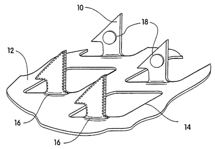
[00029] FIG. 1 illustrates one embodiment of a stratum corneum-piercing
~s microprotrusion member for use with the present invention. FIG. 1 shows a
portion
of the member having a plurality of microprotrusions 10. The microprotrusions
10
extend at substantially a 90° angle from a sheet 12 having openings 14.
The
sheet 12 may be incorporated in a delivery patch including a backing for the
sheet
12 and may additionally include adhesive for adhering the patch to the skin.
In this
2o embodiment the microprotrusions are formed by etching or punching a
plurality of
microprotrusions 10 from a thin metal sheet 12 and bending the
microprotrusions
out of a plane of the sheet. Metals such as stainless steel and titanium are
preferred. Metal microprotrusion members are disclosed in Trautman et al, U.S.
Patent 6,083,196; duck U.S. Patent 6,050,988; and Daddona et al., U.S. Patent
25 6,091,975; the disclosures of which are incorporated herein by reference.
Other
microprotrusion members that can be used with the present invention are formed
by etching silicon using silicon chip etching techniques or by molding plastic
using
etched micro-molds. Silicon and plastic microprotrusion members are disclosed
in
Godshall et al., U.S. Patent 5,879,326, the disclosures of which are
incorporated
so herein by reference.
(00030] FIG. 2 illustrates the microprotrusion member having
microprotrusions 10 having a pharmacologically active agent-containing coating
11
CA 02427381 2003-04-25
WO 02/094368 PCT/USO1/51496
16. The coating 16 may partially or completely cover the microprotrusion 10.
For
example, the coating can be in a dry pattern coating on the microprotrusions.
The
coatings can be applied before or after the microprotrusions are formed.
(00031] The coating on the microprotrusions can be formed by a variety of
known methods. One such method is dip-coating. Dip-coating can be described
as a means to coat the microprotrusions by partially or totally immersing the
microprotrusions into the drug-containing coating solution. Alternatively the
entire
device can be immersed into the coating solution. Coating only those portions
the
microprotrusion member which pierce the skin is preferred.
~o (00032] By use of the partial immersion technique described above, it is
possible to limit the coating to only the tips of the microprotrusions. There
is also a
roller coafiing mechanism that limifis the coating to the tips of the
microprotrusion.
This technique is described in a United States provisional patent ( serial
number:
60/276,762) filed 16 March 2001, which is fully incorporated herein by
reference.
(00033] Other coating methods include spraying the coating solution onto the
microprotrusions. Spraying can encompass formation of an aerosol suspension of
the coating composition. In a preferred embodiment an aerosol suspension
forming a droplet size of about 10 to 200 picoliters is sprayed onto the
microprotrusions and then dried. In another embodiment, a very small quantity
of
2o the coating solution can be deposited onto the microprotrusions as a
pattern
coating 18. The pattern coating 18 can be applied using a dispensing system
for
positioning the deposited liquid onto the microprotrusion surface. The
quantity of
the deposited liquid is preferably in the range of 0.5 to 20
nanoliters/microprotrusion. Examples of suitable precision metered liquid
dispensers are disclosed in US Patents 5,916,524; 5,743,960; 5,741,554; and
5,738,728 the disclosures of which are incorporated herein by reference.
Microprotrusion coating solutions can also be applied using ink jet technology
using known solenoid valve dispensers, optional fluid motive means and
positioning means which is generally controlled by use of an electric field.
Other
so liquid dispensing technology from the printing industry or similar liquid
dispensing
technology known in the art can be used for applying the pattern coating of
this
invention.
12
CA 02427381 2003-04-25
WO 02/094368 PCT/USO1/51496
[00034] The coating solutions used in the present invention are aqueous
solutions of the pharmacologically active agent. The solution must have a
viscosity of less than about 500 cp, and preferably less than about 50 cp, in
order
to effectively coat the tiny stratum corneum-piercing elements to an
appropriate
s thickness. As mentioned above, the pharmacologically active agent must have
an
aqueous solubility greater than about 50 mg/ml and preferably greater than
about
100 mg/ml in the coating solution.
[00035] Desired coating thickness is dependent upon the density of the
microprotrusions per unit area of the sheet and the viscosity and
concentration of
~o the coating composition as well as the coating method chosen. In general,
coating
thickness must be less than 50 micrometers since thicker coatings have a
tendency to slough off the microprotrusions upon stratum corneum piercing. A
preferred coating thickness is less than 10 micrometers as measured from the
microprotrusion surface. Generally coating thickness is referred to as an
average
~5 coating thickness measured over the coated microprotrusion. A more
preferred
coating thickness is about 1 to 10 micrometers.
[00036] The agents used in the present invention are high potency agents
requiring a dose of about 1 mg or less, preferably about 0.25 mg or less.
Amounts
within this range can be coated onto a microprotrusion array of the type shown
in
2o FIG. 1 having the sheet 12 with an area of up to 10 cm~ and a
microprotrusion
density of up to 500 microprotrusions per cm2.
[00037] Preferred pharmacologically active agents having the properties
described above are selected from the group consisting of desmopressin,
luteinizing hormone releasing hormone (LHRH) and LHRH analogs (e.g.,
25 goserelin, leuprolide, buserelin, triptorelin), PTH, calcitonin,
vasopressin, deamino
[Val4, D-ArgB] arginine vasopressin, interferon alpha, interferon beta,
interferon
gamma, menotropins (urofollotropin (FSH) and leutinizing hormone (LH),
erythrepoietrin (EPO), GM-CSF, G-CSF, IL-10, GRF and glucagon.
[00038] In all cases, after a coating has been applied, the coating solution
is
so dried onto the microprotrusions by various means. In a preferred embodiment
the
coated device is dried in ambient room conditions. However, various
temperatures
and humidity levels can be used to dry the coating solution onto the
13
CA 02427381 2003-04-25
WO 02/094368 PCT/USO1/51496
microprotrusions. Additionally, the devices can be heated, lyophilized, freeze
dried
or similar techniques used to remove the water from the coating.
[00039] Other known formulation adjuvants can be added to the coating
solution as long as they do not adversely affect the necessary solubility and
s viscosity characteristics of the coating solution and the physical integrity
of the
dried coating.
[00040] The following examples are given to enable those skilled in the art to
more clearly understand and practice the present invention. They should not be
considered as limiting the scope of the invention but merely as being
illustrated as
~o representative thereof.
Example 1
[00041] A coated microprotrusion device for transdermally delivering
desmopressin was prepared in the following manner. An aqueous desmopressin
~s solution having a concentration of 300 mg/ml was prepared by adding
desmopressin monoacetate salt (sold by Diosynth, Inc. of Des Plaines, IL) to
sterile distilled water. Tritium labeled desmopressin was added to the
desmopressin solution as a marker. A titanium microprotrusion member of the
type illustrated in FIG. 1 was used. The microprotrusion member had a circular
2o shape (1.16 cm diameter sheet with an area of 2 cm2), microprotrusions with
a
length of 360 p,m, and a microprotrusion density of 190 microprotrusions/cm2.
The
microprotrusion member was immersed briefly in the aqueous desmopressin
solution and allowed to dry overnight at room temperature. This procedure
resulted in a desmopressin coated microprotrusion member having a coating
2s containing desmopressin in the amount of 150 to 250 ~g/cm2 of the sheet.
[00042] Delivery kinetics studies were performed in twelve hairless guinea
pigs (HGPs) to evaluate the kinetics of drug absorption through the skin from
the
coated microprotrusion members prepared as described above. The system
applied is shown in FIG. 7. System 25 was comprised of the coated circular
so microprotrusion member 20 adhered to the middle portion of a low density
polyethylene (LDPE) sheet 22 having an adhesive film 24 on the skin proximal
side
of the LDPE sheet 22 between sheet 22 and microprotrusion member 20.. The
14
CA 02427381 2003-04-25
WO 02/094368 PCT/USO1/51496
LDPE sheet 22 and the adhesive film 24 act as an adhesive overlay which keeps
the microprotrusion member adhered to the animal's skin. The skin of one HGP
flank was manually stretched bilaterally (E--j and ~) at the time of applying
the
microprotrusion member to the animal. The system was impacted against the
s animals' skin using a spring-loaded impact applicator which caused the
microprotrusions to pierce the stratum corneum. Following application of the
system, the stretching tension on the skin was released, the HGP was wrapped
with a VetwrapTM bandage and housed individually in a metabolic cage for 1, 2
or 4
hours. At each time point, four of the HGPs had their systems removed and
~o residual drug was thoroughly washed from the skin and the animal was
returned to
its cage. The total amount of drug delivered systemically during these time
intervals was determined by measuring the radioactivity of excreted urine for
two
days following system removal and corrected from the percentage excreted
following IV injection (previous studies had shown that 60% of the injected
dose of
15 3H-desmopressin was excreted in urine over 48 hours). The average amount of
desmopressin delivered to the HGPs (Ma~9 ) during hours 1, 2 and 4 of wear is
presented in FIG. 3. After the first two hours, no additional amount of drug
was
absorbed. Total amount of desmopressin delivered was about 10 micrograms,
which is known to be a therapeutically effective dose in humans for treatment
of
2o nocturnal enuresis.
Example 2A
[00043] A second experiment was performed on hairless guinea pigs (HGPs).
All animals wore a system identical to those previously described in Example
1.
25 One group of animals (Group A) wore a system for 1 hour. In two other
groups
(Groups B and C), the microprotrusion device was removed 5 seconds after
application. In Group B, the treatment site was immediately washed after
removal
of the system. In Group C, the treatment site was not washed but was occluded
with an adhesive backing for 1 hour following system removal. The average
so amounts of desmopressin delivered to the animals in Groups A, B and C are
shown in FIG. 4. Group B (5 second delivery and immediate washing) resulted in
an average delivery of about 5 ~.g desmopressin. Group C (occlusion following
5
CA 02427381 2003-04-25
WO 02/094368 PCT/USO1/51496
second application) did not increase significantly the amount delivered to
Group B.
Group A (one hour delivery) resulted in an average of 18 ~,g desmopressin
delivered. These results indicate that keeping the coated microprotrusions in
piercing relation to the skin for only about 5 seconds results in substantial,
although not optimal, delivery of desmopressin and that the drug delivered
into the
skin is not removed by washing. In addition, prolonged (1 hour) contact of the
microprotrusions with the skin results in even greater amounts of desmopressin
delivered.
Example 2B
[00044] The feasibility of coating a microprotrusion array with the drug
desmopressin was evaluated. In these studies the coating was limited to the
tips
of the microprotrusions. A number of microprotrusion arrays (S250 Ti,
microprotrusion length 250 pm, 321 microprotrusions/cm~, 2 cm2 disc) were tip
~s coated using the device described in a United States provisional patent (
serial
number: 60/276,762, filed 16 March 2001 ) using a 40 wt% desmopressin acetate
solution spiked with 3H desmopressin. Analysis revealed that each
microprotrusion
array was coated with 187 ~ 30 ~,g desmopressin. SEM examination revealed that
the coating was present as a glassy amorphous matrix with good uniformity of
2o coating from microprotrusion to microprotrusion. The coating was limited to
the
first 115 ~,m of the 250 ~.m microprotrusion. The coating was found unevenly
distributed on the microprotrusion itself. Most of the solid coating appeared
to be
located in circular domed regions of the coating called a cap, centered on the
geometric center of the faces of the coated area of the microprotrusion. The
Zs maximum measured thickness of the coating was about 18 ~.m while the
average
calculated thickness over the entire coated area was only about 13 p.m.
[00045] Studies were performed in hairless guinea pigs to evaluate the
kinetics of drug absorption through the skin from desmopressin tip-coated
ao microprotrusion array systems. System application was performed on the
flank of
the animal with an impact applicator delivering an energy of 0.26 J in less
than 10
ms. The system applied comprised a coated microprotrusion array device,
adhered
16
CA 02427381 2003-04-25
WO 02/094368 PCT/USO1/51496
to the center of a LDPE backing with adhesive (7 cm2 disc). Systems remained
on
the skin for 5 seconds or 1 hour. Groups of three animals were used for both
time
points. Upon removal of the system, the application site was thoroughly
cleaned
and the washes were evaluated for radioactive content and the HGPs were
returned to their individual metabolism cages. Urine was collected for 2 days
and
counted for radioactive content. The total amount of drug delivered
systemically
was determined by measuring urinary excretion of radioactivity for two days
following system removal and corrected from the percentage excreted following
iv
injection (previous studies had shown that 60% of the injected dose of 3H-
~o desmopressin was excreted in urine over 48 hours). The used systems were
extracted for residual radioactivity. Total amounts of desmopressin delivered
systemically were 49 ~ 3 ~,g (26% drug utilization) and 97 ~ 11 ~,g (52% drug
utilization) following 5 seconds (open bar) and 1 hour (hatched bar) wearing
times,
respectively (Fig. 8). Only a small percentage of the drug was found on the
~s surface of the skin (6% at 5 seconds, and 9% at 1 hour), the balance
consisting of
desmopressin remaining on the microprotrusions.
Example 3
[00046] The properties of the desmopressin coating were evaluated in the
2o following manner. Fluorescein sodium salt was added to a 300 mg/ml solution
of
desmopressin in water. Sufficient fluorescein sodium salt was added to achieve
a
final concentration of 0.001 M.
[00047] A titanium foil (0.025 mm thick) was immersed briefly in this solution
and allowed to dry overnight at room temperature. Fluorescence microscopy
2s revealed that the dry film of desmopressin was amorphous in nature and
behaved
much like a transparent glass. A coating of about 2 p.m thick appeared to
behave
best in terms of flexibility and adherence to the titanium sheet. Coatings
thicker
than about 10 p,m were found to be brittle and susceptible to cracking.
so Example 4A
[00048] Human growth hormone (hGH) was added to sterile distilled water to
form an aqueous hGH solution having an hGH concentration of about 200 mg/ml
17
CA 02427381 2003-04-25
WO 02/094368 PCT/USO1/51496
and a viscosity of less than 50 cp. A titanium foil was immersed in the
solution,
followed by drying overnight at room temperature to form the hGH coating.
Adequate coating of the foil was demonstrated by microscopy utilizing the
method
previously discussed. Although hGH could not be used for therapeutic purposes
with this strategy because of the large therapeutic dose it requires, it is
believed to
be a good model for cytokines, particularly interferons, which require a much
smaller therapeutic dose. Similarly, titanium foil was coated with an aqueous
solution of ovalbumin, a 45,000 Dalton polypeptide containing an
oligosaccharide
side chain. The solution had an ovalbumin concentration of about 300 mg/ml and
1o a viscosity of less than 50 centipoises. Adequate coating of the titanium
foil was
demonstrated by microscopy utilizing the method previously discussed. Although
ovalbumin is not a pharmacologically active agent used in therapeutics or as
defined herein, it is a good model for large pharmacological agents such as
follicle
stimulating hormone (FSH) and erythropoietin.
Example 4B
[00049] The feasibility of coating a microprotrusion array with the drug hGH
was evaluated. In these studies the coating was limited to the tips of the
microprotrusions. Microprotrusion arrays (S250 Ti, microprotrusion length 250
~.m,
321 microprotrusions/cm~, 2 cm~ disc) were tip coated using the device
described
in a United States provisional patent application (serial number: 60/276,762,
filed
16 March 2001 ) using a 20 wt% hGH, 20 wt% sucrose coating solution. Analysis
revealed that each microprotrusion array was coated with 9.5 ~ 0.9 ~,g hGH.
SEM
revealed good uniformity of coating from microprotrusion to microprotrusion
with a
coating depth of about 100 ~.m. However, on the microprotrusion itself, the
coating
was found unevenly distributed. Most of the solid coating appeared to be
located
in caps centered on the geometric center of the faces of the coated area of
the
microprotrusion. Following two days storage in a vacuum chamber the solid
coating presented a very smooth surface with absence of cracking and it was
ao demonstrated to adhere very tightly to the microprotrusions. The maximum
measured thickness of the coating was about 4 ~m while the average calculated
thickness over the entire coated area was only about 1.7 ~.m.
18
CA 02427381 2003-04-25
WO 02/094368 PCT/USO1/51496
[00050] Studies were performed in hairless guinea pigs to evaluate the
kinetics of drug absorption through the skin from hGH tip-coated
microprotrusion
array systems. System application was performed on the flank of the
anesthetized
animals with an impact applicator delivering an energy of 0.26 J in less than
milliseconds. The system applied comprised a coated microprotrusion array
device, adhered to the center of a LDPE backing with an adhesive (7 cm~ disc).
Systems remained on the skin for 5 seconds (n=3) or 5 minutes (n=5). A group
of
animals (n = 5) received a subcutaneous injection of 10 ~,g hGH. Blood samples
~o were collected at time intervals for plasma hGH determination by ELISA. The
hGH
dose delivered was extrapolated based on an area under the curve (AUC)
calculation compared to IV administration of hGH. Results showed that hGH
delivery from the microprotrusion array was the same with 5 seconds (open
triangles) and 5 minutes (close circle) wearing times (Figure 9). On average,
5 ~.g
of hGH was delivered in each animal, which accounts for approximately 50% of
the
coated dose. This is to compare with a bioavailability of 65% following
subcutaneous administration of hGH, the results of which are shown as "X"
(Figure 9).
2o Example 5
[00051] The feasibility of coating the microprotrusion devices with ovalbumin
was evaluated. A coating solution comprising 200 mg/ml of fluorescein-tagged
ovalbumin in water was prepared. The microprotrusion member of the type used
in Example 1 was immersed briefly in the coating solution, blown dry, and
allowed
to dry overnight at room temperature. Subsequent analysis demonstrated that
this
coating procedure resulted in microprotrusions coated with ovalbumin at 200 to
250 ~.g per cm2 of the microprotrusion member.
[00052] Studies were performed in hairless guinea pigs (HGPs) to evaluate
the kinetics of ovalbumin absorption into the skin from coated
microprotrusions
so devices. The applied system comprised a coated microprotrusion device,
adhered
to the center of a LDPE backing with an adhesive housed on a 3.8 cm2 disc. The
skin of one HGP flank was manually stretched bilaterally (~ and ~) at the time
of
19
CA 02427381 2003-04-25
WO 02/094368 PCT/USO1/51496
the application of the system. Microprotrusion application was performed using
a
spring loaded applicator which impacted the system against the animal's skin.
Following application, the stretching tension was released, the HGPs were
wrapped with a VetwrapTM bandage and housed individually in a metabolic cage
for 30 minutes or 1 hour. At each time point, four HGPs had their systems
removed and residual drug was thoroughly washed from the skin and the animal
was returned to its cage. In one group of HGPs, the microprotrusion device was
removed 5 seconds after application (0 hour time point). The average total
amount
of ovalbumin delivered into the skin (Ma~g) during these time intervals was
~o determined by taking an 8 mm skin biopsy at the application site. The skin
biopsy
sample was then dissolved in hyamine hydroxide (diisobutylcresoxyethoxyethyl)
dimethyl) benzylammonium hydroxide, 1 M in ethanol, sold by J.T. Baker (NJ,
USA) and the amount of ovalbumin present was determined by fluorimetry.
Results demonstrated that up to 80 ~g ovalbumin was delivered intracutaneously
~s over the 1 hour application period. The 5 second piercing resulted in about
25 ~.g
of ovalbumin delivered intracutaneously. These results are shown in FIG. 5.
Although ovalbumin is not a pharmacological agent used in therapeutics, it is
a
good model for large potent pharmacologically active agents such as follicle
stimulating hormone and erythropoietin.
Example 6A
[00053] An experiment similar to that described in Example 1 was performed
in the HGPs using the identical microprotrusion systems which were coated with
aqueous ovalbumin solutions having ovalbumin concentrations of 200, 50, and 10
2s mg/ml ovalbumin. In all groups the microprotrusion device was removed
immediately after application. Application and analysis were performed
identically
to that described in Example 1. Results demonstrated that delivery of
ovalbumin
could be controlled by controlling the amounts coated on the microprotrusions.
The average amounts of ovalbumin delivered (Ma~g) for each of the three
solution
so concentrations ([C]) are shown in FIG. 6.
Example 6B
[00054] The feasibility of coating a microprotrusion array with the drug
CA 02427381 2003-04-25
WO 02/094368 PCT/USO1/51496
ovalbumin was evaluated. In these studies the coating was limited to the tips
of
the microprotrusions. Microprotrusion arrays (S250 Ti, microprojection length
250
p,m, 321 microprojections/cm2, 2 cm2 disc) were tip coated using the device
described in a United States provisional patent application (serial number:
60/276,762; filed 16 March 2001 ) using a 20 wt% ovalbumin tagged with
fluorescein isothiocyanate (FITC). Analysis revealed that each microprotrusion
array was coated with 4.6 ~ 0.5 ~.g ovalbumin. SEM examination revealed that
the
coating was present as a glassy amorphous matrix with good uniformity of
coating
from microprojection to microprojection. The coating was limited to the first
150
~o ~,m of the microprojection.
[00055] Studies were performed in euthanized hairless guinea pigs to
evaluate the kinetics of drug absorption through the skin from ovalbumin tip-
coated
microprotrusion array systems. System application was performed on the flank
of
the animal with an impact applicator delivering an energy of 0.26 J in less
than 10
~s ms. The applied systems comprised a coated microprotrusion array, adhered
to
the center of a LDPE backing with an adhesive (7 cm~ disc). Systems remained
on the skin for 5 seconds or 1 hour. Groups of three animals were used for
both
time points. At the end of the wearing time, the system was removed and the
skin
wiped clean of any residual drug. The total amount of ovalbumin delivered in
the
2o skin during these time intervals was determined by dissolving a 8 mm skin
biopsy
in hyamine hydroxide (10% in methanol). Quantitation was performed by
fluorimetry. Results presented in Figure 10 demonstrated that more than 80% of
the ovalbumin dose was delivered after 5 seconds wearing time (open bar).
Close
to 100% of the dose had been delivered after 1 hour application time (solid
bar).
25 [00056] Although the present invention has been described with. reference
to
specific examples, it should be understood that various modifications and
variations can be easily made by a person having ordinary skill in the art
without
departing from the spirit and scope of the invention. Accordingly, the
foregoing
disclosure should be interpreted as illustrative only and not to be
interpreted in a
so limiting sense. The present invention is limited only by the scope of the
following
claims.
21