Note: Descriptions are shown in the official language in which they were submitted.
CA 02427693 2002-08-19
METHOD I~'OR DETECTING THE PRESENCE OF
'TARGET BACTERIA OR A TARGET COMPONENT
CARBOHYDRATE ANTIGEN THEREOF
This application is a continuation-in-part of each of the following U.S.
applications, all
of which were assigned to Binax, Inc., the corporation having the rights to
receive assignment
in full of this application.
(1) Serial No. 09/139,720, filed August 25, 1998,
(2) Serial No. 09/156,486, filed September 16, 1998, now abandoned in favor of
its
continuation-in-part application,
(3) ' Serial No. 09/397,110, filed September 16, 1999,
(4) Serial No. 09/458,998, filed December 10, 1999, as a continuation-in part
of
Serial No. 09//39,720.
INTRODUCTION TO THE PRESENT INVENTION
The present invention relates to achieving rapid and accurate diagnoses, of
high sensi-.
tivity and specificity, of bacterial infections. caused by bacteria
characterized by the possession
of carbohydrate antigens. In particular, the invention involves the initial
purification of such
carbohydrate antigens to an essentially protein-free state, followed by
utilization of each so-
pur l ed car bohydratc antigen to afi-miry purify raw polyvalent antibodies to
said antigen and
the utilization of the said so-purified antibodies in diagnostic tests of high
accuracy, specificity
and sensitivity for detecting the presence of the original bacterium.
The invention is applicable to bacteria possessing carbohydrate antigens,
which bacteria
may be positive or negative to Gram's stain. The purified antibodies produced
in accordance
with this invention are of at least the same order of specificity and
sensitivity as commercially
available monoclonal antibodies and are easier to produce and to work with
than many such
CA 02427693 2002-08-19
monoclonal antibodies. They offer wide opportunities for rapid diagnostic
tests, e.g., via ICT
immunoassays, to identify bacteria that have heretofore been difficult to
identify rapidly and
accurately, whereby diagnoses of bacterial diseases they caused were often
arrived at slowly
and difficultly, using cumbersome methodology.
Each of the parent applications identified above is incorporated herein by
reference
except for now-abandoned application Serial No. 09/156,486, the disclosure of
which, in
essence, appears physically in its continuation-in-part application, U.S.
Serial No. 091397,110,
which is among the three applications incorporated herein by reference.
BACKGROUND OF THIS I7VVENTION
Gram-negative bacteria are known to have in common the possession of at least
one
lipo-polysaccharide or. other lipo-polycarbohydrate antigen, while Gram-
positive bacteria are
known to possess the common characteristic of having at least one carbohydrate
antigen that is
a lipo-teichoic acid or teichoic acid or a derivative of either. Some of both
the.Gram-positive
and Gram-negative bacteria also possess carbohydrate antigens that are
capsular - i.e., these
antigens are each enclosed in a heavy capsular layer in their native state.
This capsular layer
constitutes a slime-like substance that surrounds the bacterial cell wall of
most bacteria.
U.S. Application Serial No. 09/139,?20, which is fully incorporated herein by
refer-
ence, describes the purification io an essentially protein-free state of lipo-
carbohydrate antigens
of bacteria of the Legionella species, all of which are Gram-positive.
Emphasis is placed
therein on purifying carbohydrate antigens of Legionella pneumophila
serotypes, including
2
CA 02427693 2002-08-19
without limitation, the O-polysaccharide antigen of L. pneumophila serotype 1,
the purification
of which to an essentially protein-free state, is described in detail.
The application shows that when the essentially protein-free O-carbohydrate
antigen~of
L. pneumophila serotype 1 (which serotype is known to be the causative
organism for some 70
percent of the Legionella-caused pneumonia-like illnesses that occur), is
coupled (through a
spacer molecule) to an affinity column as described and raw polyclonal
antibodies to the un-
purified antigen are passed over the column as described, the resulting
purified antibodies are
highly antigen-specific and will readily identify the same antigen when it is
present in bodily
fluids taken from patients with disease caused by Legionella pneumophila
serotype 1. Urine is
shown to be a preferred bodily fluid for this diagnostic purpose because:
(1) The L. pneumophila serotype 1 antigen appears in urine early in the
disease state
and persists for some days even after appropriate therapeutic treatment is
initiated;
(2) The collection of the test sample is non-invasive and simple, causing a
minimum
of patient disruption as well as requiring no specially trained personnel or
specially designed
instrumentation; and
(3) Samples from, e.g.; sputum may give false negative or false positive
results due
to difficulties in obtaining or culturing the sample, possible presence of
colonies of bacteria in
the patient's nose or throat that are chronically present and were not
causative of disease, and
other similar difficulties.
The L. pneumophila serotype 1 bacterium present in urine is dead and has at
least in
part had its cell wall broken down as it is passed through the kidneys; hence
the antigen is in a
3
CA 02427693 2002-08-19
state readily accessible to the antigen-specific antibodies deposited in two
areas on the ICT test
strip.
The efficacy of the antigen-specific antibodies described in U.S. Application
Serial.No.
09/139,720 in identifying whole, and to some extent, living bacteria in
aqvueous media consti-
tuting environmental samples is further shown in the continuation-in-part
Application Serial
No. 09/458,988, also incorporated herein by reference, wherein an enzyme
immunoassay of
high specificity and sensitivity is described. This assay is based on use as
the detecting agent
for the antigen, of antigen-specific antibodies obtained by purifying raw
polyclonal antibodies
as described in detail in the parent application. The sensitivity 4-3
specificity of the so-
purified antibodies is in part illustrated by the short times within which the
enzyme immuno-
assay produced informative results, as well as by the small concentration
(0.05 ,gig per test) of
antigen-specific antibodies that gave equally informative time results with a
longer incubation
time (1 hour).
U.S. Application Serial No. 09/397,110, also incorporated herein by reference,
de-
scribes the purification to an essentially protein-free state of the C-
polysaccharide cell wall
antigen present in the pneumococcal cell wall of all S. pneumoniae serotypes.
This antigen is a
phosphocholine-containing polysaccharide derived from teichoic acid. The
Streptococcus
pneumoniae strain of bacteria are all Gram-stain positive.
The essentially protein-free antigen (which contains less than about 10
percent by
weight thereof) is covalently coupled to a spacer molecule which is in turn
covalently coupled
to an affinity column and the thus-prepared column is then used to purify raw
polyclonal
antibodies to S. pneumoniae. The resulting antigen-specific purified
antibodies showed high
4
CA 02427693 2002-08-19
sensitivity and specificity in an IC"T test for identifying S. pneumoniae in
bodily fluids,
including urine in particular.
Numerous and varied efforts have been made in the past to use raw polyvalent
antibodies to carbohydrate antigens, or monoclonal antibodies to such
antigens, of various
infectious Gram-negative or Gram-positive bacteria believed to be responsible
for diseases of
the lower respiratory tract in diverse tests, including ELISA, counter-
immunoelectrophoresis
and/or latex agglutination tests for the presence of the specific bacterium
sought. While some
of the tests have been useful in some cases, none of them has so far gained
sufficient clinical
acceptance of reliability to be used independently of cell culture tests. The
drawbacks of rail
culture tests and their tenuous reliability have been extensively documented
in the art and are
discussed in parent applications 091139,720 and 09/397,110.
To gain U.S. Food and Drug Administration ("FDA") approval of floe L.
pneumophila
serogroup 1 ICT test first described in parent Application Serial No..
09/139,720 and the S.
pneumoniae ICT test that is the subject of parent Application Serial No.
09!397,1/0 and its
parent application, Serial No. 091156,786, it was necessary for the assignee
of these appli-
rations, Binax, Inc., to conduct extensive clinical tests on each. Many of
these clinical tests
are described in the two parent applications incorporated herein by reference.
One of the
important points about the clinical tests is that FDA regulations require
extensive clinical
testing of diagnostic tests only in instances where the diagnostic test is
recognized to represent
a substantial scientific and technical departure from tests that are already
known and in
commercial use. The sensitivity and specificity of each of these two tests is
believed to be
much higher than the numbers shown in the parent applications indicate. The
reason is that the
5
CA 02427693 2002-08-19
numbers shown are based on comparison of these clinical test results with
parallel results
obtained on the same clinical samples with other earlier available assay
procedures or identi-
fication techniques (such as cell culture tests), which prior available tests
were known to be
tenuously reliable even when they were believed to be the best available
identification methods
s
for detecting the involved bacteria or their antigenic components.
In short, this invention presents the opportunity for providing highly
specific and sen-
sitive, rapid diagnostic tests for the wide spectrum of bacteria that possess
carbohydrate anti
gens, which antigens manifest themselves in human bodily fluids of patients
infected with the
corresponding bacteria, especially urine.
BRIEF DESCRFPTION OF THE INVENTION
This invention involves novel specially purified, highly antigen-specific
antibodies for
detecting the presence of bacterial carbohydrate antigens in fluids,
especially human or other
mammalian bodily fluids, and particularly urine.
These antibodies are prepared from raw polyvalent antibodies to the target
carbor-vrate
antigen by a method which comprises:
(a) purifying the raw target antigen to obtain an essentially protein-free
antigen,
r. e. , one containing not more than about 10 percent of protein,
(b) coupling the so-purified antigen to a spacer molecule by covalent binding,
(c) covalently coupling the free end of the spacer molecule to an affinity gel
packed
into a chromatographic column,
6
CA 02427693 2002-08-19
(d) passing the raw polyvalent antibodies to the raw antigen over the gel on
the
column, and
(e) eluting the gurified anri'bodies.
The purified antibodies eluted from the affinity gel are of high specificity,
sensitivity
and accuracy and may be used in any of a variety of specifically developed
immunoassay
procedures to detect the raw target antigen in fluid media, especially
mammalian bodily fluids,
and particularly urine.
A preferred ICT procedure is described in parent application 09/139,720 for
detecting
the polycarbohydrate antigens of Legioreella bacteria, and especially the O-
polysaccharide anti-
gen ofL. pneumophila serogroup 1, while applications 09/156,4$6 and 09/397,110
describe an
analogous preferred ICT procedure for detecting the C-polysaccharide cell wall
antigen present
in all serotypes of Streptococcus pneumoniae.
A similar ICT procedure for detecting the capsular polysaccharide antigen of
H. in, flu-
enzae type b is described herein in detail.
Heretofore it has not been recognized that the lipo-polycarbohydrate antigens
typically
found in Gram-negative bacteria, the antigens comprising lipo-teichoic or
teichoic acid or
derivatives thereof typically found in Gram positive bacteria and the capsular
polycarbohydrate
antigens frequently found in the heavy slime-like capsule surrounding the cell
wall of many
bacteria of both Gram-positive and the Gram-negative types may alt be detected
by a rapid,
highly specific and sensitive immunoassay of the ICT type which employs
antigen-specific
antibodies as the detecting agent, which antigen-specific antibodies are
obtained according to
the schema for purifying raw polyclonal antibodies to carbohydrate antigens
that is set forth in
7
CA 02427693 2002-08-19
the second paragraph of this section. The fact that raw polyvalent antibodies
to bacterial car-
bohydrate antigens may be rendered highly antigen-specific and sensitive by
subjecting them tc
affinity purification with a purified target bacterial carbohydrate antigen
that is essentially
protein-free likewise has not been appreciated heretofore. Likewise, the fact
that carbohydrate
antigens from both Gram-negative and Gram-positive bacteria and/or from the
capsular layer
surrounding both types of bacteria can all be purified and used to affinity
purify antibodies to
such antigens to yield antigen-specific antibodies has not been heretofore
recognized, nor has i~
been appreciated that bacterial carbohydrate antigens can be detected rapidly
with high accur-
acy, sensitivity and specificity using such antigen-specific antibodies as a
detecting agent.
BRIEF DESCRIPTION OF T>i~ DRAWING
Figure 1 and its related Figures 1A, 1B and 1C depict a typical ICT device of
the type
preferred in the performance of an assay for a bacterial carbohydrate antigen
in accordance
with this invention.
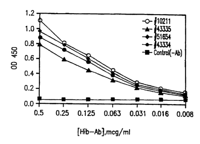
Figures 2, 3 and 4 are graphs showing, in Figure 2, the ability of antigen-
specific puri-
fled antibodies of this invention to detect other serotypes of H. in, fluenzae
type b than the one
to which the antibodies were raised. In Figures 3 and 4, the graphs reflect
that the purified
antigen-specific antibodies of H. influenzae type b were not cross-reactive
with antigens of H.
influenZae types a, c, d or f (Fig. 3) or with any of nontypical H. influenzae
NTl, NT2, NT3
or NT4 or with H. para-influenzae.
8
CA 02427693 2002-08-19
DETAILED DESCRIPTION OF THE INVENTION
The present invention represents an exceptional advance in methods for
detecting bac-
terial infection.
Because it is applicable to the detection in mammalian bodily fluids. of
bacterial carbo-
hydrate antigens of all known types - i.e., the lipo-polycarbohydrate antigens
including lipo-
polysaccharides, the antigenic lipo-teichoic acids and teichoic acids and
their antigenic deriva-
lives and the capsular polycarbohydrate antigens, including polysaccharides -
and it represents
a unified approach to the detection of bacterial infection not heretofore
envisioned, this inven-
lion holds promise for permitting the rapid diagnosis of virtually any
bacteria-caused disease
wherein the bacteria possess a carbohydrate antigen that manifests itself in
the disease state in a
bodily fluid of the patient.
Of particular importance is the opportunity that this invention affords for
rapid diag-
nosis and rapid introduction of appropriate therapy in situations where a
particular bacterially-
caused disease appears to be epidemic within a group - whether a small,
confined group, e.g.,
in a school or geriatric center, or a widespread population as, e.g., a town,
a city or a larger
region.
Broadly speaking, the preferred immunochromatographic ('ICT°) assay of
this inven-
lion may be designed and configured to be run on any known disposable ICT
device disclosed
in the art. Preferably it is designed to be conducted, and is conducted, using
an IG"f device of
the type disclosed in co-pending U.S. Patent Application Serial No. 071706,639
of Howard
Chandler, or one of its continuation-in-part applications, all of which are
assigned to Smith-
Kline Diagnostics, Inc. but are exclusively licensed to Binax, Inc. (which is
entitled to assign-
9
CA 02427693 2002-08-19
went of this application), in a wide area of use fields that includes
diagnoses of human respira
tory system diseases.
The preferred device is suitably impregnated in one region thereof with
affinity puri-
feed, highly antigen specific antibodies to the target carbohydrate antigen of
the bacterium
suspected of causing the disease. Labeled antigen-specific antibodies are
applied to another
area of the device. The test sample suspected of containing the bacterium is
contacted first
with the labeled antigen-specific antibodies, which then flow with the sample
to the device are
containing unlabeled bound antigen-specific antibodies, whereupon if the
tang::: antigen indig-
enous to the suspected bacterium is present in the sample, the labeled
antibodyaarget carbohy-
drate antigen conjugate already formed binds upon contact to the immobilized
unlabeled
antigen-specific antibodies, whereupon a visible color reaction is produced.
The label may be
any substance known in the art to produce visible color upon the reaction of a
labeled anti-
body:antigen complex with bound unlabeled antibodies. Such labels include
various finely
divided metallics, various organic molecules, and various molecular
combinations such as
enzyme combinations with another color-producing molecule. In this invention,
colloidal gold
particles constitute the preferred label.
It is of major importance in designing the preferred test device, that the
concentration
of antigen-specific antibody present at each of the two sites of the test
device where reaction
occurs be sufficient to insure that antigen present in the test sample will be
captured by the
labeled antigen-specific antibodies as the test sample contacts them and that
labeled antigen-
specific antibody:antigen conjugate will be readily captured and held by the
bound antibodies
at the sample capture line. Experimental work ur r °_rtaken in
connection with this invention
CA 02427693 2002-08-19
has shown that active antigen-specific antibody to the target carbohydrate
antigen must be
present at each site of a test device at which antigen:antibody reaction is to
occur is a concen-
tration of between 7.7 nanogramslsq. mm. of surface area and 385 nanograms/sq.
mm. of
surface area. If antigen-specific antibody concentrations lower than 7.7
nanograms/sq. mm.
are present at a site where reaction is intended to occur, false negative
results are likely to
occur.
As is known in the art, infectious bacteria frequently have multiple antigenic
com-
ponents. For example, S. pneumoniae is known to have a capsular antigen in
addition to the
polysaccharide cell wall antigen which is the target of the assay described in
parent applica-
Lions Serial Nos. 091156,486 and 09/397,110. The latter antigen was selected
as the target
antigen for the now-FDA-approved test which is described in these applications
because that
antigen is present is all known serotypes of S. pneumoniae and its relatively
miaor cross-
reactivity as described in the herein incorporated application Serial No.
091397,110 is of a
nature that allows ready clinical differentiation of S. pneumoniae-caused
infection fmm other
infections. It is noted that previous published attempts to detect S,
pneumoniae in bodily fluids
have at best yielded systems having sensitivity and specificity in the 60-70
percent range with
both polyclonal and monoclonal antibodies - a range unacceptable for reliable
diagnostic
purposes.
Among the mammalian fluids in which target carbohydrate antigens have been
shown to
be successfully detected in ongoing clinical work with the respective
described and 1.'DA-
approved ICT tests for L. pneumophila serogroup 1 and S. pneurnoniae are, in
addition to the
preferred urine, sputum, naso-pharyngeal exudates, middle ear fluid and
cerebrospinal fluid.
11
CA 02427693 2002-08-19
Other fluids in which these tests detect carbohydrate target antigens, when
present, include
blood and bronchial fluid.
- Selection of the target carbohydrate antigen for any particular bacterium is
necessarily
based upon considerations of that antigen's cross-reactivity characteristics,,
whether it.is known
to be present in all or most serotypes of a bacterial strain, whether if
peculiar to a particular
serotype of a strain, that serotype is known to be the most common source of
disease caused b5
the bacterium and like questions.
This invention offers unique capabilities in regard to ready diagnoses of
bacterial infec-
tions caused by any bacterium with one or more carbohydrate antigens of the
types already
mentioned - i. e. , lipo-polycarbohydrate antigens, antigens comprising Iipo-
teichoic or teichoic,
acid and derivatives of either, and capsular carbohydrate antigens. Among the
bacteria,
carbohydrate target antigens of which are contemplated to be within the scope
of this invention
are Flaemophilus injluenzae of various types, Mycoplasma pneccmoniae,
Chlamydia pneumo-
nice, Klebsiella pneumoniae, Staphylococcus aureus, Mycobacterium
tuberculosis, Pneudo-
moms aereiginosa, Acinetobacter, Moraxella catarrhalis, Neisseria Meningitis,
group B
Streptococci, Escherichia coli, Listeria monocytogenes, the other species of
Escherichia,
Klebsiella and Pseudomonas not specifically already named, Proteus mirabilis,
Gardnerella
vaginalis, Serratia marcescens, the various other species of Proteus and
Listeria not spe-
cifically named, the various species of Enterobacter, Xanthomonas,
Enterococcus, Bac-
teroides, Clostridium, Peptostreptococcus, Campylobacter, Salmonella and
Alcaligenes and all
other bacterial species and strains not specifically named that have one or
more carbohydrate
antigens of the types described.
12
CA 02427693 2002-08-19
The polyclonal antibodies to be purified by the techniques of the present
invention are
raised by conventional methods, by injecting an animal, e.g., a rabbit or
goat, with the crude
target antigen of the intended assay. Preferably the antigen preparation is
subjected to heat
killing of cells before injecting the animal. After an appropriate lapse of
time, the animal is
bled to obtain serum containing the desired antibodies, followed by
purification of the latter.
This serum may go through an intermediate purification step, e.g., with
ammonium sulfate or
an ion exchange resin to produce an IgG cut or may be purified directly. .
For purposes of the affinity purification, the same crude carbohydrate target
antigen
used to immunize the animal is grown in culture and then suitably purified to
an essentially
protein-free state. As used herein the expression "essentially protein-free
state" means a state
containing not more than - and preferably less than - about 10 percent
(wt./wt.) of protein.
After the antigen is purified to the essentially protein-free state, it is
coupled to a spacer
molecule by covalent binding. Examples of suitable spacer molecules include
hydrazine,
bovine serum albumen ("HSA"), the conjugate of BSA and hydrazine and like
molecules that
are capable of covalently bonding to purified carbohydrate antigens at one end
while retaining
another reactive end that is capable of bonding covalently to an affinity gel.
The purified carbohydrate antigenapacer molecule conjugate is next conjugated
to an
affinity gel and the gel is used to purify the raw polyvalent antibodies in
serum obtained by
bleeding the previously immunized animal, or an IgG cut thereof. The raw
antibodies (or their
IgG cut) are multiply applied to the affinity gel and are eluted from it as
purified, highly
antigen-specific antibodies.
13
CA 02427693 2002-08-19
The following examples illustrate the preferred mode of affinity purification
of anti-
bodies to Haemophilus influenzae type b, including the preliminary separation
and purificatio:
of the capsular carbohydrate antigen used in that purification. Many methods
for effecting ,
these separation and purification seeps are known in the literature and may be
substituted for
those herein described without departing from the scope of this invention, so
long as the puri-
fled antigen obtained is essentially protein-free as herein specified.
Example 1 ~ Culture Conditions for Culturing the Target Carbohydrate Antigen
Haeriioplulus influenzae type b (ATCC #110211) was grown in supplemented
Mueller
Hinton broth at 37° C. with 5 percent COz for 24 hours without
agitation.
The broth composition, per liter, was:
Acid hydrolyzate of casein 17.5 g.
Beef heart extract 3.0 g.
Starch 1.5 g.
Supplements as follows were also present:
Hematin 15 mg./mL
NAD (nicotine adenine dinucleotide) 15 mg./mL
Yeast extract 5 mg.lmL
The pH of this mixture was 7.3 t 0.1 as measured at 25 ° C.
Example 2 - Purification of Carbohydrate Antigen
After 24 hours, 1.82 g. of cetyltrimethylammonium bromide CAS II57-09-0 was
dis-
solved in 30 mL of distilled water and the solution was added to 500 mL of
broth supernatant
to yield a final concentration of 0.01 M cetyltrimethylammonium biomide. The
mixture was
incubated in an ice bath with stirring for one hour and left at 4 ° C.
overnight.
14
CA 02427693 2002-08-19
The mixture from Example 1 was centrifuged at 12,000 rpm and 4° C. for
20 minutes
to yield a pellet and a supernatant. Both were collected and treated,
respectively, as follows:
(1) The pellet was resuspended in 0.5 M NaCl with sonication and was then drop-
wise precipitated at 4° C. in ten times the resuspension volume of
ethanol.: The resulting solu-
tion was stored overnight at 4° C. to allow precipitation.
The solution was then centrifuged at 12,000 rpm for 20 minutes. The pellet was
dis-
solved in distilled water and then dialyzed against distilled water in
dialysis tubing having a
molecular weight cut-off of 3,500.
(2) The supernatant from the Example 1 mixture was stored at 40°
overnight, and a
precipitate was then noted to have formed. The entirety of the contents of the
container hold-
~ing this was centrifuged at 12,000 rpm for 20 minutes. A pellet was recovered
and was re-
suspended in 0.5 M NaCI with sonication. The resulting solution was dropwise
precipitated in
ten times the resuspension volume of ethanol at 4° C. The solution was
stored overnight at 4°
C. and a precipitate again formed. The solution and precipitate were
centrifuged at 12,000
rpm for 20 minutes and a pellet was recovered. The pellet was dissolved in
distilled water and
dialyzed against distilled water in dialysis tubing having a 3,500 molecular
weight cut-off.
Thereafter the dialyzed solutions from (1) and (2) above were pooled and
lyophilized.
Ninety mg. of Haemophilus inftuenzae type b polysaccharide antigen was
obtained.
A solution of this antigen of 5.3 ~cg/ml concentration was prepared and
subjected to
Lowry assay for protein and found to contain 5 percent protein (wt/wt). The
solution was also
tested for carbohydrate by the phenol-sulfuric acid method and found to
contain 36 percent
CA 02427693 2002-08-19
(wt/wt). The solution was tested for activity by both the ELISA method and SDS-
PAGE-
immunoblot and found to have requisite activity.
Example 3 - Preparation of Affinity Column
Five mg. of lyophilized Naemophilus in, fluenzae type b polysaccharide antigen
was dis-
solved in 4.52 mL of distilled water and the pH was adjusted to 5-6 with HCI;
15.64 mg. of
bovine serum albumen-hydrazine conjugate of pH S-6 was then added, followed by
mixing for
three minutes.
2.6 ~cg of 1-ethyl-3- (3-dimethylaminopropyl) carbodiimide ("EDAC") was
dissolved it
100 ~cL of distilled water. 50 ~cL of this solution was added to the
antigen/BSA-hydrazine con
jugate solution, followed by three minutes of mixing. The balance of the EDAC
solution was ,
then added to this mixture followed by two hours of mixing at room
temperature. The pH was
then adjusted to 8 with NaOH and mixed for oae hour at room temperature,
followed by stor-
age overnight at 4 ° C.
The next day the pH of the stored mixture was adjusted to 7 with HCl and a
portion
was subjected to the ELISA test, confirming its activity.
2.12 mg. of the EDAC-treated antigen/BSA hydrazine cor Gate was mixed with 2.4
g.
of washed SpheriloseT~~ gel and the resulting mixture was incubated at room
temperature for
two hours under top-to-bottom mixing conditions. 33.6 mg. of sodium
cyanoborohydride was
then dissolved in 480 ~cl of dissolved water and one-half of this solution was
added to the anti-
gen/BSA hydrazine conjugate/SpheriloseT'" gel mixture. The resulting mixture
was incubated
at room temperature far 3.5 hours under top-to-bottom mixing conditions. A
coupled anti-
genBSA hydrazinelSpheriloseT~" gel was separated and washed with 20 volumes of
distilled
16
CA 02427693 2002-08-19
water, followed by resuspension in 4.8 mL of 0.2 M Tris-HC1 blocking buffer of
pH 7. The
remaining 240 ~cL of the above-described sodium cyanoborohydride solution was
added to the
suspension and this mixture was incubated at room temperature for one hour and
then over-
night at 4° C., under top-to-bottom mixing conditions throughout.
The coupled, blocked gel was separated and washed successively with 20 to 30
volumes
of distilled water, triple strength phosphate buffered-saline of pH 7.2,
standard strength
phosphate-buffered saline of pH 9.2 and 3 M sodium thiocyanate in phosphate
buffered saline
of pH 7.5 to simukate a mock antibody purification and was packed onto an
affinity column.
Example 4 - Purification of H. influenzae type b Antibodies
To rabbit-a- Haemophilus influenzae type b serum, NaCI was added to a final
con-
centration of 0.5 M NaCI and dissolved in the serum. The mixture was
centrifuged at 5,000
XG for 20 minutes and filtered through cotton wool. Affinity geI from Example
3 was equili-
brated with normal strength phosphate-buffered saline and the serum filtrate
was applied to this
gel four times. The gel was then washed with triple strength phosphate
buffered saline, fol-
lowed by normal strength phosphate buffered saline to remove unbound serum
components.
Thereafter, ttte antibodies were eluted from the gel with 3 M sodium
thiocyanate in
phosphate buffered saline (pH = 7.~ followed by 3 M sodium thiocyanate in
distilled water
(pH 5 to ~. The recovered purified antibodies were dialyzed in normal strength
phosphate
buffered saline of pH 7.2.
Example 5 - ICT Assay for Haenwpitilus I~tf luenzae type b
A. Test Device Preparation
I7
CA 02427693 2002-08-19
A test device comprising a hinged cardboard housing equipped with a window to
allow
the viewing of both the test results and control results was prepared as shown
in Figure 1. The
device has a recess into which is placed a preformed plastic swab well on its
right-hand side
(labeled 1 in the drawing) for receiving the sample-wetted swab. An overlabel
show in Figure
1A is then placed over the entire right-hand side of the device. The overlabel
has been
equipped with two holes - a lower one (marked B on Figure 1A) into which the
saturated swal
is to be inserted and an upper one (marked B on Figure 1A) toward which the
swab will be ~~
pushed after insertion thereof into the hole B. The arrangement of the
overlabel with its holes
A and B, and the swab well cooperate to hold.the swab in a proper position
during the assay
' and to promote the expulsion of sorbed test sample liquid from the swab.
A preassembled test strip (marked B on Figure 1) described below, is inserted
into the
recess on the Ieft-hand side (labeled 2 on Figure 1) and held in place by an
adhesive applied to
the bottom thereof. An overlabel shown in Figure 1B is placed atop the left-
hand side. It has
been equipped with a single hole (marked D in Figure IB) which mates to the
right-hand side
hole A when the device is dosed for performance of the assay.
The assembled device is stored in a sealed pouch with desiccant until it is
used. Prior
to sealing the pouch and storing, a lightly adhesive tape is placed on the
outer edge of the
right-hand half of the device.
B. Test Strip Preparation and Construction
As Figure IC shows, the test strip for the assay is comprised of a pad of
sorbent
material which has been impregnated with a conjugate of gold particles and
affinity-purified
rabbit anti-Haemophilus influenzae B antibodies. In use, this conjugate is
rendered flowable
I8
CA 02427693 2002-08-19
by contact with the liquid test sample. The conjugate pad contacts a
nitrocellulose pad onto
which a capture line for sample that has reacted with the gold conjugate has
been established
by imbedding affinity-purified rabbit anti-Haemophilus influenZae B antibodies
therein. The
nitrocellulose pad also includes a downstream control line established by
striping the pad with
goat anti-rabbit immunoglobin (1gG). After passing the nitrocellulose pad, the
sample residue
passes into an absorbent pad that serves as a reservoir for liquid.
The conjugate pad may be of non-woven polyester or extruded cellulose acetate.
In
preparing the pad for use in this assay, gold particles of 45 nm. diameter are
conjugated,
according to the method of DeMay, "The Preparation and Use of Gold Probes" in
Immuno-
chemistry; Modern Methods and Application (J. M. Polak and S. Van Norden,
eds., Wright,
Bristol, England, 198 or any of various other known methods, to affinity
purified anti-
Haemophilus in, fluenZae B antibodies. The affinity-purification is preferably
achieved as
described above. See also P. Tyssen, "Affinity chromatography of
Immunoglobulins or
Antibodies" contained in Practice and Theory of Enzyme Immunosassays (R.H.
Burden and
P.H. Van ICnippedberg, eds., Elsevier, New York (1985). Any of various known
methods of
affinity purification may be substituted for the preferred method without
departing from the
present invention.
The gold conjugate particles are mixed with drying agent and embedded into a
con-
jugate pad. The drying agent used is aqueous SmM sodium tetraborate, pH 8.0,
containing 1.0
percent bovine serum albumin, 0.1 percent Triton X-100, 2.0 percent Tween 20,
8.0 percent
sucrose, and 0.02 percent sodium azide. The pad is heated sufficiently to
remove all the liquid
present and is stored in a low humidity environment pending assembly of the
test device.
19
CA 02427693 2002-08-19
These pads are especially chosen to hold the dry conjugate and to release it
when wetted by
sample.
The nitrocellulose pad is first treated by individually embedding affinity
purified anti-
Haemophilus influenzae b antibodies into a first portion of the pad. These
antibodies act as tt
capture lines. A control line is established by striping goat anti-rabbit 1gG
on the surface of
the pad. For those lines which are striped on the nitrocellulose pad, a
solution consisting of
5mM sodium phosphate, pH 7.4, containing 5 percent methanol and 0.102 percent
Intrawhite
dye is used as a carrier fluid for the antibodies. The nitrocellulose pad is
then desiccated at a
temperature of 18-25° C. to promote permanent protein absorption
thereto.
The absorbent pad used is of cellulosic material sold in commerce as Ahlstrom
243. h
requires no special treatment. All the pads are assembled in the order shown
in Figure 1C on
an adhesive strip when the test device is put together for delivery to the
customer.
C. Immunoassay Procedure
In the conduct of the assay according to the invention, finitshed test devices
having the
swab well, the overlayers with holes and the test strip arranged as shown in
the Figures ar-
utilized. A swab fashioned from fib: ous Dacron is briefly unmersed in the
urine sample and t
then removed from the sample and immediately inserted, through the overlayer
hole B on the
right-hand side of the device, into the sample well of the test device. Two or
three drops of
"Reagent A", in this case a solution of 2.0 percent Tween 20, 0.05 percent
sodium azide and
0.5 percent sodium dodecdyl sulfate in a 0.05 M sodium citrate-sodium
phosphate buffer of pf
6.5 are added to the sample through the same hole. The adhesive strip on the
edge of the
right-hand side is peeled away and the device is then closed. The sample
immediately contacts
CA 02427693 2002-08-19
the conjugate pad and flows through the immunochromatographic strip. After 15
minutes, the
test sample and control window are viewed and the results noted.
D. Results of Sample Testing
A number of urine specimens of two types were analyzed in test devices as
described
above. The two types of urine samples evaluated were urine from patients
without any
pneumonia-type infection and urine containing Haemophilus influenzae b. A11
samples were
tested in duplicate. The following chart summarizes the results of testing:
Haemophilus influenzae
B
Sample Test Line Control Line
Urine from subjects
without pneumoniaNone Positive
Haemophilus
in, flu-
enzae b-containing
urine Positive Positive
The results above were consistent for both a non-woven polyester conjugate pad
and an
extruded cellulose acetate conjugate pad. No differences were observed when
either two or
three drops of "Reagent A° were added.
Example 6 - Cross-Reactivity/Compatibility of Antigen-Spec Antibodies to H.
Influenzae Type b
A commercial preparation of synthetic H. influentae type b sold under the
label "ACT-
HIB" by Pasteur-Merieux-Connaught Laboratories as H. influenzae type b
conjugate vaccine
was injected into a rabbit and the rabbit was bled after the elapse of about
60 days.
The purified essentially protein-free capsular antigen as prepared in Example
2 was
covalently conjugated to a hydrazine-BSA conjugate as shown in Example 3 and
the
21
CA 02427693 2002-08-19
antigen:hydrazine-BSA conjugate was in turn covalently coupled to the same
affinity gel
utilized in Example 3.
The rabbit serum containing raw polyclonal antibodies to H. influenzae type b
was
purified against the purified antigen:BSA-hydrazine affinity gel in the mayner
described in
Example 4. The antigen-specific antibodies eluted from the gel were then
utilized in com-
patibility and cross-reactivity tests, the results of which are graphed in
Figures 2, 3 and 4
hereof.
A. Compatibility Tests
The compatibility tests were performed using a modified ELISA method as
follows:
96-well polystyrene microtiter plates from Dynex Technologies, Inc. were
coated with ,
100 mcl. aliquots of various strains of H. ir~uenzae cell suspension (0.5 -
0.7 x I08 cells/ml.).
The plates were incubated at 37° C. for two hours and washed four times
with PBS of pH 8.0
containing 0.02 percent Tween 20 ("PBST"). The microtiter wells were blocked
with 200 mcl.
of PBS of pH 7.2 containing BSA in a concentration of 1 mg.lml. for one hour
at room tem
perature. The plates were then again washed four times with PBST.
The purified antigen-specific antibodies obtained from the rabbit immunized
with
commercial ACT-HIB as earlier described in this example were two-fold diluted
through the
plates starting at a concentration of 0.5 mcg./ml. and ending at 0.008
mcg./ml.
The first horizontal row on the plates was used as a control. Tnstead of
antibody
solution 100 mcl. of PBS was added to each well of this row. The plates were
incubated for
one hour at room temperature and then washed four times with PBST.
22
CA 02427693 2002-08-19
Thereafter 100 ml. of goat anti-rabbit IgG conjugated to horseradish
peroxidase, diluted
1:6000 in PBST, was added to each well and the plates were incubated for 45
minutes at room
temperature. ARer again washing with PHST, 100 mcl. of TMB Peroxidase
Substrate System
from ICPL Laboratories, Gaithersburg, Maryland, was added to each well.,
The reaction in each well was stopped with 50 mcl. of IN H2S0' after three to
five
minutes of color development. The plates were counted ai 450 nm wavelength in
~a spectro-
photometric ELISA reader.
The various H. influenzae type b strains tested were products available from
American
Type Culture Collection under accession numbers #I02I I (this being the strain
utilized in
Examples 1-5 hereofj, #43335, #51654, and #43334. The results of the tests,
which are
graphed in Figure 2 hereof, show that the antigen-specific antibodies obtained
by injecting a
rabbit with ACT-HIB, bleeding the rabhit, and purifying the resultant antibody-
containing
rabbit serum with purified capsular antigen from ATCC #10211 according to the
procedures of
Examples 2 and 3 (designated as "(Hib-Ab]" in Figure 2), was most specific to
and reactive
with ATCC #10211, but still highly specific to and reactive with the capsular
antigen of each
of ATCC #43335, ATCC #51654 and #43334 at concentrations ranging from 0.063
mcg.lml.
to 0.5 mcg./ml., when compared to the control. Moreover, using instrumental
detection of the
antigen-antibody reaction, the antigen-specific antibody of this invention
produced discernible
reactivity with antigen relative to the control at lower concentrations as low
as 0.008 mg./mcl.
B. Cross-Reactivity Tests
In these tests the antigen-specific purified H. in, fluenzae type b antibodies
of this exam-
ple were tested against other species of H. influenzae, in two batches,
following the test proto-
23
CA 02427693 2002-08-19
col described for the compatibility tests of Figure 2, using the same controls
described for
those tests.
For the first batch, Figure 3 ~is a graph comparing the reactivity of the
antigen-specific
antibodies of this invention with the antigen from ATCC #10211 to which they
are specific,
against antibodies of H. in, fluenzae ( "Hi" on the figure) types a, c, d and
f. It demonstrates a
lack of cross-reactivity with all of types a, c, d and f, as compared to high
reactivity with tint
specificity for the type b H. influenzae antigen of ATCC #I0211, at
concentrations of 0.008
mcg./ml. to~0.063 mg./ml. A barely perceptible cross-reactivity with only H.
influenzae type
is observable at concentrations of antibody slightly above 0.063 mcg./ml. but
even at the high
est concentrations of 0.5 mcg.lml. the reactivity with type f antig~ is lower
than that with
ATCC #10211 type a at the lowest concentration of antibody of 0.008 mcg./mI.
The slight
cross-reactivity of type f with the antigen-specific antibody was adjudged too
minor to bt of
concern.
For the second batch, Figure 4 is a graph coraparing the reactivity of the
purified
antigen-specific antibodies of this invention with each of four non-typable H.
influenZae specie
(NTI, NTZ, NT3 and NT4) plus H. parninfluenzae as against the H. influenzne
type b strain
ATCC #10211. Figure 4 demonstrates lack of cross reactivity of the antigen-
specific anti-
bodies of the invention with all of the H. influenzae non-typable species 1,
2, 3 and 4 and ver,
slight cross-reactivity with H, parai~luenzae at antibody concentrations of
0.125 mcg./ml., to
0.5 meg./ml. This cross-reactivity, however, is of a lower order than the
reactivity of the
antibodies at a concentration of 0.008 mcg.lml. with type b H. influenzae
strains ATCC
24
CA 02427693 2002-08-19
~Y10211 and it was adjudged of negligible importance. Figure 4 also confirms
the strong speci-
ficity of the antibodies of this invention for H. influenzae type b capsular
antigen.
Clearly, the purified antigen-specific antibodies of bacterial carbohydrate
antigens can
beneficially be utilized to detect the corresponding crude carbohydrate target
antigen in any
type of immunoassay and not just in those described herein. Equally clearly,
substitution of
these purified antigen-specific antibodies for raw polyclonal antibodies in
previously described
assays for the same target carbohydrate antigen will result in greater
reliability, sensitivity and
,specificity of each such assay. Furthermore, it is believed, albeit not yet
demonstrated, that
substitution of these purified antigen-specific antibodies for monoclonal
antibodies in assays
described in the prior art will give results at least as good as and, it is
expected, in many
instances better and more reliable than those reported.
It is pointed out that the principles of this invention as herein disclosed
lend themselves
readily to a plethora of adaptations of, permutations of and combinations with
assay techniques
previously reportod by others. Many of the steps disclosed herein can be
accomplished using
different reagents or conditions from those specifically disclosed. ~ Other
methods of purifying
carbohydrate antigens to an essentially proEein free state can readily be
devised. A vast array
of literature, both patent and non-patent, discusses the design and use of
reliable, one-time-use,
disposable immunoassay test devices that could be substituted for the
preferred ICT device
described and recommended herein. It is not intended that the present
invention should be lim-
ited with respect to substitutable assay devices, materials, ingredients or
process steps except
insofar as the following claims may so limit it.
25