Note: Descriptions are shown in the official language in which they were submitted.
CA 02427916 2007-10-26
74667-227
SURFACE TRANSFECTION AND EXPRESSION PROCEDURE
FIELD OF THE INVENTION
The present invention relates to a method of cell transfection, and in
particular to the
application of cells to nucleic acids which are immobilized on a surface and
which then
transfect the cells, In one embodiment, the nucleic acids are immobilized in
an array.
BACKGROUND OF THE INVENTION
The wealth of information generated by the Human Genome Project and other
genome projects has spurred research in many traditional disciplines such as
cell biology
and has given birth to entirely new disciplines such as bioinformatics and
proteomics. The
functional analysis of the nucleotide information provided by the Human Genome
Project
will fuel research questions over the next several decades and complete
sequence
determination of the human genome should be publicly available by 2003. This
first step in
characterization of the human genome presents tremendous opportunities to
understand the
function of these genes.
An important extension of the various genome sequencing projects has been the
sequencing of short sequences of nucleotides at the 5' and 3' ends of cDNA
clones and the
generation of expressed sequence tag (EST) sequences for comparison with the
sequences
obtained from genomic DNA (Gill and Sanseau (2000) Biotechnol Annu Rev 5:25-
44). The
presence of sequences within an EST database demonstrates that some portion of
the gene is
transcribed into mRNA in a particular cell and at some relative level of
abundance. The
sequencing of ESTs has provided substantial insight into the tissue specific
and pathological
regulation of gene expression. For many individual biomedical researchers, the
partial
characterization of ESTs has greatly facilitated the cloning and expression of
genes of
interest since many of the ESTs are readily available from public or
commercial sources.
A number of techniques currently under development to understand the
regulation of
gene expression take advantage of the large genomic databases and the
availability of ESTs.
One such major new technology is the use of DNA microarrays to study
regulation of gene
transcription by quantifying gene expression (Bittner et al. (1999) Nat Genet.
22(3):213-
215; Graves DJ (1999) Trends Biotechnol. 17(3):127-34; Watson and Akil (1999)
Biol
-1-
CA 02427916 2003-05-02
WO 02/42447 PCT/US01/50426
Psychiatry 45:533-543; Brown and Botstein (1999) Nat Genet 21:33-37; Duggan et
al.
(1999) Nat Genet 21:10-14; Young (2000) Cell 102:9-15). In this approach, very
small
amounts of DNA are applied to the surface of glass microscope slides (Schena
et al. (1995)
Science 270: 467-470). Typically, the DNA sample is a short PCR-amplified
fragment
corresponding to a known gene or EST sequence. Approximately 100 nanoliters of
DNA
solution containing 10 ng of DNA is applied and fixed to the glass slide. The
application of
DNA can be automated and robotic devices can spot 10,000 individual DNA
samples onto a
single microscope slide in arrays of easily identifiable patterns. Since the
entire process is
robotic, it is possible to make tens or hundreds of replicates of such slides.
For the analysis
of gene expression, the slides are hybridized with fluorescently labeled cDNA
derived from
mRNA preparations obtained from various samples. After washing, the amount of
fluorescent DNA hybridized to the glass slide is indicative of the amount of
mRNA
complementary to the individual PCR fragment. The fluorescence intensity is
quantitated
using an array scanner to determine the fluorescence signal at the wavelengths
of the
fluorophores used to label the cDNA.
This technique has been applied to the characterization of the transcriptional
response of 8,600 individual genes in fibroblasts following serum stimulation
(Iyer et al.,
1999), and to the effect of viral infection, ionizing radiation, and cancer
chemotherapeutic
agents on transcriptional regulation (Brown and Botstein (1999) Nat Genet
21:33-37; Zhu H
et al. (1998) Proc Natl Acad Sci U. S. A. 95(24):14470-5; Amundson SA et al.
(1999)
Oncogene 18(24):3666-72; Huang F et al. (1999) Oncogene 18(23):3546-52).
Despite the wealth of information which potentially can be generated using
arrayed
DNA sequences, the information is limited to detecting the presence of nucleic
acid
sequences which are already present within a cell. Thus, DNA microarrays are
currently
used to determine gene expression. Once changes in transcription have been
characterized,
information about the relevant EST sequences is often limited to searching for
homology to
other known genes; even if such homology exists, the functionality of proteins
encoded by
the sequences is not known but can only be inferred. Thus, current
methodologies are
limited, as they do not provide any insight in the function of a particular
gene, particularly
those which encode proteins which do not show significant homology to known
genes.
Essential information for determining protein function, particularly of
uncharacterized
genes, requires expression of the protein and its characterization. An even
greater limitation
of the current techniques which employ microarrayed DNA is that major aspects
of cellular
regulation can not determined using such techniques, since most regulation of
cell function
-2-
CA 02427916 2003-05-02
WO 02/42447 PCT/US01/50426
occurs by modification of existing protein structure rather than by regulation
of gene
transcription.
What is needed is the development of a high throughput screening assay for
functional characterization of gene products; preferably, such a technique
would also take
advantage of the advances in DNA microarray technology.
SUMMARY OF THE INVENTION
Typically, determination of gene function involves transfection of cells with
a gene
under investigation. Currently, cell transfection is practiced by the addition
of nucleic acid
complexes to the media in which cells are grown; thus, there is no spatial
restriction on the
nucleic acid complexes which transfect the cells. It is an object of the
present invention to
provide a method that allows the functional characterization of proteins but
that also takes
advantage of the technological advances developed for DNA microarray
hybridization.
These objectives are met by the present invention, which provide a novel
transfection method in which nucleic acids are spatially restricted before and
at the initiation
of transfection. Thus, the present invention provides a method in which cells
are plated
directly onto immobilized nucleic acids and transfected by the immobilized
nucleic acids.
The nucleic acids are immobilized on a surface on which the cells can be
grown, and are
restricted to the original area of immobilization under normal cell culture
conditions. In
some aspects of the present invention, the spatial arrangement of the nucleic
acids is an
array; in preferred embodiments, the array is a microarray. In some
embodiments, the array
is an ordered array; in other embodiments, the array is a random array. In
preferred
embodiments of the present invention, the microarrays are generated by DNA
arrayers,
which are readily commercially available.
In one aspect, the method of the present invention further provides expression
of the
transfected nucleic acid; in yet an additional aspect, the method of the
present further
comprises detection of the expressed transfected nucleic acids. In this
additional aspect of
the present invention, the effects of transfected nucleic acids are easily
measured, as for
example by using appropriate fluorescent reporter constructs in the
transfected cells, and
detecting the fluorescence with commercially available scanners. The nucleic
acids include,
without being limited to, ESTs, PCR products, genomic DNA, cDNA, RNA,
oligonucleotides and antisense constructs; such nucleic acids may be present
within
expression vectors. The present invention in its different aspects is referred
to as Surface
Transfection and Expression Procedure (STEP).
-3-
CA 02427916 2008-07-28
53116-17
Currently, STEP is immediately applicable to the numerous existing sets of
ESTs,
many of which are in eukaryotic expression vectors. Moreover, STEP can be
utilized to
take advantage of antisense techniques so that the function of a protein can
be studied
without the availability of a full-length cDNAõ Like the differential
hybridization to EST
arrays, STEP is widely applicable to a variety of cellular regulation pathways
and is an
important and useful technique to bridge genomies and proteomics.
Thus, the present invention provides an in vitro method of transfecting cells,
comprising
providing a transfection complex immobilized on a surface, the complex
comprising nucleic
acid and at least one complexing agent, and a cell; and contacting the cell
with the nucleic
acid in the immobilized transfection complex under conditions such that the
cell is
transfected. In some embodiments, the complexing agents are selected from the
group
consisting of ligands for receptors, DNA-binding molecules, and membrane
permeable
molecules. In other embodiments, the transfection complex comprises a first
and second
complexing agents, the first complexing agent comprising a ligand for
receptors and the
second complexing agent comprising a DNA binding protein; in yet other
embodiments, the
transfection complex further comprises a third complexing agent, the third
complexing
agent comprising a membrane permeable molecule. In some preferred embodiments,
the
ligand is for a receptor which is endocytosed by cells, the DNA binding
molecule is a
cationic protein, and the membrane permeable molecule is a cationic lipid. In
other
preferred embodiments, the first complexing agent comprises transferrin and
the second
complexing agent comprises polylysine. In other preferred embodiments, the
first
complexing agent comprises viral protein, and the second complexing agent
comprises
polylysine or a histone; in even more preferred embodiments, the viral protein
is selected
from the group consisting of penton protein, HIV protein GP120, equine
rhinitis A virus
protein VP 1, human adenovirus protein E3, and Epstein-Barr virus protein
GP350. In other
embodiments, the transfection complex comprises at least two complexing
agents, wherein
at least two of the complexing agents are covalently linked to each other. In
some preferred
embodiments, the complexing agents comprise a ligand covalently linked to a
cationic
protein; in other preferred embodiments, the complexing agents comprise
transferrin
covalently linked to polylysine; in yet other preferred embodiments, the
complexing agents
comprise a viral protein covalently bound to polylysine or a historic. In yet
other preferred
embodiments, the transfection complex further comprises a third complexing
agent, the
third complexing agent comprising a membrane permeable molecule, which is
preferably a
cationic lipid. In yet other preferred embodiments, the complexing agents
comprise
-4-
CA 02427916 2003-05-02
WO 02/42447 PCT/US01/50426
transferrin, polylysine, and Lipofectamine , wherein transferrin is covalently
linked to
polylysine. In other embodiments, the transfection complex further comprises
at least one
additional agent selected from the group consisting of targeting molecules,
transcription
molecules, nucleic acid degradation inhibitors, and cell growth and integrity
modulators. In
other embodiments, the nucleic acids are selected from the group consisting of
ESTs, PCR
products, genomic DNA, cDNA, RNA, oligonucleotides and antisense constructs;
such
nucleic acids may be present within expression vectors. In yet a further
embodiment, at
least one transfection complex comprises one type of nucleic acids. In another
embodiment,
at least one transfection complex comprises more than one type of nucleic
acids.
In another aspect of the present invention, the immobilized transfection
complexes
form an array of surface immobilized transfection complexes, wherein the
transfection
complexes comprise nucleic acids and at least one complexing agent. In some
embodiments, the array is a microarray. In some embodiments, the array is
ordered; in
other embodiments, the array is random. In yet another aspect, the surface has
a
configuration selected from the group consisting of flat, concave, convex,
spherical, and
cubical. In some embodiments, the surface is a multiwell tissue culture plate;
in preferred
embodiments, the surface is a 96 well or 384 well plate. In yet a further
aspect, the surface
is selected from the group consisting of a slide, a bead, a cube, a chip, a
cube, a film, and a
membrane. In another aspect of the present invention, the surface is made from
a material
selected from the group consisting of glass, plastic, films and membranes. In
another aspect
of the present invention, the surface is precoated with a compound to which
both the nucleic
acids and the cells adhere. In one embodiment, the compound is selected from
the group
consisting of polylysine, fibronectin, and lamenin.
In other embodiments of the invention, the cells are eukaryotic cells. In some
embodiments, the cells are mammalian cells. In other embodiments, the cells
are selected
from the group consisting of cultured cells and cells freshly obtained from a
source. In yet
other embodiments, the cells are cultured cells which are selected from the
group consisting
of primary cultures, cell lines, and three-dimensional cultured cells. In yet
further
embodiments, the cells are in vivo; the cells may be selected from the group
consisting of
tissue cells, organ cells, and tumor cells.
In another aspect of the present invention, the method further comprises the
step of
expressing the nucleic acids in the transfected cells. In a further aspect of
the present
invention, the method further comprises the step of detecting the expression
of the nucleic
acids in the transfected cells. In some embodiments, detecting the expression
is monitored
-5-
CA 02427916 2008-07-28
53116-17
over a period of time. In other embodiments, detecting the expression is
assayed in intact
cells. In other embodiments, the nucleic acids encode at least one fluorescent
reporter
protein, and expression is detected by fluorescence microscopy. In yet other
embodiments,
the nucleic acids encode at least one luminescent reporter protein, and
expression is detected
by a light detector.
The present invention also provides an in vitro method of transfecting a cell,
comprising
immobilizing a transfection complex on a surface, the complex comprising
nucleic acid- and
at least one complexing agent, and contacting the cell with the immobilized
nucleic acid in
the transfection complex on the surface under conditions such that cells are
transfected. The
embodiments of the transfection complex, the form of the complexes immobilized
on the
surface, the surface, and the cells are as described above. In anoi:: aspect
of the present
invention, the method further comprises the step of expressing th. nucleic
acid in the
transfected cells, and in a further aspect of the present invention, the
method further
comprises the step of detecting the expression of the nucleic acid in the
transfected cells,
with the embodiments as described above.
The present invention also provides an in vitro method of transfecting a cell,
comprising combining
nucleic acid with at least one complexing agent so as to form at least one
transfection
complex comprising the nucleic acid and the complexing agent; immobilizing the
at least
one transfection complex on a surface so as to form immobilized nucleic acid;
and
contacting a cell with the immobilized nucleic acid under conditions such that
the cell is
transfected. The embodiments of the transfection complex, the form of the
transfection
complexes immobilized on the surface, the surface, and the cells are as
described above. In
another aspect of the present invention, the method further comprises the step
of expressing
the nucleic acid in the transfected cell, and in a further aspect of the
present invention, the
method further comprises the step of detecting the expression of the nucleic
acids in the
transfected cell, with the embodiments as described.above.
The present invention also provides an in. vitro method of transfecting a
cell, comprising
covalently linking transferrin to polylysine; combining nucleic acid and at
least one cationic
lipid with the covalently linked polylysine and hnnsferrin so as to form a
transfection
complex; immobilizing the transfection complex on a surface so as to form
immobilized
nucleic acid; and contacting the cell with the immobilized nucleic acid so as
to create a
transfected cell. In-further aspects of the invention, the method further
comprises
expressing the nucleic acid in the transfected cells; and in yet further
aspects of the
invention, the method further comprises the step of detecting the expression
of the nucleic
- 6 -
CA 02427916 2003-05-02
WO 02/42447 PCT/US01/50426
acids in the transfected cells. The embodiments of the transfection complexes,
the form of
the nucleic acids immobilized on the surface, the surface, and the cells are
as described
above.
The present invention also provides a method of transfecting a cells,
comprising
providing transfection complexes immobilized on a surface in a random array,
where the
transfection complex comprises nucleic acid and at least one complexing agent,
and a cell;
and contacting the cell with the immobilized nucleic acids under conditions
such that the
cells is transfected. The embodiments of the transfection complexes, the form
of the nucleic
acids immobilized on the surface, the surface, and the cells are as described
above. In
another aspect of the present invention, the method further comprises the step
of expressing
the nucleic acid in the transfected cell, and in a further aspect of the
present invention, the
method further comprises the step of detecting the expression of the nucleic
acid in the
transfected cell, with the embodiments as described above.
Another aspect of the present invention provides a method of immobilizing
nucleic
acid to a surface, comprising combining the nucleic acid with at least one
complexing agent
so as to form at least one transfection complex; and contacting the at least
one transfection
complex to the surface under conditions sufficient to immobilize the nucleic
acid. The
embodiments of the transfection complexes, the fonn of the nucleic acids
immobilized on
the surface, and the surface are as described above. The present invention
also provides a
surface comprising immobilized nucleic acids, wherein the nucleic acid is
immobilized in at
least one transfection complex, produced by any of the methods described
above. Thus, in
some embodiments, the surface comprises immobilized nucleic acids in an array
of surface
immobilized nucleic acids; in some preferred embodiments, the array is a
microarray. In
some embodiments, the array is ordered; in other embodiments, the array is
random. The
embodiments of the transfection complexes and the surface are as described
above.
In another aspect, the present invention also provides a transfection complex
produced by any of the methods as described above. The present invention also
provides a
composition comprising any one or more of the transfection complexes described
above.
The present invention further provides a kit comprising in one or more
containers any one or
more of the transfection complexes described above.
The present invention also provides further aspects, in which a transfection
complex
of the present invention is employed in any of several applications; several
of these aspects
are described in the following paragraphs. In these further aspects, the
embodiments of the
transfection complex, complexing agents, nucleic acids, immobilization of the
transfection
-7-
CA 02427916 2003-05-02
WO 02/42447 PCT/US01/50426
complex to a surface, a surface, and a cell are generally as described above.
In another aspect, the present invention provides a method of a detecting a
protein-
protein binding pair, comprising: providing a transfection complex comprising
a first and a
second nucleic acid and at least one complexing agent, wherein the first
nucleic acid
encodes a first protein and wherein the second nucleic acid encodes a second
protein, and
the nucleic acids are present in at least one expression vector, and a cell;
contacting the cell
with the immobilized nucleic acids under conditions such that the cell is co-
transfected with
the first and the second nucleic acids and the first and the second nucleic
acids are
expressed; and detecting the presence of a protein-protein complex, wherein at
least one
protein is a protein encoded by at least one of the nucleic acids.
In yet another aspect, the present invention provides a method of identifying
a ligand
of a receptor protein, comprising: providing a transfection complex
immobilized on a
surface, said complex comprising first and second nucleic acids and first and
second
complexing agents, said first nucleic acid encoding a receptor and said second
nucleic acid
encoding a protein, wherein said first and second nucleic acid are present in
at least one
expression vector, and said first complexing agent comprising a ligand for a
receptor, said
second complexing agent comprising a DNA binding molecule, and a cell; and
contacting
the cell with said complex under conditions such that cell is co-transfected
with the nucleic
acids and the nucleic acids are expressed; and detecting the presence of a
ligand-receptor
binding pair, wherein the receptor protein is encoded by said first nucleic
acid.
In a further aspect, the present invention provides a method of identifying
DNA
binding proteins, comprising: providing a transfection complex immobilized on
a surface,
said complex comprising a first and a second nucleic acid and at least one
complexing
agent, wherein the first nucleic acid encodes a protein and is present in an
expression vector
and wherein the second nucleic acid is not present in an expression vector,
and a cell;
contacting the cell with the immobilized nucleic acids under conditions such
that the cell is
co-transfected with the nucleic acids and the nucleic acids are expressed; and
detecting the
presence of binding between the second nucleic acid and a protein which binds
to the
second nucleic acid.
In another aspect, the present invention provides a method of analyzing the
effect of
an analyte, comprising: providing a transfection complex immobilized on a
surface, the
complex comprising nucleic acid and at least one complexing agent, wherein the
nucleic
acid encodes a protein, and the nucleic acid is present in an expression
vector, and a cell;
contacting the cell with the immobilized nucleic acid under conditions such
that the cell is
-8-
CA 02427916 2010-06-17
53116-17
transfected with the nucleic acid and the nucleic acid is expressed; adding an
analytt; to the
transfected cells under conditions such that the analyte interacts with a
protein encoded by
the transfecting nucleic acid; and detecting the effect of the analyte on the
protein.
In yet another aspect, the present invention provides a method of identifying
a post-
translational modified protein, comprising: providing a transfection complex
immobilized
on a surface, the transfection complex comprising a nucleic acid and at least
one
complexing agent, wherein the nucleic acid encodes a protein and the nucleic
acid is present
in an expression vector, and a cell; contacting the cell with the immobilized
nucleic acid
under conditions such that the cell is transfected with the nucleic acid and
the nucleic acid is
expressed; and detecting a post-transcriptional modification of the protein.
The present invention also provides a method of immobilizing nucleic acid to a
surface, comprising: combining nucleic acid with at least two complexing
agents so as to
form at least one transfection complex, wherein the complexing agents are
selected from the
group consisting of polysaccharides, lipids and dendrimers; and contacting the
at least one
transfection complex to the surface under conditions sufficient to immobilize
the nucleic
acid. These transfection complex may then be used to transfect a cell by any
of the methods
as described above; a collection of transfection complexes may also be used to
form arrays
of transfection complexes, as described above. The invention further provides
transfection
complexes comprising nucleic acid and complexing agents selected from the
group
consisting of polysaccharides, lipids and dendrimers; and surfaces comprising
such
immobilized transfection complexes.
The present invention also provides an in vitro Method of transfecting a cell,
comprising:
providing a transfection complex immobilized on a surface, said complex
comprising
nucleic acid and first and second complexing agents, said first complexing
agent comprising
a ligand for a receptor, said second complexing agent comprising a DNA binding
molecule,
and a cell; and contacting the cell with the immobilized transfection on the
surface under
conditions such that the cell is transfected using an active transport
process.
DESCRIPTION OF THE FIGURES
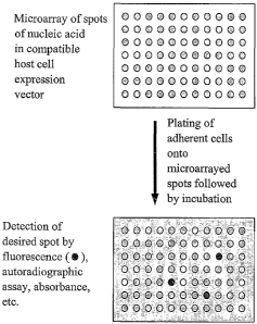
Figure 1 shows a diagram of STEP. An ordered array of nucleic acids
(preferably
cDNA clones in eukaryotic expression vectors) is immobilized to a surface,
adherent cells
are plated onto the nucleic acid array, and following STEP transfection the
transfected cells
are assayed for effects of expression of the transfected nucleic acid.
Figure 2 shows a detection of STEP transfected cells from DNA arrays spotted
with
-9-
CA 02427916 2003-05-02
WO 02/42447 PCT/US01/50426
a robotic microarray spotter.
Figure 3 shows the pathway of the activation of the dopamine 1 (Dl) re by Cl-
APB
coupled to adenylate cyclase and subsequent generation of cyclic AMP.
Figure 4 shows the transfection of two cell types where the adenoviral protein
penton is used as a complexing agent in the transfection complexes.
DEFINITIONS
To facilitate an understanding of the present invention, a number of terms and
phrases as used herein are defined below:
The term "protein kinase" refers to, proteins that catalyze the addition of a
phosphate
group from a nucleoside triphosphate to an amino acid side chain in a protein.
Kinases
comprise the largest known enzyme superfamily and vary widely in their target
proteins.
Kinases may be categorized as protein tyrosine kinases (PTKs), which
phosphorylate
tyrosine residues, and protein serine/threonine kinases (STKs), which
phosphorylate serine
and/or threonine residues. Some kinases have dual specificity for both
serine/threonine and
tyrosine residues. Almost all kinases contain a conserved 250-300 amino acid
catalytic
domain. This domain can be further divided into 11 subdomains. N-terminal
subdomains
I-IV fold into a two-lobed structure which binds and orients the ATP donor
molecule, and
subdomain V spans the two lobes. C-terminal subdomains VI-XI bind the protein
substrate
and transfer the gamma phosphate from ATP to the hydroxyl group of a serine,
threonine, or
tyrosine residue. Each of the 11 subdomains contains specific catalytic
residues or amino
acid motifs characteristic of that subdomain. For example, subdomain I
contains an
8-amino acid glycine-rich ATP binding consensus motif, subdomain II contains a
critical
lysine residue required for maximal catalytic activity, and subdomains VI
through IX
comprise the highly conserved catalytic core. STKs and PTKs also contain
distinct
sequence motifs in subdomains VI and VIII which may confer hydroxyamino acid
specificity. Some STKs and PTKs possess structural characteristics of both
families. In
addition, kinases may also be classified by additional amino acid sequences,
generally
between 5 and 100 residues, which either flank or occur within the kinase
domain.
Non-transmembrane PTKs form signaling complexes with the cytosolic domains of
plasma membrane receptors. Receptors that signal through non-transmembrane
PTKs
include cytokine, hormone, and antigen-specific lymphocytic receptors. Many
PTKs were
first identified as oncogene products in cancer cells in which PTK activation
was no longer
subject to normal cellular controls. In fact, about one third of the known
oncogenes encode
-10-
CA 02427916 2003-05-02
WO 02/42447 PCT/US01/50426
PTKs. Furthermore, cellular transformation (oncogenesis) is often accompanied
by
increased tyrosine phosphorylation activity (See, e.g., Carbonneau, H. and
Tonks, Annu.
Rev. Cell Biol. 8:463-93 (1992)). Regulation of PTK activity may therefore be
an
important strategy in controlling some types of cancer.
The terms "protein" and "polypeptide" refer to compounds comprising amino
acids
joined via peptide bonds and are used interchangeably.
As used herein, where "amino acid sequence" is recited herein to refer to an
amino
acid sequence of a protein molecule. An "amino acid sequence" can be deduced
from the
nucleic acid sequence encoding the protein. However, terms such as
"polypeptide" or
"protein" are not meant to limit the amino acid sequence to the deduced amino
acid
sequence, but include post-translational modifications of the deduced amino
acid sequences,
such as amino acid deletions, additions, and modifications such as
glycolsylations and
addition of lipid moieties.
The term "portion" when used in reference to a protein (as in "a portion of a
given
protein") refers to fragments of that protein. The fragments may range in size
from four
amino acid residues to the entire amino sequence minus one amino acid.
The term "chimera" when used in reference to a polypeptide refers to the
expression
product of two or more coding sequences obtained from different genes, that
have been
cloned together and that, after translation, act as a single polypeptide
sequence. Chimeric
polypeptides are also referred to as "hybrid" polypeptides. The coding
sequences includes
those obtained from the same or from different species of organisms.
The term "fusion" when used in reference to a polypeptide refers to a chimeric
protein containing a protein of interest joined to an exogenous protein
fragment (the fusion
partner). The fusion partner may serve various functions, including
enhancement of
solubility of the polypeptide of interest, as well as providing an "affinity
tag" to allow
purification of the recombinant fusion polypeptide from a host cell or from a
supernatant or
from both. If desired, the fusion partner may be removed from the protein of
interest after
or during purification.
The term "homolog" or "homologous" when used in reference to a polypeptide
refers
to a high degree of sequence identity between two polypeptides, or to a high
degree of
similarity between the three-dimensional structure or to a high degree of
similarity between
the active site and the mechanism of action. In a preferred embodiment, a
homolog has a
greater than 60% sequence identity, and more preferably greater than 75%
sequence
identity, and still more preferably greater than 90% sequence identity, with a
reference
-11-
CA 02427916 2003-05-02
WO 02/42447 PCT/US01/50426
sequence.
As applied to polypeptides, the term "substantial identity" means that two
peptide
sequences, when optimally aligned, such as by the programs GAP or BESTFIT
using
default gap weights, share at least 80 percent sequence identity, preferably
at least 90
percent sequence identity, more preferably at least 95 percent sequence
identity or more
(e.g., 99 percent sequence identity). Preferably, residue positions which are
not identical
differ by conservative amino acid substitutions.
The terms "variant" and "mutant" when used in reference to a polypeptide refer
to an
amino acid sequence that differs by one or more amino acids from another,
usually related
polypeptide. The variant may have "conservative" changes, wherein a
substituted amino
acid has similar structural or chemical properties. One type of conservative
amino acid
substitutions refers to the interchangeability of residues having similar side
chains. For
example, a group of amino acids having aliphatic side chains is glycine,
alanine, valine,
leucine, and isoleucine; a group of amino acids having aliphatic-hydroxyl side
chains is
serine and threonine; a group of amino acids having amide-containing side
chains is
asparagine and glutamine; a group of amino acids having aromatic side chains
is
phenylalanine, tyrosine, and tryptophan; a group of amino acids having basic
side chains is
lysine, arginine, and histidine; and a group of amino acids having sulfur-
containing side
chains is cysteine and methionine. Preferred conservative amino acids
substitution groups
are: valine-leucine-isoleucine, phenylalanine-tyrosine, lysine-arginine,
alanine-valine, and
asparagine-glutamine. More rarely, a variant may have "non-conservative"
changes (e.g.,
replacement of a glycine with a tryptophan). Similar minor variations may also
include
amino acid deletions or insertions (in other words, additions), or both.
Guidance in
determining which and how many amino acid'residues may be substituted,
inserted or
deleted without abolishing biological activity may be found using computer
programs well
known in the art, for example, DNAStar software. Variants can be tested in
functional
assays. Preferred variants have less than 10%, and preferably less than 5%,
and still more
preferably less than 2% changes (whether substitutions, deletions, and so on).
The term "gene" refers to a nucleic acid (e.g., DNA or RNA) sequence that
comprises coding sequences necessary for the production of an RNA, or a
polypeptide or its
precursor (e.g., proinsulin). A functional polypeptide can be encoded by a
full length
coding sequence or by any portion of the coding sequence as long as the
desired activity or
functional properties (e.g., enzymatic activity, ligand binding, signal
transduction, etc.) of
the polypeptide are retained. The term "portion" when used in reference to a
gene refers to
-12-
CA 02427916 2003-05-02
WO 02/42447 PCT/US01/50426
fragments of that gene. The fragments may range in size from a few nucleotides
to the
entire gene sequence minus one nucleotide. Thus, "a nucleotide comprising at
least a
portion of a gene" may comprise fragments of the gene or the entire gene.
The term "gene" also encompasses the coding regions of a structural gene and
includes sequences located adjacent to the coding region on both the 5' and 3'
ends for a
distance of about 1 kb on either end such that the gene corresponds to the
length of the
full-length mRNA. The sequences which are located 5' of the coding region and
which are
present on the mRNA are referred to as 5' non-translated sequences. The
sequences which
are located 3' or downstream of the coding region and which are present on the
mRNA are
referred to as 3' non-translated sequences. The term "gene" encompasses both
eDNA and
genomic forms of a gene. A genomic form or clone of a gene contains the coding
region
interrupted with non-coding sequences termed "introns" or "intervening
regions" or
"intervening sequences." Introns are segments of a gene which are transcribed
into nuclear
RNA (hnRNA); introns may contain regulatory elements such as enhancers.
Introns are
removed or "spliced out" from the nuclear or primary transcript; introns
therefore are absent
in the messenger RNA (mRNA) transcript. The mRNA functions during translation
to
specify the sequence or order of amino acids in a nascent polypeptide.
In addition to containing introns, genomic forms of a gene may also include
sequences located on both the 5' and 3' end of the sequences which are present
on the RNA
transcript. These sequences are referred to as "flanking" sequences or regions
(these
flanking sequences are located 5' or 3' to the non-translated sequences
present on the mRNA
transcript). The 5' flanking region may contain regulatory sequences such as
promoters and
enhancers which control or influence the transcription of the gene. The 3'
flanking region
may contain sequences which direct the termination of transcription,
posttranscriptional
cleavage and polyadenylation.
The term "heterologous gene" refers to a gene encoding a factor that is not in
its
natural environment (i.e., has been altered by the hand of man). For example,
a
heterologous gene includes a gene from one species introduced into another
species. A
heterologous gene also includes a gene native to an organism that has been
altered in some
way (e.g., mutated, added in multiple copies, linked to a non-native promoter
or enhancer
sequence, etc.). Heterologous genes may comprise plant gene sequences that
comprise
cDNA forms of a plant gene; the cDNA sequences may be expressed in either a
sense (to
produce inRNA) or anti-sense orientation (to produce an anti-sense RNA
transcript that is
complementary to the mRNA transcript). Heterologous genes are distinguished
from
-13-
CA 02427916 2003-05-02
WO 02/42447 PCT/US01/50426
endogenous plant genes in that the heterologous gene sequences are typically
joined to
nucleotide sequences comprising regulatory elements such as promoters that are
not found
naturally associated with the gene for the protein encoded by the heterologous
gene or with
plant gene sequences in the chromosome, or are associated with portions of the
chromosome
not found in nature (e.g., genes expressed in loci where the gene is not
normally expressed).
The term "polynucleotide" refers to a molecule comprised of two or more
deoxyribonucleotides or ribonucleotides, preferably more than three, and
usually more than
ten. The exact size will depend on many factors, which in turn depends on the
ultimate
function or use of the oligonucleotide. The polynucleotide may be generated in
any manner,
including chemical synthesis, DNA replication, reverse transcription, or a
combination
thereof. The term "oligonucleotide" generally refers to a short length of
single-stranded
polynucleotide chain usually less than 30 nucleotides long, although it may
also be used
interchangeably with the term "polynucleotide."
The term "nucleic acid" refers to a polymer of nucleotides, or a
polynucleotide, as
described above. The term is used to designate a single molecule, or a
collection of
molecules. Nucleic acids may be single stranded or double stranded, and may
include
coding regions and regions of various control elements, as described below.
The term "a polynucleotide having a nucleotide sequence encoding a gene" or ",
a
polynucleotide having a nucleotide sequence encoding a gene " or "a nucleic
acid sequence
encoding" a specified polypeptide refers to a nucleic acid sequence comprising
the coding
region of a gene or in other words the nucleic acid sequence which encodes a
gene product.
The coding region may be present in either a cDNA, genomic DNA or RNA form.
When
present in a DNA form, the oligonucleotide, polynucleotide, or nucleic acid
may be
single-stranded (i.e., the sense strand) or double-stranded. Suitable control
elements such as
enhancers/promoters, splice junctions, polyadenylation signals, etc. may be
placed in close
proximity to the coding region of the gene if needed to permit proper
initiation of
transcription and/or correct processing of the primary RNA transcript.
Alternatively, the
coding region utilized in the expression vectors of the present invention may
contain
endogenous enhancers/promoters, splice junctions, intervening sequences,
polyadenylation
signals, etc. or a combination of both endogenous and exogenous control
elements.
The term "recombinant" when made in reference to a nucleic acid molecule
refers to
a nucleic acid molecule which is comprised of segments of nucleic acid joined
together by
means of molecular biological techniques. The term "recombinant" when made in
reference
to a protein or a polypeptide refers to a protein molecule which is expressed
using a
-14-
CA 02427916 2003-05-02
WO 02/42447 PCT/US01/50426
recombinant nucleic acid molecule.
The terms "complementary" and "complementarity" refer to polynucleotides
(i.e., a
sequence of nucleotides) related by the base-pairing rules. For example, for
the sequence
"A-G-T," is complementary to the sequence "T-C-A." Complementarity may be
"partial," in
which only some of the nucleic acids' bases are matched according to the base
pairing rules.
Or, there may be "complete" or "total" complementarity between the nucleic
acids. The
degree of complementarity between nucleic acid strands has significant effects
on the
efficiency and strength of hybridization between nucleic acid strands. This is
of particular
importance in amplification reactions, as well as detection methods which
depend upon
binding between nucleic acids.
The term "homology" when used in relation to nucleic acids refers to a degree
of
complementarity. There may be partial homology or complete homology (i.e.,
identity).
"Sequence identity" refers to a measure of relatedness between two or more
nucleic acids or
proteins, and is given as a percentage with reference to the total comparison
length. The
identity calculation takes into account those nucleotide or amino acid
residues that are
identical and in the same relative positions in their respective larger
sequences.
Calculations of identity may be performed by algorithms contained within
computer
programs such as "GAP" (Genetics Computer Group, Madison, Wis.) and "ALIGN"
(DNAStar, Madison, Wis.). A partially complementary sequence is one that at
least partially
inhibits (or competes with) a completely complementary sequence from
hybridizing to a
target nucleic acid is referred to using the functional term "substantially
homologous." The
inhibition of hybridization of the completely complementary sequence to the
target
sequence may be examined using a hybridization assay (Southern or Northern
blot, solution
hybridization and the like) under conditions of low stringency. A
substantially homologous
sequence or probe will compete for and inhibit the binding (i.e., the
hybridization) of a
sequence which is completely homologous to a target under conditions of low
stringency.
This is not to say that conditions of low stringency are such that non-
specific binding is
permitted; low stringency conditions require that the binding of two sequences
to one
another be a specific (i.e., selective) interaction. The absence of non-
specific binding may
be tested by the use of a second target which lacks even a partial degree of
complementarity
(e.g., less than about 30% identity); in the absence of non-specific binding
the probe will not
hybridize to the second non-complementary target.
The following terms are used to describe the sequence relationships between
two or
more polynucleotides: "reference sequence", "sequence identity", "percentage
of sequence
-15-
CA 02427916 2003-05-02
WO 02/42447 PCT/US01/50426
identity", and "substantial identity". A "reference sequence" is a defined
sequence used as a
basis for a sequence comparison; a reference sequence may be a subset of a
larger sequence,
for example, as a segment of a full-length cDNA sequence given in a sequence
listing or
may comprise a complete gene sequence. Generally, a reference sequence is at
least 20
nucleotides in length, frequently at least 25 nucleotides in length, and often
at least 50
nucleotides in length. Since two polynucleotides may each (1) comprise a
sequence (i.e., a
portion of the complete polynucleotide sequence) that is similar between the
two
polynucleotides, and (2) may further comprise a sequence that is divergent
between the two
polynucleotides, sequence comparisons between two (or more) polynucleotides
are typically
performed by comparing sequences of the two polynucleotides over a "comparison
window"
to identify and compare local regions of sequence similarity. A "comparison
window", as
used herein, refers to a conceptual segment of at least 20 contiguous
nucleotide positions
wherein a polynucleotide sequence may be compared to a reference sequence of
at least 20
contiguous nucleotides and wherein the portion of the polynucleotide sequence
in the
comparison window may comprise additions or deletions (i.e., gaps) of 20
percent or less as
compared to the reference sequence (which does not comprise additions or
deletions) for
optimal alignment of the two sequences. Optimal alignment of sequences for
aligning a
comparison window may be conducted by the local homology algorithm of Smith
and
Waterman (Smith and Waterman, Adv. Appl. Math. 2: 482 (1981)) by the homology
alignment algorithm of Needleman and Wunsch (Needleman and Wunsch, J. Mol.
Biol.
48:443 (1970)), by the search for similarity method of Pearson and Lipman
(Pearson and
Lipman, Proc. Natl. Acad. Sci. (U.S.A.) 85:2444 (1988)), by computerized
implementations
of these algorithms (GAP, BESTFIT, FASTA, and TFASTA in the Wisconsin Genetics
Software Package Release 7.0, Genetics Computer Group, 575 Science Dr.,
Madison, Wis.),
or by inspection, and the best alignment (i.e., resulting in the highest
percentage of
homology over the comparison window) generated by the various methods is
selected. The
term "sequence identity" means that two polynucleotide sequences are identical
(i.e., on a
nucleotide-by-nucleotide basis) over the window of comparison. The term
"percentage of
sequence identity" is calculated by comparing two optimally aligned sequences
over the
window of comparison, determining the number of positions at which the
identical nucleic
acid base (e.g., A, T, C, G, U, or I) occurs in both sequences to yield the
number of matched
positions, dividing the number of matched positions by the total number of
positions in the
window of comparison (i.e., the window size), and multiplying the result by
100 to yield the
percentage of sequence identity. The terms "substantial identity" as used
herein denotes a
-16-
CA 02427916 2003-05-02
WO 02/42447 PCT/USO1/50426
characteristic of a polynucleotide sequence, wherein the polynucleotide
comprises a
sequence that has at least 85 percent sequence identity, preferably at least
90 to 95 percent
sequence identity, more usually at least 99 percent sequence identity as
compared to a
reference sequence over a comparison window of at least 20 nucleotide
positions, frequently
over a window of at least 25-50 nucleotides, wherein the percentage of
sequence identity is
calculated by comparing the reference sequence to the polynucleotide sequence
which may
include deletions or additions which total 20 percent or less of the reference
sequence over
the window of comparison. The reference sequence may be a subset of a larger
sequence,
for example, as a segment of the full-length sequences of the compositions
claimed in the
present invention.
When used in reference to a double-stranded nucleic acid sequence such as a
cDNA
or genomic clone, the term "substantially homologous" refers to any probe that
can
hybridize to either or both strands of the double-stranded nucleic acid
sequence under
conditions of low to high stringency as described above.
When used in reference to a single-stranded nucleic acid sequence, the term
"substantially homologous" refers to any probe that can hybridize (i.e., it is
the complement
of) the single-stranded nucleic acid sequence under conditions of low to high
stringency as
described above.
The term "hybridization" refers to the pairing of complementary nucleic acids.
Hybridization and the strength of hybridization (i.e., the strength of the
association between
the nucleic acids) is impacted by such factors as the degree of complementary
between the
nucleic acids, stringency of the conditions involved, the T., of the formed
hybrid, and the
G:C ratio within the nucleic acids. A single molecule that contains pairing of
complementary nucleic acids within its structure is said to be "self-
hybridized."
The term "T,,," refers to the "melting temperature" of a nucleic acid. The
melting
temperature is the temperature at which a population of double-stranded
nucleic acid
molecules becomes half dissociated into single strands. The equation for
calculating the Tl,
of nucleic acids is well known in the art. As indicated by standard
references, a simple
estimate of the T1, value may be calculated by the equation: T= 81.5 + 0.41(%
G + C),
when a nucleic acid is in aqueous solution at 1 M NaCl (See e.g., Anderson and
Young,
Quantitative Filter Hybridization, in Nucleic Acid Hybridization (1985)).
Other references
include more sophisticated computations that take structural as well as
sequence
characteristics into account for the calculation of T..
As used herein the term "stringency" refers to the conditions of temperature,
ionic
-17-
CA 02427916 2003-05-02
WO 02/42447 PCT/US01/50426
strength, and the presence of other compounds such as organic solvents, under
which
nucleic acid hybridizations are conducted. With "high stringency" conditions,
nucleic acid
base pairing will occur only between nucleic acid fragments that have a high
frequency of
complementary base sequences. Thus, conditions of "low" stringency are often
required
with nucleic acids that are derived from organisms that are genetically
diverse, as the
frequency of complementary sequences is usually less.
"Low stringency conditions" when used in reference to nucleic acid
hybridization
comprise conditions equivalent to binding or hybridization at 42 C in a
solution consisting
of 5X SSPE (43.8 g/l NaCl, 6.9 g/l NaH2PO4=H20 and 1.85 g/l EDTA, pH adjusted
to 7.4
with NaOH), 0.1% SDS, 5X Denhardt's reagent [50X Denhardt's contains per 500
ml: 5 g
Ficoll (Type 400, Pharmacia), 5 g BSA (Fraction V; Sigma)) and 100pg/ml
denatured
salmon sperm DNA followed by washing in a solution comprising 5X SSPE, 0.1%
SDS at
42 C when a probe of about 500 nucleotides in length is employed.
"Medium stringency conditions" when used in reference to nucleic acid
hybridization comprise conditions equivalent to binding or hybridization at 42
C in a
solution consisting of 5X SSPE (43.8 g/l NaCl, 6.9 g/l NaH2PO4=H20 and 1.85
g/l EDTA,
pH adjusted to 7.4 with NaOH), 0.5% SDS, 5X Denhardt's reagent and 100 ig/m1
denatured
salmon sperm DNA followed by washing in a solution comprising 1.OX SSPE, 1.0%
SDS at
42 C when a probe of about 500 nucleotides in length is employed.
"High stringency conditions" when used in reference to nucleic acid
hybridization
comprise conditions equivalent to binding or hybridization at 42 C in a
solution consisting
of 5X SSPE (43.8 g/l NaCl, 6.9 g/l NaH2PO4=H20 and 1.85 g/l EDTA, pH adjusted
to 7.4
with NaOH), 0.5% SDS, 5X Denhardt's reagent and 100 ig/ml denatured salmon
sperm
DNA followed by washing in a solution comprising O.1X SSPE, 1.0% SDS at 42 C
when a
probe of about 500 nucleotides in length is employed.
It is well known that numerous equivalent conditions may be employed to
comprise
low stringency conditions; factors such as the length and nature (DNA, RNA,
base
composition) of the probe and nature of the target (DNA, RNA, base
composition, present
in solution or immobilized, etc.) and the concentration of the salts and other
components
(e.g., the presence or absence of formamide, dextran sulfate, polyethylene
glycol) are
considered and the hybridization solution may be varied to generate conditions
of low
stringency hybridization different from, but equivalent to, the above listed
conditions. In
addition, the art knows conditions that promote hybridization under conditions
of high
stringency (e. g., increasing the temperature of the hybridization and/or wash
steps, the use
-18-
CA 02427916 2003-05-02
WO 02/42447 PCT/US01/50426
of formamide in the hybridization solution, etc.).
"Amplification" is a special case of nucleic acid replication involving
template
specificity. It is to be contrasted with non-specific template replication (i.
e., replication that
is template-dependent but not dependent on a specific template). Template
specificity is
here distinguished from fidelity of replication (i.e., synthesis of the proper
polynucleotide
sequence) and nucleotide (ribo- or deoxyribo-) specificity. Template
specificity is
frequently described in terms of "target" specificity. Target sequences are
"targets" in the
sense that they are sought to be sorted out from other nucleic acid.
Amplification
techniques have been designed primarily for this sorting out.
Template specificity is achieved in most amplification techniques by the
choice of
enzyme. Amplification enzymes are enzymes that, under conditions they are
used, will
process only specific sequences of nucleic acid in a heterogeneous mixture of
nucleic acid.
For example, in the case of Q_ replicase, MDV-1 RNA is the specific template
for the
replicase (Kacian et al., Proc. Natl. Acad. Sci. USA, 69:3038 (1972)). Other
nucleic acid
will not be replicated by this amplification enzyme. Similarly, in the case of
T7 RNA
polymerase, this amplification enzyme' has a stringent specificity for its own
promoters
(Chamberlin et al., Nature, 228:227 (1970)). In the case of T4 DNA ligase, the
enzyme will
not ligate the two oligonucleotides or polynucleotides, where there is a
mismatch between
the oligonucleotide or polynucleotide substrate and the template at the
ligation junction (Wu
and Wallace, Genomics, 4:560 (1989)). Finally, Taq and Pfu polymerases, by
virtue of their
ability to function at high temperature, are found to display high specificity
for the
sequences bounded and thus defined by the primers; the high temperature
results in
thermodynamic conditions that favor primer hybridization with the target
sequences and not
hybridization with non-target sequences (H.A. Erlich (ed.), PCR Technology,
Stockton
Press (1989)).
The term "amplifiable nucleic acid" refers to nucleic acids that may be
amplified by
any amplification method. It is contemplated that "amplifiable nucleic acid"
will usually
comprise "sample template."
The term "sample template" refers to nucleic acid originating from a sample
that is
analyzed for the presence of "target" (defined below). In contrast,
"background template" is
used in reference to nucleic acid other than sample template that may or may
not be present
in a sample. Background template is most often inadvertent. It may be the
result of
carryover, or it may be due to the presence of nucleic acid contaminants
sought to be
purified away from the sample. For example, nucleic acids from organisms other
than those
-19-
CA 02427916 2003-05-02
WO 02/42447 PCT/US01/50426
to be detected may be present as background in a test sample.
The term "primer" refers to an oligonucleotide, whether occurring naturally as
in a
purified restriction digest or produced synthetically, which is capable of
acting as a point of
initiation of synthesis when placed under conditions in which synthesis of a
primer
extension product which is complementary to a nucleic acid strand is induced,
(i, e., in the
presence of nucleotides and an inducing agent such as DNA polymerase and at a
suitable
temperature and pH). The primer is preferably single stranded for maximum
efficiency in
amplification, but may alternatively be double stranded. If double stranded,
the primer is
first treated to separate its strands before being used to prepare extension
products.
Preferably, the primer is an oligodeoxyribonucleotide. The primer must be
sufficiently long
to prime the synthesis of extension products in the presence of the inducing
agent. The
exact lengths of the primers will depend on many factors, including
temperature, source of
primer and the use of the method.
The term "probe" refers to an oligonucleotide (i.e., a sequence of
nucleotides),
whether occurring naturally as in a purified restriction digest or produced
synthetically,
recombinantly or by PCR amplification, that is capable of hybridizing to
another
oligonucleotide of interest. A probe may be single-stranded or double-
stranded. Probes are
useful in the detection, identification and isolation of particular gene
sequences. It is
contemplated that any probe used in the present invention will be labeled with
any "reporter
molecule," so that is detectable in any detection system, including, but not
limited to
enzyme (e.g., ELISA, as well as enzyme-based histochemical assays),
fluorescent,
radioactive, and luminescent systems. It is not intended that the present
invention be
limited to any particular detection system or label.
The term "target," when used in reference to the polymerase chain reaction,
refers to
the region of nucleic acid bounded by the primers used for polymerase chain
reaction.
Thus, the "target" is sought to be sorted out from other nucleic acid
sequences. A
"segment" is defined as a region of nucleic acid within the target sequence.
The terns "polymerase chain reaction" ("PCR") refers to the method of I.B.
Mullis
U.S. Patent Nos. 4,683,195, 4,683,202, and 4,965,188, that describe a method
for increasing
the concentration of a segment of a target sequence in a mixture of genomic
DNA without
cloning or purification. This process for amplifying the target sequence
consists of
introducing a large excess of two oligonucleotide primers to the DNA mixture
containing
the desired target sequence, followed by a precise sequence of thermal cycling
in the
presence of a DNA polymerase. The two primers are complementary to their
respective
-20-
CA 02427916 2003-05-02
WO 02/42447 PCT/US01/50426
strands of the double stranded target sequence. To effect amplification, the
mixture is
denatured and the primers then annealed to their complementary sequences
within the target
molecule. Following annealing, the primers are extended with a polymerase so
as to form a
new pair of complementary strands. The steps of denaturation, primer
annealing, and
polymerase extension can be repeated many times (i.e., denaturation, annealing
and
extension constitute one "cycle"; there can be numerous "cycles") to obtain a
high
concentration of an amplified segment of the desired target sequence. The
length of the
amplified segment of the desired target sequence is determined by the relative
positions of
the primers with respect to each other, and therefore, this length is a
controllable parameter.
By virtue of the repeating aspect of the process, the method is referred to as
the
"polymerase chain reaction" (hereinafter "PCR"). Because the desired amplified
segments
of the target sequence become the predominant sequences (in terms of
concentration) in the
mixture, they are said to be "PCR amplified."
With PCR, it is possible to amplify a single copy of a specific target
sequence in
genomic DNA to a level detectable by several different methodologies (e.g.,
hybridization
with a labeled probe; incorporation of biotinylated primers followed by avidin-
enzyme
conjugate detection; incorporation of 32P-labeled deoxynucleotide
triphosphates, such as
dCTP or dATP, into the amplified segment). In addition to genomic DNA, any
oligonucleotide or polynucleotide sequence can be amplified with the
appropriate set of
primer molecules. In particular, the amplified segments created by the PCR
process itself
are, themselves, efficient templates for subsequent PCR amplifications.
The terms "PCR product," "PCR fragment," and "amplification product" refer to
the
resultant mixture of compounds after two or more cycles of the PCR steps of
denaturation,
annealing and extension are complete. These terms encompass the case where
there has
been amplification of one or more segments of one or more target sequences.
The term "amplification reagents" refers to those reagents
(deoxyribonucleotide
triphosphates, buffer, etc.), needed for amplification except for primers,
nucleic acid
template, and the amplification enzyme. Typically, amplification reagents
along with other
reaction components are placed and contained in a reaction vessel (test tube,
microwell,
etc.).
The term "reverse-transcriptase" or "RT-PCR" refers to a type of PCR where the
starting material is mRNA. The starting mRNA is enzymatically converted to
complementary DNA or "cDNA" using a reverse transcriptase enzyme. The cDNA is
then
used as a "template" for a "PCR" reaction
-21 -
CA 02427916 2003-05-02
WO 02/42447 PCT/US01/50426
The term "gene expression" refers to the process of converting genetic
information
encoded in a gene into RNA (e.g., mRNA, rRNA, tRNA, or snRNA) through
"transcription"
of the gene (i.e., via the enzymatic action of an RNA polymerase), and into
protein, through
"translation" of mRNA. Gene expression can be regulated at many stages in the
process.
"Up-regulation" or "activation" refers to regulation that increases the
production of gene
expression products (i.e., RNA or protein), while "down-regulation" or
"repression" refers
to regulation that decrease production. Molecules (e.g., transcription
factors) that are
involved in up-regulation or down-regulation are often called "activators" and
"repressors,"
respectively.
The terms "in operable combination", "in operable order" and "operably linked"
refer to the linkage of nucleic acid sequences in such a manner that a nucleic
acid molecule
capable of directing the transcription of a given gene and/or the synthesis of
a desired
protein molecule is produced. The term also refers to the linkage of amino
acid sequences
in such a manner so that a functional protein is produced.
The term "regulatory element" refers to a genetic element which controls some
aspect of the expression of nucleic acid sequences. For example, a promoter is
a regulatory
element which facilitates the initiation of transcription of an operably
linked coding region.
Other regulatory elements are splicing signals, polyadenylation signals,
termination signals,
etc.
Transcriptional control signals in eukaryotes comprise "promoter" and
"enhancer"
elements. Promoters and enhancers consist of short arrays of DNA sequences
that interact
specifically with cellular proteins involved in transcription (Maniatis, et
al., Science
236:1237, 1987). Promoter and enhancer elements have been isolated from a
variety of
eukaryotic sources including genes in yeast, insect, mammalian and plant
cells. Promoter
and enhancer elements have also been isolated from viruses and analogous
control elements,
such as promoters, are also found in prokaryotes. The selection of a
particular promoter and
enhancer depends on the cell type used to express the protein of interest.
Some eukaryotic
promoters and enhancers have a broad host range while others are functional in
a limited
subset of cell types (for review, see Voss, et al., Trends Biochein. Sci.,
11:287, 1986; and
Maniatis, et al., supra 1987).
The terms "promoter element," "promoter," or "promoter sequence" as used
herein,
refer to a DNA sequence that is located at the 5' end (i.e. precedes) the
protein coding region
of a DNA polymer. The location of most promoters known in nature precedes the
-22-
CA 02427916 2003-05-02
WO 02/42447 PCT/US01/50426
transcribed region. The promoter functions as a switch, activating the
expression of a gene.
If the gene is activated, it is said to be transcribed, or participating in
transcription.
Transcription involves the synthesis of mRNA from the gene. The promoter,
therefore,
serves as a transcriptional regulatory element and also provides a site for
initiation of
transcription of the gene into mRNA.
Promoters may be tissue specific or cell specific. The term "tissue specific"
as it
applies to a promoter refers to a promoter that is capable of directing
selective expression of
a nucleotide sequence of interest to a specific type of tissue (e.g., seeds)
in the relative
absence of expression of the same nucleotide sequence of interest in a
different type of
tissue (e.g., leaves). Tissue specificity of a promoter may be evaluated by,
for example,
operably linking a reporter gene to the promoter sequence to generate a
reporter construct,
introducing the reporter construct into the genome of a plant such that the
reporter construct
is integrated into every tissue of the resulting transgenic plant, and
detecting the expression
of the reporter gene (e.g.,-detecting mRNA, protein, or the activity of a
protein encoded by
the reporter gene) in different tissues of the transgenic plant. The detection
of a greater
level of expression of the reporter gene in one or more tissues relative to
the level of
expression of the reporter gene in other tissues shows that the promoter is
specific for the
tissues in which greater levels of expression are detected. The term "cell
type specific" as
applied to a promoter refers to a promoter which is capable of directing
selective expression
of a nucleotide sequence of interest in a specific type of cell in the
relative absence of
expression of the same nucleotide sequence of interest in a different type of
cell within the
same tissue. The term "cell type specific" when applied to a promoter also
means a
promoter capable of promoting selective expression of a nucleotide sequence of
interest in a
region within a single tissue. Cell type specificity of a promoter may be
assessed using
methods well known in the art, e.g., immunohistochemical staining. Briefly,
tissue sections
are embedded in paraffin, and paraffin sections are reacted with a primary
antibody which is
specific for the polypeptide product encoded by the nucleotide sequence of
interest whose
expression is controlled by the promoter. A labeled (e.g., peroxidase
conjugated) secondary
antibody which is specific for the primary antibody is allowed to bind to the
sectioned tissue
and specific binding detected (e.g., with avidin/biotin) by microscopy.
Promoters may be constitutive or regulatable. The term "constitutive" when
made in
reference to a promoter means that the promoter is capable of directing
transcription of an
operably linked nucleic acid sequence in the absence of a stimulus (e.g., heat
shock,
chemicals, light, etc.). Typically, constitutive promoters are capable of
directing expression
- 23 -
CA 02427916 2007-10-26
74667-227
of a transgene in substantially any cell and any tissue. Exemplary
constitutive plan,
promoters include, but are not limited to SD Cauliflower Mosaic Virus (CaMV
SD; see e.g.,
U.S. Pat. No. 5,352,605), mannopine synthase, octopine synthase (ocs),
superpromoter (see e.g., WO 95/14098), and ubi3 (see e.g., Garbarino and
Belknap, Plant Mol. Biol. 24:119-127 (1994)) promoters. Such promoters have
been used
successfully to direct the expression of heterologous nucleic acid sequences
in transformed
plant tissue. _
In contrast, a "regulatable" or "inducible" promoter is one which is capable
of
directing a level of transcription of an operably linked nuclei acid sequence
in the presence
of a stimulus (e.g., heat shock, chemicals, light, etc.) which is different
from the level of
transcription of the operably linked nucleic acid sequence in the absence of
the stimulus.
The enhancer and/or promoter may be "endogenous" or "exogenous" or
"heterologous." An "endogenous" enhancer or promoter is one that is naturally
linked with
a given gene in the genome. An "exogenous" or "heterologous" enhancer or
promoter is one
that is placed in juxtaposition to a gene by means of genetic manipulation
(i.e., molecular
biological techniques) such that transcription of the gene is directed by the
linked enhancer
or promoter. For example, an endogenous. promoter in operable combination with
a first
gene can be isolated, removed, and placed in operable combination with a
second gene,
thereby making it a "heterologous promoter" in operable combination with the
second gene.
A variety of such combinations are contemplated (e.g., the first and second
genes can be
from the same species, or from different species.
The presence of "splicing signals" on an expression vector often results in
higher
levels of expression of the recombinant transcript in eukaryotic host cells.
Splicing signals
mediate the removal ofintrons from the primary RNA transcript and consist of a
splice
donor and acceptor site (Sambrook, et al., Molecular Cloning: A Laboratory
Manual, 2nd
ed., Cold Spring Harbor Laboratory Press, New York (1989) pp. 16.7-16.8). A
commonly
used splice donor and acceptor site is the splice junction from the 16S RNA of
SV40.
Efficient expression of recombinant DNA sequences in eukaryotic cells requires
expression of signals directing the efficient termination and polyadenylation
of the resulting
transcript. Transcription termination signals are generally found downstream
of the
polyadenylation signal and are a few hundred nucleotides in length. The term
"poly(A) site"
or "poly(A) sequence" as used herein denotes a DNA sequence which directs both
the
termination and polyadenylation of the nascent RNA transcript. Efficient
polyadenylation
of the recombinant transcript is desirable, as transcripts lacking a poly(A)
tail are unstable
-24-
CA 02427916 2003-05-02
WO 02/42447 PCT/US01/50426
and are rapidly degraded. The poly(A) signal utilized in an expression vector
may be
"heterologous" or "endogenous." An endogenous poly(A) signal is one that is
found
naturally at the 3' end of the coding region of a given gene in the genome. A
heterologous
poly(A) signal is one which has been isolated from one gene and positioned 3'
to another
gene. A commonly used heterologous poly(A) signal is the SV40 poly(A) signal.
The
SV40 poly(A) signal is contained on a 237 bp BamHl/Bcll restriction fragment
and directs
both termination and polyadenylation (Sambrook, supra, at 16.6-16.7).
The term "vector" refers to nucleic acid molecules that transfer DNA
segment(s)
from one cell to another. The term "vehicle" is sometimes used interchangeably
with
"vector."
The terms "expression vector" or "expression cassette" refer to a recombinant
DNA
molecule containing a desired coding sequence and appropriate nucleic acid
sequences
necessary for the expression of the operably linked coding sequence in a
particular host
organism. Nucleic acid sequences necessary for expression in prokaryotes
usually include a
promoter, an operator (optional), and a ribosome binding site, often along
with other
sequences. Eukaryotic cells are known to utilize promoters, enhancers, and
termination and
polyadenylation signals.
The term "transfection complex" refers to a molecular aggregate of molecules
including nucleic acid that upon entry into cells will result in changes in
gene expression.
The number of nucleic acid molecules and the type of nucleic acid molecules
can be more
than one per aggregate. Typically, a transfection complex comprises nucleic
acid with one
or more complexing agents.
The term "complexing agent" refers to a compound in a transfection complex
other
than nucleic acid whose effect is under examination; typically, such agents
facilitate
transfection with nucleic acid. Some classes of complexing agents bind to
nucleic acids
through electrostatic, hydrophobic, and/or stearic interactions to form a
molecular
aggregate; other classes bind to other molecules. Examples of such agents
include but are
not limited to ligands for receptors, DNA-binding molecules, and membrane
permeable
molecules. Additional complexing agents include but are not limited to
targeting
molecules, transcription molecules, nucleic acid degradation inhibitors, and
cell growth and
integrity modulators.
The term "ligand for receptors" refers to a first molecule, the ligand, which
is able to
bind to a second molecule, such as a protein, sugar, or lipid, which is
associated with a cell
membrane. When used in reference to STEP, the ligand binds to a receptor which
is located
-25-
CA 02427916 2003-05-02
WO 02/42447 PCT/US01/50426
in the plasmalemma and which is endocytosed by the cells; preferably, the
receptor is a
protein. Examples of such ligands include but are not limited to transferrin
and low density
lipoprotein particles, which bind to LDL receptors, and viral proteins that
are known to bind
to integrins. Other examples include but are not limited to other proteins,
carbohydrates,
hormones, small molecules and drugs.
The term "DNA-binding molecules" refers to molecules (e.g. cationic proteins)
which complex with nucleic acid to neutralize its charge and to compact its
size; these
molecules typically bind to nucleic acids through electrostatic, hydrophobic,
and/or stearic
interactions to form a molecular aggregate. DNA binding molecules include but
are not
limited to helix-loop-helix proteins (HLH), zinc finger proteins, DNA
intercalators such as
aromatic molecules, other nucleic acids, heavy metals such as platinum,
antibiotics such as
chromomycin A(3) and mithramycin (MTR), and DNA-binding peptides such as the
DNA-
binding peptide mu from adenovirus. Particularly advantageous DNA-binding
molecules
are cationic proteins.
The term "cationic protein" refers to a protein or polypeptide with an
electrostatic charge of
greater than zero at pH 7 in aqueous solution; it is in contrast to an
"anionic protein" which
is a protein or polypeptide with an electrostatic charge of less than zero
under the same
conditions. In the present invention, a "cationic protein" is a subclass of
"DNA binding
molecules," which is a subclass of "complexing agents."
The term "membrane permeable molecules" refers to molecules which are
permeable
in cell membranes, and which facilitate STEP transfection. While it is not
necessary to
understand the underlying mechanism, and while the invention is not limited to
any
particular mechanism, it is believed that these molecules facilitate
transfection by improving
the transport across the membrane of the nucleic acid in a transfection
complex into a host
cell. Particularly advantageous membrane permeable molecules are cationic
lipids.
The term "cationic lipid" refers to a hydrophobic molecule which is lipid
soluble and
which contains a positively-charged region at pH 7. The present invention
contemplates a
variety of such cationic lipids, including but not limited to LipofectamineTM,
Lipofectin ,
Lipofectamine PlusTM, Cellfectin , and LipofectaseTM (available from Life
Technologies).
In the present invention, a "cationic lipid" is a subclass of "membrane
permeable
molecules," which is a subclass of "complexing agents."
The term "targeting molecules" refers to molecules which target a transfection
complex or portions thereof which contain the nucleic acid to the appropriate
cellular
-26-
CA 02427916 2003-05-02
WO 02/42447 PCT/US01/50426
compartment in which expression or effect of the nucleic acid occurs; for
example, if the
nucleic acid is DNA, the appropriate compartment may be the cell nucleus, a
mitochondria,
or a plastid; if the nucleic acid is RNA, the appropriate compartment may be
the cytoplasm,
a mitochondria, or a plastid. Such molecules include but are not limited to
proteins, for
example the SV-40 T antigen, which contain nuclear localization signals (NLSs)
to direct
the proteins to the nucleus of the cells.
The term "transcription/translation molecules" refers to molecules which
promote
the transcription of DNA or the translation of RNA. Such molecules include but
are not
limited to proteins, which include by way of non-limiting example
transcription factors,
DNA relaxing or unwinding factors (e.g. helicases), and DNA polymerases (e.g.
TFIIA,
TFIID).
The term "nucleic acid degradation inhibitor" refers to molecules that act as
nuclease
inhibitors. Such molecules facilitate STEP by preventing degradation of the
transfected
nucleic acids. Examples of such molecules include but are not limited to
proteins (e.g.
DM122) and non-protein drugs.
The term "cell health and integrity modulators" refer to molecules that
modulate
adherence, growth, proliferation, and/or differentiation of cells; preferably,
such modulation
promotes these characteristics. These molecules facilitate STEP by modulating,
and
preferably promoting, the health and integrity of cells transfected with STEP.
Examples of
such molecules include but are not limited to proteins.
The term "dendrimer" refers to a natural or synthetic branched molecule (e.g.
polypeptides, nucleic acids, or synthetic compounds).
The term "type of nucleic acid" refers to a characteristic or property of a
nucleic acid
that can distinguish it from another nucleic acid, such as a difference in
sequence or in
physical form, such as occurs in different expression vectors, or as occurs
with the presence
of DNA and RNA, or as occurs with the presence of linear and super-coiled DNA,
or as
occurs with the presence of coding regions which encode different proteins, or
as occurs
with the presence of different control elements, or control elements which
differ amongst
themselves.
The term "immobilized" when used in reference to nucleic acid refers to a
spatial
restriction of the nucleic acid on a surface, which restriction prevents the
nucleic acid from
entering the solution in which the surface is located and becoming free in the
solution; it
involves stable complex formation, where the complex comprises the nucleic
acid and
formation of the complex is mediated at least in part by electrostatic
interactions. The term
-27-
CA 02427916 2003-05-02
WO 02/42447 PCT/US01/50426
"stable" when used in reference to a complex comprising nucleic acid refers to
maintenance
of the complex for a period of time, generally for at least 72 to 96 hours in
a solution, such
as tissue or cell culture media. The immobilized transfection complexes are
also stable
when dried upon a surface; the duration of the stability in the dried state is
usually at least
several weeks to several months.
The term "array" refers to a pattern, preferably such that the pattern can be
replicated
and/or detected by an appropriate detector. When used in reference to
immobilized
transfection complexes of the present invention, an array comprises "spots"
containing
immobilized transfection complexes. A spot is the location of a single sample
of
immobilized transfection complexes; a spot may be generated by one or more
applications
of the sample to the location. Although each spot comprises a single sample of
immobilized
transfection complexes, a single sample of transfection complexes may comprise
from one
to more than one type of nucleic acid. Moreover, different spots in an array
may comprise
the same or different transfection complexes; the transfection complexes may
differ in the
complexing agents present, the type of nucleic acid present, or both.
Typically, different
spots differ in the type of nucleic acid present. Thus, an array typically
comprises spots at
least some to most of which comprise different types of nucleic acids.
A "microarray" refers to an array which is limited to a small area. Typically,
such
arrays are limited to no more than about 1 inch by 3 inches, as they are
frequently generated
on microscope slides. Microarrays contain the maximum number of spots which
can be
created within the limits; typically, this number is less for hand-generated
arrays than it is
for robotically or machine-generated arrays. A typical machine-generated array
contains up
to about 10,800 spots.
The term "ordered array" refers to a pattern of spots of the present invention
such
that the spots are located in a pre-determined geometrical arrangement on the
surface; most
often, the geometrical arrangement is grid. The term "random array" refers to
a pattern of
spots of the present invention such that the spots are not located in a pre-
determined
geometrical arrangement on the surface. A random array can be determined by a
mathematical algorithm or by a random number generator.
The term "active transport" refers to a process by which a molecule is
transported
from outside a cell to inside the cell by any mechanism other than liposomal
mediated entry
(as for example of DNA coated with lipids), facilitated diffusion, or passive
diffusion.
Active transport includes endocytosis, particularly receptor-mediated
endocytosis. Agents
which promote the active transport of nucleic acid molecules inside the cells
to aid the
-28-
CA 02427916 2007-10-26
74667-227
transfection process include compiexing agents, including but not limited to
ligands for
receptors, DNA binding molecules, and membrane permeable molecules.
The term "transfection" refers to the introduction of foreign DNA into cells.
Transfection may be accomplished by a variety of means known to the art
including calcium
phosphate-DNA co-precipitation, DEAF-dextran-mediated transfection, polybrene-
mediated
transfection, glass beads, electroporation, microinjection, liposome fusion,
lipofection,
protoplast fusion, viral infection, biolistics (i.e., particle bombardment)
and the like.
particular types of cells. The art is well aware of these numerous
modifications.
The term "stable transfection" or "stably transfected" refers to the
introduction and
integration of foreign DNA into the genome of the transfected cell. The term
"stable
transfectant" refers-to a cell that has stably integrated foreign DNA into the
genomic DNA.
The term "transient transfection" or "transiently transfected" refers to the
introduction of foreign DNA into a cell where the foreign DNA fails to
integrate into the
genome of the transfected cell. The foreign DNA persists in the nucleus of the
transfected
cell for several days. During this time the foreign DNA is subject to the
regulatory controls
that govern the expression of endogenous genes in the chromosomes. The term
"transient
transfectant" refers to cells that have taken up foreign DNA but have failed
to integrate this
DNA.
The term "calcium phosphate co-precipitation" refers to a technique for the
introduction of nucleic acids into a cell. The uptake of nucleic acids by
cells is enhanced
when the nucleic acid is presented as a calcium phosphate-nucleic acid co
precipitate. The
original technique of Graham and van der Eb (Graham and van der Eb, Virol.,
52:456
(1973)), has been modified by several groups to optimize conditions for
The terms "infecting" and "infection" when used with a bacterium refer to co-
incubation of a target biological sample, (e.g., cell, tissue, etc.) with the
bacterium under
conditions such that nucleic acid sequences contained within the bacterium are
introduced
into one or more dells of the target biological sample.
The terms "bombarding, "bombardment," and "biolistic bombardment" refer to the
process of accelerating particles towards a target biological sample (e.g.,
cell, tissue, etc.) to
effect wounding of the cell membrane of a cell in the target biological sample
and/or entry
of the particles into the target biological sample. Methods for biolistic
bombardment are
known in the art (e.g., U.S. Pat. No. 5,584,807), and are commercially
available
(e.g., the helium gas-driven microprojectile accelerator (PDS-1000/He,
BioRad).
-29-
CA 02427916 2003-05-02
WO 02/42447 PCT/US01/50426
The term "microwounding" when made in reference to plant tissue refers to the
introduction of microscopic wounds in that tissue. Microwounding may be
achieved by, for
example, particle bombardment as described herein.
The term "transgene" as used herein refers to a foreign gene that is placed
into an
organism by introducing the foreign gene into newly fertilized eggs or early
embryos. The
term "foreign gene" refers to any nucleic acid (e.g., gene sequence) that is
introduced into
the genome of an animal by experimental manipulations and may include gene
sequences
found in that animal so long as the introduced gene does not reside in the
same location as
does the naturally-occurring gene.
The term "host cell" refers to any cell capable of replicating and/or
transcribing
and/or translating a heterologous gene. Thus, a "host cell" refers to any
eukaryotic or
prokaryotic cell (e.g., bacterial cells such as E. coli, yeast cells,
mammalian cells, avian
cells, amphibian cells, plant cells, fish cells, and insect cells), whether
located in vitro or in
vivo. For example, host cells may be located in a transgenic animal.
The terms "transformants" or "transformed cells" include the primary
transformed
cell and cultures derived from that cell without regard to the number of
transfers. All
progeny may not be precisely identical in DNA content, due to deliberate or
inadvertent
mutations. Mutant progeny that have the same functionality as screened for in
the originally
transformed cell are included in the definition of transformants.
The term "selectable marker" refers to a gene which encodes an enzyme having
an
activity that confers resistance to an antibiotic or drug upon the cell in
which the selectable
marker is expressed, or which confers expression of a trait which can be
detected (e.g..,
luminescence or fluorescence). Selectable markers may be "positive" or
"negative."
Examples of positive selectable markers include the neomycin phosphotrasferase
(NPTII)
gene which confers resistance to G418 and to kanamycin, and the bacterial
hygromycin
phosphotransferase gene (layg), which confers resistance to the antibiotic
hygromycin.
Negative selectable markers encode an enzymatic activity whose expression is
cytotoxic to
the cell when grown in an appropriate selective medium. For example, the HSV-
tk gene is
commonly used as a negative selectable marker. Expression of the HSV-tk gene
in cells
grown in the presence of gancyclovir or acyclovir is cytotoxic; thus, growth
of cells in
selective medium containing gancyclovir or acyclovir selects against cells
capable of
expressing a functional HSV TK enzyme.
The term "reporter gene" refers to a gene encoding a protein that may be
assayed.
Examples of reporter genes include, but are not limited to, luciferase (See,
e.g., deWet et al.,
-30-
CA 02427916 2007-10-26
74667-227
Mol. Cell. Biol. 7:725 (1987) andU.S. Pat Nos.,6,074,859; 5,976,796;
5,674,713; and
5,618,682), green fluorescent protein (e.g., GenBank Accession
Number U43284; a number of GFP variants are commercially
available from ClonTech Laboratories, Palo Alto, CA), chloramphenicol
aeetyltransferase,
(3-galactosidase, alkaline phosphatase, and horse radish peroxidase.
The term "wild-type" when made in reference to a gene refers to a gene which
has
the characteristics of a gene isolated from a naturally occurring source. The
term "wild-
type" when made in reference to a gene product refers to a gene product which
has the
characteristics of a gene product isolated from a naturally occurring source.
The term
"naturally-occurring" as used herein as applied to an object refers to the
fact that an object
can be found in nature. For example, a polypeptide or polynucleotide sequence
that is
present in an organism (including viruses) that can be isolated from a source
in nature and
which has not been intentionally modified by man in the laboratory is
naturally-occurring.
A wild-type gene is that which is most frequently observed in a population and
is thus
arbitrarily designated the "normal" or "wild-type" form of the gene. In
contrast, the term
"modified" or "mutant" when made in reference to a gene or to a gene product
refers,
respectively, to a gene or to a gene product which displays modifications in
sequence and/or
functional properties (i.e., altered characteristics) when compared to the
wild-type gene or
gene product. It is noted that naturally-occurring mutants can be isolated;
these are
identified by the fact that they have altered characteristics when compared to
the wild-type
gene or gene product.
The term "antisense" refers to a deoxyribonucleotide sequence whose sequence
of
deoxyribonucleotide residues is in reverse 5' to 3' orientation in relation to
the sequence of
deoxyribonucleotide residues in a sense strand of a DNA duplex. A "sense
strand" of a
DNA duplex refers to a strand in a DNA duplex which is transcribed by a cell
in its natural
state into a "sense mRNA." Thus an "antisense" sequence is a sequence having
the same
sequence as the non-coding strand in a DNA duplex. The teen "antisense RNA"
refers to a
RNA transcript that is complementary to all or part of a target primary
transcript or mRNA
and that blocks the expression of a target gene by interfering with the
processing, transport
and/or translation of its primary transcript or mRNA. The complementarity of
an antisense
RNA may be with any part of the specific gene transcript, i.e., at the 5' non-
coding
sequence, 3' non-coding sequence, introns, or the coding sequence. In
addition, as used
herein, antisense RNA may contain regions of ribozyme sequences that increase
the efficacy
of antisense RNA to block gene expression. "Ribozyme" refers to a catalytic
RNA and
-31-
CA 02427916 2003-05-02
WO 02/42447 PCT/US01/50426
includes sequence-specific endoribonucleases. "Antisense inhibition" refers to
the
production of antisense RNA transcripts capable of preventing the expression
of the target
protein.
The term "overexpression" refers to the production of a gene product in
transgenic
organisms that exceeds levels of production in normal or non-transformed
organisms. The
term "cosuppression" refers to the expression of a foreign gene which has
substantial
homology to an endogenous gene resulting in the suppression of expression of
both the
foreign and the endogenous gene. As used herein, the term "altered levels"
refers to the
production of gene product(s) in transgenic organisms in amounts or
proportions that differ
from that of normal or non-transformed organisms.
The terms "overexpression" and "overexpressing" and grammatical equivalents,
are
used in reference to levels of mRNA to indicate a level of expression
approximately 3-fold
higher than that typically observed in a given tissue in a control or non-
transgenic animal.
Levels of mRNA are measured using any of a number of teclmiques known to those
skilled
in the art including, but not limited to Northern blot analysis (See, Example
10, for a
protocol for performing Northern blot analysis). Appropriate controls are
included on'the
Northern blot to control for differences in the amount of RNA loaded from each
tissue
analyzed (e.g., the amount of 28S rRNA, an abundant RNA transcript present at
essentially
the same amount in all tissues, present in each sample can be used as a means
of
normalizing or standardizing the RAD50 mRNA-specific signal observed on
Northern
blots).
The terms "Southern blot analysis" and "Southern blot" and "Southern" refer to
the
analysis of DNA on agarose or acrylamide gels in which DNA is separated or
fragmented
according to size followed by transfer of the DNA from the gel to a solid
support, such as
nitrocellulose or a nylon membrane. The immobilized DNA is then exposed to a
labeled
probe to detect DNA species complementary to the probe used. The DNA may be
cleaved
with restriction enzymes prior to electrophoresis. Following electrophoresis,
the DNA may
be partially depurinated and denatured prior to or during transfer to the
solid support.
Southern blots are a standard tool of molecular biologists (J. Sambrook et al.
(1989)
Molecular Cloning: A Laboratory Manual, Cold Spring Harbor Press, NY, pp 9.31-
9.58).
The term "Northern blot analysis" and "Northern blot" and "Northern" as used
herein refer to the analysis of RNA by electrophoresis of RNA on agarose gels
to fractionate
the RNA according to size followed by transfer of the RNA from the gel to a
solid support,
such as nitrocellulose or a nylon membrane. The immobilized RNA is then probed
with a
-32-
CA 02427916 2003-05-02
WO 02/42447 PCT/US01/50426
labeled probe to detect RNA species complementary to the probe used. Northern
blots are a
standard tool of molecular biologists (J. Sambrook, et al. (1989) supra, pp
7.39-7.52).
The terms "Western blot analysis" and "Western blot" and "Western" refers to
the
analysis of protein(s) (or polypeptides) immobilized onto a support such as
nitrocellulose or
a membrane. A mixture comprising at least one protein is first separated on an
acrylamide
gel, and the separated proteins are then transferred from the gel to a solid
support, such as
nitrocellulose or a nylon membrane. The immobilized proteins are exposed to at
least one
antibody with reactivity against at least one antigen of interest. The bound
antibodies may
be detected by various methods, including the use of radiolabeled antibodies.
The term "antigenic determinant" as used herein refers to that portion of an
antigen
that makes contact with a particular antibody (i.e., an epitope). When a
protein or fragment
of a protein is used to immunize a host animal, numerous regions of the
protein may induce
the production of antibodies that bind specifically to a given region or three-
dimensional
structure on the protein; these regions or structures are referred to as
antigenic determinants.
An antigenic determinant may compete with the intact antigen (i.e., the
"immunogen" used
to elicit the immune response) for binding to an antibody.
The term "isolated" when used in relation to a nucleic acid, as in "an
isolated
oligonucleotide" refers to a nucleic acid sequence that is identified and
separated from at
least one contaminant nucleic acid with which it is ordinarily associated in
its natural
source. Isolated nucleic acid is present in a form or setting that is
different from that in
which it is found in nature. In contrast, non-isolated nucleic acids, such as
DNA and RNA,
are found in the state they exist in nature. For example, a given DNA sequence
(e.g., a
gene) is found on the host cell chromosome in proximity to neighboring genes;
RNA
sequences, such as a specific mRNA sequence encoding a specific protein, are
found in the
cell as a mixture with numerous other mRNA s which encode a multitude of
proteins.
However, isolated nucleic acid encoding a particular protein includes, by way
of example,
such nucleic acid in cells ordinarily expressing the protein, where the
nucleic acid is in a
chromosomal location different from that of natural cells, or is otherwise
flanked by a
different nucleic acid sequence than that found in nature. The isolated
nucleic acid or
oligonucleotide may be present in single-stranded or double-stranded form.
When an
isolated nucleic acid or oligonucleotide is to be utilized to express a
protein, the
oligonucleotide will contain at a minimum the sense or coding strand (i.e.,
the
oligonucleotide may single-stranded), but may contain both the sense and anti-
sense strands
(i.e., the oligonucleotide may be double-stranded).
-33-
CA 02427916 2003-05-02
WO 02/42447 PCT/US01/50426
The terns "purified" refers to molecules, either nucleic or amino acid
sequences, that
are removed from their natural environment, isolated or separated. An
"isolated nucleic acid
sequence" is therefore a purified nucleic acid sequence. "Substantially
purified" molecules
are at least 60% free, preferably at least 75% free, and more preferably at
least 90% free
from other components with which they are naturally associated. As used
herein, the term
"purified" or "to purify" also refers to the removal of contaminants from a
sample. The
removal of contaminating proteins results in an increase in the percent of
polypeptide of
interest in the sample. In another example, recombinant polypeptides are
expressed in plant,
bacterial, yeast, or mammalian host cells and the polypeptides are purified by
the removal of
host cell proteins; the percent of recombinant polypeptides is thereby
increased in the
sample.
The term "sample" is used in its broadest sense. In one sense it can refer to
a plant
cell or tissue. In another sense, it is meant to include a specimen or culture
obtained from
any source, as well as biological and environmental samples. Biological
samples may be
obtained from plants or animals (including humans) and encompass fluids,
solids, tissues,
and gases. Environmental samples include environmental material such as
surface matter,
soil, water, and industrial samples. These examples are not to be construed as
limiting the
sample types applicable to the present invention. The term "sample" is used in
its broadest
sense. In one sense it can refer to a biopolymeric material. In another sense,
it is meant to
include a specimen or culture obtained from any source, as well as biological
and
environmental samples. Biological samples may be obtained from animals
(including
humans) and encompass fluids, solids, tissues, and gases. Biological samples
include blood
products, such as plasma, serum and the like. Environmental samples include
environmental material such as surface matter, soil, water, crystals and
industrial samples.
These examples are not to be construed as limiting the sample types applicable
to the
present invention.
DETAILED DESCRIPTION OF THE INVENTION
The present invention provides a method of cell transfection, and in
particular to the
application of cells to nucleic acids which are immobilized on a surface and
which then
transfect the cells. In one aspect, the method of the present invention
comprises the
provision of nucleic acids immobilized on a surface; in another aspect, the
invention
comprises immobilizing nucleic acids on a surface. The nucleic acids are
immobilized in a
-34-
CA 02427916 2003-05-02
WO 02/42447 PCT/US01/50426
transfection complex comprising the nucleic acid and at least one complexing
agent.
Preferably, the complexing agents comprise ligands for a cell receptor which
is endocytosed
by the cell and DNA binding molecules; even more preferably, the transfection
complex
comprises at least two complexing agents, which are a ligand for a cell
receptor which is
endocytosed by the cell and a DNA binding molecule. The complexing agents may
further
comprise membrane permeable molecules which facilitate the passage of DNA
complexes
across cellular membranes. Additional agents which are optionally present in
the
transfection complex include targeting molecules that direct the complex or a
portion of the
complex containing the nucleic acid to an appropriate cell compartment where
the nucleic
acid can be expressed, transcription molecules that enhance transcription of
the DNA,
nucleic acid degradation inhibitors, which are molecules that inhibit nucleic
acid
degradation, and cell health and integrity modulators, which are molecules
which modulate
and preferably enhance or promote the adherence, growth, proliferation, and/or
differentiation of the cells. Thus, in other embodiments, the invention
provides transfection
complexes, and methods of forming transfection complexes. In yet other
embodiments, the
nucleic acids are immobilized in an array; preferably, the array is a
microarray. In some
embodiments, the array is an ordered array; in other embodiments, the array is
a random
array. In another aspect of the present invention, the method further
comprises expression
of the nucleic acids in the transfected cells. In yet another aspect of the
present invention,
the method further comprises detecting the expression of the nucleic acids in
the transfected
cells. The invention in its different aspects is referred to as Surface
Transfection and
Expression Procedure (or "STEP"). Additional aspects and details are as
follows; in the
following description, when the word "DNA" is used, it is used as an example
of nucleic
acids which may be used in the method of the present invention, and is not
meant to be
limiting.
The STEP method of the present invention represents an improvement over other
forms of transfection. In STEP, nucleic acids are complexed, and the complexes
are applied
to and immobilized on the surface on which cells are plated or to which cells
are exposed.
The cells thus contact nucleic acid in an immobilized state. This is in
contrast to other
methods of transfection, in which nucleic acids are applied to the media in
which cells are
grown, or are free in the media in which cells are grown. In these other
methods, the cells
contact nucleic acid which is free in solution. STEP thus allows transfection
of cells at the
same location where nucleic acid is immobilized. Because the nucleic acids are
spatially
restricted, with STEP it is possible achieve independent transfections of as
many different
-35-
CA 02427916 2003-05-02
WO 02/42447 PCT/US01/50426
nucleic acids as can be immobilized on a single surface. Because the nucleic
acids are
spatially restricted, with STEP it is possible to replicate part or all of any
particular array of
immobilized nucleic acid, as many times as is desired.
Thus, for example, in one aspect of the present invention, STEP is similar to
current
uses of DNA microarrays in that with STEP DNA can be applied to a surface
(such as a
glass slide) utilizing the same robotic devices as are currently used to
generate DNA
microarrays. Furthermore, for many STEP applications, the same fluorescence
slide
scanners can be used to quantitate experimental results. However, this is
where the
similarities end. The DNA applied to the surface, such as a glass slide, is
not used in vitro
for hybridization, as is the case for current uses of DNA microarrays.
Instead, in STEP, the
DNA applied to and immobilized on a surface is used to transfect live cells to
alter the
expression or function of proteins within the cells. It is the actual
expression or altered.
function of the proteins within the cells that is detected. Moreover, the DNA
is immobilized
as a transfection complex, which complex comprises both nucleic acid and at
least one
complexing agent; such complexing agents typically facilitate DNA transfection
and
expression. In some preferred embodiments, at least one complexing agent
comprises
ligands for a cell receptor which is endocytosed by the cell; in other
preferred embodiments,
at least a further complexing agent comprises DNA binding proteins;
preferably, the
transfection complex comprises both ligands for cell receptors and DNA binding
molecules.
The method of the present invention has the capacity to functionally screen
over
10,000 cDNAs on a single microscope slide (such a slide is typically though
not necessarily
mm x75 mm). It offers several advantages, including but not limited to that of
economy
of scale, that for many applications it allows continuous monitoring of
function in living
cells, that it is easily and completely automated, and that replication is
easily accomplished.
25 Although STEP is very simple, it is believed that the cellular processes of
STEP
encompass several aspects. Although it is not necessary to understand the
mechanism in
order to use the present invention, and it is not intended that the invention
be so limited, a
number of hypotheses are presented to explain the observed results. These
hypotheses are
presented as beliefs or thoughts. Thus, in STEP, it is believed that the first
aspect is cellular
adherence to the surface to which nucleic acids are immobilized. Some cationic
complexing
agents promote the immediate attachment of cells to the immobilized nucleic
acid in a
transfection complex, while others actually repel the cells. DNA alone,
without any
complexing agent, repels the cells, as do complexes with low molar ratios of
complexing
agent to DNA. The second aspect is believed to be cell survival. Some
complexing agents
-36-
CA 02427916 2003-05-02
WO 02/42447 PCT/US01/50426
appear to show cytotoxic effects, even if cells can adhere immediately. This
toxicity is
particularly true of certain membrane permeable molecules, such as the
lipophilic
transfection reagents such as Fugene and Lipofectamine in their pure state.
These two
particular reagents are commonly used in solution for conventional
transfection procedures,
and are not reported to be toxic at the concentrations used under these
conditions. However,
when employed in STEP, these reagents are toxic when used at high
concentrations and
dried before the cells are applied; they can, however, be used at lower
concentrations. The
third aspect is believed to be actual transfection of the DNA; the
efficiencies of transfection
appear to vary with cell type and cationic complexing agent. The fourth aspect
is believed
to be disintegration of the transfection complex, which may in part be cell
mediated.
Disintegration outside an adherent transfected cell leads to the generation of
false positive
cells outside of the immediate vicinity of area where the immobilized nucleic
acids
(transfection complexes) were added. Many complexes, as for example those
formed with
histones, are stable for 24-48 hours, and some are stable beyond 96 hours.
Optimization of
STEP for different cells and nucleic acids is thought to require optimization
of each of these
hypothetical steps through alterations in the nature of the complexing agents
and the nucleic
acids, as well as in the proportions and ratios of these components in the
transfection
complex. Guidelines for such optimization are provided subsequently.
During the discovery and development of STEP, twenty-one different experiments
were performed initially to begin characterizing the parameters thought to be
important to
STEP. Fourteen different cell lines, five different reporter plasmids and
twenty-two
different cationic complexing agents were employed. The vast majority of
experiments
were assayed by fluorescence microscopy, although luciferase measurements of
transfection
efficiency were made in some cases. Parameters which initially appeared to
affect
transfection efficiency included the manner in which the DNA is prepared, the
DNA
binding molecules, such as cationic proteins, used to prepare the transfection
complex, the
cell line used, the duration of exposure of the cells to the transfection
complexes, the
substrate on which the cells are plated which is also the surface on which the
DNA is
immobilized (glass, plastic, poly-lysine coated glass or plastic, etc), and
the density of the
cells when they are plated.
Two important variables which can be optimized through routine experimentation
are the cell line to be transfected and the DNA binding molecule (such as
cationic DNA
binding proteins). High transfection efficiency was observed initially with an
expression
vector encoding green fluorescent protein (EGFP-C 1, Clontech) using second
generation
-37-
CA 02427916 2003-05-02
WO 02/42447 PCT/US01/50426
COS-1-U3G1 cells. These cells were generated by STEP transfection of parental
COS-1-U3
cells with pNEW-NEO, a plasmid encoding the neomycin phosphotransferase gene
that
confers resistance to G-418. Subsequent selection with G418 yielded three
different cell
lines, of which COS-I-U3G1 cells possessed the best transfection efficiency
that was
approximately 10 fold higher than the COS-1-U3 parental cells. It was also
found that the
source of the cell line is important; several independent lines of COS-1 cells
obtained from
other sources did not transfect with high efficiency.
Complexing agents are necessary to immobilize the nucleic acids; for example,
DNA applied to the surface alone appeared to dissociate from the surface,
resulting in very
low transfection efficiencies. When cationic proteins alone were complexed to
nucleic
acids, histories appeared to be the best complexing agent, resulting in an
approximately 5
fold increase in transfection efficiencies when compared to poly-L-lysine (70-
150 kd) used
originally. Using COS-1-U3G1 cells and histones, a 20-30% transfection
efficiency was
initially obtained, where 100% efficiency indicates that every "spot" of DNA
applied has at
least one positive cells associated with it. However, these low transfection
efficiencies
suggested that most of the DNA in the histone:DNA complexes dissociated,
resulting in low
transfection efficiencies. Increased transfection efficiencies were obtained
by the inclusion
in the transfection complex of a ligand which binds to a cell receptor which
is endocytosed;
preferably, the ligand is conjugated to the cationic protein. For example,
when 293-HEK
cells are utilized, polylysine linked to transferrin resulted in high
transfection efficiencies.
Further increases in transfection efficiencies were observed with the
inclusion of at least one
cationic lipid. Optimization of the parameters results in each nucleic acid
"spot" having
multiple positive cells associated with it.
Immobilized Nucleic Acids
In the present invention, nucleic acids are applied to a surface as
transfection
complexes; subsequently, the nucleic acid is immobilized within the complex to
the surface.
Transfection complexes are formed by adding at least one complexing agent to
the nucleic
acids; preferentially, the complexing agents comprise ligands for a receptor
which is
endocytosed by the cell to be transfected and DNA binding molecules, such as
cationic
proteins. In other preferred embodiments, the transfection complex comprises
at least two
complexing agents, which are a ligand for a cell receptor and a DNA-binding
molecule.
Additional complexing agents include but are not limited to membrane permeable
molecules such as cationic lipids. The transfection complex may comprise
additional agents
-38-
CA 02427916 2003-05-02
WO 02/42447 PCT/US01/50426
which may modulate or enhance any of a number of additional processes which
affect
expression of the nucleic acid; such processes include but are not limited to
transfer of the
nucleic acid to the appropriate cellular location in which to exert its
effect, inhibition of
degradation of nucleic acid, modulators of transcription or translation, and
modulators of
cell growth and integrity. The nucleic acids within the transfection complex
are
immobilized by adhering to the surface to which they are applied.
Although it is not necessary to understand the mechanism in order to use the
present
invention, and it is not intended that the present invention be so limited, it
is useful to think
of the nucleic acid as a "scaffold" to which the various complexing agents are
added. When
present, DNA binding molecules such as cationic proteins of the complex adhere
to the
nucleic acids, but generally do not interact with the ligands. The ligands are
therefore
bound in some manner to the DNA alone or to the DNA binding molecules when
present;
preferably, the ligands are covalently bound to DNA binding molecules, which
are
preferably cationic proteins. The ligands also preferably bind to receptors on
the cell
membrane that are endocytosed, to facilitate endocytosis of the nucleic acids.
When
present, cationic lipids adhere or bind to the nucleic acids and also
facilitate the passage of
the nucleic acid into the cells. Finally, the cells adhere to the nucleic
acids of the
transfection complex via the ligand, as well as to the surface to which the
nucleic acids are
immobilized. Generally, the surface to which the nucleic acids are immobilized
is coated.
It is believed that the cells adhere to the surface, with or without a coating
present, with a
lower affinity than they do to the ligand of the transfection complex.
It is also believed that the presence of a ligand for a receptor results in
active
transport of the nucleic acid into the host cell. By "active transport" in the
context of the
present invention is meant a process by which a molecule is transported from
outside a cell
to inside the cell by any mechanism other than liposomal transport,
facilitated diffusion or
passive diffusion. Active transport includes endocytosis, particularly
receptor-mediated
endocytosis. Agents that promote the active transport of nucleic acid
molecules from
outside to inside the cells to aid the transfection process according to the
present invention
include but are not limited to complexing agents which comprise ligands for
receptors, such
as proteins, carbohydrates, hormones, small molecules, and drugs, DNA-binding
molecules,
and membrane permeable molecules.
A. Nucleic acids
Nucleic acids which may be employed in STEP are any sequences for which
transfection into a live cell is desired. Such nucleic acids include, but are
not limited to,
-39-
CA 02427916 2003-05-02
WO 02/42447 PCT/US01/50426
ESTs, PCR products, genoinic DNA, cDNA, RNA, oligonucleotides and antisense
constructs; such nucleic acids may be present within expression vectors. The
nucleic acids
include isolated naturally occurring as well as synthetic nucleic acids and
nucleic acids
produced by recombinant technology.
Particular useful nucleic acids in the present invention comprise genes; such
genes
include anything the expression of which can be detected, either directly or
indirectly.
Exemplary genes include transcription factors, cytoskeleton proteins,
hormones, oncogenes,
metabolic enzymes, ion channels, and reporter genes. A reporter gene may be
any
fluorescent protein, any enzyme for which immunocytochemical determination is
possible
((3-galactosidase, (3-lactamase, etc.), or any protein or epitope tagged
protein for which
specific antibodies are available. Gene products can be detected directly, as
by the products
of an enzyme or by antibody binding, or indirectly, as by linked enzyme assays
or by effects
which alter cell function. Altered cell function which can be detected include
changes in the
cell polarity, cell pH, cell morphology, or ability of a cell to bind certain
compounds.
Detection is most typically by fluorescence or luminescence.
In different embodiments of the present invention, one or more than one type
of
nucleic acid may be present in a single transfection complex. By "type of
nucleic acid" it is
meant a characteristic or property of a nucleic acid that can distinguish it
from another
nucleic acid, such as a difference in sequence or in physical form, such as
occurs in different
expression vectors, or as occurs with the presence of DNA and RNA, or as
occurs with the
presence of linear and super-coiled DNA, or as occurs with the presence of
coding regions
which encode different proteins, or as occurs with the presence of different
control
elements, or control elements which differ amongst themselves. This allows
combinatorial
analyses of sets of nucleic acid libraries, as well as analyses involving
related processes,
such as transactivators of gene expression or steps of a metabolic pathway. In
one
embodiment, four different expression vectors are present in a single
transfection complex;
an exemplary embodiment is described in Example 4.
The nucleic acids are generally though not necessarily highly purified for
transfection. An acceptable measure of purity is an absorbance ratio of 260
nm/280 nm or
greater than or equal to about 1.6, and an absorbance ratio of 260 run to 270
mn of less than
or equal to about 1. Either CsCl purification or an ion exchange
chromatography procedure
(Qiagen) generally results in isolated nucleic acids of sufficient purity.
Simple alkaline lysis
and phenol extraction of bacterial extracts containing plasmids generally
results in nucleic
acid preparations of insufficient purity. In alternative embodiments, nucleic
acids are
-40-
CA 02427916 2003-05-02
WO 02/42447 PCT/US01/50426
products of PCR reactions, which may or may not be purified from the reaction
mixture in
which the PCR products are formed.
In one embodiment of the invention, supercoiled DNA is utilized, which
produces
high STEP transfection efficiency and is typically isolated by equilibrium
density gradient
centrifugation in the presence of 1 mg/ml ethidium bromide. The resolved
supercoiled
DNA is extracted with water saturated butanol to remove the ethidium bromide
and isolated
by precipitation with ethanol in the presence of sodium acetate. DNA may also
be isolated
by ion exchange chromatography using cationic chromatography media and elution
with
NaCl.
1. Expression vectors
The nucleic acids may be contained within expression vectors. Thus, for
example, a
nucleic acid sequence may be included in any one of a variety of expression
vectors for
expressing a polypeptide, and more than one nucleic acid of interest may be
included in one
expression vector. Alternatively, parts of one gene or nucleic acid may be
included in
separate vectors. In some embodiments of the present invention, vectors
include, but are not
limited to, chromosomal, nonchromosomal and synthetic DNA sequences (e.g.,
derivatives
of SV40, bacterial plasmids, phage DNA; baculovirus, yeast plasmids, vectors
derived from
combinations of plasmids and phage DNA, and viral DNA such as vaccinia,
adenovirus,
fowl pox virus, and pseudorabies). It is contemplated that any vector may be
used as long
as it is replicable and viable in the host cells.
In some embodiments of the present invention, the constructs comprise a
vector,
such as a plasmid or viral vector, into which a desired nucleic acid sequence
has been
inserted, in a forward or reverse orientation. The desired nucleic acid
sequence is inserted
into the vector using any of a variety of procedures. In general, the nucleic
acid sequence is
inserted into an appropriate restriction endonuclease site(s) by procedures
known in the art.
Large numbers of suitable vectors are known to those of skill in the art, and
are
commercially available. Such vectors include, but are not limited to, the
following vectors:
pCDNA3.1, pCMV.5, pZEM3, pSI, pCMV.Neo and pTetOn. Any other plasmid or vector
may be used as long as it is replicable and viable in the host cells. In some
preferred
embodiments of the present invention, the expression vectors comprise an
origin of
replication, a suitable promoter and enhancer, and also any necessary ribosome
binding
sites, polyadenylation sites, splice donor and acceptor sites, transcriptional
termination
sequences, and 5' flanking nontranscribed sequences. In other embodiments, DNA
sequences derived from the SV40 splice, and polyadenylation sites may be used
to provide
-41-
CA 02427916 2003-05-02
WO 02/42447 PCT/US01/50426
the required nontranscribed genetic elements.
In certain embodiments of the present invention, the nucleic acid sequence in
the
expression vector is operatively linked to an appropriate expression control
sequence(s)
(promoter) to direct mRNA synthesis. A wide variety of promoters can be used,
depending
on the cell type which will be used in STEP. Promoters can be constitutive,
inducible, or
transactivated. Promoters useful in the present invention include, but are not
limited to, the
LTR or SV40 promoter, the E. coli lac or trp, the phage lambda PL and PR, T3
and T7
promoters, and the cytomegalovirus (CMV) immediate early, herpes simplex virus
(HSV)
thymidine kinase, and mouse metallothionein-I promoters and other promoters
known to
control expression of gene in prokaryotic or eukaryotic cells or their
viruses. The following
promoters have proved particularly useful in STEP: the human CMV promoter, the
Rous
Sarcoma Viral LTR promoter, the SV40 late promoter, the human enkephalin
promoter, the
human chorionic gonadotropin promoter, the mammalian tetracycline inducible
promoter
(Gossen et al., Science 268:1766-1769, 1995) and several synthetic promoters.
Additional
promoters include CRE-CAT and ENK72 promoters (Huggenvick et al., Mol
Endocrinol 5:
921-930 (1991)).
In other embodiments of the present invention, recombinant expression vectors
include origins of replication and selectable markers permitting
transformation of the host
cell (e.g., dihydrofolate reductase or neomycin resistance for eukaryotic cell
culture, or
tetracycline or ampicillin resistance in E. coli).
In some embodiments of the present invention, transcription of the nucleic
acid of
interest by higher eukaryotes is increased by inserting an enhancer sequence
into the vector.
Enhancers are cis-acting elements of DNA, usually about from 10 to 300 bp that
act on a
promoter to increase its transcription. Enhancers useful in the present
invention include, but
are not limited to, the S V40 enhancer on the late side of the replication
origin bp 100 to 270,
a cytomegalovirus early promoter enhancer, the polyoma enhancer on the late
side of the
replication origin, and adenovirus enhancers.
In other embodiments, the expression vector also contains a ribosome binding
site
for translation initiation and a transcription terminator. In still other
embodiments of the
present invention, the vector may also include appropriate sequences for
amplifying
expression.
2. Polyuucleotides
Any polynucleotide or oligonucleotide may be utilized in STEP; exemplary
oligonucleotides include but are not limited to straight oligonucleotides and
sugar modified
-42-
CA 02427916 2003-05-02
WO 02/42447 PCT/US01/50426
oligonucleotides which have increased intracellular stability.
Polyoligonucleotides or
oligonucleotides may be complexed in a manner similar to expression vectors,
although the
exact ratio of nucleic acids to complexing agents should be optimized
experimentally for
specific length of oligonucleotide and chemical form (phosphorothioate,
phosphate, etc.
linkages).
3. RNA
RNA may also be complexed in a manner analogous to expression vector DNA for
use with cells. In one embodiment, iRNA is utilized to transfect S2 Drosophila
cells for
iRNA inhibition of expression (Clemens et al., Proc. Natl. Acad. Sci.
97(12):6499-6503,
2000). Upon entry into a cell, iRNA results in a reduction of the
corresponding host cell
protein to about zero; thus, in this embodiment, each of about 20,000 genes
could be
examined systematically and efficiently with STEP. In yet other embodiments of
the
present invention, STEP is utilized in combinatorial analyses, in which
combinations of
different iRNAs can be used to transfect a single cell.
4. PCR products
Nucleic acids which are products of PCR may also be used directly in STEP. By
"directly" it is meant that the nucleic acids need not be purified before
being used to prepare
a transfection complex. In some embodiments, the reaction mixture in which
linear DNA is
created by PCR is used directly to prepare transfection complexes, as
described in Example
14.
B. Complexing agents
In the present invention, complexing agents are utilized to perform a number
of
functions. These include immobilizing the nucleic acids and facilitating DNA
endocytosis
by the cells; additional functions include targeting the DNA to the
appropriate cell
compartment in which the nucleic acid can be expressed, promoting expression
of the
nucleic acid, inhibiting nucleic acid degradation, and promoting host cell
growth and
integrity. A wide variety of complexing agents have been used in STEP; the
following
general classes of compounds facilitate STEP transfection.
1. Ligands for receptors
Ligands for receptors which are endocytosed by the cells of interest
facilitate the
DNA endocytosis by binding to appropriate cell surface receptors which are
endocytosed.
For this purpose, transferrin is particularly useful, although other ligands
of this class may
also be used. Other ligands include but are not limited to low density
lipoprotein (LDL)
particles, which bind to LDL receptors, and viral proteins that are known to
binding to
-43-
CA 02427916 2003-05-02
WO 02/42447 PCT/US01/50426
integrins. Integrins are transmembrane proteins which are the main receptors
for
extracellular matrix proteins. Non-limiting examples of such viral proteins
include the
penton protein, which is an adenovirus protein, HIV protein GP 120, equine
rhinitis A virus
protein VP1, human adenovirus protein E3, and Epstein-Barr virus protein
GP350. An
advantage of such viral proteins is that they exhibit less cell specificity
than do other
ligands, and thus are applicable to a wider variety of host cells.
2. DNA binding molecules
DNA-binding molecules (e.g. cationic proteins) complex with nucleic acid to
neutralize its charge and to compact its size. DNA binding molecules include
but are not
limited to helix-loop-helix proteins (HLH), zinc finger proteins, DNA
intercalators such as
aromatic molecules, other nucleic acids, heavy metals such as platinum,
antibiotics such as
chromomycin A(3) and mithramycin (MTR), and DNA-binding peptides such as the
DNA-
binding peptide mu from adenovirus. Cationic proteins include but are not
limited to
polylysines, histones, transcription factors, polyhistidines, polyarginines,
spermines, and
spermidines. Preferably, the cationic proteins are polyamines; most
preferably, they are
polylysines. Spermines and spermidines have not been as effective with HEK-293
cells; it
is hypothesized that these compounds may be too short.
3. Membrane permeable molecules
The use of membrane permeable molecules (e.g. cationic lipids) facilitates
STEP
transfectinn; the type and amounts of membrane permeable molecule present in
the complex
are preferably optimized for the cell type. Particularly advantageous membrane
permeable
molecules are cationic lipids. Cationic lipids include but are not limited to
LipofectamineTM, LipofectinOO, Lipofectamine P1usTM, Cellfectin O, and
LipofectaseTM (all
from Life Technologies). Typically, these cationic lipids comprise a mixture
of selected
subsets of about 50 naturally occurring and synthetic cationic lipids, which
are formulated
in ratios optimized for use with specific cell types. In one embodiment,
LipofectamineTM is
particularly useful, and in other embodiments, other similar compounds are
effective at
lower frequency, under the conditions described, for example, in Example 1.
4. Targeting molecules
Molecules which target the complex to the cell nucleus or to other sub-
cellular
locations also facilitate STEP; such locations are appropriate for the
expression or effect of
the transfecting nucleic acid, such as the nucleus, mitochondria, plastids, or
the cytoplasm.
Such molecules include but are not limited to proteins, for example the SV-40
T antigen,
which contain nuclear localization signals (NLSs) to direct the proteins to
the nucleus of the
-44-
CA 02427916 2003-05-02
WO 02/42447 PCT/US01/50426
cells. Polylysine contains a similar sequence that may similarly direct the
complex to the
nucleus
5. Transcription/Translation Molecules
Molecules which promote the transcription of DNA or the translation of RNA
also
facilitate STEP. Such molecules include but are not limited to proteins, which
include by
way of non-limiting example include transcription factors, DNA relaxing or
unwinding
factors (e.g. helicases), and DNA polymerases (e.g. TFIIA, TFIID).
6. Nucleic Acid Degradation Tnhihitcrs
Molecules which act as nuclease inhibitors also facilitate STEP by preventing
degradation of the transfected nucleic acids. Examples of such molecules
include proteins
(e.g. DM122) and non-protein drugs.
7. Cell Health and Tntegrty Promoters
Molecules which promote adherence, growth, and/or differentiation of cells
also
facilitate STEP by promoting the health and integrity of cells transfected
with STEP.
Examples of such molecules include but are not limited to proteins. Proteins
that promote
adherence of cells grown in culture to the culture surface include but are not
limited to
polylysine, fibronectin and collagen. Proteins that promote the growth of
cells include but
are not limited to growth factors and extracellular matrix proteins. Proteins
to promote
differentiation of cells include but are not limited to nerve growth factor
that stimulates
differentiation of PC- 12 rat pheochromocytoma cells.
One or more of the complexing agents present in the transfection complex may
be
covalently linked to one or more other complexing agent in order to promote
the association
of the desired properties of the proteins. For example, transferrin and
polylysine may be
chemically cross-linked so that the binding to the transferrin receptor and
the internalization
of transferrin will recruit the polylysine (and the associated nucleic acids)
into the same
endosomes as transferrin. Alternatively, linkage of the complexing agents may
be
accomplished by the expression of the two (or more) of the complexing agents
as fusion
proteins in bacteria or eukaryotic cells.
C. Tmmohilization
The present invention provides methods of immobilizing nucleic acid to a
surface by
forming a transfection complex comprising the nucleic acid and at least one
complexing
agent, and contacting the transfection complex to the surface such that the
nucleic acid is
immobilized in the transfection complex. Thus, the invention also provides
transfection
-45-
CA 02427916 2003-05-02
WO 02/42447 PCT/US01/50426
complexes comprising the nucleic acid and at least one complexing agent, and
the invention
provides surfaces to which nucleic acids are immobilized in such transfection
complexes.
Transfection complexes are fonned by combining nucleic acid with at least one
complexing
agent, which preferably comprises a ligand. Additional complexing agents which
are
preferably present within the transfection complex include DNA binding
molecules, and
membrane permeable molecules; preferably, such agents are cationic proteins
and cationic
lipids. Alternatively, transfection complex of the present invention comprises
at least two
complexing agents, which are a ligand and a DNA binding molecule, preferably
cationic
proteins; additional complexing agents which are preferably present within the
transfection
complex include membrane permeable molecules, preferably cationic lipids. The
ligands is
preferentially bound to the DNA binding molecule when present.
In one embodiment of the present invention, nucleic acids are immobilized
according to the following steps; these steps are optimized for use with HEIR-
293 cells
expression vectors. It is a matter of routine experimentation to optimize
immobilization for
use with other cells.
Typically, the nucleic acids, purified or otherwise, are diluted to an
appropriate
concentration in a solution. Preferred concentrations comprise range from
about 0.1 to 10
mg/ml, while most preferably the concentration is 0.12 mg/ml. The solutions
include but
are not limited to buffers such as Tris and HEPES, and other compounds, at a
pH range
from about 4 to 9; most preferably the solution is distilled water.
A volume of the diluted nucleic acid is added to a mixture chamber.
Appropriate
chambers include but are not limited to centrifuge tubes (such as
polypropylene), microliter
plates (such as polystyrene), and test tubes (such as glass). Preferably, the
chamber is a well
of a microtiter plate.
A cationic protein-ligand complex is formed, as in one embodiment by the
oxidation
of the transferrin, which results in aldehyde formation which then cross-links
with the
protein. It is important to covalently link the ligand (transferrin) with the
cationic protein
(polylysine) prior to transfection complex formation; such linkage has been
reported for
standard transfections in solutions (Wagner et al., Bioconjugate Chemistry
2:226-231,
1991). This complex is then added to the diluted nucleic acids at an
appropriate
concentration. Preferred concentrations range from about 0.1 to 10 mg/ml,
while most
preferably the concentration is about 0.4 moles of polylysine as the cationic
protein per
mole of transferrin with Fe as the ligand. An appropriate volume of the
complex added to
the nucleic acid; the volume of the complex ranges from about 0.1 to 10 times
the nucleic
-46-
CA 02427916 2003-05-02
WO 02/42447 PCT/US01/50426
acid volume; preferably, about an equal volume of the complex is added to the
nucleic
acids. This first nucleic acid mixture is mixed and incubated for an
appropriate time at an
appropriate temperature. The time ranges from about 30 seconds to about 4
hours, but is
preferably about 5 minutes; the temperature ranges from about 0 to 37 C, but
is preferably
about room temperature (about 18-22 C).
Alternatively, other cell surface ligands may be used to transfect cells which
have
low levels of transferrin receptor or when levels of transferrin in the
culture media compete
with the STEP transfection complexes. A non-limiting example of such proteins
is the
adenoviral penton protein, which binds to cell surface integrins and which can
be used
instead of transferrin to transfect many cell types which have less than
optimal transfection
efficiencies using transferrin in the transfection complex. In these
embodiments, the penton
protein is used at concentrations of about 0.02mg/ml to 1.Omg/ml. When
present, the
penton protein is preferably bound or linked to a DNA binding molecule;
preferably, the
DNA binding molecule is a cationic protein; most preferably, the cationic
protein is
polylysine or histone.
i
When present, a membrane permeable molecule is then added to the first nucleic
acid mixture at an appropriate concentration, forming a second nucleic acid
mixture;
preferably, the membrane permeable molecule is a cationic lipid. Preferred
concentrations
of cationic lipids range from about 0.2 to 4 mg/ml; preferably, the
concentration is about 1
mg/ml when lipofectamine is the cationic lipid. An appropriate volume of the
cationic lipid
added to the second mixture, where the volume of the lipid ranges from about
0.1 to 10
volumes of the first nucleic acid mixture; preferably about an equal volume is
added to the
mixture. This second mixture is then mixed and incubated for an appropriate
time at an
appropriate temperature. The time ranges from about 30 seconds to 4 hours, but
is
preferably about 5 min; the temperature ranges from about 0 to 37 C, but is
preferably
room temperature (about 18-22 C). This second nucleic acid mixture comprises
transfection complexes.
The transfection complex mixture is then applied to a surface. Various surface
configurations are contemplated; in the present invention, surfaces include
but are not
limited to a range from flat to concave to convex to spherical to cubic. The
type of
configuration depends upon the subsequent application. In one embodiment, the
surface is a
flat slide. In another embodiment, the surface is a bead. In yet another
embodiment, the
surface is a cube; in a related embodiment, different transfection complexes
are immobilized
on different faces or surfaces of the cube, and in yet another related
embodiment, different
-47-
CA 02427916 2003-05-02
WO 02/42447 PCT/USO1/50426
cell types are plated on different faces or surfaces of a cube. In yet another
embodiment, the
surface is a multiwell tissue culture plate, and the transfection complex is
immobilized to
the surface of at least one of the wells; in a preferred embodiment, the
surface is a 96 well or
384 well tissue culture plate.
Various surface materials are also contemplated; in the present invention,
materials
include but are not limited to glass, plastic (such as polypropylene,
polystyrene), films (such
as cellulose acetate) and membranes (such as nylon sheets). The type
ofmaterial depends
upon the subsequent application.
The surface is generally though not necessarily coated with a compound to
which
both the nucleic acids and cells will adhere. Various coatings are
contemplated; in the
present invention, coatings include but are not limited to polylysine,
fibronectin, and
lamenin. The type of coating depends upon both the nucleic acids and the
cells. Preferably,
for HEK-293 cells and expression vector, the coating is polylysine.
The transfection mixture may be applied by a number of means, including but
not
limited to direct pipetting, aerosol spraying, electrostatic deposition, and
mechanical
deposition, as with solid pins. Applications include a single application and
multiple
applications of a single transfection complex mixture to a single spot.
Multiple applications
appear to result in multiple layers of transfection complex, and result in
increased
transfection efficiency. It is believed that the increase in efficiency is due
in part to the
higher affinity of the cells to the transfection complex, when compared to the
affinity of the
cells for the surface alone; with multiple layers of transfection complex, it
is believed that as
one layer of transfection complex is endocytosed, cells bind to the next lower
level of
transfection complexes, and begin to endocytose these complexes. Preferably,
the
transfection complex mixture is applied onto a slide using solid pins and
multiple
applications (2-5 applications). The amount of nucleic acid within a spot
depends upon the
initial nucleic acid concentration and the volume applied in each application,
and the
number of applications; preferably, the amount of nucleic is 2 to 500 ng, and
most
preferably 20-150 ng. The conditions of applying the transfection complex
mixture are
preferably high humidity; most preferably, the humidity is 70-80%.
The spots of the transfection complex mixture are then dried. Conditions for
drying
vary and include but are not limited to drying at room temperature (such as in
a chamber or
in a tissue culture hood), under a vacuum, drying upon application of infrared
light, and
drying by heating to about from 50 to 200 C. Preferably, the spots dry onto
the surface
such as a glass slide in a 10 cm tissue culture dish in a tissue culture hood
without
-48-
CA 02427916 2003-05-02
WO 02/42447 PCT/US01/50426
ultraviolet light.
Typically, more than one sample of transfection complex mixture is applied to
a
single surface, where each mixture is applied in a spot and each spot
comprises only one
mixture, which may be singly or multiply applied. The result is an array of
spots of
immobilized transfection complexes on the surface, where the array is a
pattern of spots,
preferably such that the pattern can be replicated and/or detected by an
appropriate detector.
Although each spot generally comprises a single sample of immobilized
transfection
complexes, a single sample of transfection complexes may comprise from one to
more than
one type of nucleic acid. Moreover, different spots in an array may comprise
the same or
different transfection complexes; the transfection complexes may differ in the
complexing
agents present, the type of nucleic acid present, or both. Typically,
different spots differ in
the type of nucleic acid present. Thus, an array typically comprises spots at
least some to
most of which comprise a unique type of nucleic acid per spot. These unique
and different
types of nucleic acids will then typically have different effects in the cells
which are
transfected by them; the effects vary, depending upon the use to which STEP is
put. The
effects of the transfected nucleic acids are then measured by a detector, and
the
identification of the nucleic acids which have any particular effect is
determined by the
location of the nucleic acid within the array.
Cells: Types, Preparations, Plating, and Culture
A. Types of Cells
Cells which are applied to immobilized nucleic acids in STEP may be considered
host cells. The present invention is directed to both cultured cells and cells
freshly obtained
from a source (as, for example, freshly dissected out from a tissue or organ).
Cultured cells
include both primary cultures, cell lines, and three dimensional cultured
cells. The present
invention is also directed to cells in vivo.
In some embodiments of the present invention, the host cell is a higher
eukaryotic
cell (e.g., a mammalian cell). In other embodiments of the present invention,
the host cell
is a lower eukaryotic cell (e.g., a yeast cell). In still other embodiments of
the present
invention, the host cell can be a prokaryotic cell (e.g., a bacterial cell).
Specific examples
of host cells include, but are not limited to, Escherichia coli, Salmonella
typhinaurium,
Bacillus subtilis, and various species within the genera Pseudomonas,
Streptomyces, and
Staphylococcus, as well as Saccharomycees cerivisiae, Schizosaccharomycees
pombe,
Drosophila S2 cells, Spodoptera Sf9 cells, Chinese hamster ovary (CHO) cells,
COS-7 lines
-49-
CA 02427916 2003-05-02
WO 02/42447 PCT/US01/50426
of monkey kidney fibroblasts, (Uluzman, Cell 23:175 (1981)), 293T, C127, 3T3,
HeLa and
BHK cell lines, NT-1 (tobacco cell culture line), root cell and cultured roots
in
rhizosecretion (Gleba et al., Proc Natl Acad Sci USA 96: 5973-5977 (1999)).
Utilization
of plant cells in step may require removal of cell walls, by techniques which
are well known
in the art.
High transfection efficiencies have been observed with HEK-293T cells, HEK-293
cells, and NIH-3T3 cells. Other cell types such as COS-1 cells may also be
used.
B. Cell culture and culture p base,
ase
In the present invention, cells are cultured prior to transfection according
to methods
which are well known in the art, as for example by the preferred methods as
defined by the
American Tissue Culture Collection or as described (for example, Morton, H.J.,
In Vitro 9:
468-469 (1974). In one aspect of the invention the cells are then typically
treated before
they are added to the immobilized transfection complexes; preferably,
treatment is
trypsinization.
In one embodiment of the present invention, HEK-293T cells are maintained in
Delbecco's Modified Eagle's Medium (DMEM) containing 10% fetal calf serum at
37 C in
a humidified tissue culture incubator at 5% CO2. Cells are grown on plastic or
glass prior to
their use in STEP transfection. When cells reach a confluency of 80% they are
passaged by
treatment with 0.25% trypsin in 1 mM EDTA to lift the cells off of the growth
substrate.
Cells are pelleting by centrifugation at 1000 x g and the trypsinization media
is removed.
The cell pellet is resuspended in DMEM and the cells are diluted to
approximately four
times their original growth volume to give a confluency of 20%. In other
embodiments,
NIH 3T3 and COS-1 cells are treated in a similar manner.
Cells in the G2/M phase transfect with highest efficiency, so in some
embodiments,
transfection efficiency is highest with cells that have be synchronized by
double-thymidine
blockage, aphidocolin treatment or nocodazole treatment as described (Mortimer
et al.,
Gene Ther 6: 401-411 (1999); Tseng et al., Biochiin Biophys Acta 1445: 53-
64)).
C. Cell density
Cell density is an important factor in STEP transfection; in embodiments in
which
non-three dimensional cell cultures are utilized, initial plating densities of
10 to 105
cells/cm2 are preferred. The higher the cell density, the earlier peak
expression will occur,
which is thought to be due to contact inhibition at higher densities.
D. Transfected cells
Cells lines previously transfected using STEP and selected with the
appropriate
-50-
CA 02427916 2003-05-02
WO 02/42447 PCT/US01/50426
selection agent have shown enhanced transfection efficiencies (approximately 5
to 10 fold).
Such cells are preferentially employed in the present invention.
E. Plating
Prepared cells are added to the immobilized nucleic acids by conventional
means
well known in the art. Typically, in some aspects of the invention which
utilize non-three
dimensional cell cultures and freshly obtained cells, the cells are present in
a media at a
particular density; the amount of media and cell density are determined for
each cell type
and nucleic acid. Preferably, the amount of media added ranges from about 5 to
30 ml/10
cm tissue culture dish; most preferably, about 20 ml of media are added. The
cell
concentration ranges from about 103 to 10$/20 ml plated; preferably 106 cells
per 20 ml are
added. The number of cells applied to each spot of immobilized transfection
complex will
depend upon the concentration of cells plated onto the immobilized
transfection complexes,
the number of spots of immobilized transfection complex, and the density of
the
immobilized transfection complex spots over which the cells are plated.
Preferably, about 1
to 1000 cells are plated per spot of transfection complex; more preferably,
about 20 to 100
cells are plated per spot. Preferably, HEK-293 are freshly trypsinized before
they are added
to the immobilized nucleic acid spots.
The cells are cultured for an appropriate period of time at an appropriate
temperature
under appropriate atmospheric conditions. The temperature and the atmospheric
conditions
depend upon the type of cell and the nucleic acids; for HEK-293 cells, the
incubation
temperature is preferably 37 C at 5% CO2.
The cells are transfected at an appropriate time during culture; this time
depends
upon the type of transfection utilized. Typically, the time ranges from about
one hour to 30
days, but is preferably about 24 to 72 hours.
In other aspects of the invention that utilize three-dimensional cultured
cells, the
surface to which nucleic acids are immobilized is applied to the cells. Both
the surface and
the three dimensional cellular structure are marked so that the array of the
immobilized
nucleic acids can be correlated with the pattern of detected effects. The
cells are transfected
under conditions appropriate for such cell culture; preferably, transfection
occurs passively.
In yet other aspects of the invention, the surface to which nucleic acids are
immobilized are applied to a tissue or organ or other implantable surface in
vivo. Such
application includes but is not limited to surgical implantation. In some
embodiments, the
surface is a film or membrane; both the surface and the tissue or organ are
marked so that
the array of the immobilized nucleic acids can be correlated with the pattern
of detected
-51-
CA 02427916 2003-05-02
WO 02/42447 PCT/US01/50426
effects. The cells are transfected under conditions appropriate for the
specific organ or
tissue in vivo; preferably, transfection occurs passively. In one embodiment,
the tissue is a
tumor, and the detected effect is growth of tumor cells after transfection
with the nucleic
acids.
Transfection
A. Methods
In some embodiments, various methods are used to enhance transfection of the
cells.
These methods include but are not limited to osmotic shock, temperature shock,
and
electroporation, and pressure treatment. In pressure treatment, plated cells
are placed in a
chamber under a piston, and subjected to increased atmospheric pressures (for
example, as
described in Mann et al., Proc Natl Acad Sci USA 96: 6411-6 (1999)).
Electroporation of
the cells in situ following plating may be used to increase transfection
efficiency. Plate
electrodes are available from BTX/Genetronics for this purpose.
In embodiments utilizing 293-HEIR cells, the cells are preferably passively
transfected by the immobilized nucleic acid complexes.
B. Enhancements
In some embodiments, compounds are included during transfection to increase
expression. Such compounds include but are not limited to lysosomal inhibitors
such as
chloroquine and nuclease inhibitors such as DM122.
Gene Expression: Detection and Quantitation
In various aspects of the present invention, gene expression is detected by
any of
several methods, at appropriate times after transfection. The time after
transfection depends
upon the cells and the nucleic acids; for HEIR-293 cells, the cells are
cultured undisturbed
for at least 16 hours after plating, at which point gene expression can be
detected.
A. ' Fluorescence
Fluorescence of a wide variety of proteins (GFP, DsRed , aqueorin) is measured
directly using fluorescence microscopy or microarray slide scanners following
appropriate
fixation of the slides. Fluorescence microscopy allows continuous monitoring
of the same
cells over a time course to assay for protein expression, while scanners allow
more rapid
and accurate quantitation of fluorescence but the cells must be fixed.
Enzyme activities may also be measured using chromogenic or fluorescent
substrates in living or fixed cells.
-52-
CA 02427916 2003-05-02
WO 02/42447 PCT/US01/50426
B. Antibodies
Antibodies (M2 Flag as well as others) are also be used to detect the
expression of
STEP transfected proteins.
C. Reporter Assays
Reporter assays generally must be optimized for STEP transfection and optimal
conditions may be different from those used in more standard transfection
procedure.
Important parameters to alter are the amount of reporter vector, the time
course of reporter
expression, and the proteolytic half-life of the reporter protein used.
D. Selection
Genetic selection is used to isolate stably transfected cells using STEP.
Hygromycin, G418 and puromycin selection have all be used with high
efficiency.
Selection for stable transformants in HEK-293 cells can begin at about 48
hours after
plating if desired.
Applications of STEP
The method of the present invention has numerous applications. The following
are
given by way of illustration, and are not meant to be limiting.
A. Screening novel eDNA clones for function
In one aspect of the present invention, STEP arrays of thousands of expression
vectors encoding novel members of the protein kinase family are easily
screened for their
ability to regulate expression from specific enhancer elements using specific
fluorescent
reporter constructs. In other aspects of the present invention, novel
transcription factors are
screened in a similar manner. In yet other aspects of the present invention,
the function of
many different classes or proteins are assessed using STEP transfection. In
one
embodiment, a typical analysis of a small family of protein kinases and
transcriptional
response elements is described in Example 13.
B. Dnig screening
In one aspect of the present invention, STEP arrays of expression vectors for
protein
tyrosine kinases are treated with various candidate drugs and the in vivo
activity of the
kinases are determined by fixing and staining the cells with anti-
phosphotyrosine antibodies.
Since thousands of slide "copies" of the array are easily generated
robotically using DNA
arrayers, thousands of drugs are screened for in vivo inhibition. Subsets of
the kinases are
activated. by treatment of the STEP transfected cells in culture with various
growth factors.
-53-
CA 02427916 2003-05-02
WO 02/42447 PCT/US01/50426
In other aspects of the present invention, hundreds of different drug assays
employing STEP
transfection in a similar manner are contemplated.
In still other aspects of the present invention, STEP is used to analyze the
metabolism of drugs. If drugs are identified that alter a pathway that is
measured using
STEP, then expression vectors for various enzymes that are known to be
responsible for
drug metabolism, e.g. the cytochrome P450 family, can be included in STEP. If
a particular
cytochrome P450 was responsible for metabolism of the drug, then co-
transfection of the
P450 enzyme should attenuate the effect of the drug on the STEP assay. By way
of a non-
limiting example, the effect of cytochrome P450 on drug PD 098059, a potent
inhibitor of
the MAP kinase cascade, is measured. Overexpression of RasV12 activates the
Elk-1
reporter in STEP transfected cells, and PD 098059 inhibits this activation.
Transfection
with various members of the cytochrome P450 family in combination with RasV12
and the
Elk-1 reporter reverse the PD098059 inhibition of the Elk-1 reporter if the
transfected
cytochrome P450 is able to metabolize the PD098059 to an inactive compound.
In further aspects of the present invention, STEP can be used to identify
ligands and
drugs that act as agonists and antagonists to known or orphan receptors.
C. Mutagenesis stu lies
In another aspect of the present invention, STEP arrays are used for screening
random mutations of proteins with sufficiently sensitive reporter assays for
determining the
activity of the mutant proteins. In one embodiment of the present invention,
mutagenesis of
the autoinhibitory domain of cGMP-dependent protein kinase is investigated, as
mutagenesis of this kinase leads to constitutive activation, and a
transactivation assay
involving the transcriptional regulation of a cyclic AMP-response element -
green
fluorescent protein (CRE-GFP) reporter construct is utilized to identify
constitutively active
mutants. In this way, thousands of mutants are screened on a single slide and
multiple
replicate experiments are easily generated. The collection of mutants are used
to define an
inhibitory domain within the amino terminus of cGK. In other embodiments of
the present
invention, many different types of proteins, for which single-cell assays are
available or
devised for functional readout, are subjected to mutagenesis and analysis in a
similar
fashion.
In another aspect of the present invention, STEP arrays are used to identify
proteins
-54-
CA 02427916 2003-05-02
WO 02/42447 PCT/US01/50426
which affect DNA repair. In one embodiment, reporter molecules which contain a
single
base mismatch at or near the initiation codon (ATG) for GFP reporter construct
are
generated. The reporter molecule contains the proper base (ATG) in the coding
strand of
the DNA but a mutant base (CAC in contrast to the normal CAT) in the non-
coding strand.
Repair of the mismatch on the non-coding strand leads to transcription of
rnRNAs with the
proper RNA sequence to generate a functional GFP molecule. If the DNA repair
reporter is
co-transfected with a potential DNA repair enzymes using STEP, then the
ability of the
DNA repair enzyme to repair the DNA mismatch is indicated by cell
fluorescence.
D. Antisense screening
In another aspect of the present invention, STEP arrays containing thousands
of
antisense oligonucleotides and antisense expression vectors are screened for
the ability to
inhibit expression of individual proteins. In an embodiment of the present
invention, a test
system for this application is developed using fluorescent proteins and
antisense
oligonucleotides as well as antisense constructs as described in Example 12.
In other
embodiments of the present invention with more widespread applicability,
fusion protein
constructs between target proteins and fluorescent reporters are used in the
screening
process. Utilization of the present invention to screen for and identify
effective antisense
tools has dramatic and positive impacts on the practical use of antisense
technology.
E. In vivo protein interactions
Fluorescence Resonance Energy Transfer (FRET) has been reported for the in
vivo
detection of protein interactions and is easily detected in the DNA microarray
format using
microscopy. A number of in vivo methods have been reported to determine
protein-protein
association using FRET from genetically encoded variants of the green
fluorescent proteins
(Zaccolo et al. (2000) Nat Cell Biol 2:25-29; Pollack and Heim (1999) Trends
Cell Biol
9:57-60). In yet another aspect of the present invention, libraries of
expression vectors for
fusion proteins between uncharacterized sequences of interest and a
fluorescence donor
protein are generated, then in vivo interactions are detected by
cotransfection of an
expression vector with an appropriate "bait" protein fused to a fluorescence
acceptor protein
from such a library of fusion proteins.
F. Identification of pro ,in-protein complexes and post-translational
modifications.
In yet another aspect of the present invention, STEP is used to identify post-
translational modifications of proteins, and to identify protein-protein
interactions. In this
aspect, a DNA encoding a protein which can be easily purified, preferably in
situ, and the
mass measured, also preferably in situ, is transfected into cells using STEP.
In one
-55-
CA 02427916 2003-05-02
WO 02/42447 PCT/US01/50426
embodiment, STEP is performed on a poly-lysine coated cellulose acetate
membrane, and at
least one transfecting DNA encodes a protein with a hexahistidine epitope tag.
The
expressed protein is then purified by in situ transfer to a Nickel/NTTA
affinity membrane;
only the hexahistidine tagged protein (and proteins bound to it) binds to the
Nickel/NTA
affinity membrane, while all the other cellular proteins are washed away. The
molecular
weight of the purified protein (and any associated proteins) is then
determined by MALDI
mass spectrometry. Post-translational modifications of the hexahistidine-
tagged protein
(including but not limited to phosphorylation, glycosylation, proteolytic
cleavage) are
indicated by an increased molecular weight. In another embodiment, at least a
second DNA
encoding a second protein is co-transfected with a first DNA encoding a first
protein with a
hexahistidine tag, and the expressed proteins purified, and the molecular
weights
determined, as described above. Binding of at least a second protein to the
first
hexahistidine-tagged protein is also indicated by increased molecular weight.
G. IiLa iell-hansfesetiQn
The utility of STEP transfection is widespread, as it is not restricted to
transfection
of cell lines in culture. In yet another aspect of the present invention, STEP
is applied to
transfection of primary cultures of cells from a. wide variety of tissues and
organisms by
standard culture methods used for primary cultures. In further aspects of the
invention,
STEP transfection is used in vivo by implantation of surfaces, such as
cellulose acetate
membranes, to which transfection complexes have been immobilized. In one set
of
embodiments, the transfection complexes comprise expression vectors or
antisense
oligonucleotides; the membranes are implanted into solid humors in whole
organisms, and
the effect of STEP transfection on localized tumor cell growth or viability is
determined at
various periods after implantation.
EXPPRIMEN'I'AL
The following examples are provided'in order to demonstrate and further
illustrate
certain preferred embodiments and aspects of the present invention and are not
to be
construed as limiting the scope thereof.
In the experimental disclosure which follows, the following abbreviations
apply: N
(nornr.al); M (molar); mM (millimolar.); 1a1V1(micromolar); mol (moles); rnmol
(millimoles);
iimol (micromoles); mnol (nanomoles): pmol (picomoles); g (grams); mg
(milligrams); tag
(micrograms); ng (n.anograms);1 or L (liters); ml (milliliters); pl
(microliters); cm
-56-
CA 02427916 2003-05-02
WO 02/42447 PCT/US01/50426
(centimeters); mm (millimeters); }gym (micrometers); nm (nanometers); C
(degrees
Centigrade); Sigma (Sigma Chemical Co., St. Louis, MO); CRE (CAMP response
element);
CREB (cAMP response element binding protein); ATP (adenosine 5' triphosphate);
STK
(protein serine-threonine kinase); PTK (protein tyrosine kinase); mRNA
(messenger RNA);
hnRNA (heteronuclear RNA); cDNA (complementary DNA); DEAE (diethylaminoethyl);
G418 (geneticin); GFP (green fluorescent protein); EGFP (enhanced green
fluorescent
protein); FRET (fluorescence resonance energy transfer); DMEM (Dulbecco's
modified
Eagle's Medium); CMV (cytomegalovinis); VASP (vasodilator- and A kinase-
stimulated
phosphoprotein); PEST (proline, glutamate, serine and threonine rich); Neo
(neomycin
phosphotransferse); Ca (alpha isoform of the catalytic subunit of cAMP-
dependent protein
kinase); PKA (CAMP-dependent protein kinase); PKG (cGMP-dependent protein
kinase);
RRC (ratiometrically responsive cells); SGK (serum- and glucocorticoid-induced
protein
kinase); PKCa, (alpha isoform of protein kinase C); CaMKII (the type II
isoform of
calcium/calmodulin dependent protein kinase).
EXAMPLES
EXAMPLE I
STEP: Surface Transfection and Expression Procedure
In one embodiment, the present invention provides the following method; this
method is used in the subsequent examples, unless otherwise noted:
1. Preparation of transfection complexes
a.. Dilute plasmid DNAs to 0.12 mg/ml in dH20.
b. Add 1 volume of plasmid DNA to a well of a microtiter plate.
c. Add 1 volume of transferrin-polylysine complex at 1 mg/ml (0.4 mole
polylysine per mole of transferrin with Fe), mix and incubate for 5 min at
room temperature.
d.. Add 1 volume of 2 mg/ml lipofectamine, mix and incubate for 20 min at
room temperature.
2. Immobilization of nucleic acids
a. Spot mixture onto slide at high humidity (70-80%) using solid pins and
multiple spotting (2-5 spottings).
b. Allow complexes to dry onto microscope slide in 10 cm tissue culture dish
in
tissue culture hood without ultraviolet light.
-57-
CA 02427916 2003-05-02
WO 02/42447 PCT/US01/50426
3. Plating and cnltnre of HF,K-293 cells
a. Add 20 ml of media containing 106 freshly trypsinized, exponentially
growing HEK-293 cells
b. incubate at 37 C at 5 % C02.
c. Culture cells without disturbing for at least 16 hours after plating.
4. Detection of expression
Expression of proteins can be detected as early as 16 hours.
5. Selection of transformants (if desired)
Select for stable transfectants beginning within 48 hours after plating.
Prior to their use in STEP, HEK-293T cells are maintained in Dulbecco's
Modified
Eagle's Medium (DMEM) containing 10% fetal calf serum at 37 C in a humidified
tissue
culture incubator at 5% C02. The cells are grown on plastic or glass prior to
their use in
STEP transfection. When cells reach a confluency of 80% they are passaged by
treatment
with 0.25% trypsin in 1 mM EDTA to lift the cells off of the growth substrate.
Cells are
pelleting by centrifugation at 1000 x g and the trypsinization media is
removed. The cell
pellet is resuspended in DMEM and the cells are diluted to approximately four
times their
original growth volume to give a confluency of 20%. NIH 3T3 and COS-1 cells
are treated
in a similar manner. (Morton, H.J. In Vitro 9:468-469, (1974)).
The nucleic acids are preferably supercoiled DNA, which produces the highest
STEP transfection efficiency and is typically isolated by equilibrium density
gradient
centrifugation in the presence of 1 mg/ml ethidium bromide. The resolved
supercoiled
DNA is extracted with water saturated butanol to remove the ethidium bromide
and isolated
by precipitation with ethanol in the presence of sodium acetate. DNA may also
be isolated
by ion exchange chromatography using cationic chromatography media and elution
with
NaCl.
EXAMPLE 2
Development of the STEP transfection protocol
Green fluorescent protein (GFP) expression vectors (pEGFP-C 1, Clontech) and
COS-1 or HEK-293 cells were used initially to develop a STEP transfection
protocol.
Initial attempts to transfect cells in culture with DNA added directly to
polylysine coated
plates resulted in sporadic, low transfection efficiencies of less than 1 in
10' cells. This was
-58-
CA 02427916 2003-05-02
WO 02/42447 PCT/US01/50426
largely due to loss of the DNA from the surface of the plate or slide, as
determined by
monitoring the fate of fluorescently labeled DNA during the spotting and
culture
procedures. Complex formation of the DNA with cationic proteins such as poly-
lysine or
histories resulted in higher transfection efficiencies of 1 in 103 to 1 in
104' however a large
number of false positive cells were also observed. These false positive cells
were found
outside of the areas to which the DNA complex was applied. From careful
observation over
the time course of transfection, it was determined that the false positive
cells resulted from
the fragmentation of the DNA complexes and subsequent transfection of cells
outside the
DNA application area. Chemical cross-linking of the DNA complexes resulted in
a
decrease in the number of false positive clones, however it also dramatically
reduced
transfection efficiencies for cells plated on the complex.
The use of cationic lipid/DNA complexes resulted in toxicity to the cells and
a lack
of expression from cells plated onto the DNA. This toxicity was reduced in a
ternary
complex of DNA, cationic lipid and histone or poly-lysine. The transfection
efficiencies
were still low, in the range of 1 in 102 to 1 in 103. However, as previously
reported by
others, the inclusion of transferrin in the complex and its covalent coupling
to the complex
resulted in a large increases in solution phase transfection efficiency as
determined by
reported gene transfection (Zenke, et al., Proc Natl Aead Sci US A 87:3655-9
(1990);
Cheng, P. W., Hum Gene Ther 7:275-82 (1996).
The cell lines shown to be efficiently transfected by the STEP protocol
include
NIH-3T3 fibroblasts, HEK-293 and HEK-293T cells and with lower efficiency COS-
1 and
COS-7 cells. Cell lines that have not yet shown efficient transfection include
C6 glioma,
NIE-115 neuroblastoma, NG-108 neuroblastoma-glioma, C361 and SY5Y cells.
Increasing
the number of cell lines that are transfected with high efficiency using STEP
is described in
Example 11. The condition of the cells to be plated and their density are both
important to
the efficiency of STEP. The cells to be plated are preferably exponentially
growing at a
confluency of 30-50% prior to trypsinization and are preferably plated at a
density of 1-5
x104 cells/cm2 on the applied DNA complexes.
Finally, in this Example, efficient STEP transfection requires that the
surface to
which the DNA complex is applied be pretreated with poly-lysine and that the
DNA
complex be applied under conditions of controlled humidity of 70 to 80% and
temperature
of about 18 to 22 C. If formed properly, the DNA complexes are stable under
tissue
culture conditions in media for 72 hours or longer.
-59-
CA 02427916 2003-05-02
WO 02/42447 PCT/US01/50426
Using the optimized STEP conditions, transfection efficiencies of 20 to 70% of
the
cells plated onto the DNA, with very low incidence of false positives (<1%),
are routinely
achieved. Specific examples are shown below.
EXAMPLE 3
Detection of STEP transfected cells using DsRed reporter expression
HEK-293T cells were transfected with an expression vector for DsRed according
to
STEP in Example 1 and as follows. HEK-293T cells are HEK-293 cells expressing
an
SV40 T antigen, which allows for high copy replication of expression vectors
which contain
an SV40 origin of replication. Two hundred nanoliters of a solution consisting
of
pDsRed-C1 plasmid DNA (Clontech, 20 ng), lipofectamine (130 ng), transferrin
(20 ng) and
polylysine (40 ng) was applied to the surface of a poly-lysine coated
microscope slide. The
solution was allowed to dry for 30 minutes and the microscope slide was
transferred to a 10
cm tissue culture dish. In this case, HEK-293T cells were plated onto the
microscope slide
in DMEM containing 10% FCS and the cells were incubated in a humidified 5% CO2
incubator for 48 hours. The expression of DsRed (a red fluorescent protein
from marine
coral; Fradkov, et al., FEBS Lett 479:127-30 (2000)) was determined by
fluorescence
microscopy. The cells were photographed under brightfield or fluorescence
using a
rhodamine filter. The outline of the DNA spot can be seen in the brightfield
image and the
DNA spot itself occupies the lower half of the image. Cell density over the
DNA spot is
lower than the cell density outside of the spot, in part because cells adhere
to the DNA spot
less effectively than to polylysine surrounding the spot and in part because
cellular
replication is inhibited in the transfected cells. In this experiment,
transfection efficiency
was determined to be 30% and the false positive rate was less than 0.1%.
EXAMPLE 4
Simultaneous expression and detection of more than one gene
A. Two proteins: \TFP and DsReT)
Expression vectors for both GFP and DsRed (Fradkov, et al., FEBS Lett 479:127-
30
(2000)) were used to determine the efficiency of co-expression during STEP
transfection
according to the procedure in Example 1 and as follows. Transfection complexes
were
formed from either the two expression vectors separately, or from a mixture of
the two
expression vectors. Three DNA spots were applied to a standard microscope
slide using
-60-
CA 02427916 2003-05-02
WO 02/42447 PCT/US01/50426
STEP. The left spot contained only pDsRedC1 expression vector (20 ng), the
center spot
contained only pEGFPC1 expression vector (20 ng) and the third spot contained
an equal
mixture ofpEGFPCI and pDsRedCl vectors (10 ng each). Cells were plated onto
the DNA
spots and after 24 hours fluorescence photomicrographs were generated using
the
rhodamine filter set (A) or the fluorescein filter set (B) to detect DsRed or
GFP expression,
respectively. Both fluorescent proteins were detected in greater than 50% of
the cells over
the DNA spots and 100% of the DsRed positive cells were also GFP positive.
Only 85% of
GFP positive cells were also DsRed positive, because of the greater intrinsic
fluorescence of
GFP protein compared to DsRed protein. Thus, these results show that cells co-
express
both fluorescent proteins at 100% efficiency, although the sensitivity of
detection for EGFP
is higher than that for the DsRed expression vector. This high efficiency of
co-transfection
demonstrates that transactivation assays and other assays that require the
interaction of two
or more transfected proteins in the same cells can utilize STEP transfection.
B. Four proteins- FFcP, DsResl,~i-,d, and nn romycin resistance
At least four different expression vectors have been simultaneously introduced
into
cells using STEP transfection. Transfection complexes were formed from a
mixture of all
four expression vectors together. Individual cells were simultaneously
transfected with
EGFP (pEGFP-C1; Clontech), DsRed (pDsRed-C1; Clontech), 1i-galactosidase
(CMV.Rgal;
Huggenvik et al., Mol Endocrinol 5: 921-930 (1991)) and puromycin resistance
(pPUR;
Clontech) by STEP; expression of all four proteins was then observed following
transfection. Expression of these four proteins was detected by simultaneous
green
fluorescence, red fluorescence, P-galactosidase cytochemical staining, and
growth in the
presence of puromycin.
EXAMPLE 5
Detection of STEP transfected cells using non-fluorescent techniques
Although fluorescence is among the most rapid and sensitive of techniques for
detection of gene expression, STEP transfected cells can also be detected
using a number of
different methods. In one method, DNA complexes containing the pTK-Hyg plasmid
directing the expression of the hygromycin resistance gene were spotted onto a
glass slide
and cells were plated onto the slide. Forty-eight hours after plating of
cells, hygromycin
(100 mg/ml) was added to the media and cells were incubated for an additional
ten days
with media changes every 3 days. The majority of cells died and were washed
away but a
photomicrograph showed a "colony" of live cells directly over the STEP
transfected spot.
-61-
CA 02427916 2003-05-02
WO 02/42447 PCT/USO1/50426
Thus, the results showed that transfected cells can be selected using the
common selectable
markers used for establishing stable transformants including hygromycin
resistance, G-418
resistance and puromycin resistance. In another method, the CMV.3gal plasmid
(prepared
as described in Angelotti et al., Journal of Neuroscience 13: 1418-1428
(1993)) which
directs the expression of R-galactosidase was used in STEP transfection. After
48 hours of
incubation, the cells were fixed and stained with X-gal (as described in Sanes
et al. EMBO
J. 5: 3133-3142 (1986)). A photomicrogaph showed both a portion of the spot on
the left
side of the image and the edge of the DNA spot. The expression of beta-
galactosidase was
indicated by the dark blue staining of cells within the area of the DNA spot.
These results
showed that enzymatic detection methods employing cytochemical staining
techniques such
as beta-galactosidase staining can also be used to demonstrate STEP
transfection.
EXAMPLE 6
Immunocytochemical detection of protein expression using STEP
In order to assay the in vivo function of proteins and to compare the efficacy
of
effector proteins such as protein kinases in transactivation assays, it will
be necessary to
demonstrate and quantitate the expression of the effector proteins. One method
of doing so
involves detecting the proteins immunocytochemically. Such techniques can be
effectively
utilized in STEP transfection, as is demonstrated by the following experiment.
DNA complexes were formed with either pCMV.Neo empty vector DNA (Vector)
or with pFlagVASP DNA (pFlagVASP) which encodes a flag-tagged VASP protein
(Collins, et al., JBiol Chem 274:8391-404 (1999)), a substrate for
phosphorylation by
cGMP-dependent protein kinase. For the purposes of this experiment, the
pFlagVASP
served only as an expression vector directing the expression of a protein
carrying the Flag
epitope tag. Forty-eight hours after transfection by STEP, the cells were
fixed and stained
with primary M2 monoclonal antibody followed by a rhodamine conjugated
secondary Goat
anti-mouse antibody. In a brightfield image of the cells, the DNA spots were
clearly visible
for both the pFlagVASP and Vector spots. The same set of spots could also be
observed in
a second image using fluorescence illumination and a rhodamine filter set to
detect the
rhodamine conjugated secondary antibody. Fluorescence was detected only in
cells over
spots containing the pFlagVASP expression vector, as was determined by
comparing the
two images. It was also seen that the expression of the STEP transfected
pFlagVASP was
highest at the periphery of the spots because these spots were generated at
lower than
optimal humidity. Thus, the results demonstrated that the expression of a Flag-
tagged
-62-
CA 02427916 2003-05-02
WO 02/42447 PCT/US01/50426
protein can be specifically detected in cells using STEP transfection, a M2
monoclonal
primary antibody and a rhodamine-conjugated secondary antibody. This detection
of
epitope tagged proteins is used subsequently in Example 8to establish and
quantitate
expression of proteins in transactivation assays.
EXAMPLE 7
Transactivation assay using a tetracycline inducible system
To determine whether STEP transfection could be modified to generate inducible
expression of a protein, the tetracycline inducible system developed by Bujard
and
coworkers was employed (Baron et al. (2000) Proc Natl Acad Sci U S A 96:1013-
1018).
These experiments demonstrate the induction of EGFP expression by doxycycline
in HEK
TetOn cells following STEP transfection.
Two DNA complexes were prepared for these experiments, one containing the pBi-
EGFP plasmid (Clontech) directing the expression of EGFP under the control of
a
tetracycline responsive element and another complex containing pEGFP-C 1
plasmid DNA
(Clontech) with EGFP expression under the control of the strong human
cytomegalovirus
early promoter. Spots for each of these complexes were applied next to each
other on each
of two different microscope slides and HEK TetOn cells were plated onto each
slide in
separate 10 cm culture dishes. Twenty-four hours after transfection, one plate
was
incubated in DMEM and 10% FCS while the other was incubated in the same media
containing 10 mg/ml of doxycycline. Fluorescence photomicrographs were
prepared 48
hours after plating of the cells. One photomicrograph showed the fluorescence
image from
the control plate which did not receive doxycycline and two spots were
visible; the left spot
corresponded to complexes formed with pBiEGFP and the right spot corresponded
to the
complex formed with pEGFP. In the absence of doxycycline, none of the cells on
the
pBiEGFP spot were fluorescent while approximately 30% of the cells on the
pEGFP-C1
spot were fluorescent. A second photomicrograph showed the fluorescence of
cells from the
slide treated with doxycycline. Treatment with doxycycline resulted in
detectable GFP
expression in 20% of the cells on the pBi-EGFP spot which was comparable to
the GFP
expression seen for the pEGFP-Cl spot on the same slide.
These results showed that expression of GFP could be induced in HEK TetON
cells
using the tetracycline analog doxycycline. In this experiment, the TetOn
transcription factor
was stably expressed in all cells plated and the reporter plasmid pBI-EGFP was
included
specifically in the STEP complex that was applied to the slide. The results
showed a clear
-63-
CA 02427916 2003-05-02
WO 02/42447 PCT/US01/50426
induction of GFP fluorescence by doxycycline
EXAMPLE 8
Transactivation of a cyclic AMP responsive promoter by a constitutively active
cAMP-dependent protein kinase
A transcriptional activation assay (Hall, et al., JBiol Chem 274:3485-95
(1999);
Taylor, et al., JBiol Chem 275:28053-62 (2000)) for measurement of in vivo
kinase activity
has been adapted and modified for use with STEP transfection. The catalytic
(C) subunit of
cAMP-dependent protein kinase has been shown by numerous investigators to
phosphorylate the cAMP response element binding protein ("CREB") transcription
factor
and lead to increases in transcription from gene promoters containing the cAMP
response
element ("CRE") to which CREB binds as a dimer. The canonical CRE nucleotide
sequence consists of the palindromic nucleotide sequence TGACGTCA. A reporter
plasmid
designed to detect increases in CREB activity designated pCRE-d2EGFP
(Clontech) has
been described (Li, et al., JBiol Chem 273:34970-5 (1998)) which contains a
CRE enhancer
and encodes a destabilized derivative of EGFP (d2EGFP). This destabilized
derivative
contains a PEST sequence derived from ornithine carboxylase which alters the
normal
proteolytic half-life of EGFP from 24 hours to 2 hours (Li, et al., JBiol Chem
273:34970-5
(1998)). This destabilized EGFP allows for more quantitative measurements of
transcriptional regulation without the problems inherent to a long half-life
protein.
The pCRE-d2EGFP was used as a reporter plasmid to determine whether
co-transfection of the constitutively active catalytic subunit of cAMP-
dependent protein
kinase would regulate the transcription of pCRE-d2EGFP and result in increased
fluorescence compared to control cells which did not receive the C subunit
vector.
The following experiments describe transcriptional regulation of a CRE-
containing
expression vector in STEP assays. Transfection complexes were formed with a
mixture of
pCMV.Neo (2 ng, prepared as described by Huggenvick et al., Mol Endocrinol 5:
921-930
(1991)) and pCRE-d2EGFP (18 ng) or pCMV.Ca (2 ng, prepared as described by
Huggenvick et al., Mol Endocrinol 5: 921-930 (1991)) encoding the C subunit of
cAMP-dependent protein kinase and pCRE-d2EGFP (18 ng) and the complexes were
applied to the surface of a poly-lysine coated microscope slide. HEK-293T
cells were
plated and 24 hours later fluorescence micrographs were obtained using a 4X
objective
using brightfield illumination, or fluorescence illumination using a
fluorescein filter set.
Fluorescence images using a lOX objective of a spot containing pCMV.Neo and
another
-64-
CA 02427916 2003-05-02
WO 02/42447 PCT/US01/50426
containing pCMV.Ca and individual positive cells could be identified. These
two
fluorescence images were analyzed by pixel density histogram analysis to
demonstrate a 16
to 20-fold increase in fluorescent intensity in the STEP transfection with
pCMV.Ca
compared to that for pCMV.Neo.
These results showed that co-transfection of the constitutively active
catalytic
subunit of cAMP-dependent protein kinase does indeed regulate the
transcription of pCRE-
d2EGFP and result in increased fluorescence compared to control cells which
did not
receive the C subunit vector. Cells plated onto STEP spots containing pCRE-
d2EGFP and
empty vector pCMV.Neo show a low average fluorescence. However, cells plates
onto
STEP spots containing the pCMV.Ca expression vector encoding the C subunit of
cAMP-
dependent protein kinase as well as pCRE-d2EGFP show a high average
fluorescence. The
fluorescence from the cells transfected with pCMV.Ca and pCRE-d2EGFP is
similar to that
seen for the pEGFP-C1, containing the strong constitutive CMV promoter.
Detailed
examination of the brightfield images shows that there are equal numbers of
cells adherent
to both DNA spots. Quantitation of the increase in GFP fluorescence using the
MicroComputer Imaging Device (MCID) software suggests that cellular
fluorescence signal
was increased 16 to 20-fold.
EXAMPLE 9
Use of fluorescence slide scanners for detection of STEP transfection
Most of the previous Examples describing aspects of STEP transfection
efficiencies
have involved fluorescence microscopy. GFP positive cells were not detected
because the
scanners available did not have the blue Argon excitation laser for optimal
GFP detection.
However, the DsRed fluorescent protein has excitation and emission maxima of
558 nm and
583 nm which overlaps well with the Cy3 label commonly used for hybridization
to DNA
arrays for quantitation of gene expression.
The expression of DsRed in STEP transfected cells was detected using an
automated
scanning fluorescence microarray analyzer as described in the following
experiments. DNA
complexes were prepared for STEP transfection using the pDsRed-C1 expression
vector.
Eight DNA spots were observed where the fluorescence intensities for both Cy5
filter sets
(ex 649 nm, em 670 nun) and Cy3 filter sets (ex 550 nm, em 570 rim) were
shown. The
spots are approximately 0.5 to 1 mm in diameter. The DNA complexes spotted
differed in
their ratios of polylysine, transferrin and Lipofectamine to the DNA, by
weight. These
ratios were, for each 120 ng of DNA: 200 to 20 ng polylysine, 800 to 80 ng
transferrin, and
-65-
CA 02427916 2003-05-02
WO 02/42447 PCT/US01/50426
2000 to 200 ng Lipofectamine . Only two DNA spots resulted in efficient STEP
transfection of cells; these spots contained the ratios, to 120 ng of DNA:
200, 800, and
2000 ng, and 100, 400, and 1000 ng of polylysine, transferrin, and
Lipofectamine ,
respectively. The fluorescence signal from these cells was observed only with
the Cy3 filter
set. A 5X magnification of one of the spots was generated from a TIFF
document.
Fluorescence photomicrographs of the same spot and a micrograph shows that
individual
fluorescent cells are discernible. The same fluorescent cells were clearly
detected both
microscopically and with the slide scanner.
These results demonstrate that STEP transfected cells can be detected using
DNA
array fluorescence analyzer and fluorescence microscopy. The fluorescence
detected from
individual cells was specific for the Cy3 filter set and was not seen using
the Cy5 filter set.
The same cells were detected microscopically using the Rhodamine filter set on
a
fluorescence microscope. These results demonstrate that quantitation of STEP
transfected
cells can be adapted to microarray fluorescence analysis for high throughput
data analysis of
STEP experiments
EXAMPLE 10
Generation of STEP transfected cells using robotic arrayers
DNA complexes have been successfully applied using robotic arrayers to spot
the
complexes to slides, as described in the following experiment. A 4x4 grid of
16 spots was
generated using a robotic spotting station (Genomic Solutions Flexisys). After
drying,
HEK-293T cells were plated onto the microscope slides and 48 hours later
fluorescence
micrographs were generated. The results are shown in Figure 2. In panel(A),
the
fluorescence of EGFP was detected using a fluorescein filter set at 40X
magnification. In
panel (B), the fluorescence of DsRed was detected using a rhodamine filter set
at 40X
magnification. In panel (C), the detection of EGFP and some minimal DsRed
"bleed
through" fluorescence using a wide bandpass fluorescein filter set at 10OX
magnification.
Arrows indicate the outer circumference of the DNA spots that are just barely
visible due to
the inclusion of trace amounts of fluorescein in the DNA complex. In panel
(D), a
schematic shows the type of DNA spots generated with the arrayer the four
spots in the first
and third rows contained pDsRed-Cl plasmid DNA and the four spots in the
second and
fourth rows contained pEGFP-C1
The results demonstrate that about 90% of the spots showed at least one
positive cell
and 50% showed at least 5 or more positive cells. Each spot was in contact
with
-66-
CA 02427916 2003-05-02
WO 02/42447 PCT/US01/50426
approximately 25 to 30 cells when brightfield images are examined. There are
several
important parameters in the use of robotic spotters in this experiment. First,
the humidity
for spotting should be at least 70% or the liquid at the tip of the spotting
pin will dry before
it can be transferred efficiently to the glass slide. Second, multiple
applications of DNA
complex to the same spot gives significantly greater transfection efficiency,
perhaps due to
the formation of laminae of DNA complexes. Third, solid pins are in general
more efficient
and reproducible than slotted pins in the generation of transfecred cells,
possibly because the
DNA complexes are viscous enough to prevent efficient liquid transfer down the
slot
EXAMPLE 11
STEP transfection applied to mutational analysis of protein function
Optimization of STEP transfection and quantitation
STEP transfection can be applied to the study of protein structure and
function.
Currently, the majority of protein structural studies involve the deletion of
predicted
domains and the characterization of these deletions on the in vitro and, less
often, the in vivo
function of the protein. Typically, the role of individual amino acids within
a domain of a
protein are inferred from homology to other proteins. In this Example, a
domain of the
cGMP-dependent protein kinase (PKG) is randomly mutagenized and selected for
"gain of
function" mutants in order to define an inhibitory region of the kinase. STEP
allows the
functional screening of 1,000 mutants for mutational activation in vivo using
a
transcriptional activation assay. This Example also outlines the optimization
of the STEP
method for the application to a multitude of other structure/function studies.
A. Optimization of STEP transfection and quantitation
STEP transfection is easily optimized for numerous applications. The
experiments
in this Example identify important areas that can be optimized.
Such optimization of the STEP procedure take advantage of what is currently
known about
molecular events surrounding transfection. Transfection has generally been
thought to
consist of three stages (Bally, et al., Adv Drug Deliv Rev 38:291-315 (1999)).
In the first
stage, DNA is taken into the cell by endocytosis. During endocytotic entry the
DNA may
be either in the fluid phase or adsorbed to the surface of the cell membrane.
Inclusion of
transferrin in STEP increases the likelihood that the DNA is adsorbed to
transferrin
receptors on the membrane and will enter endocytic vesicles. The second phase
of
transfection involves escape of the DNA from the normal lysosomal degradation
that occurs
-67-
CA 02427916 2003-05-02
WO 02/42447 PCT/US01/50426
with most lysosomal contents. Again, transferrin may help direct the DNA to a
subpopulation of endocytic vesicles that are more likely to escape fusion with
lysosomes
and polylysine may aid in the protection of DNA from lysosomal nuclease.
Finally, the last
step in transfection is the transport of the DNA to the nucleus where it can
be transcribed by
RNA polymerases. The efficiency of each of these steps is highly dependent on
the form of
the DNA complex and the type of cell being transfected.
1. Influence of call cycle on STEP transfection
It is preferable to have near 100% transfection efficiency for all cells on
the DNA
complex. The following strategies increase the transfection efficiency
further. Initial
experiments with STEP have indicated that cells plated for STEP transfection
are preferably
\in a phase of exponential growth, which is in agreement with other reports
(Mortimer, et
al., Gene Ther 6:401-411 (1999); Tseng, et al., Biochim Biophys Acta 1445:53-
64 (1999);
Brunner, et al., Gene Tier 7:401-7 (2000)) that peak transfection efficiencies
are obtained
with cells in the G2/M phase of the cell cycle. In these studies, transfection
efficiency
varied as much as 500-fold over the course of the cell cycle. Therefore, HEK-
293T cells are
enriched in the G2/M phase by different methods. In one method, centrifugal
elutriation is
used to fractionate cells based on their size and enrich for the larger G2/M
phase cells
(Brunner, et al., Gene Ther 7:401-7 (2000)). Cell fractions are collected and
used for
plating directly in STEP transfection experiments. In another method, HEK-293T
cells are
synchronized with a double thymidine block treatment to synchronize cells at
Gl phase and
then plated onto STEP transfected cells at different times following the
removal of the
second thymidine block (Tseng, et al., Biochiin Biophys Acta 1445:53-64
(1999)). In yet
another method, use is made of either nocodazole (1 mg/ml), which disrupts
G2/M
transition by disrupting microtubules, or of aphidicolin (5 mg/ml), which
inhibits DNA
polymerase and arrests cells in S phase (Mortimer, et al., Gene flier 6:401-
411 (1999)).
These cell-cycle enriched populations then used in STEP transfection
experiments with
pEGFP-C 1 or pDsRedC 1 in a time course used to assay for expression as shown
in
previous Examples 3 and 4. A 4- to 5-fold increase in transfection efficiency
using cells
that are enriched in the G2/M phase compared to asynchronously growing cells
is preferably
observed (Mortimer, et al., Gene Then 6:401-411 (1999)). For HEK and NIH-3T3
cells this
means that a 80-90% of the cells are STEP transfected routinely.
2. Treatment during transfection
It may be preferable to increase transfection efficiencies by treating the
cells during the
transfection process. Such treatment methods include eletroporation.
Electroporation is
-68-
CA 02427916 2003-05-02
WO 02/42447 PCT/US01/50426
commonly used as a transfection technique and functions to transiently
permeabilize the cell
membrane to allow entry of the DNA (Neumann, et al., Bioelectrochem Bioenerg
48:3-16
(1999)). In most standard applications, cells are electroporated in cuvettes
in the presence
of DNA. However, plate electrodes are available for electroporation of cells
while they are
adherent on surfaces (BTX/Genetronics) and this technique has been used to
transfect
human umbilical vein cells (HUVECs) with efficiencies comparable to
electroporation in a
cuvette (Lewis, et al., Gene Ther 6:1617-25 (1999)). HEK-293T cells are plated
on slides
using the standard STEP protocol and then are subjected to electroporation at
1, 4, 12, and
24 hours after plating to determine enhancement of EGFP expression. The
electroporation
conditions are essentially those defined previously (Lewis, et al., Gene Ther
6:1617-25
(1999)), although parameters such as pulse length and voltage around the
reported optima
are varied (450V and 20 msec). Positive cells are identified by cell counting
using
fluorescence microscopy and the efficiency of transfection is determined by
counting the
total cells on brightfield illumination.
Transfection efficiency is also increased by preventing the degradation of DNA
in
lysosomes. In one method, chloroquine diphosphate, which enters the lysosome
and
prevents acidification of the lysosomes so that the degradative activity of
enzymes is
reduced, is used. Chloroquine is added to a final concentration of 100 mM to
the medium
following cell plating for a period of 0.5 to 4 hours. In another method, the
nuclease
inhibitor DMI-2, which has been reported previously to increase transfection
efficiencies by
10-fold (Ross, et al., Gene Ther 5:1244-50 (1998)), is used. DMI-2 is a
polyketide
metabolite of Streptomyces and its utilization requires purification of DMI-2,
the procedure
for which is straightforward and takes about three days (Nagao,et al., JEnzyme
Inhib
10:115-24 (1996)). The purity of the compound is determined by mass
spectrometry with
an expected molecular weight of 854 Da. A 10-fold enhancement of transfection
at DMI-2
concentrations of 250-750 ng/ml is observed, in accordance with results
reported by Ross, et
al., Gene Ther 5:1244-50 (1998).
3. Cell 1)vPe
STEP transfection is optimized for application to a great variety of cell
lines. Each
cell line represents a different milieu for protein expression and comparison
of distinct cell
types yields maximum amounts of information from STEP experiments.
In quantitation of the efficiency of STEP transfection, efficiency is defined
as the
percentage of total cells over the applied DNA that are detectably
fluorescent. HEK-293T,
HEK-293 and NIH-3T3 cells routinely show transfection efficiencies above 30%.
COS-1,
-69-
CA 02427916 2003-05-02
WO 02/42447 PCT/US01/50426
COS-7 and CV-1 cells have shown transfection efficiencies of approximately 1-5
%. Other
cell lines tested (C6 glioma, N1E-l15, NG-108, C361 and SH-SY5Y neuroblastoma
cells)
have shown less than 1%. STEP transfection was optimized for HEK-293T cells
which
showed initial high efficiencies. Generally, optimization involves using two-
dimensional
arrays of spots in which components of the DNA complex are varied in
concentration of
type along one of the dimensions of the array. Thus, in a typical experiment,
an array of
100 spots is generated that vary the concentration of expression vector DNA
and cationic
lipid each by a factor of 100, while the concentrations of transferrin and
polylysine remain
constant. These grids can be been hand-spotted, with each spot approximately 1-
2 mm in
diameter, so that after plating of the cells we have only 200-400 cells on
each spot. With
these procedures, it is believed that for many of the lower efficiency cell
lines, transfection
efficiencies in the range of .01 to 1 % exist but are not detectable with
these procedures.
Once transfection efficiencies are detectable, it is possible to optimize
transfection by
varying parameters such as the time of pre-incubation of various components or
by
changing the cell density.
Cell lines are screened for high efficiency STEP transfection using minor
variation
of protocol described in Example 1. These cell lines include but are not
limited to those
described above, as well as other cell types including CHO, HeLa, MCF-7, A43
1, BHK and
AtT-20 cells. For these assays, larger volumes of DNA complex solution are
added so that
the area of the DNA spots are 1-2 cm in diameter and 10,000 cells are plated
onto each
DNA spot. This will allows determination of the transfection efficiency with
greater
sensitivity in the range of 0.01% to 1%. For HEK-293T cells, the ratio of DNA,
cationic
protein, cationic lipid and transferrin for complex formation have been
optimized. Similar
optimizations are carried out for other cell lines: The optimal conditions
determined for
HEK-293 cells are used as a starting point to screen other cell lines
including those observed
initially to have lower transfection efficiency (COS-1, COS-7 and CV-1).
4. Genetic selection of cells competent for STEP transfection
Finally, genetic selection is used to select clonal cells with high STEP
transfection
efficiencies following STEP transfection. For example, HEK-293T, NIH-3T3, CV-
1, and
CHO cells are plated onto DNA spots containing the expression vector pTK-Hyg
which
allows the selection of stably transfected cells in the presence of
hygromycin. Stable cells
have been selected following STEP transfection and treatment with hygromycin
(see
previous examples). The process of transfection and selection is believed to
enrich for a
subpopulation of cells that are more competent for STEP transfection than the
parental cell
-70-
CA 02427916 2003-05-02
WO 02/42447 PCT/US01/50426
population. Prior to the inclusion of transferrin in the STEP complexes, COS-1
cells were
isolated using a G418 selection which transfected with a higher efficiency;
however this
enhanced efficiency was not maintained as it was rapidly lost between 5 and 10
passages.
A second set of experiments generates cells stably transfected with a
constitutive
expressed DsRed construct and the pCRE-d2EGFP plasmid. These transfections
result in
isolation of a cell line with a moderate level of DsRed expression and barely
detectable
expression of d2EGFP in the basal state. These cells have the potential to
provide a much
more sensitive measure of induction of the CRE-EGFP reporter, the ratio of
fluorescence at
the maximum wavelength for EGFP to the fluorescence at the maximum wavelength
for
DsRed can be used. These cells, referred to as Ratiometrically Responsive
Cells (RRCs),
normalize for differences in cell morphology that can lead to variation in the
intensity of
fluorescence observed in STEP transfected cells. The RRCs are used to
determine the
degree of sensitivity to, or the range of dynamic range of response to
secondary transfection
with the C subunit of PISA, of a cell stably expressing the CRE-d2EGFP
plasmid. The
results with RRCs are compared to expression experiments where both the
reporter
(pCRE-d2EGFP) and the C subunit expression vector are transiently transfected
(as
described in Example 8). The RRC cell lines are used for quantitation of the
fluorescence
induction as described below in Section B.
5. Increasing the efficiency of detection beyond \ TFP
GFP, its mutants with altered spectral properties and other fluorescent
proteins have
dramatically changed the way that many experiments in gene expression and
cellular
localization are performed (Tsien, R. Y., Annu Rev Biochem 67:509-44 (1998)).
However,
at the cellular level these fluorescent proteins are relatively inefficient in
their detection,
since they must attain approximately micromolar levels before they are
detectable within
cells. Reporter molecules that require cellular disruption, such as
luciferase, generally can
be detected in vitro at 10 to 100-fold lower levels of expression than those
required to detect
GFP expression in vivo. It is believed that more cells are transfected during
the STEP
procedure than we are detectable using fluorescent protein detection. Thus, an
alternative
reporter system for use in STEP transfection is developed as follows.
Recently, Tsien and coworkers described a novel reporter expression and
detection
system that employs the E. coli b-lactamase enzyme as a reporter (Zlokarnik,
et al., Science
279:84-8 (1998)). The novel aspect of this system was the mechanism of the
enzyme
detection, which involved a new substrate molecule for beta-lactamase named
CCF2/AM, a
cell-permeant acetoxymethyl (AM) ester. Once inside the cell, the AM groups
are cleaved
-71-
CA 02427916 2003-05-02
WO 02/42447 PCT/US01/50426
by cellular esterases to trap the CCF2 molecule at high concentrations in the
cells. CCF2
itself has two fluorescent moieties (a 7-hydroxycoumarin donor fluor and a
fluorescein
acceptor fluor) that are in close proximity and interact to undergo
fluorescence resonance
energy transfer (FRET) to generate a green emission (520 nm). However, when
CCF-2 is
cleaved by beta-lactamase, FRET no longer occurs and the fluorescence emission
from the
7-hydroxycoumarin fluor is now in the blue wavelengths (447 nm). The detection
of beta-
lactamase using CCF2/AM was reported to be 1,000 fold more sensitive than
detection of
green fluorescent protein on a molecule per cell basis. Furthermore, the beta-
lactamase
protein has a half-life of approximately 3 hours and allows greater
sensitivity to changes in
gene transcription than GFP (half-life of 24 hours).
Therefore, cells are STEP transfected with a CMV-beta-lactamase expression
vector
(Aurora Biosciences), and at 24 or 48 hours later are incubated at room
temperature with the
CCF2/AM (Aurora Biosciences). Fluorescence determinations employ fluorescence
microscopy with excitation at 409 nm, and the ratio of emission at 447 nm
(product) is
compared with the emission at 520 nm (substrate) to determine the amount of b-
lactamase
expression. Fixation of the cells with formaldehyde, glutaraldehyde or other
reagents may
improve quantitative determination of CCF2 fluorescence in fixed cells, so
that the CCF2
cleavage can be used in conjunction with DNA microarray slide scanners. Those
conditions
under which the sensitivity of b-lactamase is shown to be significantly
greater than the GFP
reporters described in the preceding Examples and in which CCF2 quantitation
is adapted to
fluorescence scanners are employed when increased sensitivity is desired.
6. Quantitation of reporter fluorescence induction by PKA
The quantitative aspects of the STEP are developed as follows. Image analysis
programs have been used to characterize transactivation assays with the C
subunit of
cAMP-dependent protein kinase and a constitutively active form of the cGMP-
dependent
protein kinase (PI(G) with the MicroComputer Imaging Device (MCID) and NIH
Image
imaging analysis applications. Using pixel density histogram analysis from
these programs,
fluorescence intensities over the STEP DNA spots are increased 16 to 20 fold
by inclusion
of a constitutively active kinase with the CRE-d2EGFP reporter plasmid. Two
different
constitutively active kinases have been used for these experiment, either the
C subunit of
PKA (Gamin, et al., JBiol Cli.enz 271:15736-42 (1996)) or the cGKIbS79D mutant
of PING
(Collins, et al., JBiol Chein 274:8391-404 (1999)). Large amounts of the
reporter
expression vector (90-95% of the total DNA) may be required for significant
induction by
the C subunit expression vector (see preceding Examples).
-72-
CA 02427916 2003-05-02
WO 02/42447 PCT/US01/50426
The transcriptional response following STEP transfection are characterized in
transfection experiments which include varying amounts of C subunit expression
vector
(from 0.1% of the total DNA to 5%). The linearity of the response to
increasing amounts of
C subunit expression vector is determined by quantitating the increase in
cellular
fluorescence using density histogram analysis. Quantitation of the signal from
STEP
transfection is significantly different from that for DNA array hybridization
experiments,
since only a minor fraction of the STEP spot area generates fluorescence
signal. In analyses
of STEP spots, density histograms are generated for all of the pixels within
the two DNA
spots to be compared. These histograms are compared and the 2% of pixels with
the highest
intensity are chosen from each image for quantitation. A roughly linear
increase with C
subunit expression vector is observed, with perhaps a decline at higher
concentrations due to
cell morphology changes specifically induced by the C subunit (Huggenvik, et
al., Mol
Endocrinol 5:921-30 (1991); Collins, et al., JBiol Chem 274:8391-404 (1999)).
The same
analysis is also performed with a kinase defective form of the C subunit to
ensure that the
effect is due to the kinase activity of the C subunit (Brown, et al., JBiol
Chem 265:13181-
13189 (1990)). Once the RRCs are generated as described above, the same
analysis is
carried out using only the C subunit expression vector and ratiometric imaging
using the
DsRed fluorescence as an internal standard for expression.
B. The inhibitory domain of cGMP-dependent protein kinase is identified using
STEP transfection to screen a mutational expression library
These experiments describe the application of STEP to the study of protein
structure
and function. Currently, a majority of studies employ simple deletion analysis
of proteins in
order to define functional domains, and then use homology between these
domains and
known proteins to predict which amino acids within are important to fiznction.
STEP
transfection and analysis can be used to allow more extensive mutagenic
analysis of protein
structure and function.
The cGMP-dependent protein kinase (PKG) is selected as an exemplary target for
mutagenesis and STEP mediated functional screening. It is a particularly
useful target as
there is a dearth of knowledge concerning the structure of this protein
relative to the
cAMP-dependent protein kinases (PKAs). The following paragraphs present a
short
background to the PKAs and PKGs.
A large number of ligands for seven transmembrane receptors (e.g.,
epinephrine)
-73-
CA 02427916 2003-05-02
WO 02/42447 PCT/US01/50426
alter transcription in their target cells by increasing the intracellular
concentration of cAMP.
The effects of cAMP are mediated by cAMP-dependent protein kinase (PKA). CAMP
binds to the regulatory (R) subunits of PKA causing release of the active
catalytic (C)
subunit so that it may phosphorylate cellular proteins. A great deal is known
about the
interactions between the R and C subunits of PKA and how cAMP binding relieves
the
inhibitory effect of the R subunit (Taylor, et al., Pharmacol Ther 82:133-41
(1999)).
Many genes regulated by cAMP contain a palindromic sequence of nucleotides
(TGACGTCA) that mediates the transcriptional induction and is known as the
cyclic AMP
response element (CRE). The CRE binding protein (CRE) binds as a dimer to the
CRE and
mediates transcriptional regulation only when it is phosphorylated by the C
subunit on Ser
133. This pathway has been well established in many cell types (Shaywitz, et
al., Annu Rev
Biochem 68:821-61 (1999).
Atrial natriuretic peptides and nitric oxide do not alter cAMP levels but
rather
increase in the levels of cGMP in smooth muscle cells and neurons. The
majority of
cellular effects of cGMP are mediated by the cGMP-dependent protein kinase
(PKG) which
is similar in structure and function to the cAMP-dependent protein kinase,
except that the
catalytic component of the kinase is actually fused to the regulatory
component as part of
the same polypeptide chain. Although much is known about the interactions
between the R
and C subunits of PKA, little is known about the interactions between the
regulatory and
catalytic domains of PKG. However, once cGMP binds to PKG it is able to
phosphorylate
proteins including CREB to mediate changes in gene transcription in a manner
analogous to
but quantitatively different PKA (Collins, et al., JBiol Chem 274:8391-404
(1999)).
The experiments in this Example delineate the inhibitory region of the
regulatory
domain of PKG. This information is also useful for the design of specific
inhibitors for
PKG that do not inhibit PKA. The pCMV.Flag-cGKIb expression vector encoding
the
Flag-tagged murine cGMP-dependent protein kinase (Collins, et al., JBiol Chem
274:8391-
404 (1999)) is mutagenized using a combination of sodium nitrite and formic
acid
treatments as described previously (Orellana, et al., Proc Natl Acad Sci USA
89:4726-30
(1992)). Following mutagenesis, the DNA is used as a template for
amplification using
primers directed against the initiation codon and the codon for Tyr 135, which
represents the
transition between the amino terminal regulatory domain and the cyclic
nucleotide binding
domain. The PCR amplified fragments are subcloned into Bg1II/NheI digest of
pCMV.cGKIb and the resulting plasmids are used as a mutational library for
screening.
Before screening the library, 12 clones are selected at random for sequencing
to determine
-74-
CA 02427916 2003-05-02
WO 02/42447 PCT/US01/50426
the mutational frequency in the library. From previous characterizations of
the mutagenesis
procedure, an average of about 2-3 nucleotide substitutions are observed in
each mutant
clone. Approximately 5-10 % of the mutants contain nonsense mutations, and
these
plasinids do not express functional kinase because translation terminates
before the coding
region of the catalytic domain. About 80-90 % of the clones contain missense
mutations.
Since perhaps 15 residues make up the autoinhibitory domain, 4-5 % of the
total number of
clones show constitutive kinase activation. A pool of 1,000 clones is screened
using STEP
transfection, resulting in the observation of about 40-50 individual mutants
with constitutive
kinase activity. For the clones that demonstrate constitutive activation, the
location of the
mutations is determined by sequencing the mutagenized region and verifying the
constitutive activation of the mutants using standard kinase activity
measurements and in
vivo luciferase assays.
In the process of screening the mutant library using STEP transfection, the
STEP
protocol is optimized for high throughput plasmid DNA purification. The 96-
well format is
used to isolate plasmid DNAs from the mutant clones for transfection, using
the QlAwell 96
Ultra Plasmid Kits (Qiagen). Plasmid DNAs are quantitated by UV absorbance and
used to
generate STEP spots on microscope slides. All 1000 mutant expression vectors
along with
positive and negative controls for the STEP transfection and EGFP fluorescence
quantitation will be spotted. Based on the results of the experiments in
Section A, the
mutant vectors are either mixed with pCRE-EGFPC 1 reporter vector prior to
spotting, or the
RRCs stably expressing the pCRE-EGFPC1 construct are used (RRCs are described
further
in Example 11 A 4). The pCMV.Neo parental expression vector and the expression
vector
encoding the kinase deficient mutant (mCGKIbK404R; Collins, et al., JBiol Chem
274:8391-404 (1999)) are used as negative controls for the screening. The
expression
vector encoding the constitutively active mutant mCGKIbS79D and the expression
vector
for the C subunit of cAMP-dependent protein kinase serve as positive controls.
Preliminary results with the mCGKIbS79D in STEP transfections using the
pCRE-d2EGFPC1 result in a 16 to 20-fold induction of EGFP fluorescence with
the
constitutively active mutants. Other mutations may not produce as great an
activation, but
several other mutations produce a similar effect.
EXAMPLE 12
The use of STEP in development of effective antisense oligo-nucleotides
The down regulation of gene expression using antisense strategies has a wide
variety
-75-
CA 02427916 2003-05-02
WO 02/42447 PCT/US01/50426
of applications from basic research to clinical treatments. This technique has
had several
notable successes, including the delivery in clinically approved drugs
(Nemunaitis, et al., J
Clin Oncol 17:3586-95 (1999); Yuen, et al., Clin CancerRes 5:3357-63 (1999)).
However,
it is not widely used because of the difficulty of identifying effective
antisense sequences.
The mechanism of action of antisense oligonucleotides is unclear in most cases
(Crooke,
Biochim Biophys Acta 1489:31-44 (1999)), although the action of RNase H in
degradation
of RNA/DNA duplexes has been implicated for many effective antisense
oligonucleotides.
There is evidence in some cases for additional mechanisms including inhibition
of 5' cap
formation on mRNAs and translational arrest (Baker, et al., Biochim Biophys
Acta 1489:3-
18 (1999)).
A rapid and efficient means to screen for effective antisense oligonucleotide
sequences would have a wide applicability in biomedical research. Such a
screening
technique would make it possible to develop antisense reagents for any
particular gene of
interest, allowing the down regulation of protein levels for which no other
inhibitory agents
are available. STEP transfection has the capacity to allow the screening of
thousands of
antisense sequences for their efficacy in down regulation of protein levels
given the recent
advances in oligonucleotide synthesis (Lipshutz, et al., Nat Genet 21:20-4
(1999)).
Random sequences of antisense oligonucleotides are screened in STEP format to
determine which sequences are capable of interfering with a particular
process. For
example, antisense oligonucleotides against adenylate cyclase, the catalytic
subunit of
cAMP-dependent protein kinase, and CREB all have the potential to interfere
with the
increase in a CRE-EGFP reporter seen in response to isoproterenol acting on
the
b-adrenergic receptor. Thus, a random library of antisense oligonucleotides is
efficiently
introduced into cells using STEP transfection, and sequences which interfere
with the
induction of fluorescence by isoproterenol include sequences complementary to
adenylate
cyclase, the catalytic subunit of PKA and CREB. As long as a microscopically
detectable
readout is available for any regulatory pathway of interest, this technique
can be used to
identify novel components of a signal transduction cascade or any other
cellular pathway.
In this Example, STEP transfection techniques are optimized for the entry of
oligonucleotides into cells from fixed complexes using a well-characterized
control protein
and a novel assay for antisense inhibition of expression. Following the
optimization of
oligonucleotide efficacy using STEP, oligonucleotide sequences are identified
which
inhibit the production of a target protein kinase for which antisense reagents
have not
previously been described.
-76-
CA 02427916 2003-05-02
WO 02/42447 PCT/US01/50426
A. Optimization of STEP transfection for cellular delivery of antisense
oligonucleotides
A large number of successful therapeutic applications of antisense have been
reported, but the most rigorously tested are those applications that made it
to the stage of
clinical trials. ISIS3521 is a phosphorothioate antisense oligonucleotide drug
based on the
sequence of protein kinase C which has had significantly positive effects on
the clinical
outcome for patients with ovarian and other cancers (Nemunaitis, et al., J
Clin Oncol
17:3586-95 (1999); Yuen, et al., Clin Cancer Res 5:3357-63 (1999)). In this
section, an
antisense oligonucleotide corresponding to the targeted sequence of human PKCa
and a
PKCa-EGFP fusion protein is used to identify optimal conditions for STEP
mediated
inhibition of PKCa protein levels.
An indicator cell line is constructed first. This cell line expresses a PKCa-
EGFP
fusion protein as well as the DsRed fluorescent protein. A human PKCa-EGFP
expression
vector is available (Clontech). The reporter cell line is generated by
transfecting the
pPKCa-EGFP plasmid into HEK-293T cells and selecting for stably expressing
clones using
G418 resistance and the neomycin phosphotransferase gene contained on the
pPKCa-EGFP
vector. Several stable cell lines are selected that express high, medium and
low levels of the
PKCa-EGFP protein; these are then transfected secondarily with a mixture of
the pDsRedC1
expression vector and the pTK-Hyg expression vector which encodes resistance
to
hygromycin (10:1 molar ratio). The pTK-Hyg plas are obtained which differ in
the
magnitude of expression of both PKCa-EGFP and DsRed. Cell lines that express
very high
levels of PKCa-EGFP do not show significant reduction in fluorescence but
generate the
most reproducible results with antisense experiments.
These cell lines are used to determine conditions in which a control antisense
PKCa
phosphorothioate oligonucleotide (GTTCTCGCTGGTGA@TTTCA; ISIS3521), included in
STEP complexes, results in a decrease in expression of the PKCa-EGFP fusion
protein. The
efficacy of the oligonucleotide is first confirmed using standard antisense
delivery methods
(Dean, et al., JBiol Chem 269:16416-24 (1994)) to treat 60 mm dishes of normal
HEK-293T cells followed by western blot analysis of PKCa protein levels. PKCa
antibodies
are commercially available for this purpose (Upstate Biotechnology, Inc.).
Following
confirmation of the efficacy of the PKCa antisense oligonucleotide, the same
two-dimensional array analysis of the factors that alter transfection
efficiency is employed
as was utilized for plasmid DNA transfection (see Preliminary Results and
Specific Aim
-77-
CA 02427916 2003-05-02
WO 02/42447 PCT/US01/50426
1A). Basically, the type of cationic lipid and protein included in the DNA
complex is
varied, as is the ratio of the various DNA complex components. Increased
pressure enhances
the effect of antisense oligonucleotides following STEP, similar to previous
reports that
pressure treatment increases the uptake of oligodeoxynucleotides (Mann, et
al., Proc Natl
Acad Sci USA 96:6411-6 (1999)). For applying increased pressure, a small
Plexiglas
chamber with a sealed piston and a pressure gauge is constructed. The chamber
is
prewarmed to 37 C and filled with 5% CO2. Each 10 cm tissue culture plate is
treated at 1
to 3 atm pressure for 1 to 10 min, and the effect on STEP transfection
efficiency is
determined as described above.
The conditions for optimal STEP complex formation are generally similar to
that for
plasmid DNA.
B. Effective antisense oligonucleotides for the serum- and glucocorticoid
regulated
kinase are developed using STEP
The procedure for introduction of antisense oligonucleotides into living cells
is
optimized as described under Section A. The utility of STEP for actually
screening
antisense oligonucleotides for their ability to down regulate expression is
demonstrated by
the use of a novel target for antisense down regulation.
Serum- and glucocorticoid-induced protein kinase (SGK) was originally
identified in
a differential screen to identify mRNAs induced in response to glucocorticoids
(Webster, et
at., Mol Cell Biol 13:2031-40 (1993)). Glucocorticoid or serum stimulation
results in a
10-fold elevation of both SGK mRNA and protein. Among the protein kinases, SGK
is
most homologous to Akt/PKB where it shows 54% amino acid homology over the
catalytic
domain. Three different isoforms of SGK are expressed widely (Kobayashi, et
at., Biochenz
J 344 Pt 1:189-97 (1999)) and all are activated by the phosphoinositide-
dependent protein
kinase-1 (PDK-1) that is responsive to a multitude of growth factors and cell
stimuli.
Because many cell stimuli also induce the expression of SGK and the induction
is so rapid,
SGK has been classified as an immediate early gene. SGK is the only
serine/threonine
kinase to fall under this classification (Buse, et al., JBiol Chenz 274:7253-
63 (1999)).
However, there are no known physiological substrates for this protein kinase
and no specific
inhibitors of the SGK kinase activity have been reported.
The properties of SGK make it an ideal target for antisense down regulation.
First,
an effective antisense oligonucleotide would prove very useful in the
characterization of
downstream effects of SGK and the identification of substrate proteins.
Secondly, the short
-78-
CA 02427916 2003-05-02
WO 02/42447 PCT/US01/50426
half-life of the protein makes it an ideal target for antisense
oligonucleotides because
antisense oligonucleotides are most effective against mRNAs encoding proteins
with a short
proteolytic half-life (Baker, et al., Biochina Biophys Acta 1489:3-18 (1999)).
Finally, it may
be possible to develop antisense oligonucleotides that would discriminate
between isoforms
of SGK in order to identify isoform-specific functions.
To screen for antisense oligonucleotides, an expression vector encoding a
SGKl/EGFP fusion protein is generated in a manner analogous to the PKCa/EGFP
expression vector used in Specific Aim 2A. The mouse SGK1 cDNA is obtained
either
from the laboratory of Dr. Eiten Reuveny at the Weizmann Institute in Rehovot,
Israel or by
PCR amplification based on the published mouse SGK1 sequence (Genbank
accession
number AF205855). The half-life of the encoded SGKl/EGFP fusion protein is
determined
by conventional transient transfection of the vector into HEK-293T cells, then
treatment
with serum to induce SGK followed by treatment with cycloheximide to inhibit
protein
translation. Cellular extracts are made at 0, 30, 60, 120 and 240 minutes
following
cycloheximide treatment and the extracts are analyzed by western blot analysis
with
antibodies against EGFP. The amount of protein remaining at each time point is
detennined
and a half-life for the protein is calculated. The half-life of the fusion
protein is
approximately 20-30 minutes, similar to the half-life of SGK. Under some
circumstances,
EGFP fusion stabilizes the protein. For these circumstances, a second
expression vector is
generated with SGK fused to the destabilized d2EGFP coding region (Li, et al.,
JBiol Chein
273:34970-5 (1998)), and the half-life of the destabilized construct
determined. As
described above for the PKCa-EGFP reporter cell line, stable cell lines are
generated which
express the SGK1-EGFP fusion protein as well as the internal standard DsRed
fluorescent
protein so that ratiometric imaging can be used to increase the sensitivity of
fluorescence
scanning.
Originally, 10 different oligonucleotide sequences for the SGK1 mRNA are
selected
based on their lack of propensity to form hairpin structures and on the
predicted stability of
the hybrid with the SGKI mRNA. The length of the oligonucleotides varies from
18 to 24
nucleotides depending on the base composition. Based on our current analysis
of the mouse
SGK mRNA sequence, the following nucleotide sequences are targeted for the
synthesis of
the first ten oligonucleotides: 23-43 (21-mer); 38-60 (23-mer); 275-298 (24-
mer); 366-389
(24-mer); 826-849 (24-mer); 1252-1270 (19-mer); 1626-1647 (22-mer); 1690-1709
(20-finer); 1859-1880 (22-mer); and 2243-2266 (24-mer). The first two
oligonucleotides and
the last four oligonucleotides are targeted to the 5' untranslated and 3'
untranslated regions
-79-
CA 02427916 2003-05-02
WO 02/42447 PCT/US01/50426
which are poorly conserved between the SGK1, SGK2 and SGK3 mRNAs (Kobayashi,
et
al., Biochem J344 Pt 1:189-97 (1999)). The antisense oligonucleotides possess
simple
phosphorothioate linkages that have been shown to be effective in many cases.
As described above, the oligonucleotides are complexed with the optimal
concentrations of cationic lipid, cationic protein, and transferrin that are
found to
downregulate the PKCa-EGFP fusion protein as described in Section A. Minor
variations
of these parameters are optimal for a different oligonucleotide against a
different mRNA;
therefore, the STEP transfection is optimized for the SGK1 mRNA. Certain
conditions are
determined such that one of the oligonucleotides above is shown to
significantly reduce the
fluorescence signal of the SGK1-EGFP (greater than 90% reduction); these
conditions are
then utilized in the experiments described below to establish the efficacy of
the
oligonucleotide on down regulation of the native SGK1 mRNA. If conditions
which down
regulate the SGKl-EGFP reporter with single nucleotides are not easily
determined, pools
of the oligonucleotides are examined for their effectiveness relative to that
of individual
nucleotides. A second set of ten additional antisense oligonucleotide sequence
are targeted if
no combination of the first ten are easily found to be effective. The second
set of
oligonucleotides will target other regions of the mRNA and will likely include
additional
modifications to the oligonucleotides such as self-stabilization (Agrawal, S.,
Biochim
Biophys Acta 1489:53-68 (1999)).
Once an effective antisense oligonucleotide sequence is defined, the efficacy
of the
oligonucleotide in the down regulation of the endogenous SGK1 mRNA is
determined. For
this purpose, NMuMg nontransformed mouse mammary epithelial cells that have
recently
been developed as a model system for studying the response of SGK1 (Bell, et
al., JBiol
Cheni 275:25262-72 (2000)) are utilized. NMuMg cells are plated on control
plates or on
plates treated to form STEP complexes with the SGK1 antisense
oligonucleotides.
Following plating, cells are shocked for 3 minutes with 0.3 M sorbitol to
induce SGK1
mRNA and protein levels (Bell, et al., JBiol Chein 275:25262-72 (2000)). The
induction of
SGK levels and time course of degradation of the SGK1 protein in the presence
of
cycloheximide are determined by western blotting using antibody against the
SGK1 protein
(Upstate Biotechnology). The antisense nucleotide directed against PKCa serves
as a
negative control for this experiment. The cells plated on the STEP
precipitates show a
decrease in the induction of SGK1 protein and a decrease in the half-life of
the protein
following treatment with cycloheximide.
-80-
CA 02427916 2003-05-02
WO 02/42447 PCT/US01/50426
If it is difficult to obtain evidence that the antisense oligonucleotide for
SGK1
downregulates the SGK1 protein in NMuMg cells, the NMuMg cells are transfected
and
stable cell lines expressing the SGKl-EGFP fusion protein are isolated to
identify the
optimal conditions for antisense treatment with the SGK1 antisense
oligonucleotide.
NMuMg cells are transfected with moderate efficiency (Bell, et al., JBiol
Chein 275:25262-
72 (2000)). The identification of effective SGK1 antisense oligonucleotides
allows their use
in further studies characterizing the role of SGK1 in NmuMg cells as well as
in other cell
lines.
EXAMPLE 13
Conditions for functional screening of cDNA expression libraries using STEP
In this Example, STEP transfection is applied to the functional screening of
proteins
on a high throughput scale. The exemplary results from protein kinases and the
regulation
of transcription demonstrate that the high throughput functional screening of
proteins using
STEP is adaptable to many different areas of research. As another example,
STEP is
effectively used in a large scale screening of signal transduction pathway
components to
define functional "modules" important to various aspects of cell metabolism in
a manner
analogous to that proposed by Hartwell, et al., Nature 402:C47-C52 (1999).
A. A small library of constitutively active protein kinases are screened for
their
regulation of cAMP-response element (CRE) dependent transcription
The classical PKA/CREB/CRE mechanism for cAMP regulation of gene expression
was established over a decade ago (Gonzalez, et al., Nature 337:749-52
(1989)). Since that
time, it has been demonstrated that a number of protein kinases are able to
regulate gene
expression through phosphorylation of CREB or other factors which are able to
bind to the
CRE. The experiments in this Example determine the ability of a group of 25
different
protein kinases to regulate transcriptional activity through the CRE.
Constitutively active
mutations of all of the protein kinases employed in these experiments are
identified and
listed in Table 1. These protein kinases were chosen for the diversity of
signal transduction
pathways that they represent, as well as the extent to which the
constitutively active
mutations have been characterized in vitro and in vivo.
-81-
CA 02427916 2007-10-26
74667-227
Table 1.
Constitutively active kinases and their relevant transcription factors
Kinase Transcription Response
PKA CREB, c-fos, NF-1 CRE, AP-1, NF-1
PKG CREB CRE
PrKX Unknown Unknown
c-raf Elk-1 SRE
MEKK-1 p53, c-jun p53, AP-1
SEK-1 c jun, HSF-1 SRE, HSF-1
MKK-6 ATF2 CRE
MKK-3 ATF2 CRE
ERK-2 Elk-1, HSF-1 SRE, HSF-1
PE'-Ca c-jun AP-1
PKD (PKCmu) Unknown Unknown
Akt CREB CRE
GSK3-R HSF-1 HSF-1
CaMKIIo: CREB CRE
CaMKIV ATF2 CRE
ASK-1 c jun, Elk-1 AP-1, SRE
TAK-1 Elk-1 SRE
PAK ATF2 CRE
RSK-2 CREB CRE
ALK-2 c-myc E-box
IKK-a NF-KB NF-KB
ILK CREB CRE
c-src CREB, CTF CRE, CTF
c-Abl E2F E2F
EGFRvIII Elk-l, c jun SRE, AP-1
Trk-A Elk-1 SRE
82 -
CA 02427916 2003-05-02
WO 02/42447 PCT/US01/50426
In these experiments, each of the constitutively active forms of the kinases
is
subcloned into an expression vector that provides an amino-terminal or carboxy-
terminal
Flag epitope tag. This epitope is used to quantitate the amount of protein
kinase produced
following STEP transfection. The expression vector contains the human CMV
promoter
directing expression of the kinases, and each of the vectors are tested in
normal transfection
assays to demonstrate that the appropriate sized protein is synthesized in
vivo. The
constitutively active expression vectors are prepared according to the
references cited in
Table 1.
Once confinned in conventional transient expression experiments, the
expression
vectors for the constitutively active kinases are used in STEP transfection to
determine the
efficacy with which they regulate CRE-dependent gene expression. Two different
modes of
transfection are employed. First, STEP complexes are formed with a mixture of
an
individual kinase vector with varying amounts of the pCRE-d2EGFP reporter
plasmid.
These complexes are then spotted and HEK-293T or NIH-3T3 cells plated to
determine if
co-expression of the kinase results in transcriptional activation of the pCRE-
d2EGFP
reporter plasmid. Additional cell lines developed as described in Example 11,
Section A,
are used to investigate the role of cell specific transcription factors in the
induction of
CRE-dependent transcription. The STEP transfected cells are fixed at various
times
following plating (6, 12, 24, 48 and 72 hours). A set of triplicate slides are
used at each
time point for determination of GFP fluorescence (as described in Example 8)
and a second
set of triplicate slides are used for iminunocytochemical staining with the M2
monoclonal
antibody to estimate the abundance of the Flag-tagged protein kinase (as
described in
Example 6). From these two determinations, the relative efficiency of each
kinase for
stimulation of the CRE-EGFP reporter is determined at each time point. The
resulting
kinetic profile of transcriptional regulation for each kinase is compared for
the 25 different
kinases shown in Table 1. Constitutively active forms of PKA, PKG and CaMKII
give the
strongest induction; some induction is also observed with many of the other
kinases (see
Table 1), in line with published reports.
In a second series of experiments, the same set of 25 constitutively active
kinases are
used in STEP transfection with the RRC lines developed as described in Example
11,
Section A. The intracellular concentration of CRE sites are much lower in STEP
transfections with the RRC lines because the reporter plasmid is not co-
transfected with the
-83-
CA 02427916 2003-05-02
WO 02/42447 PCT/US01/50426
kinase but is already stably expressed in the reporter cell line. The result
is a much more
sensitive assay to activation of transcription by constitutively active
kinases. In the case of
RRC cell lines, varying amounts of expression vectors for the constitutively
active kinases
are included in the STEP complexes so that increasing amounts of protein
kinase are
produced. In this way, the minimal amount of kinase required for a
transcriptional response
is determined by comparing the ratiometric imaging of GFP with the M2
monoclonal
antibody staining.
The data obtained is used to generate an induction profile for each kinase
over the
time course of 72 hours. These profiles are compared on both quantitative and
qualitative
bases. The result is the identification of novel kinases that may regulate CRE-
dependent
transcription, as well as the grouping of the kinases into clusters defined by
the kinetics of
the CRE response. Any differences in such kinetic profiles which are not
explained
mechanistically in the literature, then serve as motivation investigate that
particular kinase
pathway in greater detail.
B. The functional analysis of the constitutively active protein kinases
extended to a
larger set of 21 different transcriptional response elements
Once the response of the CRE to the constitutively active kinases has been
determined, the microarray format of STEP transfection is used to determine
the response
of a set of 21 different characterized transcriptional response elements to
the set of 25
constitutively active protein kinases. The response elements employed in these
experiments
are shown in Table 2.
-84-
CA 02427916 2007-10-26
74667-227
Table 2.
Selected Reporter Sequences for Functional Screening of Constitutively Active
Protein
Kinases
Reporter/Sequence Transcription Factor
AP-1* (TGACTCA) c-fos, junB, junD
CRE* (TGACGTCA) CREB, CREM, etc.
NF-kB*(GGGAATTCC) NF-kB
SRE* (60 nucleotides) Elk-I
p53* (GAAACTGAAACT) p53
ISRE*(AAACTGAAACTG) Statl, Stat2, IRF
GAS*(AGTTTCATATTTACTCTAAATC) Statl
NFAT* (GGAGGAAAAACTGTTCATACAGAAGGCGT) NF-ATc; NF-ATp
E-box* (CACGTCCACGTC)
E2F* (CTTGGCGGGAGATAGAA) c-myc
pRb* (60 nucleotides) E2F-1,E2F-2,E2F-3
Ets-1 (CCAGGAAG) pRb
Oct-1 (ATGCAAATGATAT) Bts-1
HNF3(CTAAGTCAATAAT) Oct-1, Oct-2
C/EBPb (tgcagATTGCGCAATctgca) HNF3
CTF (gccAGCCAATgagcgc) C/EBPb
Egr-I (CGCCCTCGCCCCCGCGCCGGG) CTF/NF1
Delta Factor Egr-1, WT1
(CCCCOCTGCCATC)
NF-1 YY-1, F-ACTI, etc
(GTTATGGCGACTATCCAGCTTTGTG)
HSFI (GAAacCCCtgGAAtaTTcccGAC) NF-I
SIB (TTCCCGTAA) HSFI
Stat1,2,3
All of these response elements have been well characterized previously and
corresponding reporter vectors have been described. Furthermore, reporter
vectors for the
majority of these response elements are available commercially (Stratagene and
Clontech).
Most of the reporter vectors have been designed to employ luciferase as the
reporter gene,
so that the luciferase coding region is first replaced with the d2-EGFP coding
region before
they are utilized. Alternatively, the luciferase coding region is first
replaced with the
b-lactamase reporter system for use in those conditions under which the b-
lactamase has
-85-
CA 02427916 2003-05-02
WO 02/42447 PCT/US01/50426
substantial advantage over EGFP in terms of sensitivity or quantitation (as
described in
Example 11, Section A). All of the transfections in these experiments involve
co-transfection of the reporter vectors and constitutively active kinases.
Alternatively, RRC
cell lines are developed for each of the response elements. As described above
in Section A,
we a time profile for each of the kinase/response element partners is
developed in order to
characterize the kinetic response of the particular reporter vector.
Over 500 different kinase/response element interactions are tested in a
systematic
manner. Only 20 % of these interactions have been studied previously, so that
the majority
of these results represent novel information about kinase regulation of gene
transcription.
Detection of novel positive regulation of transcription for a protein
kinase/response element
pair is confirmed using standard transfection techniques and a luciferase
assay reporter to
determine the magnitude of induction.
Several technical questions are addressed by these experiments. First, the
various
reporters have significantly different basal and stimulated levels of
transcription. In this
regard, the beta-lactamase expression system is an important alternative to
detection
because of the greater dynamic range of this reporter system (Zlokarnik, et
al., Science
279:84-8 (1998)). Furthermore, the basal expression of the reporter is
controlled to some
extent by altering the amount of reporter plasmid present in the STEP
complexes that are
spotted. Those cell lines that have the highest transfection efficiencies in
the STEP system
are preferably utilized in these experiments. Alternatively, expression
vectors for particular
transcription factors are included into the STEP complex itself; such
transcription factors
include but are not limited to CREB, c-jun and fos. These expression vectors
are
commercially available or prepared as described in the references listed for
response
elements in Table 2.
EXAMPLE 14.
Use of PCR products in STEP Transfection
Typically, transient transfection is more efficient using supercoiled DNA than
linear
DNA. However, bacterial growth and plasmid isolation require a significant
time
commitment if large numbers of expression vector constructs are to be assayed
for protein
function. An unexpected advantage of STEP is that it can also be performed
with DNA
fragments generated by PCR, which need not be purified before use in STEP.
This results
in significant savings of time, supplies, and effort.
In this Example, primers that flank the CMV promoter and SV40 polyA addition
-86-
CA 02427916 2003-05-02
WO 02/42447 PCT/US01/50426
sequence of pEGFP-C 1 were used to amplify a 1.8 kb fragment corresponding to
the
expression cassette for EGFP. Following isolation using a Qiaquik kit
(Qiagen), this PCR
fragment was used in STEP transfection, resulting in transfection efficiencies
of 50 %.
Similar results have been obtained with expression of the pDsRed-C 1 plasmid.
Subsequently, it was determined that it was not necessary to purify the PCR
fragment prior
to using it to form transfection complexes, such that PCR reaction mixtures
can be added
directly to complexing agents to form transfection complexes which are then
used to form
arrays.
Methods:
Oligonucleotides corresponding to sequences 5' of the CMV promoter
(ATTACGGGGTCATTAGTTCATA) and 3' of the SV40 poly(A) addition sequence
(TCTCGGTCTATTCTTTTGATTT) were used to amplify a 1.8 kb fragment corresponding
to nucleotides 4721-1770 of pEGFP-C1 using Vent polymerase (New England
Biolabs).
Following agarose gel electrophoresis, the PCR fragment was isolated using
QlAquick
purification (Qiagen).
PCR fragments (purified or crude) were diluted to 0.12 mg/ml in water. Ten
microliters of plasmid DNA were added to one well of a microtiter plate. Ten
microliters of
a transferrin-poly-L-lysine complex (1 mg/ml, Sigma) were then added and the
mixture
incubated for 5 minutes at room temperature. Ten microliters of a 2 mg/ml
lipofectamine
(Life Technologies, Inc.) were added to this mixture and the resulting
solution incubated for
20 min at room temperature. The transfection complex solution was then spotted
by hand
using a micropipetter to deliver 100 nanoliters. After spotting, slides were
allowed to dry
for 30 min in a tissue culture hood. The microscope slides were placed into a
tissue culture
plate (10, cm diameter) and 106 exponentially growing cells in 20 ml of DMEM
with 10%
FCS were added. The cells were incubated at 37 C in 5% CO2 following plating.
Results
Using STEP transfection and expression of proteins encoded on linear PCR
fragments as described above, approximately 50 % of the cells showed EGFP
expression.
-87-
CA 02427916 2003-05-02
WO 02/42447 PCT/US01/50426
EXAMPLE 15.
Application of STEP to Assays of Transmembrane Receptor Function
In order to demonstrate the application of STEP to the study of membrane
receptor
function, the STEP transfection protocol was used to transfect HEK-293T cells
with an
expression vector for the human D1 dopamine receptor (pCMV.D1) and a cyclic
AMP
responsive promoter driving the expression of a destabilized green fluorescent
protein
(pCRE-d2EGFP). The purpose of the experiment was to measure activation of the
D1
dopamine receptor by a Dl receptor agonist, Chloro-APB (CI-APB). Activation of
the DI
receptor by CI-APB could be measured by virtue of it's coupling to adenylate
cyclase and
subsequent generation of cyclic AMP, as indicated by the pathway shown in
Figure 3.
Three hundred nanograms of pCMV.D 1 (or control vector pCMV.Neo) and 300 ng
of pCRE-d2EGFP in 10 microliters were mixed with 10 microliters of transferrin-
polylysine
complex (1 mg/ml). After 10 minutes, 10 microliters of lipofectamine (1 mg/ml)
were
added and the mixture incubated for an addition 10 minutes. Spots of
approximately 100
nanoliters were placed onto four different polylysine coated microscope
slides, and the spots
allowed to dry under ambient room conditions. HEK-293T cells were then plated
onto three
slides, while one slide was grown only in media containing serum. The three
slides plated
with cells were grown in the presence of either CI-APB or the
phosphodiesterase inhibitor
IBMX or both CI-APB and IBMX.
After 48 hours, the cells were examined using fluorescence microscopy. The
cells
expressing the Dl receptor and treated with CI-APB (1 micromolar) showed
significantly
greater expression of the green fluorescent protein reporter. Quantitation of
these results
using the MCID image analysis software generated the results shown in Table 3
below.
Table 3
Pixels per spot in STEP D1 activation experiment
DNA/Treatment CI-APB + Cl- IBMX Control
IBMX APB
CMV.D1 476+57 447+38 165+35 44+16
CMV.Neo 45+24 46+38 65+32 21+13.
-88-
CA 02427916 2003-05-02
WO 02/42447 PCT/US01/50426
Cells transfected with the D1 receptor expression vector and treated with Cl-
APB
showed a ten fold higher level of GFP expression than cells which were
transfected with the
empty parental vector pCMV.Neo. These results clearly demonstrate that STEP
can be used
to measure activation of a membrane receptor by a specific ligand and that the
activation
can be quantitated by determination of GFP fluorescence. The STEP method can
be
similarly applied to the identification of ligands and drugs that act as
agonists or antagonists
at other known and orphan receptors.
EXAMPLE 16.
Use of additional cell surface ligands for increasing STEP transfection
efficiency
Other cell surface ligands may be used to transfect cells which have low
levels of
transferrin receptor or when levels of transferrin in the culture media
compete with the
STEP transfection complexes. One non-limiting example is a protein such as the
adenoviral
penton protein, which binds to cell surface integrins and which can be used
instead of
transferrin to transfect many cell types which have less than optimal
transfection
efficiencies using transferrin in the transfection complex.
For this purpose, the adenoviral penton protein is expressed in either
bacteria or in
baculovirus-infected Sf9 cells and purified using the methods and techniques
as described
above. The penton protein may be used at concentrations of about 0.02 mg/ml to
1.0
mg/ml. The purified protein is mixed with the nucleic acid to be transfected
along with
polylysine or histones and a cationic lipid such as lipofectamine or
lipofectamine 2000.
After spotting of the complex, cell lines (such as rat PC-12 pheochromocytoma,
NG-108
neuroblastoma-glioma hybrid cells, and SH-SY5Y neuroblastoma cells), which
normally
show low transfection efficiencies (less than 10%) using transferrin, are
transfected with
efficiencies of 50 to 80% if the adenoviral penton protein is used.
Transfection efficiencies
may be increased even further by producing fusion proteins containing the
penton protein at
the amino terminus and DNA binding proteins such as histones at the carboxy
terminus.
In the following experiments, transfection complexes were formed as described
in
Example 1, except that in the preparation of some transfection complexes,
purified penton
protein was used instead of transferrin; the penton protein was used at a
concentration of
0.64mg/ml. Transfection complexes were thus prepared with either transferrin
or penton
protein. After immobilizing the complexes as described in Example 1, the cell
lines rat PC-
12 pheochromocytoma and HEK-293T were transfected as described in Example 1.
The
-89-
CA 02427916 2007-10-26
74667-227
transfected cells were then examined by microscopy under brightfield, which
showb all the
cells, and under fluorescence, which shows just those cells which are
expressing GFP.
The results are shown in Figure 4 as sets of images; the cell line is
indicated at the
top of the images, and the ligand used in the transfection complex is
indicated to the right of
the images. The images under each indicated cell line are either brightfield
images (to the
left) or fluorescence images (to the right). These results demonstrate that PC-
12 was
transfected at low efficiencies using transferrin (less than 10%), and that an
increase in
transfection efficiency (of 50% to 80%) was observed when the ligand was
adenoviral
penton protein.
Various modifications and variations of the described method
and system of the invention will be apparent to those skilled in the art
without departing
from the scope and spirit of the invention. Although the invention has been
described in
connection with specific preferred embodiments, it should be understood that
the invention
as claimed should not be unduly limited to such specific embodiments. Indeed,
various
modifications of the described modes for carrying out the invention that are
obvious to those
skilled in the relevant fields are intended to be within the scope of the
following claims.
-90-