Note: Descriptions are shown in the official language in which they were submitted.
CA 02428515 2003-05-12
WO 02/38201 PCT/USO1/43800
IMPROVED HEMODIALYSIS TREATMENT APPARATUS AND METHOD
FIELD OF THE INVENTION
The present invention relates to an improved apparatus and methods for
hemodialysis treatment of patients with renal disease.
BACKGROUND OF THE INVENTION
Hemodialysis is a lifesaving treatment for many patients with renal disease.
Hemodialysis replaces the function of the kidneys for purifying the blood by
removing
waste products and excess fluid from the blood in patients whose kidney
function has
been permanently or temporarily disabled. The patient's blood is pumped
through a
membrane or hollow fiber dialyzer where it exchanges fluid and dissolved
substances
with a dialysate solution by diffusion through a semipermeable membrane.
Hemodialysis
treatments take approximately three to four hours to perform and the
treatments are
usually repeated three times a week. This treatment regimen is very time
consuming and
disruptive of the patient's ability to lead a normal life. Improvements to
speed up the
hemodialysis process would be very beneficial to the patients and would allow
more
efficient use of medical resources. Further improvements to the hemodialysis
process can
be realized by a reduction in the necessity for anticoagulation during
hemodialysis
treatments.
CA 02428515 2003-05-12
WO 02/38201 PCT/USO1/43800
SUMMARY OF THE INVENTION
In keeping with the foregoing discussion, the present invention takes the form
of
an improved hemodialysis apparatus and methods for hemodialysis treatment of
patients
with renal disease. The apparatus is configured for use with a hemodialysis
treatment
system, which typically includes a membrane or hollow fiber dialyzer where
fluid and
dissolved substances are exchanged between the patient's blood and a dialysate
solution
by diffusion across a semipermeable membrane. The apparatus includes an
ultrasonic
module, which is configured to deliver ultrasonic energy to the dialyzer to
improve the
efficiency of the hemodialysis treatment system. The ultrasonic module can be
a separate
unit with means to attach it to the dialyzer or, alternatively, it can be
permanently
integrated with the dialyzer into a single unit. The ultrasonic module can be
constructed
as a piece of durable equipment that is reusable with many disposable or
reusable
dialyzers for a multiplicity of patients, as a single-patient reusable product
or as a single-
use disposable product.
In a first embodiment, the ultrasonic module includes an ultrasonic transducer
and
an acoustic coupling, which is configured to efficiently transmit ultrasonic
vibrations
from the ultrasonic transducer to the body of the dialyzer. The ultrasonic
transducer may
utilize any known ultrasonic transducer technology, such as piezoelectric
transducers,
magnetostrictive transducers or silicon ultrasound transducers. The acoustic
coupling is
split into a first half and a second half with semicylindrical cutouts that
are sized to fit
around the body of the dialyzer for good acoustic coupling. The first and
second halves of
the acoustic coupling are hinged together to facilitate insertion of the
dialyzer and a
closure device is provided to fasten the ultrasonic module around the
dialyzer. Other
2
CA 02428515 2003-05-12
WO 02/38201 PCT/USO1/43800
geometries of the ultrasonic transducer and acoustic coupling may be used with
noncylindrical dialyzers, such as flat membrane dialyzers.
In an alternate embodiment, the ultrasonic module includes one or more
ultrasonic
transducers that transmit ultrasonic waves into the chamber of the hollow
fiber dialyzer
by way of one or more waveguide rods. The waveguide rods may be textured or
faceted
or have other geometrical features to promote uniform dispersion of the
ultrasonic energy
within the chamber. Additionally, the waveguide rods may be constructed in the
configuration of a tapered ultrasonic amplifying horn to increase the
amplitude of the
ultrasonic waves produced by the ultrasonic transducers.
In either embodiment, the ultrasonic transducer is connected to the output of
an
ultrasonic waveform generator, which may operate in one of several possible
modes. The
ultrasonic waveform generator may produce a simple narrowband sine wave at a
desired
frequency, or it may produce a variable or sweeping frequency sine wave. The
ultrasonic
waveform generator may sweep the frequency within a desired range to find a
resonance,
and lock onto the resonant frequency. Alternatively, the ultrasonic waveform
generator
may produce a broadband waveform, such as a square wave or a sawtooth wave.
The
ultrasonic waveform generator may be made switchable between these various
modes for
different purposes. The ultrasonic waveform generator may operate over a wide
range of
frequencies, including sonic frequencies and ultrasonic frequencies in the
kilohertz and
megahertz ranges. The ultrasonic waveform generator preferably includes a
variable
power output, with a low power setting for continuous use to increase the
diffusion rate
across the semipermeable membranes of the dialyzer and a high power setting
for
intermittent application to break up thrombus that may form within the
dialyzer.
3
CA 02428515 2003-05-12
WO 02/38201 PCT/USO1/43800
In use, the ultrasonic waveform generator energizes the ultrasonic transducer
to
produce ultrasonic waves at a desired frequency and amplitude and with a
desired
waveform to increase the diffusion rate across the semipermeable membrane of
the
dialyzer. The increased diffusion rate significantly reduces the amount of
time required
for hemodialysis treatments. Intermittently, the power output of the
ultrasonic waveform
generator may be increased to a higher level to break up any thrombus that may
form
within the dialyzer and to remove any platelets or fibrin that may have
deposited on the
surfaces of the semipermeable membrane. The frequency and the waveform, as
well as
the amplitude of the ultrasonic waves may also be changed. This will keep the
dialyzer
working at maximum efficiency for a longer period of time. This feature also
provides an
advantage by reducing or eliminating the necessity for anticoagulation during
hemodialysis treatments.
Optionally, the invention may also include a thrombus detection and
thrombolysis
module. An emboli detector, which may be an ultrasonic or optical detector,
detects
thrombi or other emboli exiting the dialyzer. When an embolus is detected, a
control
module energizes an ultrasonic transducer that is focused on a chamber below
the
dialyzer to break up the embolus. Thrombi and emboli larger than a certain
size are
prevented from entering the patient's circulatory system by a screen or filter
at the exit of
the chamber.
BRIEF DESCRIPTION OF THE DRAWINGS
FIG 1 is a schematic diagram showing the improved hemodialysis treatment
apparatus of the present invention.
4
CA 02428515 2003-05-12
WO 02/38201 PCT/USO1/43800
FIG 2 is a side view showing the ultrasonic module of the improved
hemodialysis
treatment apparatus applied to a hollow fiber dialyzer.
FIG 3 is a cross section of the ultrasonic module and the hollow fiber
dialyzer of
FIG 2.
FIG 4 shows a second embodiment of the improved hemodialysis treatment
apparatus of the present invention.
FIG 5 is a cross section of the ultrasonic module and the hollow fiber
dialyzer of
FIG 4.
DETAILED DESCRIPTION OF THE INVENTION
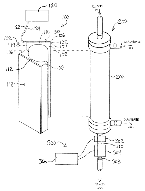
The improved hemodialysis treatment apparatus of the present invention is
shown
schematically in FIG 1. The apparatus of the present invention is intended for
use with a
standard hemodialysis treatment system. The construction and operation of such
systems
are well known in the art and thus need not be described in detail here. The
hemodialysis
1 S treatment system will typically include a membrane or hollow fiber
dialyzer 200 where
fluid and dissolved substances are exchanged between the patient's blood and a
dialysate
solution by diffusion across a semipermeable membrane. A cross section of a
typical
hollow fiber dialyzer 200 can be seen in FIG 3. A semipermeable membrane in
the form
of a multiplicity of hollow fibers 204 passes through the cylindrical body 202
of the
dialyzer 200. Blood flows through the hollow fibers 204 and the dialysate
solution flows
within a chamber 206 surrounding the hollow fibers 204. Diffusion takes place
between
the patient's blood and the dialysate solution across the walls of the hollow
fibers 204.
CA 02428515 2003-05-12
WO 02/38201 PCT/USO1/43800
The invention includes an ultrasonic module 100, which is configured to attach
to
the dialyzer 200 and to improve the efficiency of the hemodialysis treatment
system. The
ultrasonic module 100 and the dialyzer 200 are shown assembled together in a
side view
in FIG 2 and in a cross section in FIG 3. In one preferred embodiment of the
invention,
the ultrasonic module 100 can be constructed as a piece of durable equipment
that is
reusable with many disposable or reusable dialyzers 200 for a multiplicity of
patients. In
this case, since the ultrasonic module 100 does not directly contact the
patient's blood or
the dialysate solution, the ultrasonic module 100 would not need to be
sterilized between
uses. In an alternate preferred embodiment, the ultrasonic module 100 can be
permanently integrated with the dialyzer 200 into a single unit. The combined
ultrasonic
module 100 and dialyzer 200 unit can be constructed as a disposable product or
as a
single-patient reusable product. In this case, the combined ultrasonic module
100 and
dialyzer 200 unit would be constructed so that it can be sterilized before
use.
The ultrasonic module 100 includes an ultrasonic transducer 130 and an
acoustic
coupling 132. The ultrasonic transducer 130 is preferably constructed as of a
layer of
piezoelectric material 102, which is coated on a first side with a first
conductive electrode
104 and on a second side with a second conductive electrode 106. An insulating
layer
may be coated over the electrodes 104, 106. The piezoelectric material 102
used in the
ultrasonic transducer 130 may be a polymeric piezoelectric material, such as
polyvinylidene difluoride (PVDF), or a ceramic piezoelectric material, such as
lead
zirconium titanate (PZT), or other known piezoelectric materials. If desired,
the
ultrasonic transducer 130 may be constructed with multiple layers of
piezoelectric
material 102 to increase the amplitude and/or power of the ultrasonic waves
produced.
6
CA 02428515 2003-05-12
WO 02/38201 PCT/USO1/43800
Alternatively, the ultrasonic transducer 130 may utilize other known
ultrasonic transducer
technologies, such as magnetostrictive transducers or silicon ultrasound
transducers.
Suitable silicon ultrasound transducers, which are produced on silicon wafers
using
MEMS (Micro Electro Mechanical Systems) technology, are available from Sensant
Corporation, 14470 Doolittle Drive, San Leandro, CA, USA 94577 and are
described in
U.S. Patent 6,246,158, which is hereby incorporated by reference.
The acoustic coupling 132 is configured to efficiently transmit ultrasonic
vibrations from the ultrasonic transducer 130 to the body 202 of the dialyzer
200. For
convenience, the acoustic coupling 132 is split into a first half 114 and a
second half 116
that hinge apart or separate to facilitate insertion of the body 202 of the
dialyzer 200 into
the ultrasonic module 100, as shown in FIG 1. The first and second halves 114,
116 of the
acoustic coupling 132 are made primarily of an acoustic coupling material 108
having an
acoustic impedance that is matched approximately to the acoustic impedance of
the blood
and the dialysate solution. Various materials, such as polyurethane, low
density
polyethylene and gel materials, are suitable for use as an acoustic coupling
material 108.
The first and second halves 114, 116 of the acoustic coupling 132 have
semicylindrical
cutouts 110, 112, which are sized to fit tightly around the cylindrical body
202 of the
dialyzer 200 for good acoustic coupling when the ultrasonic module 100 is in a
closed
position, as shown in FIGS 2 and 3. A latch, clamp or other closure device may
be
provided to hold the ultrasonic module 100 in the closed position.
Other geometries of the ultrasonic transducer 130 and acoustic coupling 132
are
possible, for example for use with noncylindrical dialyzers, such as flat
membrane
dialyzers. Alternatively, the ultrasonic transducer 130 may be coupled
directly to the
7
CA 02428515 2003-05-12
WO 02/38201 PCT/USO1/43800
body 202 or to the chamber 206 of the dialyzer 200 without an additional
acoustic
coupling 132. For example, this can be accomplished by utilizing an ultrasonic
transducer
130 having an acoustic impedance that is matched approximately to the acoustic
impedance of the blood and the dialysate solution. Additionally, the
ultrasonic transducer
I30 may be configured as an array of transducers in any desired geometry. Such
a
transducer array may be coupled directly to the body 202 or to the chamber 206
of the
dialyzer 200 or indirectly through one or more acoustic couplings.
The ultrasonic transducer 130 is attached to the first half 114 of the
acoustic
coupling 132. Preferably, the second half 116 of the acoustic coupling 132 has
an
acoustically reflective surface 118 positioned opposite to and parallel with
the ultrasonic
transducer 130. The acoustically reflective surface I I8 may be backed with a
Iugh
acoustic impedance material, such as a metal, which will produce a positive
reflection of
the ultrasonic waves, or a low acoustic impedance material, such as air, which
will
produce a negative reflection of the ultrasonic waves. The acoustically
reflective surface
118 allows the acoustic coupling 132 to be designed as a resonant structure,
which will
increase the efficiency of the ultrasonic transducer 130. In a preferred
configuration, the
ultrasonic transducer 130 and the acoustic coupling 132 extend substantially
the full
length of the body 202 of the dialyzer 200. If desired, the ultrasonic
transducer 130 and
the acoustic coupling 132 may be enclosed in a protective and esthetic
housing.
The first electrode 104 and the second electrode I06 of the ultrasouc
transducer
I30 are connected to the output of an ultrasonic wavefonn generator I20 by a
first
electrical lead 122 and a second electrical lead 124, respectively.
Preferably, the first
electrical lead 122 and the second electrical lead 124 are configured as a
coaxial cable.
8
CA 02428515 2003-05-12
WO 02/38201 PCT/USO1/43800
The ultrasonic waveform generator 120 may operate in one of several possible
modes.
The ultrasonic waveform generator 120 may produce a simple narrowband sine
wave at a
desired frequency, or it may produce a variable or sweeping frequency sine
wave. The
ultrasonic waveform generator 120 may sweep the frequency within a desired
range to
find a resonance, indicated by a local minimum in the electrical impedance,
and lock onto
the resonant frequency. Alternatively, the ultrasonic waveform generator 120
may
produce a broadband waveform, such as a square wave or a sawtooth wave. The
ultrasonic waveform generator 120 may be made switchable between these various
modes for different purposes. The ultrasonic waveform generator 120 may
operate over a
wide range of frequencies, including sonic frequencies and ultrasonic
frequencies in the
kilohertz and megahertz ranges. Ultrasonic frequencies in the range of 20 to
40 kilohertz
are thought to be particularly effective for use in the present invention. The
ultrasonic
waveform generator 120 will preferably include a variable power output, with
at least a
low power setting for continuous use to increase the diffusion rate across the
semipermeable membranes of the dialyzer 200 and a high power setting for
intermittent
application to break up thrombus that may form within the dialyzer 200.
In use, the ultrasonic waveform generator 120 energizes the ultrasonic
transducer
130 to produce ultrasonic waves at a desired frequency and amplitude and with
a desired
waveform to increase the diffusion rate across the semipermeable hollow fiber
membranes 204 of the dialyzer 200. The increased diffusion rate significantly
reduces the
amount of time required for hemodialysis treatments. The ultrasonc waves are
transmitted from the ultrasonic transducer 130 into the body 202 of the
dialyzer 200 by
the acoustic coupling 132, preferably producing a uniform acoustic field
within the
9
CA 02428515 2003-05-12
WO 02/38201 PCT/USO1/43800
chamber 206 of the dialyzer 200. Intermittently, the power output of the
ultrasonic
waveform generator 120 may be increased to a higher level to break up any
thrombus that
may form within the dialyzer 200 and to remove any platelets or fibrin that
may have
deposited on the surfaces of the hollow fiber membranes 204. The frequency and
the
waveform, as well as the amplitude of the ultrasonic waves may also be
changed. This
will keep the dialyzer 200 working at maximum efficiency for a longer period
of time.
This feature also provides an advantage by reducing or eliminating the
necessity for
anticoagulation during hernodialysis treatments.
Optionally, the invention may also include a thrombus detection and
thrombolysis
module 300, as shown in FIG 1. An emboli detector 302, which may be an
ultrasonic or
optical detector, detects thrombi or other emboli exiting the dialyzer 200.
When an
embolus is detected, a control module 306 energizes an ultrasonic transducer
304 that is
focused on a chamber 310 below the dialyzer 200 to break up the embolus.
Thrombi and
emboli larger than a certain size are prevented from entering the patient's
circulatory
system by a screen or filter 308 at the exit of the chamber 310.
FIG 4 shows a second embodiment of the improved hernodialysis treatment
apparatus, which includes an ultrasonic module 100 and a hollow fiber dialyzer
200. FIG
5 is a cross section of the ultrasonic module 100 and the hollow fiber
dialyzer 200 of FIG
4. In this embodiment, the ultrasonic module 100 takes the form of one or more
ultrasonic transducers 150, 152 that trailsmit ultrasonic waves into the
chamber 206 of the
hollow fiber dialyzer 200 by way of one or more waveguide rods 154, 156. The
ultrasonic transducers 150, 152 may utilize piezoelectric transducers,
magnetostrictive
CA 02428515 2003-05-12
WO 02/38201 PCT/USO1/43800
transducers, silicon ultrasound transducers or other known ultrasonic
transducer
technologies. The waveguide rods 154, 156 are preferably constructed of a
metal or other
material that will efficiently conduct the ultrasonic energy into the chamber
206 of the
hollow fiber dialyzer 200 and transfer the ultrasonic waves to the dialysate
solution.
Suitable materials for the waveguide rods 154, 156 include, but are not
limited to,
stainless steel, titanium, titasuum alloys and cobalt alloys. The waveguide
rods 154, 156
may be textured or faceted or have other geometrical features to promote
uniform
dispersion of the ultrasonic energy within the chamber 206. The waveguide rods
154, 156
may also be constructed in the configuration of a tapered ultrasonic
amplifying horn to
increase the amplitude of the ultrasonic waves produced by the ultrasonic
transducers
150, 152.
By way of example, FIG 4 shows the apparatus with two such ultrasonic
transducers 150, 152 connected to two waveguide rods 154, 156. Alternatively,
the
apparatus may be constructed with a single ultrasonic transducer connected to
one or
more waveguide rods or with multiple ultrasonic transducers and waveguide
rods.
Preferably, the waveguide rods 154, 156 are arranged to produce a relatively
uniform
acoustic field within the chamber 206.
In one preferred embodiment of the invention, the ultrasonic module 100 can be
permanently integrated with the dialyzer 200 into a single unit. The combined
ultrasonic
module 100 and dialyzer 200 unit can be constructed as a disposable product or
as a
single-patient reusable product. In this case, the combined ultrasonic module
100 and
dialyzer 200 unit would be constructed so that it can be sterilized before
use. In an
alternate preferred embodiment, the ultrasonic module 100 can be constructed
as a piece
11
CA 02428515 2003-05-12
WO 02/38201 PCT/USO1/43800
of durable equipment that is reusable with many disposable or reusable
dialyzers 200 for
a multiplicity of patients. In this case, since the waveguide rods 154, 156 of
the ultrasonic
module 100 do contact the dialysate solution, the ultrasonic module 100 would
be
constructed so that it could be sterilized between uses.
The ultrasonic transducers 150, 152 are connected to the output of the
ultrasonic
waveform generator 120 by electrical leads 158 & 160 and 162 & 164,
respectively.
Preferably, the electrical leads are configured as coaxial cables. Preferably,
the ultrasonic
transducers 150, 152 operate at the same frequency and in phase with one
another to
produce a relatively uniform and constant acoustic field. Alternatively, the
ultrasonic
transducers 150, 152 may be operated at different frequencies and/or out of
phase with
one another to produce different acoustic effects within the chamber 206. As
described
above, the ultrasonic waveform generator 120 may operate in one of several
possible
modes, including nanrowband and broadband modes, and with low and high power
settings.
In use, the ultrasonic waveform generator 120 energizes the ultrasonic
transducers
150, 152 to produce ultrasonic waves at a desired frequency and amplitude and
with a
desired waveform to increase the diffusion rate across the semipermeable
hollow fiber
membranes 204 of the dialyzer 200. The increased diffusion rate significantly
reduces the
amount of time required for hemodialysis treatments. The ultrasonic waves are
transmitted from the ultrasonic transducers 150, 152 into the chamber 206 of
the dialyzer
200 by the waveguide rods 154, 156, which serve as an acoustic coupling to the
dialysate
solution. Intermittently, the power output of the ultrasonic waveform
generator 120 may
be increased to a higher level to break up any thrombus that may form within
the dialyzer
12
CA 02428515 2003-05-12
WO 02/38201 PCT/USO1/43800
200 and to remove any platelets or fibrin that may have deposited on the
surfaces of the
hollow fiber membranes 204. The frequency and the waveform, as well as the
amplitude
of the ultrasonic waves may also be changed. This will keep the dialyzer 200
working at
maximum efficiency for a longer period of time. This feature also provides an
advantage
by reducing or eliminating the necessity for anticoagulation during
hemodialysis
treatments. Optionally, the invention may also be used with the thrombus
detection and
thrombolysis module 300 described above in connection with FIG 1.
While the present invention has been described herein with respect to the
exemplary embodiments and the best mode for practicing the invention, it will
be
apparent to one of ordinary skill in the art that many modifications,
improvements and
subcombinations of the various embodiments, adaptations and variations can be
made to
the invention without departing from the spirit and scope thereof.
13