Note: Descriptions are shown in the official language in which they were submitted.
CA 02428537 2003-05-09
WO 03/032824 PCT/US02/32811
METHOD FOR DETERMINING ACCOMMODATION
Background of the Invention
This invention relates generally to the field of ophthalmic diagnostic devices
and,
more particularly, to wavefront sensors used as diagnostic devices.
The human eye in its simplest terms functions to provide vision by
transmitting
light through a clear outer portion called the cornea, and focusing the image
by way of a
crystalline lens onto a retina. The quality of the focused image depends on
many factors
including the size and shape of the eye, and the transparency of the cornea
and the lens.
The optical power of the eye is determined by the optical power of the cornea
and
the crystalline lens. In the normal, healthy eye, sharp images are formed on
the retina
(emmetropia). In many eyes, images are either formed in front of the retina
because the
eye is abnormally long (axial myopia), or formed in back of the retina because
the eye is
abnormally short (axial hyperopia). The cornea also may be asymmetric or
tonic, resulting
in an uncompensated cylindrical refractive error referred to as corneal
astigmatism. In
addition, due to age-related reduction in lens accommodation, the eye may
become
presbyopic resulting in the need for a bifocal or multifocal correction
device.
In the past, axial myopia, axial hyperopia and corneal astigmatism generally
have
been corrected by spectacles or contact lenses, but there are several
refractive surgical
procedures that have been investigated and used since 1949. Barraquer
investigated a
procedure called keratomileusis that reshaped the cornea using a microkeratome
and a
cryolathe. This procedure was never widely accepted by surgeons. Another
procedure
that has gained widespread acceptance is radial and/or transverse incisional
keratotomy
(RK or AK, respectively). In the 1990s, the use of photablative lasers to
reshape the
surface of the cornea (photorefractive keratectomy or PRK) or for mid-stromal
photoablation (Laser-Assisted In Situ Keratomileusis or LASIK) have been
approved by
regulatory authorities in the U.S. and other countries.
In the past, the amount of tissue removed by the laser was determined by
taking
pre-operative measurements of the optical errors of the eye, sphere, cylinder
and axis,
termed "low order" optical aberrations. These measurements were manually
loaded into
the refractive laser and a proposed corrective "recipe" was calculated by the
laser software.
More recently, the use of wavefront sensor technology, which measures both the
low order
CA 02428537 2003-05-09
WO 03/032824 PCT/US02/32811
2
optical aberrations and the "higher" order aberrations, such as coma, trefoil
and spherical
aberrations, have been investigated. See for example U.S. Patent Nos.
5,777,719,
5,949,521, 6,095,651 (Williams, et al.), U.S. Patent Application Serial Nos.
09/566,409 and
09/566,668, both filed May 8, 2000, and in PCT Patent Publication No. WO
00/10448, the
entire contents of which being incorporated herein by reference. These
wavefront sensors
are particularly useful when used in combination with a high-speed eye
movement tracker,
such as the tracker disclosed in U.S. Patent Nos. 5,442,412 and 5,632,742
(Frey, et al.),
the entire contents of which being incorporated herein by reference. The
ultimate goal of
these devices is to link the wavefront sensor to the laser and eye movement
tracker to
provide real-time diagnostic data to the laser during surgery. In the past, as
best seen in
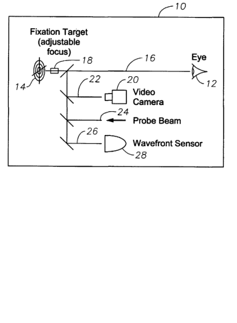
FIG. l, in order to focus wavefront sensing device 10, the patient was seated
at the device
so that the patient's eye 12 views fixation target 14 though optical pathway
16 that
includes adjustable focus mechanism 18. Mechanism 18 compensates for defocus
error
(and possibly astigmatism) to allow the patient to see fixation target 14
relatively clearly
regardless of the refractive error in the patient's eye 12. Video camera 20,
disposed along
optical pathway 22 allows device 10 operator (not shown) to position eye 12
relative to
device 10. Once the patient is in the correct position and is viewing fixation
target 14,
probe beam 24 of optical radiation is sent into eye 12. A fraction of the
radiation is
scattered by the retina exits the eye in the form of a re-emitted wavefront.
Optical
pathway 26 conveys this wavefront to the entrance face of wavefront sensor 28.
The lens of the eye is a dynamic element, capable of changing the effective
focal
length of the eye through accommodation. In performing wavefront measurements,
it is
important to take this accommodative ability into account. Normally, the
wavefront is
measured with the lens as relaxed as possible, so that the eye is minimally
refracting (most
hyperopic). Relaxing the lens is typically achieved by adjusting the focus
mechanism in
the fixation pathway so that the fixation target appears to lie just beyond
the patient's most
hyperopic focal point. The fixation target in this instance will appear
slightly out of focus
to the patient. This process is known as "fogging".
Prior to the present invention, wavefront sensors were typically only used to
measure the refractive error of an eye when the eye was in its most relaxed or
hyperopic
state. The inventors have discovered that a wavefront sensor can be used to
measure the
range of accommodation of an eye, and to measure optical aberrations over the
CA 02428537 2003-05-09
WO 03/032824 PCT/US02/32811
3
accommodative range. This capability may have diagnostic utility in
characterizing age-
related changes in performance of the crystalline lens and in interpreting
certain visual
symptoms. This capability may also allow for customization of the ablation
pattern of the
refractive laser to optimize the patient's vision at any accommodative state
(i.e., at any
desired focal point in front of the eye).
Accordingly, a need continues to exist for a method of determining the
accommodative range of an eye.
Brief Summary of the Invention
The present invention improves upon the prior art by providing an automated
objective method for determining the accommodative range and aberration
profile of an
eye using a wavefront sensor that iteratively determines the change in
accommodation of
the lens of an eye.
Accordingly, one objective of the present invention is to provide an automated
objective accommodative measurement method.
Another objective of the present invention is to provide an automated
aberration profile measurement method.
Another objective of the present invention is to provide an accommodation
measurement method for a wavefront sensor that does not require subjective
determination
of accommodation.
Another objective of the present invention is to provide an accommodation
measurement method for a wavefront sensor that does not require participation
by a skilled
operator.
These and other advantages and objectives of the present invention will become
apparent from the detailed description and claims that follow.
Brief Description of the Drawing
FIG. 1 is a simplified schematic representation of a prior art wavefront
sensing
device.
FIG. 2 is a flow chart illustrating a fogging method that may be used with the
present invention.
CA 02428537 2003-05-09
WO 03/032824 PCT/US02/32811
4
FIG. 3 is a flow chart illustrating the accommodation measurement method of
the
present invention.
Detailed Description of the Invention
The method of the present invention may be practiced on any commercially
available wavefront sensor having appropriate software controls. Suitable
devices are
disclosed in U.S. Patent Nos. 5,777,719, 5,949,521, 6,095,651, U.S. Patent
Application
Serial Nos. 09/566,409 and 09/566,668, both filed May 8, 2000, and in PCT
Publication
No. WO 00/10448. The method of the present invention requires that the eye
initially be
fogged, which can include an iterative process wherein the wavefront sensor
first
calculates a stable effective clinical prescription for the eye being
measured. The
prescription is then moved in the hyperopic direction until the emanating
wavefront is
stable at the eye's most hyperopic state. These steps "fog" the eye in
preparation for the
method of the present invention.
As best seen in FIG. 2, one preferred method of fogging the eye involves
initially
having the patient view fixation target 14 with focus mechanism 18 in its
nominal
position, which is the appropriate position for an eye with no significant
defocus or
astigmatic error. As patient eye 12 attempts to view this target, initial
wavefront
measurement is taken at step 100. The effective clinical prescription for eye
12 is
calculated at step 102 from the measurement taken at step 100 and focus
mechanism 18 is
adjusted at step 104 based on the prescription calculated at step 102 to
correct for any
detected defocus or astigmatic error. A second wavefront measurement is taken
at step
106 and the effective clinical prescription for eye 12 is calculated at step
108 from the
measurement taken at step 106. The difference between the prescription
calculated at step
102 and the prescription calculated at step 106 is determined during step 110.
If the two
prescriptions are not within allowed tolerances, such as approximately 0.25
diopters (other
tolerances may also be used), steps 104 through 110 are repeated iteratively
until a stable
prescription is obtained (e.g., the difference between the two prescriptions
is within the
allowed tolerance for defocus and astigmatic errors, including the axis of any
detected
astigmatism).
CA 02428537 2003-05-09
WO 03/032824 PCT/US02/32811
Once a stable prescription is obtained at step 110, focusing mechanism 18 is
adjusted in discrete, defined steps (for example, 0.5 diopters or other
suitable amount) in
the hyperopic direction during step 112, and an initial wavefront measurement
is taken at
step 114. This, in effect, applies an accommodative stimulus to eye 12. The
effective
clinical prescription for eye 12 is calculated at step 116 from the
measurement taken at
step 114. The prescription calculated at step 116 is analyzed at step 118 to
see if the
prescription has also moved in the hyperopic direction by more than a
threshold amount,
such as approximately 0.25 diopters. If the prescription has not moved in the
hyperopic
direction by more than a threshold amount, then the prescription is deemed
stable and eye
12 suitably fogged for accurate wavefront measurements. If the prescription
has moved in
the hyperopic direction by more than the threshold amount, the prescription is
deemed not
to be stable, and steps 112 through 118 are repeated iteratively until a
stable prescription is
obtained.
As best seen in FIG. 3, once eye 12 has been appropriately fogged at step 200,
focusing mechanism 18 is adjusted in discrete, defined steps in the myopic
direction
during step 212, and a wavefront measurement is taken at step 214. The
effective clinical
prescription for eye 12 is calculated at step 216 from the measurement taken
at step 214.
The prescription calculated at step 216 is analyzed at step 218 to see if the
prescription has
also moved in the myopic direction by more than a threshold amount, such as
approximately 0.25 diopters. If the prescription has not moved in the myopic
direction by
more than a threshold amount, then the prescription is deemed stable and the
accommodative range of eye 12 is known to lie between the stable myopic
prescription
calculated at step 216, and the stable hyperopic prescription calculated at
step 116. If the
prescription has moved in the myopic direction by more than the threshold
amount, the
prescription is deemed not to be stable, and steps 212 through 218 are
repeated iteratively
until a stable prescription is obtained. In this manner, the entire
accommodative range of
eye 12 can be determined.
In some cases, patients may have difficulty viewing target 14 and complying
with
the test. In order to facilitate measurement of such patients, additional
logic may be
added. For example, a sudden large hyperopic change in the wavefront may
indicate that
the patient has lost fixation. Adjusting focus mechanism 18 by a substantial
amount (e.g.,
CA 02428537 2003-05-09
WO 03/032824 PCT/US02/32811
6
one diopter) in the hyperopic direction may allow the patient to regain
fixation and resume
the accommodation testing.
In addition to measuring the accommodative range of eye 12, the method of the
present invention can also be used to customize the outcome of laser
refractive surgery.
For example, since the wavefront and corrective prescription for eye 12 is
known
throughout the entire accommodative range, the laser refractive surgery can be
optimized
at any particular focal length depending upon the desires of the patient.
Therefore, if the
patient desires optimum focus at one meter, the prescription that will
optimize the
refractive correction at a one meter focal length may be used. Similarly, if
the patient
desires optimum focus at infinity, or at the point of maximal accommodation,
the final
prescription calculated at step 116 or at 216, respectively, may be used.
This description is given for purposes of illustration and explanation. It
will be
apparent to those skilled in the relevant art that changes and modifications
may be made to
the invention described above without departing from its scope or spirit.
