Note: Descriptions are shown in the official language in which they were submitted.
CA 02429049 2003-05-15
WO 02/080998 PCT/US01/51651
VEIN HARVESTING SYSTEM AND METHOD
Field of the Invention
This invention relates to a system for harvesting a generally cylindrical
shaped tissue structure from the body of a patient. More particularly, the
invention is directed to a system for harvesting a section of a blood vessel
from
a patient.
Background of the Invention
In certain circumstances. it is desirable to remove sections of tubular
tissue structure from a patient's body. Such tissue may be used in another
part
of the patient's body, may be transplanted into a second patient's body or may
be discarded. As used herein, the term "tubular tissue structure" includes
blood
vessels, tendons, bile ducts and any other similar tissue formation which is
generally tubular in structure and capable of being separated from surrounding
tissue. Although the invention herein will be discussed in terms of harvesting
blood vessels it should be understood that the apparatus and method described
are equally applicable to harvesting other tubular tissue structures from a
human or animal body.
Vein harvesting is commonly done in connection with coronary artery
bypass surgery. The saphenous vein is a subcutaneous vein which is often used
for coronary artery bypass grafting, infra-inguinal bypass grafting and vein-
vein
bypass grafting. Other veins may also be used including the mammary vessel
and the lessor saphenous vein. Previously, it has been necessary to make an
incision along the full length of the vein section to be removed. The vein is
then freed by severing and ligating the branches of the vein, after which the
section of the vein can be removed from the patient. The full length incision
must then be closed, for example by suturing or stapling. Obviously, the
1
CA 02429049 2003-05-15
WO 02/080998 PCT/US01/51651
harvesting of the vein in this manner leaves disfiguring scars which are
cosmetically undesirable. Additionally, the large incision creates a risk of
infection to the patient and may not heal properly, especially with those
patients
who have poor circulation in their extremities. Such an incision may create a
chronic wound.
Devices for harvesting a section of a blood vessel without creating a full-
length incision have been suggested. U.S. Patent No. 4,793,346 (Mindich)
discloses a device for harvesting a section of a blood vessel by making only
small incisions at opposite ends of the blood vessel section. The device
includes a guide rod which fits inside the vein section and a tube having an
inner diameter slightly larger than the outer diameter of the vein section to
be
harvested. The tube has one or more knife blades at the leading edge which are
connected to an electrical supply. The vein section is removed by making the
incision sufficiently deep so as to expose the ends of the blood vessel to be
harvested. The blood vessel is cut to expose one end, the guide rod is
inserted
inside the blood vessel section, and the tube is placed over the end of the
blood
vessel section to be removed. The tube is then pushed along the blood vessel
(into the patient) while rotating the tube to sever the branches of the blood
vessel with the knife blade mounted at the leading edge of the tube.
Electrical
current is supplied to the knife blades to heat the blades and thereby
cauterize
the ends of the severed branches of the blood vessel. The procedure is
continued until the tube has reached the second of the two incisions. The
blood
vessel is exposed and cut from the patient at the second incision. The tube is
then removed from the patient with the blood vessel section inside the tube.
The blood vessel section is then removed from the tube for further treatment
and used as desired.
UK Patent No. GB 20 82 459A discloses a device for harvesting a
section of a blood vessel similar to that disclosed in the Mindich patent.
Again,
2
CA 02429049 2003-05-15
WO 02/080998 PCT/USO1/51651
two incisions are made, one at each end of the blood vessel section to be
harvested. A guide rod is inserted into the blood vessel section through one
of
the incisions and a tube having a cutting element having a cutting tool at its
operative end is passed over the blood vessel section and guide rod assembly.
The tube is rotated as it passes over the blood vessel section to sever the
connecting branches. After the tube has passed the entire length of the blood
vessel section, the section is cut away through the second incision and the
tube
is removed from the patient with the harvested section inside the tube.
Blood vessel harvesting devices of this type have certain distinct
disadvantages. While they eliminate the need for a full-length incision to
remove the blood vessel segment, two incisions, one at each end of the segment
to be harvested, are required in order to remove the blood vessel segment. For
patients likely to develop chronic wounds, each additional incision increases
the risk to the patient, and it is desirable to keep such incisions as close
to the
patient's trunk as possible and to minimize the number and size of such
incisions. Additionally, such devices are unable to adequately close off
several
branches of the blood vessel and thus are unable to adequately control
bleeding.
As a result, the patient suffers greater blood loss than is necessary. These
prior
devices may also remove more tissue than is necessary because the size of the
cutting device is not readily adaptable to the changes in the size of the
blood
vessel.
In U.S. Patent No. Re 36,043 (Knighton), an improved device and
method for vein removal is disclosed which solve some of the problems
associated with the use of prior art devices. Knighton discloses an endoscope
having a lumen extending longitudinally through the scope body. The
endoscope includes means for viewing an area adjacent the distal end of the
lumen. The lumen has a lateral dimension large enough to accommodate the
blood vessel being harvested and at least one tool for use in harvesting the
3
CA 02429049 2003-05-15
WO 02/080998 PCT/US01/51651
blood vessel. A first end of the blood vessel section to be harvested is
exposed
through an incision in the patient's body. A dissecting tool and a gripping
tool
are inserted through the lumen of the endoscope and used to dissect the blood
vessel away from the surrounding connective tissue of the patient's body.
Additional tools are provided for use through the lumen of the endoscope to
remove body fluids and coagulate bleeding tissue, to ligate and sever side
branches from the blood vessel to be harvested, and to ligate and sever a
distal
end of the blood vessel to be harvested when a desired length of blood vessel
has been dissected. Only a small incision in the patient's body is necessary
to
harvest a relatively long length of blood vessel in a precise and controlled
manner using this device and procedure.
U.S. Patent No. 5,772,576 (Knighton et al.) also describes a device and
method for vein removal. The device has one or more lumens extending
through a body portion. One lumen is sized to accommodate a blood vessel and
at least one tool for use in removing the vessel. The device may also include
viewing means so that the operator may remotely view an area adjacent the
distal end of the body portion. The device protects the vessel being removed
from damage by the tools used in the procedure, which is critical since the
blood vessel is destined for reuse (as in arterial bypass). In addition, a
single
operator can use the device.
However, there is still room for improvement in curreni vein harvesting
devices. It is difficult to see the vein to be harvested, especially near the
distal
end, even with the use of optical fiber devices. Moreover, it can be difficult
to
position the tools needed to harvest a vein in a fast and efficient ma.nner
and
minimize damage to vein and to the patient. Thus a need in the art remains for
a convenient method that would effectively harvest a vein without causing any
damage to it
4
CA 02429049 2003-05-15
WO 02/080998 PCT/US01/51651
Summary of the Invention
This invention is a system for the harvesting of a blood vessel. The
system comprises a housing having a removable lower portion. In a preferred
embodiment, the housing is configured to contain a body portion having at
least
one lumen. In a more preferred embodiment, the housing is configured to
contain a multi-lumen body portion. Various tools are used in conjunction with
the housing and the body portion for removal of a blood vessel.
In one aspect the invention is a method of removing a section of a
generally cylindrical tissue structure from the body of a human or animal. The
method includes providing a device having a first portion and a second
portion,
the first and second portions defining a working space therebetween, the
second
portion being movable with respect to the first portion; exposing a proximal
end
of the tissue structure section to be harvested through an incision in the
body;
inserting the device through the incision such that the second portion is
positioned between the first portion and the tissue structure; moving the
second
portion with respect to the first portion to expose the working space between
the first portion and the tissue structure; inserting at least one tool
through the
incision into the working space; dissecting the tissue structure away from
surrounding tissue with the at least one tool; cutting a proximal end of the
tissue
structure section; cutting a distal end of the tissue structure section; and
removing the tissue stiucture section through the incision.
The method may include providing a device wherein the first portion has
a non-planar shape and the second porlion is substantially planar, wherein the
second portion is slidably engaged to the second portion and wherein the step
of moving the second portion with respect to the first portion includes
sliding
the second portion out of engagement with the first portion.
In another aspect the invention is a system for removing a generally
cylindrical tissue structure from a human or animal body. The system includes
5
CA 02429049 2003-05-15
WO 02/080998 PCT/US01/51651
a device having an elongate nonplanar housing defining a longitudinal axis; a
movable portion configured to be movable with respect to the housing from an
engaged position where an at least partially enclosed working space is defined
between the housing and the movable portion and a worlcing position where the
working space is exposed, the housing and movable portion being sized to be
insertable through the incision and positioned adjacent the tissue structure;
and
at least one tool used in removing the tissue structure, the tool having a
distal
operative tip and being sized to be accommodated within the working space.
The system may further comprise an elongate element having a first lumen
sized for accommodating the tissue structure and a second lumen sized for
accommodating at least one tool.
In one aspect the invention is a method of removing a generally
cylindrical tissue steacture from the body of a human or animal. The method
comprises providing a means for creating a working space; exposing a proximal
end of the tissue structure to be harvested through an incision in the body;
inserting the means for creating a working space through the incision adjacent
the tissue structure section; manipulating the means for creating a worldng
space to form a working space adjacent the tissue stracture section; inserking
at
least one tool through the incision into the worlang space between the tissue
structure section and the means for creating a working space; dissecting the
tissue structure away from surrounding tissue with the at least one tool;
cutting
a proximal end of the tissue structure section; cutting a distal end, of the
tissue
structure section; and removing the tissue structure section through the
incision.
The method may further comprise providing an elongate element having first
and second lumens and inserting a proximal end of the tissue structure section
through the first lumen of the elongate device. Further, the method may
comprise providing a substantially cylindrical dissection tool within the
first
lumen of the elongate member, the dissection tool having a third lumen,
6
CA 02429049 2003-05-15
WO 02/080998 PCT/US01/51651
inserting a proximal end of the tissue structure section through the third
lumen
of the dissection tool, providing a cutting tool, and inserting the cutting
tool
through the second lumen of the elongate element.
In a further aspect the invention is a system for removing a generally
cylindrical tissue structure from a lumen or animal body. The system
comprises means for creating a worldng space adjacent the tissue structure in
the body, the means being sized to be insertable into the body adjacent the
tissue structure through an incision in the body, and at least one tool used
in
removing the tissue structure, the tool being sized to be accommodated within
the worldng space.
The system may further comprise an elongate element having a first
lumen sized for accommodating the tissue structure and a second lumen sized
for accommodating at least one tool.
In another aspect the invention is a method for creating a worldng space
for at least one tool over a generally cylindrical tissue stracture in a human
or
animal body. The method comprises providing a device having a first portion
and a second portion, the first and second portions defining a working space
therebetween, the second portion being movable with respect to the first
portion; making an incision in the body above the tissue structure; inserting
the
device through the incision such that the second portion is positioned between
the first portion and the tissue structure; moving the second portion with
respect
to the first portion to expose the worldng space between the "first portion
and the
tissue structure; and inserting the at least one tool through the incision
into the
working space.
In a further aspect the invention is a device for insertion through an
incision for creating a working space for at least one tool adjacent a
generally
cylindrical tissue structure in a human or animal body. The device comprises
7
CA 02429049 2006-09-14
75391-34
an elongate nonplanar housing defining a longitudinal axis;
and a movable portion configured to be movable with respect
to the housing from an engaged position where an at least
partially enclosed working space is defined between the
housing and the movable portion and a working position where
the working space is exposed, the housing and movable
portion being sized to be insertable through the incision
and positioned adjacent the tissue structure.
According to a further aspect of the invention
there is provided a system for removing a generally
cylindrical tissue structure from a human or animal body
comprising: a device having an elongate nonplanar housing
defining a longitudinal axis; a substantially planar movable
portion configured to be movable with respect to the housing
from an engaged position where an at least partially
enclosed working space is defined between the housing and
the movable portion and a working position where the working
space is exposed, the housing and movable portion being
sized to be insertable through the incision and positioned
adjacent the tissue structure; and at least one tool used in
removing the tissue structure, the tool having a distal
operative tip and being sized to be accommodated within the
working space.
According to a further aspect of the invention
there is provided a device for insertion through an incision
for creating a working space for at least one tool adjacent
a generally cylindrical tissue structure in a human or
animal body comprising: an elongate nonplanar housing
defining a longitudinal axis; a substantially planar movable
portion configured to be movable with respect to the housing
from an engaged position where an at least partially
enclosed working space is defined between the housing and
the movable portion and a working position where the working
8
CA 02429049 2006-09-14
75391-34
space is exposed, the housing and movable portion being sized to be insertable
through the incision and positioned adjacent the tissue structure.
Bnef Description of the Drawings
Aspects of the present invention will be appreciated with reference to the
description of the invention when read in conjunction with the accompanying
drawings.
FIG. lA is a perspective view of the device of the present invention.
FIG. 1B is a partial bottom view of the distal end of the device of FIG. lA.
FIG. 1C a transverse cross-sectional view of the device of FIG. 1A along Iine
i C-1 C.
FIG. 2A is a perspective view of the housing of FIG. lA containing a
body portion.
FIG. 2B is a transverse cross-sectional view of a preferred embodiment
of the present invention, along line 2B-2B of FIG. 2A.
FIG. 3 is a side elevational view of a gripping tool for use witli the
present invention.
FIG. 4 is a side elevational view of bipolar scissors for use with the
present invention.
FIG. 5 is a schematic view of the vei11 haivesting device of the present
invention being used in the renzoval of the saphenous vein of a patient.
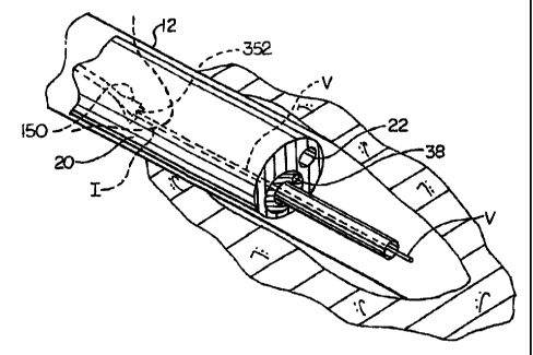
FIGS. 6 and 7 are enlarged perspective illustrations showing the distal
end of the system in use during the hanTesting of a blood vessel.
8a
CA 02429049 2003-05-15
WO 02/080998 PCT/US01/51651
Detailed Description of the Preferred Embodiments
The present invention is a system for harvesting a section of a vessel
from a patient's body for use in another part of a patient's body or for
transplanting into a second patient's body. For example, a section of the
saphenous vein may be removed for use in coronary bypass surgery. The
saphenous vein travels along the medial side of the foot, leg, and thigh,
where it
joins with the femoral vein near the groin.
The terms "distal" and "proximal" as used in this specification refer to
the method use of the device. "Proximal" refers to a location closer to the
physician and "distal" refers to a location farther from the physician.
"Upper"
and "lower" are terms that refer to an orientation with respect to the use of
the
device, that is, relative to the physician.
Turning now to the figures, the components of the vein harvesting
system are illustrated. FIG. IA shows device 10 comprising nonplanar housing
12 and removable (lower) portion 14. Housing 12 has an elongate shape
defining a longitudinal axis. An arcuate tubular shape is shown in the
drawings
although it will be appreciated that other shapes could be usedl FIG. 1B shows
a bottom view of distal end 16 of device 10. As best seen in FIG. 1C
removable portion 14 is held in position in housing 12 by slots 13a and 13b
which extend longitudinally, nearly to distal end 16, through housing 12.
Removable portion 14 preferably extends from proximal end 17 of device 10 to
near distal end 16. Device 10 is inserted into an incision in the patient's
leg (as
illustrated in FIG. 6). The proximal end of device 10 remains outside of the
incision and the physician grasps and renioves removable portion 14. A cavity,
space, or tunnel is thus created, peinlitting the insertion of tools for
harvesting
the vein. The housing is shaped and configured not only for insertion of
tools,
but for ease of insertion into the incision. It should be understood that
additional means for creating a working space above the vein are also
9
CA 02429049 2006-09-14
75391-34
conteinplated within the scope of this invention. For example, the device
could
comprise a unitaiy housing having a first profile upon insertion adjacent the
vein and opening to a second profile once positioned. The opening froin the
first profile to the second profile creating a working space adjacent the
vein.
Such a configuration is disclosed in commonly assigned United States Patent
No. 6,558,313 entitled "Vein Harvesting System and Method".
In a preferred embodiment, a body portion having at least one lumen is
used with the device. After removable portion 14 is removed, a body portion
can be inserted into the housing and used with various tools to haivest the
vein.
Use of a body portion is preferred in order to protect the vessel from
potential
dainage during the harn7esting procedure. In a more preferred enibodiment, the
device is used with a inulti-lumen body portion. The nlulti-lumen body portion
is configured to be used in conjunction with one or more tools.
Various multi-lumen endoscopes and similar devices have been
described in the art and may be useful with the device of this invention. For
example, an endoscope configured to be used with various tools in harvesting
blood vessels is described in U.S. Patent No. RE 36,043 (Knighton). U.S.
Patent
No. 5,772,576 (Knighton et al.) describes a multi-lumen body portion capable
of
isolating the blood vessel from the tools used for removal of a blood vessel.
F1G. 6 illustrates the position of preferred 2nulti-lumen body portion 20
within the space created by housing 12 when the system is in use for vein
harvesting. FIGS. 2A and 2B show housing 12 and Ynulti-lunien body portion
20 in detail.
The components of preferred multi-luinen body portion 20 are shown in
CA 02429049 2003-05-15
WO 02/080998 PCT/US01/51651
FIG. 2B in a transverse cross-sectional view, taken along line 2B-2B of FIG.
2A. Multi-lumen body portion 20 is positioned within housing 12 and has two
lumens, 22 and 24 extending longitudinally therethrough. Lumen 22 is
configured to accept scissors, preferably bipolar gcissors, and lumen 24 is
configured to receive rotatable tube 26, itself conpprising two lumens 28 and
30.
The lumens extend longitudinally throughout boqly portion 20. Tube 26 freely
rotates within lumen 24. Lumen 28 is configured to receive an optical fiber
device or endoscope and lumen 30 is configured ito receive a gripping tool, as
described further below. In the embodiment illustrated in FIG. 2B, the lumens
are generally circular in cross-section although tey may be any shape suitable
for the insertion of the tools.
Device 10 (i.e., housing 12 and removable portion 14) and multi-lumen
'body portion 20 may be constructed of a rigid material such as metal or
plastic.
In the method of this invention, an incisioii is made in the area from
which the vein is to be harvested For example, xhe incision is made in the
groin area for harvesting the saphenous vein. Ariother incision can be made
near the knee if a long section of vein is needed: Device 10 is inserted
through
the incision and positioned over the top of the saphenous vein. Removable
portion 14 is withdrawn from housing 12, leaving housing 12 in place over the
vein, forming a workspace or tannel. This workspace is now ready to receive
tools, a body portion having at least one lumen, cir, preferably, a multi-
lumen
body portion and associated tools and components, such as viewing devices.
The blood vessel (e.g., the saphenous veiri) is cut and the end of the
vessel is held by means of a gripping tool and pulled into vein dissecting
lumen
30, as described further below.
The blood vessel is protected from damagle by multi-lumen body portion
20 which isolates the vessel from the tools used tb harvest the vessel. In
addition, there is room to operate tools and remoive the vessel without
causing
Il
CA 02429049 2006-09-14
75391-34
dainage because of the worlcspace created by upper portion of housing 12.
A viewing device, such as an endoscope, is used so that the vein and side
branches of the vein can be seen and cut. Preferably, these side branches are
cut an.d cauterized with bipolar scissors (sucll as illustrated in FIG. 4),
inserted
tlzrough scissors lumen 22 of multi-lumen body portion 20. Alternatively, side
branches can be cut with scissors and ligated with a clip, whiclz serves the
san-ie
function as eauterization. The advantage to the use of bipolar scissors is
that
cutting and cauterizing of the side branches occurs in one step.
Lumen 30 (in rotatable tube 26) is of a size large enougll to
accoinznodate the blood vessel that is to be harvested as well as gripping
tool
150. Rotatable tube 26 is inserted into lumen 24 of multi-lunlen body portion
20. Gripping tool 150 is inserted into lumen 30 of rotatable tube 26 and the
entire inulti-lumen body portion 20 is moved distally until a desired length
of
vein is dissected. The distal end of the vein is cut, preferably with bipolar
scissors, that are inserted into lumen 22 of multi-lumen body portion 20. The
section of vein is then removed through lumen 30.
A second incision can be made below tl-ie knee and the process repeated
if a longer piece of vein is needed.
Multi-lumen body portion 20 also has lumen 28, sized to accommodate a
fiber optics viewing device 38 which includes an appropriate fiber optics
illumination source, as illustrated in FIGS. 5 and 6. Device 38 is positioned
such that the area inunediately adjacent the distal end of body portion 20 can
be
illuininated and viewed by the operator. Device 38 is operably connected to an
external monitor 40 that includes a suitable light source by conduit 42.
Conduit
42 enters the endoscope lumen at endoscope poi-t 28 of rotatable tube 26 (as
shown in FIG. 2B). Multi-lumen body portion 20 could also be provided with
an irrigation channel and/or a smoke evacuation channel if deemed necessary.
12
CA 02429049 2003-05-15
WO 02/080998 PCT/US01/51651
TOOLS
Gripping tool 150 in FIG. 3 is used to hold and retain the vessel being
harvested. Gripping too1150 has an elongated shaft 155, with handle 156
attached to a proximal end of shaft 155 and gripping mechanism 160 attached
to a distal end of shaft 155. Handle 156 is preferably a scissors-type handle
to
actuate gripping mechanism 160 at the distal end of shaft 155 and includes
latching mechanism 157 which allows the gripping mechanism to be locked in
a set position (e.g., in a gripping position). Shaft 155 transmits the
actuating
movement from handle 156 to gripping mechanism 160. Gripping mechanism
160 includes first jaw 162 and second jaw 164 that oppose each other. When
gripping handle 156 is operated by the physician, first jaw 162 and second jaw
164 are moved toward each other and may be used to grip blood vessel 20
between gripping surfaces 166 and 168. Jaws 162 and 164 are small enough to
fit through lumen 30 of rotating tube 26.
During the entire procedure, the blood vessel is held in tension by the
physician via gripping too1150. Multi -lumen body portion 20 is advanced
distally into the patient's body and the blood vessel is moved into lumen 30
of
multi-lumen body portion 20, thus dissecting the blood vessel.
When a side branch is encountered during a vein dissection, bipolar
scissors are inserted to cut and cauterize the side branch. Preferred bipolar
scissors 400 are illustrated in FIG. 4. Blades 410 are positioned at the end
of
shaft 412. Shaft 412 can be turned by turning knob 414 in order to position
the
blades at the desired location. Handle 418 is connected to shaft 412 and
provided with lever 416. Lever 416 is depressed to close blades 410. Handle
418 is connected to, power cord 420 that has bipolar connectors 422 and 424.
During use, the connectors preferably are activated by a foot switch (not
shown) that is depressed to activate current flow to the blades (thus
cauterizing
the side branches) at the desired site.
13
CA 02429049 2003-05-15
WO 02/080998 PCT/1JS01/51651
Method of Operation
The vein harvesting device 10, multi-lumen body portion 20 and
accompanying tools 150 and 400 are used in combination for harvesting a
vessel. After proper preparation of the incision site, the physician makes a
small incision I (e.g., about 3cm long) over the proximal aspect of the blood
vessel to be harvested (as illustrated in FIGS. 5 and 6). Device 10, with
removable portion 14 in place, is inserted into incision I and moved distally.
After insertion to the desired position, removable portion 14 is removed and
housing 12 is left in place, forming a workspace for the insertion of tools.
As
seen in FIG.'6, blood vessel V is then severed to expose free end 352 and free
end 353 (which may be clipped as shown in FIG. 6). Free end 352 is grasped
by gripping tool 150 which extends through lumen 30. Rotatable tube 26 is
then moved down the vessel to dissect it from surrounding tissue. ' Tlus is
done
for a short length under direct vision. For example, to remove a saphenous
vein, an incision will be made at the groin over the saphenous vein and the
vein
will be dissected free from the junction of the common femoral vein. As shown
in FIGS. 5 and 6, gripping too1150 is inserted through lumen 30 of tube 26
such that the distal end of gripping tool 150 extends beyond the distal end of
lumen 30. Free end 352 of blood vessel V is held by gripping tool 150 such
that it is held under tension in the manner previously described. Rotatable
tube
26 :s then advanced distally over gripping tool 150 and blood vessel V is
dissected away from surrounding connective tissue. Side branches V' are cut
(and cauterized) as necessary during the dissection.
As illustrated in FIG. 7, the dissection process proceeds distally along
blood vessel V. The patient's body is not shown in order to show the
harvesting
system in operation, and incision I is indicated by a dotted line. Multi-lumen
body portion 20 is advanced along with the rotatable tube 26 into the
incision.
Gripping tool 150 continues to grasp the vein. Until this point, the operator
has
14
CA 02429049 2003-05-15
WO 02/080998 PCT/USO1/51651
been viewing the procedure under direct vision. Now, the operator switches to
viewing the dissection process (occurring at the area immediately adjacent the
distal end of the lumen 24) through the fiber optic viewing device 38 located
at
the distal end multi-lunien body portion 20. Viewing device 38 is positioned
in
lumen 28 of rotatable tube 26. Alternatively, the viewing device could be
provided by a separate scope. As previously discussed, viewing device 38
provides adequate lighting for the operator to view the dissection and tool
operations occurring within the patient via the monitor. Irrigant may be
introduced as necessary through an irrigation channel (not shown) to keep
blood or other body tissue from obscuring vision adjacent the distal end of
multi-lumen body portion 20.
During the dissection process, bipolar scissors 400 are used as needed to
cut and cauterize side branches V'. Typically, the bipolar scissors remain in
lumen 22 and are advanced (to expose and use the blades) when needed. The
dissection proceeds until a desired length of vein is cut. The maximum length
of the vein typically is limited by the length from the incision near the
groin to
the knee. The vein is then removed through the proximal end of lumen 30,
though the entire multi-lumen body portion could be removed proximally with
the vein in lumen 30.
The harvested blood vessel is then ready for use in, for example,
coronary bypass surgery.
Although particular embodiments have been disclosed herein in detail,
this has been done for purposes of illustration only, and is not intended to
be
limiting with respect to the scope of the claims. In particular, it is
contemplated
that various substitutions, alterations, and modifications may be made to the
invention without departing from the spirit and scope of the invention as
defined by the claims.