Note: Descriptions are shown in the official language in which they were submitted.
CA 02430024 2003-05-27
WO 02/43796 PCT/USO1/44272
MEDICAL DEVICE FOR DELIVERY OF
A BIOLOGICALLY ACTIVE MATERIAL TO A LUMEN
FIELD OF THE INVENTION
This invention relates generally to medical devices and methods for
delivering a biologically active material to a desired location within the
body of a patient.
More particularly, the invention is directed to medical devices having a
catheter and a
balloon with a plurality of micro-needles at its outer surface for delivering
a biologically
active material to a body lumen. Additionally, the invention is directed to
medical devices
having a catheter, a balloon and a sheath surrounding the balloon. Also, the
invention is
directed to medical devices having a catheter, a balloon and a shockwave
generator for
delivery of biologically active materials.
BACKGROUND OF THE INVENTION
When a disease is localized to a particular part of the body, in particular a
body lumen, such as, without limitation, a blood vessel, direct administration
of biologically
active materials for the treatment of the disease may be more preferred than
systemic
administration. Systemic administration requires larger amounts and/or higher
concentrations of the biologically active materials because of inefficiencies
associated with
the indirect delivery of such materials to the afflicted area. Also, systemic
administration
may cause side effects which may not be a problem when the biologically active
material is
locally administered.
However, such localized delivery of biologically active materials to a body
lien is difficult since body lumens are involved in the transport of body
fluids, which tend
to carry the biologically active material away from the afflicted area. Thus,
there is a need
for devices and methods for the localized delivery of biologically active
materials to
afflicted tissue, especially body lumens.
A number of devices for delivering biologically active materials to body
lumens or vessels involve the use of catheters having expandable portions,
such as a
balloon, disposed on the catheter. To overcome the problem that the delivered
biologically
active material is washed away from the applied area by the blood-flow, there
are generally
two kinds of prior art balloon catheters: one kind is a balloon catheter which
temporarily
occludes blood-flow and infuses a biologically active material to the occluded
area, and the
other kind is a balloon catheter which directly administers the biologically
active material to
a vessel wall by the use of macro-needles. However, the former still has the
problem of
-1-
CA 02430024 2003-05-27
WO 02/43796 PCT/USO1/44272
systemic leakage around the balloon, allowing for systemic distribution of the
biologically
active material. On the other hand, although the latter type of balloon
catheters do not cause
siguficant systemic leakage of the biologically active material, because of
the large size of
the macro-needles used to inject the biologically active material into the
tissue, there is still
back-leakage at the needle track. Also, the large size of the needles cause
damage in the
tissue of the vessel wall. Thus, the prior art balloon catheters cannot
deliver a biologically
active material quickly and accurately to a wall of body lumen without causing
damage in
the body lumen tissue and/or systemic leakage.
In addition, rapid advances in DNA technologies have increased the
necessity for a device or method which realizes more accurate and uniform
delivery of
genetic materials. Therefore, there is still a need for devices and methods
which cause
minimum tissue damage while ensuring accurate and uniform localized delivery
of
biologically active materials including genetic materials to body lumens.
SUMMARY OF THE INVENTION
These and other objectives are accomplished by the present invention. To
achieve the aforementioned objectives, we have invented a medical apparatus
and a method
for delivery of a biologically active material to a surface of a body lumen.
The apparatus for delivery of biologically active materials of the invention
comprises a catheter and a balloon having micro-needles.
In an embodiment of the invention, the apparatus comprises a catheter, a
balloon, with a biologically active material disposed on an outer surface of
the balloon, and
micro-needles disposed upon the outer surface of the balloon. The micro-
needles contact a
body lumen as the balloon is expanded, and the biologically active material is
delivered into
the body lumen. The biologically active material can be delivered by fluid
convection along
the outer surface of the micro-needles. Alternatively, biologically active
material can,
instead of being disposed on the outer balloon surface, be expelled from an
inner
compartment of the balloon through pores in the outer balloon surface. The
balloon is
optionally surrounded by a sheath.
In another embodiment of the invention, the apparatus comprises a catheter
having at least one lumen in fluid communication with an internal compartment
of the
balloon, and micro-needles having a lumen in fluid communication with the
compartment,
wherein the micro-needles are disposed upon an outer surface of the balloon.
The
micro-needles contact a body lumen as the balloon is expanded, and the
biologically active
material is delivered to the body lumen through the micro-needle lumen. The
balloon is
optionally surrounded by a sheath.
CA 02430024 2003-05-27
WO 02/43796 PCT/USO1/44272
Further, another embodiment of the apparatus of the invention comprises a
catheter, a balloon disposed upon the catheter, and a shockwave generator for
delivery of a
biologically active material.
Moreover, in another embodiment, the apparatus of the invention comprises
a catheter, a balloon having micro-needles disposed upon or within an outer
surface of the
balloon, and a triggering source for rupturing the micro-needles. The micro-
needles are
ruptured by the triggering source, which can be a shockwave, and a
biologically active
material is delivered through the micro-needles to an afflicted area of a body
lumen.
The present invention also includes a method for delivering a biologically
active material to a body lumen. In one embodiment, the method is carned out
by inserting
a catheter with a balloon disposed thereon into a body lumen. The balloon has
micro-
needles disposed upon or within its outer surface. Once the catheter is
inserted, the balloon
is inflated so that the micro-needles contact the surface of the body lumen.
The biologically
active material is then delivered to the surface of the body lumen.
In another embodiment, the method of the invention involves inserting a
catheter having a balloon disposed upon it into a body lumen. The balloon is
inflated to
contact a body lumen. A shockwave is then applied to the afflicted area of the
body lumen
to allow delivery of the biologically active material into the body lumen.
DESCRIPTION OF THE FIGURES
Figure 1 illustrates a configuration of an embodiment of a balloon catheter of
the invention.
Figures 2A and 2B depict cross-sectional views along the longitudinal axis
of embodiments of a balloon catheter of the invention, wherein hollow micro-
needles with
apertures are disposed upon a plate which is disposed on a balloon surface or
within the
balloon wall. The catheter has a lumen for containing the biologically active
material and
mother lumen for inflating the balloon. Details of the micro-needles are shown
in the
enlarged portion of the balloon.
Figure 3 depicts a cross-sectional view along the longitudinal axis of another
embodiment of a balloon catheter of the invention, wherein solid micro-
needles, i.e., micro-
needles without lumens, are disposed upon a plate which is disposed upon a
balloon surface
or within the balloon wall. The outer surface of the balloon is coated with a
polymer
containing a biologically active material. Details of the micro-needles are
shown in the
enlarged portion of the balloon.
Figures 4A and 4B depict a cross-sectional view along the longitudinal axis
of another embodiment of a balloon catheter of the invention, wherein solid
micro-needles
-3-
CA 02430024 2003-05-27
WO 02/43796 PCT/USO1/44272
wall and the micro-needles project through a porous outer surface of the
balloon. In Figure
4A, the micro-needles are disposed upon a porous plate, and the plate is
attached to the
inner surface of the balloon wall. In Figure 4B, the micro-needles may be
mounted on a
solid plate and the solid plate is attached to the inner surface of the
balloon wall. Details of
the micro-needles are shown in the enlarged portions of the balloon.
Figures SA and SB depict cross-sectional views of another embodiment of a
balloon catheter of the invention, wherein a sheath surrounds a balloon having
hollow
micro-needles. In Figure SA, the balloon is in its deflated state. In Figure
SB, the balloon is
in its inflated state. Details of the micro-needles are shown in the enlarged
portions of the
balloon.
Figure 6A depicts a cross-sectional view along the longitudinal axis of
another embodiment of a balloon catheter of the invention in its deflated
state, wherein a
sheath surrounds a balloon having solid micro-needles. Figure 6B depicts a
cross-sectional
view of the same embodiment which is cut along the line I-I in Figure 6A. A
portion of the
Figure 6B is enlarged to Figure 6B'. Figure 6C shows the same portion as in
Figure 6B'
and the balloon is in its inflated state.
Figures 7A and 7B depict cross-sectional views along the longitudinal axis
of a portion of another embodiment of a balloon catheter of the invention,
wherein a sheath
surrounds a balloon having micro-needles. In Figure 7A, the balloon is in
deflated state,
and in Figure 7B, the balloon is in its inflated state.
Figures 8A and 8B depict cross-sectional views along the longitudinal axis
of other embodiments of balloon catheters of the invention where a shockwave
generator is
used to deliver the biologically active material.
Figures 9A, 9B and 9C each depict an embodiment of a micro'-needle which
is capable of being ruptured.
DESCRIPTION OF THE PREFERRED EMBODIMENTS
The medical apparatus suitable for the present invention include those having
at least an inflatable portion such as a balloon. The term "balloon" is
defined as an
inflatable bag-like object made of a balloon wall. The balloon wall may be
made of one or
more layers. The balloon may contain one or more internal walls in addition to
the balloon
wall. Also, the balloon can have more than one compartment.
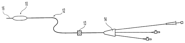
A configuration of an embodiment of an apparatus of the present invention is
illustrated in Figure 1. The embodiment includes a catheter 12 which has a
proximal end 14
and distal end 16 and includes a guide wire and a lumen for inflation (both
not shown in
Figure 1). A balloon 10 with micro-needles (not shown) disposed upon its outer
surface is
-4-
CA 02430024 2003-05-27
WO 02/43796 PCT/USO1/44272
a shockwave generator 13 disposed upon the proximal portion I~4 of the
catheter~l2. ~ Ohce
the catheter is introduced into a body lumen in a manner known to the skilled
artisan, the
balloon is positioned to a targeted area in the body lumen and then inflated
to contact a
surface of the body lumen in a way known in the art. After a biologically
active material is
delivered, the balloon is deflated and removed.
Figures 2A and 2B show two embodiments of the present invention. A
balloon 20A in Figure 2A is disposed upon a catheter 29 having a guidewire 2~.
The
balloon 20A is made of a balloon wall 27 having an inner and outer surface and
has an
internal wall 23, an inflation compartment 26, and an internal compartment 25a
surrounded
by the balloon wall 27 and the internal wall 23. A plate 102, having pores
102a and a
pl~ality of micro-needles 21 disposed upon it, is positioned on the inner
surface 27a of the
balloon wall 27. The micro-needles 21 project through the balloon wall 27 and
are disposed
on the outer surface of the balloon 27b. It should be noted that the micro-
needles do not
have to be actually resting on the outer surface in order to be considered as
being disposed
thereon. As long as the micro-needles protrude through or from the surface,
they are
considered as being disposed upon the surface. The micro-needles 21 have an
aperture 24
and a lumen 22. The lumen 22 is aligned with the pores 102a such that the
lumen 22 is in
fluid communication with the interior compartment 25a of the balloon 20A. The
interior
compartment 25a can be in fluid communication with a lumen 29b of the catheter
29.
The term "micro-needle" is a term of art. Generally, a "micro-needle" is
construed as a needle having a diameter at most about 100 p,m, preferably
about 10 pm or
less and a length at most about 1 mm. The micro-needles applicable to this
invention
include hollow ones 21 such as those in Figure 2A. The term "hollow" means
having one or
more lumens) 22 running through the interior of the micro-needle 21, wherein
fluid and/or
solid materials can pass through the lumens) 22. These hollow micro-needles 21
can
preferably have an aperture 24 connected to the lumen 22 of the micro-needle
21. The term
"aperture" means an opening in the outer surface of the micro-needle 21 which
are
sufficiently large to allow passage of fluid and/or solid materials out of the
micro-needles.
The aperture can be at the tip of the micro-needles or located at other places
in the
micro-needle outer surface. In other embodiments as discussed below, the micro-
needles
c~ be solid or capable of being ruptured.
In the embodiment shown in Figure 2A, the balloon 20A is inflated by
infusing a liquid or gas into the inflation compartment 26 of the balloon 20A
using inflation
lumen 29a of the catheter 29. As the balloon 20A is inflated, the micro-
needles 21 contact a
surface of the body lumen, and a biologically active material located in the
interior balloon
compartment 25a is delivered through the micro-needle lumens 22 and aperture
24 to the
-5-
CA 02430024 2003-05-27
WO 02/43796 PCT/USO1/44272
systemic leakage. After the delivery is completed, the balloon 20A is deflated
and removed
from the body lumen. In other embodiments, the lumen 29b for the biologically
active
material and the inflation lumen 29a can be the same; and/or the inflation
compartment 26
and the interior compartment 25a containing the biologically active material
can be the
same.
Figure 2B shows another embodiment, a balloon 20B, which is similar to the
balloon 20A in Figure 2A, except that the interior compartment 25b is not
connected to a
catheter lumen. In this embodiment, the interior compartment 25b contains the
biologically
active material, and expansion of the inflation compartment 26 will squeeze
the biologically
active material out of the interior compartment 25b through the micro-needles
21 into the
body lumen surface.
Another embodiment of the present invention is shown in Figure 3. A
balloon 30 is made of balloon wall 37, and the balloon wall is made of an
outer layer 37b
and an inner layer 37a. In this embodiment, a plurality of solid micro-needles
31 are
disposed upon a plate 302 which is disposed between the outer layer 37b and
the inner layer
37a. The micro-needles project through the outer layer 37b and are disposed on
an outer
surface of the balloon. The micro-needles can also be hollow in other
embodiments. The
outer surface of the balloon 30 is coated with a polymer containing a
biologically active
material 32. The balloon 30 is inflated by infusing a liquid or gas into an
inflation
compartment 33 using an inflation lumen 39a of the catheter 39 having a
guidewire 38. The
needles 31 pierce the body lumen and create micro-pores or nano-pores, i. e.,
spaces of the
size of the micro-needle, in the body lumen. The biologically active material
contained in
the coating 32 is squeezed by the balloon 30 and forced into or allowed to
seep into the
micro- or nano-pores created by the micro-needles. After a predetermined time,
the balloon
is deflated and removed from the body lumen. The time during which the balloon
is
25 inflated is determined by the type of body lumen tissue, the biologically
active material
used, Garner material or coating if used and the size and number of needles.
For example, if
the body lumen is a coronary artery, the time is generally between about 5
seconds and
about 2 minutes, preferably between about 10 and about 30 seconds.
Figure 4A shows another embodiment of an apparatus of the present
30 invention. A plurality of solid micro-needles 41 are disposed upon a porous
plate 402,
which is disposed on a porous balloon wall 47 of the balloon 40A. In some
embodiments,
the needles can be hollow. The porous plate 402 has a plurality of pores 44,
and the porous
balloon wall 47 has a plurality of pores 42. The balloon 40A also includes an
interior
compartment 45 defined by an internal wall 43. The balloon 40A of this
embodiment is
inflated in a body lumen by inserting a liquid or gas into an inflation
compartment 46 using
-6-
CA 02430024 2003-05-27
WO 02/43796 PCT/USO1/44272
micro-needles 41 contact a surface of the body lumen piercing the surface and
create micro-
or nano-poxes in the surface. Then, a biologically active material, which is
placed into the
interior compartment 45 of the balloon 40A using a first catheter lumen 49b,
is expelled
from the interior compartment 45 through the pores 44 of the plate 402 and the
pores 42 of
the balloon wall 47. The biologically active material is delivered into the
micro- or nano-
pores created by the micro-needles 41. After the biologically active material
is delivered,
the balloon is deflated and removed from the body lumen.
In the other embodiments, the interior compartment 45 is not in fluid
communication with catheter lumen 49b. Instead, the interior compartment 45
contains the
biologically active material, and expansion of the inflation compartment 46
will squeeze the
biologically active material out of the interior compartment 45 through the
pores 42 and
pores 44.
Figure 4B shows another embodiment of an apparatus of the present
invention. A plurality of solid micro-needles 41 are disposed upon a plate
403. The plate
403 is disposed between an outer layer 47b and an inner layer 47a of an
balloon wall 47 of a
balloon 40B. The outer layer 47b has a plurality of pores 42. The micro-
needles 41 are
positioned such that they project through the outer layer 47b and are disposed
on an outer
surface of the balloon. The plate 403 and the outer layer 47b define a
compartment 45,
which contains a biologically active material. The biologically active
material is placed into
the compartment 45 using a first catheter lumen 49b. The balloon 40B of this
embodiment
is inflated in a body lumen by inserting a liquid or gas into an inflation
compartment 46
using an inflation lumen 49a of the catheter 49 having a guidewire 48. Upon
inflation, the
micro-needles 41 contact a surface of the body lumen piercing the surface and
create micro-
or nano-pores in the surface. The biologically active material is expelled
from the pores 42
and is delivered into the micro- or nano-pores created by the micro-needles
41. After the
biologically active material is delivered, the balloon is deflated and removed
from the body
lumen.
Another embodiment of the apparatus of the present invention is shown in
Figures SA and SB. A balloon 50 is in its deflated state in Figure SA and in
its inflated state
in Figure SB. The balloon 50 has generally the same structure as that shown in
Figures 2A
~d 2B except that it is surrounded by a sheath 52. The sheath is used to
protect both the
micro-needles and the surface of a body lumen, while the balloon with micro-
needles are
being positioned in or withdrawn from the body lumen. The sheath can also
prevent the
biologically active material from being inadvertently delivered during
catheter placement or
withdrawal. A plurality of micro-needles, in this case hollow micro-needles 54
having
apertures, are disposed upon a plate 502, which is disposed on the inner
surface 57a of a
_7_
CA 02430024 2003-05-27
WO 02/43796 PCT/USO1/44272
the balloon wall 57 and an interior wall 53. The micro-needles 54 project
through the
balloon wall 57 and are disposed upon an outer surface of the balloon. The
sheath 52 has a
plurality of ports or openings 51 through which the micro-needles 54 are able
to project.
The balloon 50 is inflated by infusing a liquid or gas into the inflation
compartment 56 of
the balloon 50 using an inflation lumen 59a of the catheter 59 having a
guidewire 58. As
the balloon 50 is inflated, the balloon wall 57 contacts the sheath 52 and the
micro-needles
54 project out through the ports 51 of the sheath 52. The sheath 52 may expand
by being
pushed outwardly by the balloon 50 as in Figure SB. The micro-needles 54
contact a
surface of the body lumen piercing the surface. A biologically active
material, which is
placed into the interior compartment 55 using a first catheter lumen 59b, is
delivered,
quickly and accurately without systemic leakage, through the micro-needles 54
into the
body lumen. After the injection is completed, the balloon 50 is deflated, and
the sheath 52
may collapse, or return to its original state. In other embodiments, a similar
type of sheath
can be used with a balloon catheter shown in Figure 3. In another embodiment,
the sheath
52 may not have any ports and is removed after the balloon 50 has been placed
at the target
area of the body lumen where the biologically active material is to be
deliver.
Figure 6A is a cross-sectional view of a balloon 60 along its longitudinal
axis, and Figure 6B is a cross-sectional view of the balloon 60 along line I-I
in Figure 6A.
A portion of Figure 6B is enlarged and referred to as Figure 6B'. Figure 6C
shows the same
portion of the balloon as in Figure 6B' and in its inflated state. The balloon
60 has
generally the same structure as that shown in Figures SA and SB except that it
has solid
micro-needles 64 instead of micro-needles having lumens and does not have an
interior
compartment. A plurality of micro-needles 64 are disposed upon a plate 602
which is
disposed on the outer surface 67a of a balloon wall 67. The balloon 60
comprises an
inflation compartment 66. When the balloon 60 is in its deflated state, the
micro-needles 64
lay along the outer surface 67a of the balloon wall 60 and are covered by a
sheath 62 as
shown in Figure 6B'. The sheath 62 has a plurality of ports or openings 61.
The balloon 60 is inflated by placing a fluid into the inflation compartment
66 of the balloon 60 using an inflation lumen 69a of the catheter 69 having a
guidewire 68.
As the balloon 60 is inflated, the micro-needles 64 become erect such that
they protrude
from an outer surface 67a of the balloon 60. Upon inflation of the balloon 60,
the outer
surface 67a contacts the sheath 62 and the micro-needles 64 project out
through the ports 61
of the sheath 62 as shown in Figure 6C. The sheath 62 may expand by being
pushed
outwardly by the balloon 60. A biologically active material may be placed on
the outer
surface 67a of the balloon 60 (not shown). When the micro-needles 64 contact a
surface of
the body lumen, piercing the surface and create micro- or nano-pores in the
body lumen, the
_g_
CA 02430024 2003-05-27
WO 02/43796 PCT/USO1/44272
created by the micro-needles 64. After a predetermined time, the balloon 60 is
deflated, and
the sheath 62 may collapse, or return to its original state, and those are
removed from the
body lumen.
In other embodiments, the balloon 60 may have an internal compartment for
delivering a biologically active material, and either the micro-needles 64
have lumens or the
balloon 60 is porous to allow delivery of the biologically active material.
A portion of another embodiment of the apparatus of the present invention is
shown in Figures 7A and 7B. A balloon 77 is in its deflated state in Figure 7A
and in its
inflated state in Figure 7B. Since the balloon 77 has generally the same
structure as that
shown in Figures SA and SB, the figures do not show the entire balloon. In
this
embodiment, the sheath 72 surrounding the balloon 72 has a plurality of micro-
needle-
channels 71 a for guiding the micro-needles 74 through the ports 71 of the
sheath 72. The
length of the channel 71 is shorter than that of the micro-needles 74, and the
internal
diameter of the channel 71 is larger than the external diameter of the micro-
needles 74. As
shown in Figure 7A, when the balloon 77 is deflated, only a part of the micro-
needles 74 are
surrounded by the channels 71 a. As the balloon 77 is inflated, the micro-
needles 74 are
guided by the channels 71a through the ports 71. When the balloon 77 is in its
most inflated
state, as shown in Figure 7B, the tips of the micro-needles 74 project out
through the ports
71 and contact the surface of the body lumen piercing its surface.
Figures 8A and 8B depict cross-sectional views of another embodiment of
the invention in which a shockwave source or a shoclcwave generator is
employed. In the
embodiment shown in Figure 8A, the balloon 80A is disposed upon a distal
portion of a
catheter 89 having a guidewire 88. The balloon 80A has an inflation
compartment 86 and
an interior compartment 85 for containing a fluid. The inflation compartment
is in fluid
communication with an inflation lumen 89a of the catheter 89 and the interior
compartment
85 is in fluid communication with a first catheter lumen 89b. A shockwave
source (not
shown in Figure 8A; see Figure 1, number 13) can be disposed upon a proximal
portion of
the catheter 89. The biologically active material is contained in a polymer
coating 81 on the
outer surface 87a of the balloon 80A. When the balloon 80A is positioned and
inflated to
contact a surface of the body lumen, a shockwave is generated by the source
and is
propagated through the first catheter lumen 89b and through the interior
compartment 85.
The shockwave disrupts the cell lipid bilayer of the cells at the body lumen
and can
simultaneously create a compressive force which delivers the biologically
active material
through the disrupted cell lipid bilayer into cell cytoplasm. A shockwave is a
wave formed
of a traveling zone of pressure within a fluid.
In Figure 8B, the balloon 80B is disposed upon a distal portion of a catheter
'~ 5
-9-
CA 02430024 2003-05-27
WO 02/43796 PCT/USO1/44272
interior compartment 85. The inflation compartment"86 is m fluid communication
with an
inflation lumen 89a of the catheter 89 and the interior compartment 85 is in
fluid
communication with a first Lumen 89b of the catheter. The interior compartment
85
contains a biologically active material that is preferably suspended or
dissolved in a fluid.
Preferably, the balloon wall 87 contains a plurality of pores 82. When the
balloon 80B is
positioned and inflated to contact a surface of the body lumen, the
biologically active
material is infused into the interior compartment 85 and slowly expelled from
pores 82 in
the balloon wall 87. A shockwave is generated by a shockwave generator and
propagates
through the first lumen 89b and the biologically active material in the
interior compartment
85. The shockwave disrupts the cell lipid bilayer of the cells in the body
lumen and also
creates a compressed force which delivers the biologically active material
through the
disrupted cell Lipid bilayer into cell cytoplasm.
Further, in another embodiment, a balloon catheter, for either expelling a
biologically active material through pores in a balloon wall or delivering the
biologically
active material contained in a polymer coated on the outer surface of the
balloon, is inserted
In a body lumen of an afflicted area. Then, a shockwave source generates a
shockwave
from outside the patient's body, wherein the shockwave is focused on a body
lumen. The
shockwave disrupts the cell lipid bilayer of the cells in the body lumen and
also creates a
compression force which delivers the biologically active material through the
disrupted cell
lipid bilayer into cell cytoplasm.
The following is a more detailed description of suitable materials and
methods useful in producing and using the apparatus of the invention.
One can use the apparatus of the present invention to apply a biologically
active material to a surface of a body lumen.
The term "biologically active material" encompasses therapeutic agents, such
as drugs, and also genetic materials and biological materials. The genetic
materials mean
DNA or RNA, including, without limitation, of DNA/RNA encoding a useful
protein stated
below, intended to be inserted into a human body including viral vectors and
non-viral
vectors. Viral vectors include adenoviruses, gutted adenoviruses, adeno-
associated virus,
retroviruses, alpha virus (Semliki Forest, Sindbis, etc.), lentiviruses,
herpes simplex virus,
ex vivo modified cells (e.g., stem cells, fibroblasts, myoblasts, satellite
cells, pericytes,
cardiomyocytes, sketetal myocytes, macrophage), replication competent viruses
(e.g.,
ONYX-015), and hybrid vectors. Non-viral vectors include artificial
chromosomes and
mini-chromosomes, plasmid DNA vectors (e.g., pCOR), cationic polymers (e.g.,
polyethyleneimine, polyethyleneimine (PEI)) graft copolymers (e.g., polyether-
PEI and
polyethylene oxide-PEI), neutral polymers PVP, SP 1017 (SUPRATEK), lipids or
- 10-
CA 02430024 2003-05-27
WO 02/43796 PCT/USO1/44272
the protein transduction domain (PTD). The biological materials include cells,
yeasts,
bacteria, proteins, peptides, cytokines and hormones. Examples for peptides
and proteins
include growth factors (FGF, FGF-1, FGF-2, VEGF, Endotherial Mitogenic Growth
Factors, and epidermal growth factors, transforming growth factor a and Vii,
platelet derived
endothelial growth factor, platelet derived growth factor, tumor necrosis
factor a, hepatocyte
growth factor and insulin like growth factor ), transcription factors,
proteinkinases, CD
inhibitors, thymidine kinase, and bone morphogenic proteins (BMP's), such as
BMP-2,
BMP-3, BMP-4, BMP-5, BMP-6 (Vgr-1), BMP-7 (OP-1), BMP-8. BMP-9, BMP-10, BMP-
11, BMP-12, BMP-13, BMP-14, BMP-15, and BMP-16. Currently preferred BMP's are
BMP-2, BMP-3, BMP-4, BMP-5, BMP-6, BMP-7. These dimeric proteins can be
provided
as homodimers, heterodimers, or combinations thereof, alone or together with
other
molecules. Cells can be of human origin (autologous or allogeneic) or from an
animal
source (xenogeneic), genetically engineered, if desired, to deliver proteins
of interest at the
transplant site. The delivery media can be formulated as needed to maintain
cell function
and viability. Cells include whole bone marrow, bone marrow derived mono-
nuclear cells,
progenitor cells (e.g., endothelial progentitor cells) stem cells (e.g.,
mesenchymal,
hematopoietic, neuronal), pluripotent stem cells, fibroblasts, macrophage, and
satellite
cells.
Biologically active material also includes non-genetic therapeutic agents,
such as:
~ anti-thrombogenic agents such as heparin, heparin derivatives, urokinase,
and PPack
(dextrophenylalanine proline arginine chloromethylketone);
~ anti-proliferative agents such as enoxaprin, angiopeptin, or monoclonal
antibodies
capable of blocking smooth muscle cell proliferation, hirudin, and
acetylsalicylic
acid, amlodipine and doxazosin;
~ anti-inflammatory agents such as glucocorticoids, betamethasone,
dexamethasone,
prednisolone, corticosterone, budesonide, estrogen, sulfasalazine, and
mesalamine;
~ antineoplastic/antiproliferative/anti-miotic agents such as paclitaxel, 5-
fluorouracil,
cisplatin, vinblastine, vincristine, epothilones, methotrexate, azathioprine,
adriamycin and mutamycin; endostatin, angiostatin and thymidine kinase
inhibitors,
taxol and its analogs or derivatives;
~ anesthetic agents such as lidocaine, bupivacaine, and ropivacaine;
~ anti-coagulants such as D-Phe-Pro-Arg chloromethyl keton, an RGD
peptide-containing compound, heparin, antithrombin compounds, platelet
receptor
antagonists, anti-thrombin anticodies, anti-platelet receptor antibodies,
aspirin
(aspirin is also classified as an analgesic, antipyretic and anti-inflammatory
drug),
~5
-11-
CA 02430024 2003-05-27
WO 02/43796 PCT/USO1/44272
dipyridamole, protamine, hirudin, prostaglandin inhibitors, platelet
inhibitors and
tick antiplatelet peptides;
vascular cell growth promotors such as growth factors, Vascular Endothelial
Growth
Factors (FEGF, all types including VEGF-2), growth factor receptors,
transcriptional
activators, and translational promotors;
~ vascular cell growth inhibitors such as antiproliferative agents, growth
factor
inhibitors, growth factor receptor antagonists, transcriptional repressors,
translational repressors, replication inhibitors, inhibitory antibodies,
antibodies
directed against growth factors, bifunctional molecules consisting of a growth
factor
and a cytotoxin, bifunctional molecules consisting of an antibody and a
cytotoxin;
~ cholesterol-lowering agents; vasodilating agents; and agents which interfere
with
endogenous vasoactive mechanisms;
~ anti-oxidants, such as probucol;
~ antibiotic agents, such as penicillin, cefoxitin, oxacillin, tobranycin
~ angiogenic substances, such as acidic and basic fibrobrast growth factors,
estrogen
including estradiol (E2), estriol (E3) and 17-Beta Estradiol; and
~ drugs for heart failure, such as digoxin, beta-blockers, angiotensin-
converting
enzyme (ACE) inhibitors including captopril and enalopril.
The biologically active material can be used with (a) biologically non-active
materials) including a solvent, a carrier or an excipient, such as sucrose
acetate isobutyrate
(SABERTM commercially available from SBS) ethanol, n-methyl pymolidone,
dimethyl
sulfoxide, benzyl benxoate and benzyl acetate.
The device of the present invention can be used to apply the biologically
active material to any surface of a body lumen where a catheter can be
inserted. Such body
lumen include blood vessels, urinary tract, coronary vasculature, esophagus,
trachea, colon,
and biliary tract.
The present invention is generally directed to a balloon catheter device that
includes an elongate, flexible catheter which extends along a longitudinal
axis between a
proximal end and distal end. The catheter includes an outer wall which
surrounds an
interior passageway, or a lumen which extends along the longitudinal axis from
the
proximal end. Any catheter of the type generally in use can be used for the
present
invention. The catheter can be extruded from a polymer, such as polyethylene,
polyamides,
polyimides, PEBAX or similar material.
The catheter of the present invention may have more than one lumen
including one for liquid or gas for inflating the balloon and one or more
lumens) for
-12-
CA 02430024 2003-05-27
WO 02/43796 PCT/USO1/44272
dissolved or suspended in a fluid such as a liquid or gas. Also, the catheter
can have one or
more lumens) for guide wire(s). The guide wires) may extend through the whole
catheter
shank and balloon. Alternatively, a guide wire may be disposed outside the
catheter and
extends through an inner lumen only in the area of the balloon as shown in
U.S. Patent No.
5,154,725. Also, the catheter can include a lumen for blood perfusion.
A balloon is disposed coaxially about the catheter on the distal portion of
the
catheter. A material for the balloon of the type generally in use can be
employed for the
present invention. Non-limiting examples for the balloon material include
polyethylene
terephthalate (PET), polyamides, polyimides, PVC, polyurethanes and similar
materials. In
one embodiment of the present invention, the balloon has a porous balloon
wall, i. e., the
balloon has a plurality of pores through which the biologically active
materials can travel to
the place of delivery or afflicted area.
In another embodiment, the balloon outer surface has a polymer coating
containing a biologically active material. Hydrogel polymers are often used to
contain the
biologically active material and are disclosed in U.S. Patent No. 5,304,121,
PCT publication
W0951030~3 and U.S. Patent No. 5,120,322 which are incorporated by reference.
However, a non-hydrogel, such as proteins, synthetic and natural polymers,
lipids, and
micro-spheres, can be also used for the present invention. The coating may be
formed by an
appropriate method known in the art.
The balloons of the present invention can have one or more compartments)
including an inflation compartment. When the balloon has a coating of a
polymer
containing a biologically active material on its outer surface, the balloon
may have only an
inflation compartment, i.e., a compartment for containing liquid or gas for
inflating the
balloon. Also, when the balloon is used to contain the biologically active
material, the
balloon may have only one compartment which is for both containing the
inflation
gas/liquid for inflation of the balloon and containing the biologically active
material. When
the balloon has more than one compartment, one of the compartments can be used
for
inflation of the balloon, and the others) can be used for containing the
biologically active
material. The balloon compartment for inflation is preferably located closer
to the center of
the balloon than the compartments) for containing the biologically active
material which
is/are preferably located directly inside of the balloon wall or is in fluid
communication
with either pores or micro-needles in the balloon. However, the compartment
for
containing the biologically active material does not have to necessarily
surround the whole
inflation compartment.
In one embodiment of the present invention, the balloon includes a plurality
of micro-needles. As shown in Figures 2A, 2B, 3, 4A, 4B, SA, SB, 6A -C, 7A and
7B the
-13-
CA 02430024 2003-05-27
WO 02/43796 PCT/USO1/44272
stated above, the term "micro-needle" is a term of art, and generally
construed as a needle of
a diameter at most 100 ~,m and of a length at most 1 mm. An appropriate size
of the
micro-needles depends on the thickness of the balloon, the size of body lumen
where the
balloon catheter is introduced, how deep the targeted site is located from the
top of the body
lumen surface, and also the size of the biologically active material to be
delivered.
Generally, the outer diameter of the micro-needles is between about 10 nm and
about 100
p,m, preferably about 10 p,m and below. The length of the micro-needles is
typically
between about 1 ~,m and about 1 mm, preferably about 10 ~m and about 500 ~,m,
more
preferably between about 30 ~m and about 200 Vim. The minute size of the micro-
needles
will permit inter-cell penetration at controllable depths minimizing tissue
damages.
F~hermore, the balloon can include micro-needles of different sizes, i.e.,
diameters and/or
length.
The micro-needles may be solid or porous, and hollow or non-hollow, i. e.,
solid. The term "porous" means having sufficient pores or voids where fluid
and/or solid
materials can pass through. The hollow micro-needle may have a tip having an
aperture
connected to a lumen through the micro-needle or a porous tip having a
plurality of pores
where at least one lumen running through the micro-needle is connected to one
of the pores.
The porous tips allow radial distribution of biologically active material from
an individual
micro-needle.
The micro-needles can be ruptured. When a micro-needle is capable of
being ruptured, the micro-needle preferably has at least one weak spot which
is easily
broken to give an opening without creating any debris. Figures 9A, 9B and 9C
each depict a
cross-sectional view along the longitudinal axis of embodiments of micro-
needles capable
of being ruptured. The micro-needles 90A, 90B and 90C, have an interior lumen
92. The
wall of the needle 94 has at least one weak spot 96a, 96b and 96c which breaks
upon
application of a triggering energy or triggering source to the micro-needles.
Upon rupture,
an opening in fluid communication with the needle lumen 92 is formed. In
Figures 9A and
9C, the weak spots 96a and 96c are spots where a part of the needle wall 94 is
thinner than
the rest of the needle wall. In Figure 9B, the weak spot 96b is a tip where
the needle wall
94 is made of a material which is weaker than that from which the rest of the
needle wall is
made. In another embodiment, micro-needles have an aperture which is weakly
sealed with
a quickly dissolving, bioabsorbable material.
The rupturing of the micro-needles can be triggered by various sources, such
as a shockwave, ultrasound energy, radio-frequency, light, temperature and
other energy
conducting sources, such as those used for detaching the coils disclosed in
U.S. Patent No.
5,569,245 to Guglielini, which are incorporated by reference. Examples of
suitable
-14-
CA 02430024 2003-05-27
WO 02/43796 PCT/USO1/44272
5,725494 to Brisken, PCT publications WO00/16704:; x"0007184.6'8; WO00/00f95;
WO00/07508 and W099/33391, which are all incorporated by reference. The
triggering
source, such as a shockwave, generates the needed pressure to rupture the
micro-needle tips
and to expel a dose of the biologically active material at sufficient velocity
to penetrate the
cell lipid bilayer of the cells of the body lumen.
The micro-needles capable of being ruptured may be made from a
bioabsorbable material, such as poly(L-lactic acid), polycaprolactone,
poly(lactide-co-glycolide), poly(hydroxybutyrate), poly(hydroxybutyrate-co-
valerate),
polydioxanone, polyorthoester, polyanhydride, poly(glycotic acid), poily(D, L-
lactic acid),
poly(glycotic acid-co-trimethylene carbonate), polyphosphoester,
polyphosphoester
urethane, poly(amino acids), cyanoacrylates, poly(trimethylene carbonate),
poly(iminocarbonate), copoly(ether-ester) (e.g. PEO/PLA), polyalkylene
oxalates,
polyphosphazenes and biomolecules such as fibrin, fibrinogen, cellulose,
starch, collagen
and hyaluronic acid.
When the micro-needles are non-hollow, i. e., solid, the micro-needles may
have gutters on their exterior surface along their longitudinal axis. The
biologically active
material can travel along the gutter to the afflicted area by capillary
action. A biologically
active material is either contained in a coating on the surface of the balloon
or expelled from
the pores in the balloon wall, and is delivered into the micro- or nano-pores,
i.e., the
punctures created by the micro-needles. Also, hollow micro-needles and solid
micro-needles can be used together in a single balloon.
The micro-needles of the present invention can be made from a number of
appropriate materials, such as metals, ceramics, semiconductors, organics,
polymers and
composites. Preferred materials are stainless steel, nickel, iron, tin,
chromium, copper,
gold, titanium, alloys of these or other metals, glass, silicon, silicon
dioxide and polymers,
such as PET, polyurethane, PVC, polyamides, polycarbonates, polyethylene, and
high-
density UPE. Bioabsorbable polymers are preferable in case the micro-needles
are broken
and left in a body lumen or tissue.
The micro-needles are micro-fabricated by processes known to the skilled
artisans, e.g., etching techniques, such as photoresist, wet, and dry removal;
electroplating
and electroless plating; diffusion processes, such as boron, phosphorus,
arsenic, and
~timony diffusion; film deposition, such as evaporation, sputtering, chemical
vapor
deposition (CVD), epitaxy, electroplating, screen printing, lamination
stereolithography,
laser machining, and laser ablation; lithography; thermal oxidation of
silicon. Those
methods are explained in greater detail in PCT publication W099164580 and
Micromechanical Devices for Intravascular Drug Dlivery, Michael L. Reed,
Journal of
'~ 5
-15-
CA 02430024 2003-05-27
WO 02/43796 PCT/USO1/44272
Pharmaceutical Sciences, vol. 87, no. 11, (Nov. 1998), 1387=1394 which are
incorporated
by reference.
For example, metal micro-needles disposed on a plate can be made by
physical vapor deposition of appropriate metal layers on solid needle forms
made by silicon,
by embossing, or by injection molding. The silicon solid needle form can be
made, e.g., by
using thermal oxidation of silicon, a wafer is thermally oxidized to grow a
thin layer of
Si02, and then, the Si02 layer is patterned by photolithography. When the Si02
layer is
immersed in an aqueous solution of potassium hydroxide, the wafer surface not
covered
with Si02 is etched, and the areas covered with Si02 becomes the solid micro-
needles.
Particularly, methods for making hollow micro-needles disposed on a plate is
also known in the art (see W099/64580). Examples for such methods are a
micromold
plating method:
(1) A photo-defined mold is first produced by, e.g., spin casting a thick
layer,
about 150 ~,m, of an epoxy onto a substrate, such as glass or silicon, that
has
been coated with a thin sacrificial layer (e.g., copper laminate), typically
about 10 to 50 nm thick. A plurality of cylindrical holes are then
photolithographically defined through the epoxy layer.
(2) The sacrificial layer, is partially removed at the bottom of the
cylindrical
holes in the photoresist by e.g., wet etching.
(3) A seed layer, such as Ti/Cu/Ti (e.g., 30nxn1200nm/30nm) is conformally DC
sputter-deposited onto the upper surface of the epoxy mold and onto the side
walls of the cylindrical holes. The seed layer should be electrically isolated
from the substrate.
(4) One or more electroplatable metals or alloys, such as Ni, Ni, Fe, Au, Cu,
or
Ti are electroplated onto the seed layer.
(5) The surrounding epoxy is removed, leaving a plate having hollow
micro-needles disposed upon it.
Tapered hollow micro-needles can be made in the same way by making a
tapered mold e.g., by using a mold-insert or laser-ablated mold. Porous micro-
needles can
be made either by etching a porous material or rendered solid micro-needles
porous, for
example, by means of electrochemical oxidation.
Various methods can be used to displace the plate upon which the
micro-needles are disposed, to the balloon. In one embodiment, the plate is
secured to the
inner surface of the balloon wall in a manner such that the plate and balloon
wall will move
as a single unit upon expansion. Furthermore, a balloon having micro-needles
can be
prepared by, first forming a balloon by a conventional method and then
attaching plates
-16
CA 02430024 2003-05-27
WO 02/43796 PCT/USO1/44272
using heat or chemical treatment, the plate can be secured to the-bal~oon by
covering it with
another balloon or a sheath. In one embodiment, a second balloon is formed
over a first
balloon on which a plate with micro-needles is attached, and the micro-needles
project
through the second balloon. In another embodiment, the plate is secured to the
inner surface
of the balloon wall so that the micro-needles are retracted when the balloon
is deflated and
will protrude through the balloon wall when the balloon is inflated. The
balloon wall can be
punctured by the micro-needles or it can have openings for micro-needles to
protrude
through. Also, the plate can be located within the balloon wall, such that it
is sandwich
between two layers of material that make up the balloon wall.
Alternatively, a balloon having micro-needles can be prepared from a
p°l~er sheet having micro-needles. A sheet having micro-needles can be
prepared by
attaching a plate having micro-needles to a polymer sheet by, e.g., heat or
chemical
treatment or using an adhesive prior to formation of a balloon. In one
embodiment, instead
of heat or chemical treatment, the plate having micro-needles can be pressed
to the polymer
sheet so that the micro-needles project through the polymer sheet. The plate
may be
weed so that the micro-needles can easily project through the polymer sheet.
In another
embodiment a second layer of balloon wall material is attached to the
underside (i. e., side
without needles) of the plate. Once the plate is attached to the polymer
sheet, the sheet is
folded to form a balloon by a conventional method.
The micro-needles can be oriented perpendicular or at an angle to the outer
balloon surface. Hollow micro-needles may be in fluid communication with an
internal
compartment of the balloon. The micro-needles may be distributed uniformly
within the
area of the balloon surface which contacts the body lumen when the balloon is
expanded.
Alternatively, the micro-needles can be disposed upon a limited area of the
balloon surface,
such as its one half side or its center. The number of the micro-needles
distributed in a
given area of the balloon depends on the targeted tissue. Generally, about one
micro-needle
per ten (10) cells is preferred. Typically, the number of the micro-needles is
more than
about ten (I0) per cm2, preferably between about 1x10² and about 1x10⁶, more
preferably
between about Ix103 and about Ix105 per cm2. The numerous micro-needles ensure
uniform delivery over the area of targeted tissue.
In one embodiment of the device of the present invention, a sheath (see
Figure 5) surrounding at least a part of the balloon is included. The sheath
of the present
invention can be made from various materials, for example, metals, such as
nitinol,
platinum, stainless steel, and various alloys, and polymers. Preferably, the
sheath is
expandable; and the expandable sheath may be attached to the balloon so that
the sheath
expands as the balloon expands and collapses as the balloon is deflated.
-17-
CA 02430024 2003-05-27
WO 02/43796 PCT/USO1/44272
Furthermore, the sheath has a plurality of ports. The ports can be any shape,
such as a round, oval or square hole and a slit, so long as the micro-needles
can project
through the ports. The sheath can also be formed of a mesh. Generally, the
thickness of the
sheath is between about 0.01 and about 0.1 mm.
Alternatively, a sheath without ports may be made of a material which is
capable of being punctured by the micro-needles, such as a polymer. When the
balloon is
inflated, the micro-needles disposed upon the balloon puncture the sheath and
project
through the sheath. Examples of polymers suitable for the sheath are
polyurethanes,
polyesters, PTFE and FEP; polyethylene and nylon are preferable. One of
ordinary skill can
select the polymer material and thickness for the sheath as well as the
material and sizes of
the needles in order to obtain the desired results.
Moreover, another embodiment of the device of the present invention
involves a shockwave generator for delivery of the biologically active
material to the body
lumen (see Figures 6A and 6B). Shockwave sources known in the art can be used
for the
present invention; preferably the source generates a shockwave having enough
energy to
disrupt the cell lipid bilayer of the cells in the body lumen. Examples of
such sources are
disclosed in U.S. Patent No. 5,233,972, U.S. Patent No. 5,374,236, and U.S.
Patent No.
4,610,249. Shockwave sources are used extensively in lithotripsy, i.e. methods
of treating
kidney stones. The same types of sources used in such technique can be applied
to the
present invention. Generally the energy of the shockwave used is less
intensive than that
used in lithotripsy. Also, the area of the body lumen exposed to the shockwave
in the
present invention is smaller than that in lithotripsy. To disrupt the cell
lipid bilayer,
generally, the shockwave should have a pressure of between about 10 atm and
about 5,000
atm, preferably between about 75 atm and about 150 atm pressure are useful.
Also, the
shockwave should be applied for relatively short periods of time, such as
between about 1
nsec and about 1 msec.
In one embodiment, wherein the biologically active material is contained in a
polymer coating on the balloon's exterior surface, when the balloon is
positioned and
inflated to contact a surface of the body lumen, the shockwave is created at
the proximal
portion of the catheter. The shockwave travels to the distal portion of the
catheter where the
biologically active material is to be delivered, and disrupts the cell lipid
bilayer of the cells
in the body lumen. The shockwave creates a compassion force which delivers the
biologically active material through the disrupted cell lipid bilayer into the
cell cytoplasm.
The balloon in this embodiment may also have the micro-needles discussed
above.
A balloon catheter of the present invention can be used with a shockwave
~5
source which is not attached to the catheter. When the balloon catheter
contacts a surface of
_18_
CA 02430024 2003-05-27
WO 02/43796 PCT/USO1/44272
a body lumen, the shockwave source is focused on the surface from outside the
patient's
body. Skilled artisans in the art know how to focus on a surface of a body
lumen.
In another embodiment of the present invention, the balloon may have
micro-needles without any openings but capable of being ruptured as explained
above and the
micro-needles have an interior lumen in fluid communication with an interior
compartment of
the balloon, which contains a biologically active material. When the balloon
is positioned and
inflated, micro-needles contact a surface of the body lumen piercing it. A
triggering source is
applied to rupture the micro-needles to deliver the biologically active
material from the
interior compartment.
The description contained herein is for purposes of illustration and not for
p~°ses of limitation. Changes and modifications may be made to the
embodiments of the
description and still be within the scope of the invention. Furthermore,
obvious changes,
modifications or variations will occur to those skilled in the art. Also, all
references cited
above are incorporated herein, in their entirety, for all purposes related to
this disclosure.
20
30
-19-