Note: Descriptions are shown in the official language in which they were submitted.
CA 02430060 2013-01-03
IDENTIFICATION AND USE OF CELL SURFACE RECEPTOR-BINDING
LIGANDS FOR THE DIAGNOSIS AND TREATMENT OF CANCER
Field of the Invention
The invention relates to assays using shed cell surface receptor interchain
binding regions and cleavage products for cancer diagnosis, and for the
evaluation of cancer treatment and using the portion of the receptor that
remains on the cell as a molecular target for cancer therapeutics.
Background of the Invention
Many of the biomolecular interactions that promote tumorigenesis involve
cell surface proteins that mediate both intra- and intercellular signaling.
"Tumor markers" are proteins on the surface of a cell that are exclusively
expressed, over-expressed or show an altered expression pattern as a
result of transformation to a neoplastic state. The surface concentration of
certain tumor markers has been correlated to the progression of cancer.
For example, the interaction between the cell surface receptor aV63 and
the cell adhesion molecule vitronectin has been implicated in angiogenesis
(Varner J, Cheresh D: Integrins and Cancer. Curr Opin Cell Biol, 1996,
8(5): 724-730; Vailhe B, Ronot X, Tracqui P, Usson Y, Tracqui L: In vitro
angiogenesis is modulated by the mechanical
CA 02430060 2003-05-27
WO 02/056022 PCT/US01/44782
-2-
properties of fibrin gels and is related to aVf13 integrin localization. In
Vitro Cell Dev
Biol Anim, 1997, 33(10): 763-773; Horton M: The aVb3 integrin "vitronectin
receptor".
Int J Biochem Cell Biol, 1997, 29(5): 721-725) and the increased concentration
of ocVP3
on melanoma cells has been correlated with poor prognosis (Hieken T, Farolan
M,
Ronan S, Shilkaitis A, Wild L, Das Gupta T: 33 integrin expression in melanoma
predicts subsequent metastasis. J Surg Res, 1996, 63(1): 169-173).
Cell surface receptors, that have been linked to cancer, make up an important
class of therapeutic targets. Many pharmaceutical companies are actively
involved in
screening drug libraries for compounds that bind to and block these cell
surface
receptors. For example, an important drug used to treat breast cancer is
Herceptin
(Pegram M, Lipton A, Hayes D, Webber B, Baselga J, Tripathy D, Baly D,
Baughman S,
Twaddell T, Glaspy J, Slamon D: Phase II study of receptor-enhanced
chemosensitivity
using recombinant humanized anti-p185 Her2/neu monoclonal antibody plus
cisplatin, in
patients with Her2/neu-overexpressing metastatic breast cancer refractory to
chemotherapy treatment, J Clin Oncol, 1998, 16(8): 2659-2671). This drug binds
to and
blocks HER2/neu (Ross J, Fletcher J: review, The Her2/neu oncogene in breast
cancer:
prognostic factor, predictive factor, and target for therapy. Stem Cells,
1998, 16(6): 413-
428) which is a cell surface receptor that is over-expressed on 30% of breast
tumors.
Another cell surface receptor, called MUC1 (Treon S, Mollick J, Urashima M,
Teoh G, Chauhan D, Ogata A, Raje N, Hilgers J, Nadler L, Belch A, Pilarski L
and
Anderson K: MUC1 core protein is expressed on multiple myeloma cells and is
induced
by dexamethasone. Blood, 1999, 93(4): 1287-1298), is especially interesting
since it is
aberrantly expressed on many human tumors, including 80% of breast tumors, and
on a
significant percentage of prostate, lung, ovarian, colorectal and perhaps
brain, cancers.
On healthy secretory epithelium, MUC1 is clustered at the apical border and is
not
expressed over other portions of the cell. However, in tumor cells, the
receptor is
homogeneously over-expressed over the entire cell surface (Kufe D., Inghirami
G., Abe
M., Hayes D, Justi-Wheeler H, Schlom J: Differential reactivity of a novel
monoclonal
antibody (DF3) with human malignant versus benign breast tumors. Hybridoma,
1984, 3:
223-232), rather than just at the apical border. It is also known that women
with breast
cancer have elevated levels of shed MUC1 receptor in their blood stream.
Extracellular
portions of the MUC1 receptor are cleaved or "shed", by at least one enzyme,
and
CA 02430060 2003-05-27
WO 02/056022 PCT/US01/44782
-3-
released into the blood stream. Levels of shed MUC1 receptor in serum are
measured to
track breast cancer patients for recurrence. However, the method is too
variable and
insensitive to be used as a general diagnostic.
Until now, the mechanistic link between the MUC1 receptor and tumorigenesis
has not been understood. Attempts to correlate the number of repeat units,
which varies
from person to person, and susceptibility to cancer failed. Investigations of
a possible
connection, between glycosylation of the MUC1 receptor and cancer, produced
conflicting results. Importantly, until now, a functional ligand(s) for the
extracellular
portion of the MUC1 receptor has not been identified.
Absent an understanding of the mechanism of the MUC1 receptor, and how it
triggers tumorigenesis, it has not been possible to design or identify
therapeutics that
interfere with the disease-associated function of this receptor. Indeed,
currently there is
no drug in use or, to our knowledge, in clinical trials that is known to
target the MUC1
receptor.
The present invention describes discoveries that elucidate critical aspects of
the
mechanism by which MUC1 triggers cell proliferation and tumorigenesis. These
discoveries provide novel molecular targets for drug screening assays which
the
inventors have used to identify compounds that inhibit the MUC1-dependent
tumorigenesis. These discoveries also enable an early diagnostic assay.
Summary of the Invention
The present invention provides a variety of kits, methods, compositions,
peptide
species and articles associated with cell proliferation, specifically cancer.
The invention
involves primarily techniques and components for the diagnosis and treatment
of cancer.
In one aspect, the invention provides a series of kits. One kit includes a
first
article having a surface, and a peptide sequence immobilized relative to or
adapted to be
immobilized relative to the surface. The peptide sequence includes a portion
of a cell
surface receptor that interacts with an activating ligand such as a growth
factor to
promote cell proliferation. Also included in the kit is a candidate drug for
affecting the
ability of the peptide sequence to bind to other identical peptide sequences
in the
presence of the activating ligand. The portion includes enough of the cell
surface
receptor to interact with the activating ligand and the portion is free of
interchain binding
region to the extent necessary to prevent spontaneous binding between the
portions.
CA 02430060 2003-05-27
WO 02/056022 PCT/US01/44782
-4-
Another kit of the invention comprises a species able to become immobilized
relative to a shed cell surface receptor interchain binding region, and a
signaling entity
immobilized relative to or adapted to be immobilized relative to the species.
Another kit of the invention comprises a species able to bind to a portion of
a cell
surface receptor that remains attached to the cell surface after shedding of a
cell surface =
receptor interchain binding region, and a signaling entity immobilized
relative to or
adapted to be immobilized relative to the species.
Another kit of the invention comprises a species able to bind to a portion of
a cell
surface receptor that includes the interchain binding region, and a signaling
entity
0 immobilized relative to or adapted to be immobilized relative to the
species.
Another kit of the invention comprises an article (which can be a particle),
and at
least a fragment of the sequence that corresponds to that portion of a cell
surface receptor
that interacts with an activating ligand such as a growth factor to promote
cell
proliferation, the fragment being detached from any cell, fastened to or
adapted to be
fastened to the article.
Another kit of the invention comprises an article having 'a surface, and a
biomolecule that binds to a portion of a cell surface receptor that interacts
with an
activating ligand such as a growth factor to promote cell proliferation. The
biomolecule
is fastened to or adapted to be fastened to the surface of the article.
In another aspect, the invention provides a series of methods. One method
comprises providing a peptide including a portion of a cell surface receptor
that interacts
with an activating ligand such as a growth factor to promote cell
proliferation, exposing
the peptide to a candidate drug for affecting the ability of the activating
ligand to interact
with the peptide, and to the activating ligand, and determining the ability of
the candidate
drug to prevent interaction of the activating ligand with the peptide. The
portion
includes enough of the cell surface receptor to interact with the activating
ligand and the
portion is free of interchain binding region to the extent necessary to
prevent
spontaneous binding between portions.
Another method of the invention involves treating a subject having cancer or
being at risk for developing cancer, the method comprises administering to the
subject an
agent that reduces cleavage of a cell surface receptor.
CA 02430060 2003-05-27
WO 02/056022 PCT/US01/44782
-5-
Another method of the invention for treating a subject having cancer or at
risk for
developing cancer comprises administering to the subject an agent that reduces
cleavage
of a cell surface receptor interchain binding region from the cell surface.
Another method of the invention comprises determining an amount of cleavage
of a cell surface receptor interchain binding region from a cell surface, and
evaluating
indication of cancer or potential for cancer based upon the determining step.
Another method of the invention comprises determining a site of cleavage of a
cell surface receptor in a sample from a subject, and evaluating an indication
of cancer or
potential for cancer based upon the determining step.
Another method of the invention involves determining a cleavage site of a cell
surface. The method comprises contacting a cell with an agent that binds
specifically to
one potential cell surface receptor cleavage site and another agent that binds
specifically
to another potential cell surface receptor cleavage site. The ratio of binding
of the two
agents to the cell surface is compared in the method.
Another method of the invention comprises determining a first amount of
cleavage of a cell surface receptor interchain binding region from a cell
surface of a
sample from a subject. A second amount of cleavage of cell surface receptor
interchain
binding region from a cell surface of a sample from the subject is also
determined, and
the first amount is compared to the second amount.
Another method of the invention involves treating a subject to reduce the risk
of
or progression of cancer. The method comprises administering to a subject, who
is
known to be at risk for cancer or is diagnosed with cancer, an agent for
inhibiting
interaction of an activating ligand with a portion of a cell surface receptor
that interacts
with the activating ligand to promote cell proliferation.
Another method of the invention involves treating a subject to reduce the risk
of
or progression of cancer. The method comprises administering to a subject, who
is
known to be at risk of cancer or is diagnosed with cancer, an agent for
preventing
clustering of portions of cell surface receptors that interact with an
activating ligand such
as a growth factor to promote cell proliferation.
Another method of the invention comprises exposing a ligand capable of binding
with a portion of a cell surface receptor that remains attached to the cell
after shedding of
the cell surface receptor interchain binding region, and an agent capable of
blocking this
CA 02430060 2003-05-27
WO 02/056022 PCT/US01/44782
-6-
binding, to a candidate drug for disruption of interaction between the ligand
and the
agent. Disruption of the interaction by the candidate drug is determined.
Another method of the invention comprises exposing a portion of a cell surface
receptor that remains attached to the cell surface after shedding of a cell
surface receptor
interchain binding region which is capable of binding with a ligand, and an
agent capable
of blocking this binding, to a candidate drug for disruption of interaction
between the
portion and the agent, and determining disruption of the interaction by the
candidate
drug.
Another method of the invention comprises exposing a synthetic drug, and a
biological target of the synthetic drug, to a candidate drug which may
interact with a
biological target to a degree greater than the interaction between the
synthetic drug and
the target, and determining disruption of the interaction by the candidate
drug.
Another method involves diagnosing a physiological state indicative of cancer
or
potential for cancer. The method comprises determining a specific cleavage
site of
MUC 1 distinguishable from a different cleavage state of MUCl.
Another method of the invention involves treating a subject having a cancer
characterized by the aberrant expression of MUC1, comprising administering to
the
subject etomoxir in an amount effective to reduce tumor growth.
Another method of the invention involves treating a subject having a cancer
characterized by the aberrant expression of MUC1, comprising administering to
the
subject L-a-methyl-dopa in an amount effective to reduce tumor growth.
Another method of the invention for treating a subject having cancer
characterized by the aberrant expression of MUC1, comprises administering to
the
subject calcimycin in an amount effective to reduce tumor growth.
Another method for treating a subject having a cancer characterized by the
aberrant expression of MUC1, comprises administering to the subject
butylindazole in an
amount effective to reduce tumor growth.
In another aspect, the invention provides compositions. One composition of the
invention comprises at least a portion of a shed cell surface receptor
interchain binding
region, and a signaling entity immobilized relative to or adapted to be
immobilized
relative to the portion.
CA 02430060 2003-05-27
WO 02/056022 PCT/US01/44782
-7-
The invention also provides peptide species. One peptide species of the
invention
comprises at least a fragment of a sequence that corresponds to that portion
of a cell
surface receptor that interacts with an activating ligand such as a growth
factor to
promote cell proliferation, the portion being detached from any cell, and an
affinity tag.
Brief Description of the Drawings
Fig. 1 is a schematic illustration of the MUC1 receptor;
Fig. 2 is a black and white photocopy of an image from a colloid-based, color
change
binding experiment that shows which portions of soluble MUC1 bind to each
other or
self-aggregate;
Fig. 3 is a black and white photocopy of an image from a colloid-based color
change
experiment in which the ability of peptides to self-aggregate was used to help
determine
a boundary between a portion of the MUC1 receptor that self-aggregates the
cell-
proximal portion that does not; results imply a disease-related cleavage site
on the
MUC1 receptor;
Fig. 4 is a graph of percent cell proliferation that shows that an antibody
against an
epitope of the MUC1 receptor which is proximal to the cell surface, and that
dimerizes
the receptor, enhances cell proliferation in a manner typical of a growth
factor/receptor ¨
antibody interaction;
Fig. 5 is a graph of percent cell proliferation that shows that an antibody
against an
epitope of the MUC1 receptor which is proximal to the cell surface, and that
dimerizes
the receptor, dramatically enhances cell proliferation;
Fig. 6 is a black and white photocopy of an image of a section of a 96-well
plate
illustrating a color change assay in which a ligand(s) present in the lysates
of cells that
express MUC1, binds to and dimerize the His-PSMGFR peptide, derived from MUC1,
which is immobilized on gold colloids, while lysates from cells that do not
express
MUC I do not;
Fig. 7 is a black and white photocopy of an image of 96-well plate
illustrating a colloid-
based color-change binding assay between a MUC 1-derived peptide and a
ligand(s)
present in a crude cell lysate; addition of imidazole, which releases the
probe peptide
from the colloid, causes a reversal of the color change, which argues that the
color
change is the result of a specific interaction rather than random colloid
aggregation;
CA 02430060 2003-05-27
WO 02/056022 PCT/US01/44782
-8-
Figs. 8A-D is a black and white photocopy of an image that shows a colloid-
based color
change assay in 96-well plates in which a ligand present in a cell lysate
caused
dimerization of a MUC 1-derived peptide and that the degree of color change,
which
indicates an amount of ligand present, was a function of which cell line
supplied the
__ lysate;
Fig. 9 is a black and white photocopy of a silver-stained gel showing ligands
that were
fished out of cell lysates using the PSMGFR peptide, in the presence of the
protease
inhibitor PMSF;
Fig. 10 is a black and white photocopy of a silver-stained gel showing ligands
that were
__ fished out of cell lysates using the PSMGFR peptide, in the absence of the
protease
inhibitor PMSF;
Fig. 11 is a black and white photocopy of an image of a 96-well plate
illustrating a color-
change binding assay between a MUC1-derived peptide and a ligand(s) present in
a
crude cell lysate from cells that overexpress MUC1;
__ Fig. 12 is a black and white photocopy of an image of a 96-well plate
illustrating a
color-change drug-screening assay used to detect inhibitors of the MUCl-Ligand
interaction;
Fig. 13 shows a histogram illustrating the selective inhibition of
proliferation of tumor
cells that aberrantly express the MUC1 receptor, in response to treatment with
__ compounds of the invention, and lack of an effect on cells that do not
express MUC1;
Fig. 14 is a black and white photocopy of an image of a 96-well plate
illustrating a
color-change drug-screening assay identifying several compounds that intefere
with the
interaction of the MGFR portion of the MUC1 receptor and a multimerizing
ligand(s);
Fig. 15 shows a histogram illustrating the selective inhibition of
proliferation of tumor
__ cells that aberrantly express the MUC1 receptor, in response to treatment
with drugs that
specifically inhibit MUC1 positive cells;
Fig. 16 shows a histogram illustrating the nonselective inhibition of
proliferation of cells
in response to treatment with drugs that non-specifically inhibit cell
proliferation;
Fig. 17 shows a histogram illustrating that drugs that selectivly inhibit
proliferation of
__ tumor cells that aberrantly express the MUC1 receptor bind to the PSMGFR,
while drugs
that non-selectively inhibit cell proliferation do not;
Fig. 18 is a graph showing that the inhibition of MUC1-dependent cell
proliferation
induced by an anti-tumor drug identified in accordance with the invention, is
modulated
CA 02430060 2003-05-27
WO 02/056022 PCT/US01/44782
-9-
when a synthetic peptide, corresponding to the portion of MUC1 that remains at
the cell
following cleavage, competitively inhibits the drug-cell surface receptor
interaction;
Fig. 19 is a black and white photocopy of a comassie blue-stained gel showing
that the
PSMGFR peptide runs at an apparently higher molecular weight after incubation
with
cell;
Fig. 20 is a black and white photocopy of an image of a 96-well plate
illustrating a
color-change ligand binding assay illustrating that inhibiting enzymatic
modification of
PSMGFR Prevents it binding to ligands.
Detailed Description of the Invention
Definitions:
The term "MUC1 Growth Factor Receptor" (MGFR) is a functional definition
meaning that portion of the MUC1 receptor that interacts with an activating
ligand, such
as a growth factor, to promote cell proliferation. The MGFR region of MUC1 is
that
portion that is closest to the cell surface and is defined by most or all of
the PSMGFR.
The MGFR is inclusive of both unmodified peptides and peptides that have
undergone
enzyme modifications, such as, for example, phosphorylation, glycosylation,
etc.
Results of the invention are consistent with a mechanism in which this portion
is made
accessible to the ligand upon MUC1 cleavage at a site associated with
tumorigenesis that
causes release of the IBR from the cell.
The term "Interchain Binding Region" (IBR) is a functional definition meaning
that portion of the MUC1 receptor that binds strongly to identical regions of
other MUC
1 molecules giving MUC1 the ability to aggregate (i.e. self-aggregate) with
other MUC1
receptors via the IBRs of the respective receptors. This self-aggregation may
contribute
to MUC1 receptor clustering, observed in healthy cells.
In a preferred embodiment, the IBR may be approximately defined as a stretch
of
at least 12 to 18 amino acid sequence within the region of the human MUC1
receptor
defined as comprising amino acids 507 to 549 of the extracellular sequence of
the MUC1
receptor, with amino acids 525 through 540 and 525 through 549 especially
preferred
(numbers refer to Andrew Spicer etal., J. Biol. Chem Vol 266 No. 23, 1991 pgs.
15099-
15109; these amino acid numbers correspond to numbers 1067, 1109, 1085, 1100, -
1085,
CA 02430060 2003-05-27
WO 02/056022 PCT/US01/44782
-10-
1109 of Genbank accession number P15941; PID G547937, SEQ ID NO: 10) or
fragments, functional variants or conservative substitutions thereof.
The term "cleaved IBR" means the IBR (or a portion thereof) that has been
released from the receptor molecule segment which remains attached to the cell
surface.
The release may be due to enzymatic or other cleavage of the IBR. As used
herein, when
the IBR is "at the surface of a cell", it means the IBR is attached to the
portion of the cell
surface receptor that has not been shed, or cleaved. The cleaved IBR of
interest is a
"disease-associated cleavage", i.e. that type of cleavage that can result in
cancer.
The term "Constant Region" (CR) is any non-repeating sequence of MUC1 that
exists in a 1:1 ratio with the IBR and forms part of the portion of MUC1 that
is shed
upon cleavage in healthy and tumorigenesic cells.
The term "Repeats" is given its normal meaning in the art.
The term "Primary Sequence of the MUC1 Growth Factor Receptor" (PSMGFR)
is a peptide squence, defined below (See Table 1 ¨ SEQ ID NO: 7), that defines
most or
all of the MGFR. The PSMGFR is inclusive of both unmodified peptides and
peptides
that have undergone enzyme modifications, such as, for example,
phosphorylation,
glycosylation, etc. The histidine-tagged PSMGFR (See Table 1 ¨ SEQ ID NO: 2)
is
abbreviated herein as His-PSMGFR.
The term "Extended Sequence of the MUC1 Growth Factor Receptor"
(ESMGFR) is a peptide squence, defined below (See Table 1 ¨ SEQ ID NO: 3),
that
defines all of His-PSMGFR plus 9 amino acids of the proximal end of PSIBR.
PSIBR is a peptide squence, defined below (See Table 1 ¨ SEQ ID NO: 8), that
defines most or all of the IBR.
The term "separation" means physical separation from a cell, i.e. a situation
in
which a portion of MUC 1 that was immobilized with respect to a cell is no
longer
immobilized with respect to that cell. E.g. in the case of cleavage of a
portion of MUC
1, the portion that is cleaved is "separated" if it is free to migrate away
from the cell and
thereafter may be detected in a bodily fluid, or immobilized at a location
remote from the
cell from which it was cleaved such as another cell, a lymph node, etc.
The term "binding" refers to the interaction between a corresponding pair of
molecules that exhibit mutual affinity or binding capacity, typically specific
or
non-specific binding or interaction, including biochemical, physiological,
and/or
pharmaceutical interactions. Biological binding defines a type of interaction
that occurs
CA 02430060 2003-05-27
WO 02/056022 PCT/US01/44782
-11 -
between pairs of molecules including proteins, nucleic acids, glycoproteins,
carbohydrates, hormones and the like. Specific examples include
antibody/antigen,
antibody/hapten, enzyme/substrate, enzyme/inhibitor, enzyme/cofactor, binding
protein/substrate, carrier protein/substrate, lectin/carbohydrate,
receptor/hormone,
receptor/effector, complementary strands of nucleic acid, protein/nucleic acid
repressor/inducer, ligand/cell surface receptor, virus/ligand, etc.
The term "binding partner" refers to a molecule that can undergo binding with
a
particular molecule. Biological binding partners are examples. For example,
Protein A
is a binding partner of the biological molecule IgG, and vice versa.
The term "aggregate" (noun) means a plurality of cell surface receptors or
fragments thereof (e.g. MUC 1) immobilized with respect to each other with or
without
= an intermediate auxialliary to the host system. This includes self-
aggregation of healthy
receptors at a cell surface; self-aggregation of cleaved receptors or
fragments bound to
each other; cleaved receptors or fragments bound to receptors or fragments
attached to a
cell surface; receptors or fragments, whether attached to a cell or cleaved,
immobilized
with respect to each other via an intermediate auxialliary to the host.
"Intermediate
auxialliary to the host system" includes a synthetic species such as a
polymer, dendrimer,
etc., or a naturally-occurring species, for example an IgM antibody, which is
not simply
naturally present in the host system but is added to the host system from a
source
external to the host system. This excludes aggregation that is the result of
an
intermediate naturally present in the host system such as a growth factor that
can cause
disease-associated aggregation ("Inductive multimerization"). "Aggregate"
(verb) or
"aggregation" means the process of forming an aggregate (noun).
"Inductive multimerization" refers to aggregation wherein the aggregate formed
can act to induce the cells to grow or proliferate. Inductive multimerization
typically
involves dimerization or tetramerization of cell surface receptors, for
example by a
growth factor or other activating ligand, but can also involve higher order
multimerization, so long as the degree of multimerization is not so great as
to mimic
natural receptor clustering, in a particular cell type, which prevents
receptors from
signalling the cell to grow or proliferate.
"Preventative clustering" refers to multimerization of receptors to form an
aggregate involving a sufficient number of receptors to mimic natural receptor
clustering,
CA 02430060 2003-05-27
WO 02/056022 PCT/US01/44782
-12-
in a particular cell type, which prevents receptors from signalling the cell
to grow or
proliferate, for example with an intermediate auxialliary to the host system.
A "ligand" to a cell surface receptor, refers to any substance that can
interact with
the receptor to temporarily or permanantly alter its structure and/or
function. Examples
include, but are not limited to binding partners of the receptor and agents
able to alter the
chemical structure of the receptor (e.g. modifying enzymes).
An "activating ligand" refers to a ligand able to effect inductive
multimerization
of cell surface receptors. Activating ligands can include, but are not limited
to, a single
molecular species with greater than one active site able to bind to a
receptor; a dimer, a
tetramer, a higher multimer, or a complex comprising a plurality of molecular
species. In
the context of MUC1 tumor cells, an activating ligand can be a species
produced by the
cells that interacts with the MGFRs on the surface of the MUC1 tumor cells in
a manner
that effects inductive multimerization.
A "growth factor" refers to a species that may or may not fall into a class of
previously-identified growth factors, but which acts as a growth factor in
that it acts as
an activating ligand.
A "MUC1 presenting cell" refers to both non-cancerous and cancerous cells
expressing MUC1 and/or MGFRs on the surface. A "MUC1 tumor cell" or "MUC1
cancer cell" or "cancerous MUC1 cell" refers to a cancerous tumor cell that
aberrantly
expresses MUC1 and/or MGFR on its surface.
"Colloids", as used herein, means nanoparticles, i.e. very small, self-
suspendable
or fluid-suspendable particles including those made of material that is, e.g.,
inorganic or
organic, polymeric, ceramic, semiconductor, metallic (e.g. gold), non-
metallic,
crystalline, amorphous, or a combination. Typically, colloid particles used in
accordance
with the invention are of less than 250 nm cross section in any dimension,
more typically
less than 100 nm cross section in any dimension, and in most cases are of
about 2-30 nm
cross section. One class of colloids suitable for use in the invention is 10-
30 nm in cross
section, and another about 2-10 nm in cross section. As used herein this term
includes
the definition commonly used in the field of biochemistry.
As used herein, a component that is "immobilized relative to" another
component
either is fastened to the other component or is indirectly fastened to the
other component,
e.g., by being fastened to a third component to which the other component also
is
fastened, or otherwise is transitionally associated with the other component.
For
CA 02430060 2003-05-27
WO 02/056022 PCT/US01/44782
-13-
example, a signaling entity is immobilized with respect to a binding species
if the
signaling entity is fastened to the binding species, is fastened to a colloid
particle to
which the binding species is fastened, is fastened to a dendrimer or polymer
to which the
binding species is fastened, etc. A colloid particle is immobilized relative
to another
colloid particle if a species fastened to the surface of the first colloid
particle attaches to
an entity, and a species on the surface of the second colloid particle
attaches to the same
entity, where the entity can be a single entity, a complex entity of multiple
species, a cell,
another particle, etc.
"Signaling entity" means an entity that is capable of indicating its existence
in a
to particular sample or at a particular location. Signaling entities of the
invention can be
those that are identifiable by the unaided human eye, those that may be
invisible in
isolation but may be detectable by the unaided human eye if in sufficient
quantity (e.g.,
colloid particles), entities that absorb or emit electromagnetic radiation at
a level or
within a wavelength range such that they can be readily detected visibly
(unaided or with
a microscope including an electron microscope or the like), or
spectroscopically, entities
that can be detected electronically or electrochemically, such as redox-active
molecules
exhibiting a characteristic oxidation/reduction pattern upon exposure to
appropriate
activation energy ("electronic signaling entities"), or the like. Examples
include dyes,
pigments, electroactive molecules such as redox-active molecules, fluorescent
moieties
(including, by definition, phosphorescent moieties), up-regulating phosphors,
chemiluminescent entities, electrochemiluminescent entities, or enzyme-linked
signaling
moieties including horseradish peroxidase and alkaline phosphatase.
"Precursors of
signaling entities" are entities that by themselves may not have signaling
capability but,
upon chemical, electrochemical, electrical, magnetic, or physical interaction
with another
species, become signaling entities. An example includes a chromophore having
the
ability to emit radiation within a particular, detectable wavelength only upon
chemical
interaction with another molecule. Precursors of signaling entities are
distinguishable
from, but are included within the definition of, "signaling entities" as used
herein.
As used herein, "fastened to or adapted to be fastened", in the context of a
species
relative to another species or to a surface of an article, means that the
species is
chemically or biochemically linked via covalent attachment, attachment via
specific
biological binding (e.g., biotin/streptavidin), coordinative bonding such as
chelate/metal
binding, or the like. For example, "fastened" in this context includes
multiple chemical
CA 02430060 2003-05-27
WO 02/056022 PCT/US01/44782
-14-
linkages, multiple chemical/biological linkages, etc., including, but not
limited to, a
binding species such as a peptide synthesized on a polystyrene bead, a binding
species
specifically biologically coupled to an antibody which is bound to a protein
such as
protein A, which is attached to a bead, a binding species that forms a part
(via genetic
engineering) of a molecule such as GST or Phage, which in turn is specifically
biologically bound to a binding partner covalently fastened to a surface
(e.g., glutathione
in the case of GST), etc. As another example, a moiety covalently linked to a
thiol is
adapted to be fastened to a gold surface since thiols bind gold covalently.
Similarly, a
species carrying a metal binding tag is adapted to be fastened to a surface
that carries a
molecule covalently attached to the surface (such as thiol/gold binding) which
molecule
also presents a chelate coordinating a metal. A species also is adapted to be
fastened to a
surface if a surface carries a particular nucleotide sequence, and the species
includes a
complementary nucleotide sequence.
"Covalently fastened" means fastened via nothing other than one or more
covalent bonds. E.g. a species that is covalently coupled, via EDC/NHS
chemistry, to a
carboxylate-presenting alkyl thiol which is in turn fastened to a gold
surface, is
covalently fastened to that surface.
"Specifically fastened" or "adapted to be specifically fastened" means a
species is
chemically or biochemically linked to another specimen or to a surface as
described
above with respect to the definition of "fastened to or adapted to be
fastened", but
excluding all non-specific binding.
Certain embodiments of the invention make use of self-assembled monolayers
(SAMs) on surfaces, such as surfaces of colloid particles, and articles such
as colloid
particles having surfaces coated with SAMs. In one set of preferred
embodiments,
SAMs formed completely of synthetic molecules completely cover a surface or a
region
of a surface, e.g. completely cover the surface of a colloid particle.
"Synthetic
molecule", in this context, means a molecule that is not naturally occurring,
rather, one
synthesized under the direction of human or human-created or human-directed
control.
"Completely cover" in this cotitext, means that there is no portion of the
surface or
region that directly contacts a protein, antibody, or other species that
prevents complete,
direct coverage with the SAM. I.e. in preferred embodiments the surface or
region
includes, across its entirety, a SAM consisting completely of non-naturally-
occurring
molecules (i.e. synthetic molecules). The SAM can be made up completely of SAM-
CA 02430060 2003-05-27
WO 02/056022 PCT/US01/44782
-15-
forming species that form close-packed SAMs at surfaces, or these species in
combination with molecular wires or other species able to promote electronic
communication through the SAM (including defect-promoting species able to
participate
in a SAM), or other species able to participate in a SAM, and any combination
of these.
Preferably, all of the species that participate in the SAM include a
functionality that
binds, optionally covalently, to the surface, such as a thiol which will bind
to a gold
surface covalently. A self-assembled monolayer on a surface, in accordance
with the
invention, can be comprised of a mixture of species (e.g. thiol species when
gold is the
surface) that can present (expose) essentially any chemical or biological
functionality.
For example, they can include tri-ethylene glycol-terminated species (e.g. tri-
ethylene
glycol-terminated thiols) to resist non-specific adsorption, and other species
(e.g. thiols)
terminating in a binding partner of an affinity tag, e.g. terminating in a
chelate that can
coordinate a metal such as nitrilotriacetic acid which, when in complex with
nickel
atoms, captures a metal binding tagged-species such as a histidine-tagged
binding
species. The present invention provides a method for rigorously controlling
the
concentration of essentially any chemical or biological species presented on a
colloid
surface or any other surface. Without this rigorous control over peptide
density on each
colloid particle, co-immobilized peptides would readily aggregate with each
other to
form micro-hydrophobic-domains that would catalyze colloid-colloid aggregation
in the
absence of aggregate-forming species present in a sample. This is an advantage
of the
present invention, over existing colloid agglutination assays. In many
embodiments of
the invention the self-assembled monolayer is formed on gold colloid
particles.
The kits described herein, contain one or more containers, which can contain
compounds such as the species, signaling entities, biomolecules, and/or
particles as
described. The kits also may contain instructions for mixing, diluting, and/or
administrating the compounds. The kits also can include other containers with
one or
more solvents, surfactants, preservative and/or diluents (e.g. normal saline
(0.9% NaCl,
or 5% dextrose) as well as containers for mixing, diluting or administering
the
components to the sample or to the patient in need of such treatment.
The compounds in the kit may be provided as liquid solutions or as dried
powders. When the compound provided is a dry powder, the powder may be
reconstituted by the addition of a suitable solvent, which also may be
provided. Liquid
forms of the compounds may be concentrated or ready to use. The solvent will
depend
CA 02430060 2003-05-27
WO 02/056022 PCT/US01/44782
-16-
on the compound and the mode of use or administration. Suitable solvents for
are well
known for drug compounds and are available in the literature.
The term "cancer", as used herein, may include but is not limited to: biliary
tract
cancer; bladder cancer; brain cancer including glioblastomas.and
medulloblastomas;
breast cancer; cervical cancer; choriocarcinoma; colon cancer; endometrial
cancer;
esophageal cancer; gastric cancer; hematological neoplasms including acute
lymphocytic
and myelogenous leukemia; multiple myeloma; AIDS-associated leukemias and
adult T-
cell leukemia lymphoma; intraepithelial neoplasms including Bowen's disease
and
Paget's disease; liver cancer; lung cancer; lymphomas including Hodgkin's
disease and
lymphocytic lymphomas; neuroblastomas; oral cancer including squamous cell
carcinoma; ovarian cancer including those arising from epithelial cells,
stromal cells,
germ cells and mesenchymal cells; pancreatic cancer; prostate cancer; rectal
cancer;
sarcomas including leiomyosarcoma, rhabdomyosarcoma, liposarcoma,
fibrosarcoma,
and osteosarcoma; skin cancer including melanoma, Kaposi's sarcoma,
basocellular
cancer, and squamous cell cancer; testicular cancer including germinal tumors
such as
seminoma, non-seminoma (teratomas, choriocarcinomas), stromal tumors, and germ
cell
tumors; thyroid cancer including thyroid adenocarcinoma and medullar
carcinoma; and
renal cancer including adenocarcinoma and Wilms tumor. Preferred cancers are;
breast,
prostate, lung, ovarian, colorectal, and brain cancer.
The term "cancer treatment" as described herein, may include but is not
limited
to: chemotherapy, radiotherapy, adjuvant therapy, or any combination of the
aforementioned methods. Aspects of treatment that may vary include, but are
not limited
to: dosages, timing of administration, or duration or therapy; and may or may
not be
combined with other treatments, which may also vary in dosage, timing, or
duration.
Another treatment for cancer is surgery, which can be utilized either alone or
in
combination with any of the aforementioned treatment methods. One of ordinary
skill in
the medical arts may determine an appropriate treatment.
An "agent for prevention of cancer or tumorigenesis" means any agent that
counteracts any process associated with cancer or tumorigenesis described
herein. For
example, an agent that interacts with (e.g. binds to) to MGFR thereby reducing
or
preventing interaction, with MGFR, of an agent that promotes tumorigenesis by
its
interaction with MGFR.
CA 02430060 2003-05-27
WO 02/056022 PCT/US01/44782
-17-
An "agent that reduces cleavage of a cell surface receptor interchain binding
region" as used herein is any composition that prevents or reduces cleavage of
the
MUC1 receptor between the MGFR and the IBR that would otherwise occur in the
absence of the agent. Cleavage of the receptor between the MGFR and the IBR
can be
caused by activity of enzymes that are membrane-associated or soluble. Some of
these
enzymes are directly responsible for cleavage. Other enzymes can affect
cleavage, (e.g.
prevent cleavage at a particular location) by modifying MUC1 with sugar groups
or
phosphates that mask a recognition epitope associated with cleavage. Other
enzymes can
promote cleavage at a particular location by modifying MUC1 with sugar groups
or
phosphates that create a recognition motif for cleavage at that location. One
way to
select agents that reduce cleavage of a cell surface receptor IBR is to first
identify
enzymes that affect cleavage as described above, and screen agents, and their
analogs,
for their ability to alter the activity of those enzymes. Another way is to
test agents that
are known to affect the activity of similar enzymes (e.g. from the same
family) for their
ability to alter the site of cleavage of MUC1, and to similarly test analogs
of these
agents. Alternatively, agents are screened in a cell-free assay containing the
enzyme and
MUC1 receptors, and the the rate or position of cleavage measured by antibody
probing,
Polymerase Chain Reaction (PCR), or the like. Alternatively, without first
identifying
enzymes that affect MUC I, agents are screened against cells that present MUC1
for the
agents' ability to alter cleavage site or the rate of cleavage of MUC1. For
example,
agents can be screened in an assay containing whole cells that present MUC1
and
aggregation potential of the cell supernatant can be measured, an indication
of the
amount of IBR that remains attached to the cleaved portion of MUC1, i.e. the
degree of
cleavage between MGFR and IBR. In another technique, agents can be screened in
an
assay containing whole cells that present MUC1, the supernatant removed, and
the cell
remain tested for accessibility of the MGFR portion, e.g. using a labeled
antibody to the
MGFR. Agents can be identified from commercially available sources such as
molecular
libraries, or rationally designed based on known agents having the same
functional
capacity and tested for activity using the screening assays.
An "agent that reduces cleavage of the MUC1 receptor" is any composition that
prevents or reduces cleavage of the MUC1 receptor at any location. Such an
agent can
be used to treat a subject having cancer or at risk for developing cancer
because if
cleavage is prevented, then the accessibility of the MGFR, a functional
receptor
CA 02430060 2013-01-03
-18-
associated with cancer, is reduced or prevented. Such agents can be selected
by
exposing cells to a candidate agent and determine, in the supernatant, the
amount of cleaved MUC1 receptor, relative to a control.
A subject, as used herein, refers to any mammal (preferably, a human),
and preferably a mammal that may be susceptible to tumorigenesis or cancer
associated with the abherrant expression of MUC1 . Examples include a human,
non-human primate, cow, horse, pig, sheep, goat, dog, or cat. Generally, the
invention is directed toward use with humans.
The samples used herein are any body tissue or body fluid sample
obtained from a subject. Preferred are body fluids, for example lymph, saliva,
blood, urine, and the like. Blood is most preferred. Samples of tissue and/or
cells
for use in the various methods described herein can be obtained through
standard methods including, but not limited to: tissue biopsy, including punch
biopsy and cell scraping, needle biopsy, and collection of blood or other
bodily
fluids by aspiration or other methods.
The following patent applications and publications referred to herein:
international patent application serial no. PCT/US00/01997, filed 01/25/00,
entitled "Rapid and Sensitive Detection of Aberrant Protein Aggregation in
Neurodegenerative Diseases", published as no. WO 00/43791, international
patent application serial no. PCT/US00/01504, filed 01/21/00, entitled "Assays
involving Colloids and Non-Colloidal Structures", published 07/27/00 as
international patent publication no. WO 00/34783, U.S. patent application
serial
no. 09/631,818, filed 08/03/00, entitled "Rapid and Sensitive Detection of
Protein
Aggregation", a U.S. provisional patent application by Bamdad, et al., serial
no.
60/248,865, filed 11/15/00, entitled "Endostatin-Like Angiogenesis
Inhibition,"
and a U.S. Utility Application No. 10/003681 of same title filed 11/15 2001.
The present invention involves, generally, novel molecular targets for
drug screening, therapeutics and diagnostics related to cancers that are
characterized by the aberrant expression of a class of cell surface receptors
characterized by interchain binding regions. One such set of cancers are those
characterized by the aberrant expression of MUC 1. Much of the description of
the invention herein involves cells that aberrantly express MUC1 . It is to be
understood that in these instances the description is to be considered
exemplary,
and that the principles of the invention apply to other cell surface receptors
that
CA 02430060 2013-01-03
-19-
function by a similar mechanism. With the disclosure herein, those of ordinary
skill in the art will readily be able to identify other cell surface receptors
that
function by this or a similar mechanism, and to apply the invention to those
cancers characterized by aberrant expression of receptors. The invention is
based
on a novel mechanism involving cell surface receptors that have regions that
self-
aggregate, exemplified by MUC1, which was elucidated by the inventors.
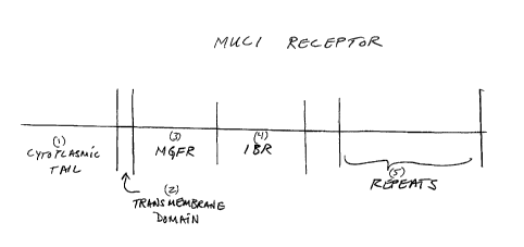
MUC1 comprises several regions termed herein as follows, recited in an
order starting from the region closest to the cell surface and progressing
away
from the cell. The basic structure of the MUC1 receptor is illustrated in
FIG.1
The receptor, as illustrated comprises: 1) cytoplasmic tail; 2) transmembrane
section; 3) MGFR; 4) IBR, and 5) repeats.
One aspect of the present invention features the discovery that a specific
region of the MUC1 receptor, i.e., the IBR, binds strongly to identical
regions of
other MUC 1 molecules. That is, the MUC1 receptor has the ability to aggregate
(i.e. self-aggregate) with other MUC1 receptors via the IBR of the respective
receptors. This self- aggregation may contribute to MUC1 receptor clustering,
observed in healthy cells. The discovery that the IBR portion of the MUC1
receptor self-aggregates is consistent with the following mechanistic model
for
which the inventors present supporting evidence. Mechanistic model: (1)
receptor
clustering is associated with the healthy state because the aggregated IBR
portions block access of ligands, such as growth factors, modifying enzymes
and
the like to the neighboring extracellular portions of the MUC1 receptor that
act
as the functional receptor; clustering also blocks access of intracellular
tails to
intracellular modifying enzymes and signaling ligands; (2) when the MUC1
receptor is cleaved at a position that releases the IBR, the critical force
that
keeps the receptors clustered is lost and receptors are free to migrate within
the
cell membrane or interact with modifying enzymes, secreted ligands such as
activating ligands or growth factors or other cell surface receptors; these
interactions could involve a new, inductive multimerization state, such as
dimerization, that triggers a cell proliferation signaling cascade.
Cleavage of MUC1 may occur at a site at or near the C-terminal boundary
of the IBR in tumor or cancer cells (between the cell and the IBR), releasing
the
IBR from the cell. Alternatively, cleavage of MUC1 may occur within the IBR
CA 02430060 2013-01-03
-20-
itself to cause sufficient disrupting of the IBRs such that the ability to
self-
aggregate is lost with the result that the MGFR becomes accessible to agents
or
ligands. As described in Example lb, the addition of 9 amino acids of the IBR
region to a peptide from the MGFR region (which does not self-aggregate),
confers some ability to self-aggregate. Alternatively, loss of aggregation of
MUC1
receptors need not necessarily be the result of cleavage. For example, an IBR
can
be absent as a result of alternative splicing of the MUC1 gene.
Before the present invention, a ligand(s) for the MUC1 receptor had not
been conclusively identified. Research articles suggested that the shed
portion of
the MUC1 receptor becomes a ligand for the portion of the receptor that
remains
attached to the cell surface after cleavage. In the present invention, this
hypothesis was tested, and it was determined not to be the case. Further, it
was
determined that altered sites of cleavage of the MUC1 receptor could result in
altered function. In a colorimetric colloid binding assay (described in
PCT/US00/01997, referenced above, as well as some other assays of the above-
referenced patent applications/publications) various fragments of the MUC1
receptor were tested for their ability to interact with each other fragment of
MUC1 as well as for their ability to self-aggregate. We found that one portion
of
the receptor, fairly close to the cell surface, aggregated with itself in a
high
affinity interaction. This suggested that this portion of the MUC1 receptor,
which
we termed the interchain binding region, kept the receptors tightly clustered
in a
healthy cell, and that enzyme cleavage of MUC1 at a site that released the
IBR,
would cause the receptors not remain clustered. This clustering may affect
cell
signaling in two ways. First, the clustering of the receptors on the cell
surface
may restrict access to portions of the receptor that are binding sites for
ligands
such as modifying enzymes or growth factors. Secondly, as is appreciated by
those skilled in the art, the intracellular portions (cytoplasmic tails) of
many cell
surface receptors are involved in signaling cascades that control programmed
cell
growth (proliferation) as well as programmed cell death (apoptosis). Receptors
that
CA 02430060 2003-05-27
WO 02/056022 PCT/US01/44782
-21-
are tightly clustered on the cell surface also have clustered cytoplasmic
tails within the
cell, which may prevent them from interacting with intracellular proteins
involved in
intracellular signaling.
In some cases, the MUC1 receptor may be cleaved to release the IBR, from the
cell surface. Alternatively, cleavage can result in a release of a sufficient
portion of the
IBR that causes the MUC1 receptor to lose the ability to self-aggregate. Loss
of
aggregation of MUC1 may have several ramifications. Release of the IBR or
sufficient
portion of the IBR from the cell surface allows the receptors to evenly
distribute on the
cell surface, leaving the cytoplasmic tails free to associate with
intracellular signaling
proteins. External agents, such as modifying enzymes and/or activating
ligands, are then
able to bind to the remaining extracellular portion of the receptor and induce
disease-
associated signals, either via a change in the multimerization state, i.e.,
inductive
multimerization, or as an induced conformational change. As is appreciated by
those of
ordinary skill in the art, ligands such as growth factors and hormones often
induce
receptor dimerization which triggers, in turn, an intracellular signaling
cascade.
Cell proliferation may result from accessibility of the MGFR portion to an
activating ligand which can interact with the MGFR portion. For example, the
self-
aggregating IBR of the MUC1 receptor may form a dense reticulum which
sterically
prevents a ligand such as a growth factor from intreacting with the MGFR
portion of the
receptor, which is proximal to the cell relative to the IBR. In a cancerous or
tumor cell,
this reticulum may be lost, allowing ligand interaction with the MGFR.
The above mechanistic model is consistent with a mechanism whereby the
portion of the MUC1 receptor, that remains attached to the cell surface after
shedding of
the IBR region, i.e. the MGFR, functions as a receptor for ligands that
trigger cell
proliferation. Evidence is also presented herein that indicates that this
portion of the
receptor is enzyme modified before it is able to be recognized by at least one
of its
ligands (See Example 8). This mechanism is demonstrated herein with a showing
that:
(a) an interaction between a ligand and this portion of the MUC1 receptor
(MGFR),
which dimerizes the receptor, triggers cell proliferation; and (b) blocking
the interaction
of this portion of the MUC1 receptor (MGFR) with its ligand(s), blocks cell
proliferation. When tumor cell lines, in which the MUC1 receptor is
homogeneously
expressed across the entire cell surface, are treated with an IgG antibody
raised against
the MGFR portion of the MUC1 receptor, the rate of cell proliferation is
greatly
CA 02430060 2003-05-27
WO 02/056022 PCT/US01/44782
-22-
enhanced, see Fig. 5. Since IgG antibodies are bivalent, i.e. one antibody
simultaneously
binds to two adjacent MGFR portions on the cell surface, these results
demonstrate that
the antibody acts as an activating ligand, mimicing the effect of a growth
factor, which
dimerizes MGFR portions, and thus triggers a cell proliferation signaling
cascade which
is consistent with signaling via the cytoplasmic tails of the receptors. This
finding leads
to two conclusions. First, an activating ligand(s) that binds to the MGFR
portion of the
MUC1 receptor causes inductive multimerization of the receptor. Secondly, an
effective
therapeutic strategy is therefore to block the MGFR portion of the receptor
with a
monomeric composition, thus preventing inductive multimerization and
subsequent
signaling cascades. For example, a single chain, or monovalent, antibody
raised against
the MGFR portion of the MUC1 receptor would function as an effective anti-
cancer
therapeutic. Another therapeutic strategy is to block the activity of enzymes
that modify
the receptor, which may be required for some ligand binding
The inventors have also discovered that cells that overexpress the MUC1
receptor
also have increased levels of a ligand(s) that dimerizes the MGFR present in
the lysates
and supernatants of these cells, see Example 3b and Fig. 8A-D for details. In
the colloid-
colloid interaction experiment, described in Example 3b, ligands that
simultaneously
bind more than one colloid-immobilized receptor, i.e. dimerize, cause a
solution color
change from pink to blue. Gold colloids that presented synthetic peptides
derived from
the MGFR portion of the MUC1 receptor (His-PSMGFR) were incubated with
lysates/supernatants from various cells lines known to overexpress, express,
or not
express the MUC1 receptor. Lysates from HTB-133 (T47D) cells, which
overexpresses
MUC1 (see Table 2), caused colloid suspensions to turn blue within 15 minutes,
indicating a high concentration of a ligand(s) in the lysate that interacts
with the colloid-
immobilized MGFR-derived peptides. In experiments with lysates from other cell
lines,
the rate of color change of the colloid solution, which indicates the amount
of ligand
present, correlated to the degree of expression of the MUC1 receptor in those
cell lines
with cells.
More than one species may be a physiologically relevant ligand for this
portion of
the MUC1 receptor. Enzymes that modify the receptor may be relevant ligands of
this
portion of the receptor. For example, one ligand may bind monomerically to an
unmodified MGFR portion of the MUC1 receptor, while another ligand, with a
different
function, such as inductive multimerization, may recognize an enzyme-modified
version
CA 02430060 2003-05-27
WO 02/056022 PCT/US01/44782
-23-
of the receptor. Because the experiment described above, Example 3b, was
performed in
cell lysate/supernatants, it is important to note that several receptor-ligand
interactions,
including enzymatic modifications to the receptor, may be taking place,
wherein only the
ligand(s) dimerization (or multimerization) of the MGFR portion, results in a
solution
color change. In an experiment similar to Example 3b, (Example 8) the enzyme
inhibitor,
PMSF was added to the lysate prior to the introduction of the colloids bearing
the
synthetic peptide His-PSMGFR, see Table 1 SEQ ID # 2. Referring now to Figure
20,
solutions that contained PMSF did not undergo the solution color change. This
result is
consistent with a mechanism in which the MGFR portion of the MUC1 receptor is
first
enzyme mofdified before it is recognized by the ligand(s) that dimerize or
multimerize
the receptor
The inventors reasoned that prior attempts by others to identify ligands for
the
MUC1 receptor were hampered by the self-aggregation properties of the
receptor.
Therefore, only the MGFR portion of the receptor was used as bait to fish
ligands out of
lysates and supernatants. The His-PSMGFR sequence of Table 1, was immobilized
on
NTA-nickel beads via the histidine tag of the peptide, the beads were then
incubated with
lysates and supernatants from a variety of cell types, including cancer cell
lines that
overexpress MUC1. Enzyme inhibitors such as PMSF were added to some of the
lysates
and supernatants to circumvent problems of alternative ligands binding to
modified
versions of the peptide. Following incubation of the cell supernatants with
the
PSMGFR-presenting beads, the beads were washed, then the peptide-ligand
complexes
eluted from the NTA-Nickel beads by adding excess imidazole. Captured ligands
from
the probe peptides, eluates, were separated using standard SDS-PAGE methods.
Protein
bands were excised from the gels and analyzed to identify the target ligands.
Standard
methods for protein analysis including peptide micro sequencing and tandem
mass spec
were used in these studies. Other methods can be used to identify MUC1
ligands,
including ligand fishing with beads, MALDI mass spec, and the like.
Accordingly, another aspect of the invention involves the identification of
ligands, derived from lysates from a cell line selected from the group
consisting of HTB-
133, CRL-1504, and CRL-1500, that bind to the MGFR portion of the MUC1 cell
surface receptor, see Figs. 9 and 10. In some embodiments, the ligands may
include
sequence(s) from: Metastasis Inhibition Factor NM23, 14-3-3, Cathepsin D,
annexin,
Beta lipotropin (Beta-LPH), beta-melanotropin or Beta-MSH. In other
embodiments, the
CA 02430060 2003-05-27
WO 02/056022 PCT/US01/44782
-24-
biomolecule that binds to the MGFR portion may be a cleavage product of
proopiomelanocortin (POMC). In all embodiments, the preferred cell surface
receptor is
MUCl. In one embodiment, the MGFR portion includes some or all of the sequence
from the PSMGFR peptide (SEQ ID NO: 7). These ligands may exist in a
multimeric
state including dimers, tetramers, or complexes containing some or all of
these ligand
species. In one aspect, the invention involves modification and use of the
above species
as anti-cancer agents.
In one embodiment the ligand is a 23kD protein. In another embodiment, the
ligand is approximately 17kD. In a preferred embodiment, the ligand identified
is a
protein that migrates through a gel with an apparent molecular weight of
351(D. Since
this species is much more apparent when the protease inhibitor PMSF is not
added to the
lysate (Fig. 10), this protein may be an enzyme that modifies the MUC1
receptor or a
ligand that recognizes the unmodified version of the receptor. Experiments
were
performed as described in more detail below in Example 4b to characterize
these species.
Peptide sequences contained within both the 17 kD and the 23 kD bands (PMSF
added to lysate) corresponded to a protein known as Metastasis Inhibition
Factor NM23,
which has been implicated in both the promotion and inhibition of metastasis
of human
cancers. Whether the role of NM23 is a tumor supressor or promoter may depend
on the
type of cancer. In ovarian, colon and neuroblastoma tumors, NM23
overexpression has
been linked to a more malignant phenotype (Schneider J, Romero H, Ruiz R,
Centeno
MM, Rodriguez-Escudero FJ, "NM23 expression in advanced and borderline ovarian
carcinoma", Anticancer Res, 1996; 16(3A): 1197-202). However, breast cancer
studies
indicate that reduced expression of NM23 correlates with poor prognosis (Mao
H, Liu H,
Fu X, Fang Z, Abrams J, Worsham MJ, "Loss of NM23 expression predicts distal
metastases and poorer survival for breast cancer", Int J Oncol 2001
Mar;18(3):587-91).
Because NM23 exists as a hexamer, in MUCl-presenting cells, it may function to
hold
MUC1 receptors in a clustered configuration to restrict access of the MGFR to
modifying and activating ligands. The sequences that were identified from the
protein gel
band described in Figures 9 and 10 and that also exist in Metastasis
Inhibition Factor
NM23 are shown below in Table 4
Peptide sequences that are associated with the 35 kD gel band (PMSF NOT
added to lysate) corresponded to more than one protein species, including 14-3-
3, which
is a signaling protein implicated in many cancers, and cathepsin D, which is a
protease
CA 02430060 2003-05-27
WO 02/056022 PCT/US01/44782
-25-
and is also implicated in tumor progression. 14-3-3 exists as a dimer and can
simultaneously bind to two, identical phospho-serine peptides. This protein
has been
shown to be involved in intracellular signaling, but particularly relevant to
this invention
is the fact that 14-3-3 has been shown to be secreted by some cell types,
including
dendritic cells. Ligands that bind to the MGFR portion of the receptor to
induce
inductive multimerization would tend to be secreted factors. This protein
would
dimerize the MUC1 receptor to trigger cell proliferation, which is consistent
with the
mechanism presented herein. Cathepsin D is a protease and may be involved in
the
cleavage of the MUC1 receptor.
Yet another aspect of the invention involves the identification of other
ligands,
also derived from supernatants from a cell line selected from the group
consisting of
HTB-133, CRL-1504, and CRL-1500, that bind to the MGFR portion of the MUC1
cell
surface receptor. In one embodiment, the ligand identified is a protein that
migrates
through a gel with an apparent molecular weight of 55kD. In another
embodiment, the
ligand is approximately 70kD; 80kD; or 100kD. In a particular embodiment, the
ligand
present in cell supernatants is a 13kD protein, see Fig. 9., which migrates
though gel with
apparent molecular weight of 13kD. The protein with an apparent molecular
weight of
about 13 kD appears upon polyacrylamide gel electrophoresis as a smeared band
which
may indicate that the protein is glycosylated, enzyme modified, or that the
band contains
more than one protein species. Using mass spec and maldi mass spec techniques,
combined with homolgy to peptide sequences in the Genbank database, it was
determined that two fragments derived from the 13 kD corresponded with a high
degree
of homology to beta-lipotropin (Odell W, Wolfsen A, Bachelot I, and Hirose F,
(1979)
"Ectopic production of lipotropin by cancer" The American Journal of Medicine
66; pgs.
631-638), which was previously known as beta-MSH (beta-melanotropin). Beta-
lipotropin (beta- LPH: 98 amino acids or about 10 kd) and ACTH (aa' 130-169)
are
cleavage products of proopiomelanocortin (Publisher Williams & Wilkins,
chapter
authors: Faye W, Lemke T, Williams D, Text book ¨ Principles of Medicinal
Chemistry;
Fourth edition 1995) (POMC: 260 amino acids). Because these peptides are
glycosylated, their apparent molecular weights can be altered from their
actual molecular
weights. These cell surface receptor binding ligands can be purified or
produced using
techniques known to those of skill in the art. It is also possible that in
certain situations
an external ligand does not bind to and dimerize the MGFR portions of the MUC1
CA 02430060 2003-05-27
WO 02/056022 PCT/US01/44782
-26-
receptor to trigger cell proliferation, but rather the receptors become
covalently coupled
to each other, for example by an enzyme that covalently links the two. One way
this
could be accomplished is by an enzyme that attaches an entity, such as a sugar
group, to
both receptors.
Because the portion of the MUC1 receptor that self-aggregates (IBR), and in
doing so may protect intracellular signaling sequences from participating in
signaling
cascades that induce proliferation, can be cleaved and shed from the cell
surface, it can
be beneficial to identify small molecules that interact with the MGFR portion
of MUC1
that remains cell-attached (MGFR). These small molecules that bind to the
portion of
1() MUC 1 that remains cell-attached (MGFR) can then be used in two ways.
First, they can
be used to block the remaining, cell-attached portion of the MUC1 receptor so
that it
cannot interact with activating ligands that induce proliferation and
metastasis. For
example, a ligand to the cell-attached portion of MUC 1 may effect inductive
multimerization of the receptor, causing a signaling cascade inside the cell.
Blocking
binding of the ligand to the MGFR region can inhibit the signaling cascade
that causes
proliferation. Secondly, as discussed in more detail below, the small
molecules can be
polymerized or attached to dendrimers to artificially cause preventative
clustering the
cell-attached MGFR portions of the MUC1 receptors and thus shield the
cytoplasmic
tails from interaction with intracellular signaling proteins.
The findings of the invention have important implications for diagnostic and
therapeutic procedures. For instance our finding that a fragment of the MUC1
receptor
that is close to the cell surface aggregates with itself, indicates that the
position of
enzyme cleavage is associated with receptor clustering, accessibility of
adjacent portions
of the receptor to putative ligands, and thus cancer. Agents that modulate the
activity of
this enzyme may be potent anti-cancer agents. Additionally, an early
diagnostic test for
cancers that aberrantly express MUC1 may be based on detecting the portion of
MUC1
that self-aggregates (IBR) circulating in bodily fluids. This portion of MUC1
includes
part or all of the PSIBR (sequence shown in Table 1). Agents that bind to the
portion
(some or all of the PSMGFR sequence) of MUC1 that remains attached to the cell
surface after the release of the portion that self-aggregates (IBR ¨ some or
all of the
PSIBR sequence) may be potent anti-cancer drugs. In addition, agents that
block binding
of the natural ligand to the remaining portion after the release of the IBR,
may also be
useful as anticancer drugs. Drug candidates that target portions of the MGFR,
ie.,
CA 02430060 2003-05-27
WO 02/056022 PCT/US01/44782
-27-
sequences including some or all of the amino acids contained within the
PSMGFR, of the
receptor can be used either as monomers, to block the interaction of the MGFR
portion
of MUC1 with extracellular agents or biomolecules, or as a polymer, dendrimer,
etc. to
both block the interaction with external biomolecules and to artificially
cluster the
MGFRs. Another alternative agent, which can be used to artificially cluster
the MGFRs
is an IgM antibody raised against the MGFR or PSMGFR. This artifically-induced
clustering may serve to keep the cytoplasmic tails clustered to prevent
interaction with
intracellular signaling agents, thereby effecting preventative clustering.
One aspect of the invention involves s novel drug screening assays, that
identify
therapeutics that interfere with the proliferation of tumor cells that
aberrantly express
MUC1. The drug screen makes use of the new molecular target for cancer that is
disclosed herein. Another aspect of the invention involves therapeutics
identified by the
drug screen. Yet another aspect of the invention involves methods for
diagnosing
MUC1 cancers, which is based upon the mechanism elucidated by the inventors.
One embodiment of the invention involves a drug screening assay which can
rapidly identify agents that interrupt the interaction between the MGFR and
its ligand(s)
and thus can be used as cancer therapeutics, (see Example 5a and Fig. 12 for
details).
The fact that an activating ligand(s) that binds to the MGFR portion of the
MUC1
receptor can result in inductive multimerization of the receptor, allows us to
construct a
convenient drug-screening assay to identify compounds that inhibit this
interaction or
inhibit enzymes that modify the MGFR portion required for ligand binding and
thus
inhibit the proliferation signal. In one assay of the invention, synthetic
peptides which
include much or all of the MGFR sequence are attached to nanoparticles such as
gold
colloids. Gold colloids have the intrinsic optical property that when in a
disperse,
homogeneous solution, the solution appears pink, but when the colloids are
forced into
close proximity, the solution turns blue. When cell lysates or supernatants,
which contain
a ligand(s) that dimerizes the colloid-attached peptides, are added, the
colloids becomes
aggregated and the solution turns blue. Drug candidates that interrupt this
ligand-
receptor interaction are easily identified because they cause the solution to
remain pink.
As discussed above, it appears that the ligand that binds to the MGFR portion
of
the receptor to trigger inductive multimerization may recognize an enzyme-
modified
form of the receptor. Therefore the above-described drug screening assay can
identify
compounds that: a) inhibit enzyme modification of the MGFR portion of the MUC1
CA 02430060 2003-05-27
WO 02/056022 PCT/US01/44782
-28-
receptor; b) bind to the MGFR region and block its interaction with an
activating ligand
of the MGFR portion; or c) bind to an activating ligand, such as a growth
factor and
block its interaction with the MGFR portion. Drugs that act according to (a)
or (b) are
predicted to selectively inhibit MUC1-dependent cell proliferation, whereas
drugs that
act according to (c), on agents such as growth factors, are expected to
inhibit
proliferation of a variety of cell types, see Examples 5a-d.
Drugs that have been found by the above protocol to disrupt an interaction
between the MGFR portion of the MUC1 receptor and its ligand(s) are described
in
much greater detail below and include, for example, calcimycin, fusaric acid,
L-a-
methyl-dopa, butylindazone, NS1619 or etomoxir. Additional drugs are described
in
greater detail in applicant's co-pending U.S. Provisional Patent Applications
Serial Nos.
60/317,302 and 60/317,314, both filed on September 5, 2001 and entitled
COMPOSITIONS AND METHODS OF TREATMENT OF CANCER.
Agents so identified may be potent anti-cancer agents either in monomeric form
or as polymers or dendrimers. Drug libraries and peptide libraries can be
screened for
molecules that inhibit binding of the ligand to its target.
Alternatively, standard methods can also be used to identify agents that
interrupt
the interaction between the MGFR and its ligand(s). These methods can be used
to
identify agents that bind: (1) to the MGFR portion of the MUC1 receptor, or
(2) to one
or more of its ligands. These methods include but are not limited to phage
display
methods, yeast two-hybrid system, sandwich assays, surface plasmon resonance-
based
assays, antibody-based assays, peptide bead assays for testing with drug
libraries, bead
assays, GFP-reporter assays, and the like. Ligands to the MGFR portion of the
MUC1
receptor can be identified by a number of methods including using a peptide
whose
sequence corresponds to some or all of the MGFR, ie. the PSMGFR peptide
(Sequence
ID 7), as bait to fish out ligands. Another way to do this is to attach a
signaling entity,
such as GFP (green fluorescent protein), directly or indirectly to the ligand
and attach the
receptor-derived peptide to a solid support. Compounds that interfere with the
interaction will cause a loss of signal.
As an alternative to the natural ligand competition assays described above,
direct
binding assays can be employed to identify drug agents able to interact with
the MGFR.
Small molecules that bind to the remaining extracellular portion of cleaved
MUC1
CA 02430060 2003-05-27
WO 02/056022 PCT/US01/44782
-29-
(MGFR region:exemplified by most or all of the PSMGFR peptide sequence), can
be
identified using standard methods (MALDI, western blotting, gel
electrophoresis,
ELISA, etc) or using colloid-colloid or colloid-bead binding assays. For
example, in one
embodiment, small molecules can be synthesized on beads or attached to
colloids either
by attachment to a thiol and direct incorporation into a SAM on the colloid or
by
EDC/NHS coupling of molecules containing a primary amine. A histidine-tagged
peptide including the desired MUC1 (e.g. His-PSMGFR) sequence can be bound to
colloids and assayed for interaction with a second set of colloids presenting
a candidate
drug. A color change from pink to blue would indicate an interaction between
the
MUC1 peptide and the small molecule. Alternatively, the MUC1-bearing colloids
can
be assayed for an interaction with a small molecule attached to a bead. Red
coloration of
the bead surface would signify that the MUC1 peptide bound to the bead-
immobilized
small molecule.
Any drugs or small molecules identified as binding to the MUC1 sequence can
potentially be used to block binding of the remaining extracellular portion of
cleaved
MUC1 to its natural ligand, and can potentially inhibit cancer growth.
In another embodiment, the above-described competition assays are employed to
identify "second generation" drug candidates by assaying such candidates for
their
ability to disrupt an interaction between the MGFR portion of the MUC1
receptor and a
synthetic ligand found, according to the inventive methods described above, to
bind to
the MGFR, e.g. calcimycin, fusaric acid, L-a-methyl-dopa, butylindazone,
NS1619 or
etomoxir. In this way, drugs that bind the MGFR with higher affinity than the
"first
generation" drug can be identified. For example, in performing the drug
screen, a
synthetic ligand such as calcimycin, fusaric acid, L-a-methyl-dopa,
butylindazone,
NS1619 or etomoxir is modified to facilitate attachment to surfaces, such as
colloids. A
peptide derived from the MGFR region of the MUC1 receptor, such as the His-
PSMGFR, is attached to a second set of colloids. Direct binding between the
synthetic
ligand and the MGFR peptide is confirmed, a library of drug candidates are
then assayed
for their ability to disrupt the interaction. It is not intended that this
aspect of the
invention be limited to the competition techniques or assays explicitly
described herein.
Several techniques, including standard methods, could potentially be used to
detect
CA 02430060 2003-05-27
WO 02/056022 PCT/US01/44782
-30-
binding between the MGFR and a synthetic ligand or drug, then competitive
inhibition of
binding by a drug candidate.
Another aspect of the invention is a drug screening assay for identification
of
drugs that can be useful for prevention and/or treatment of cancer by altering
the
cleavage state of MUC 1 receptors on cells. In such assays, described in more
detail
below, cultured cells are exposed to candidate drugs. The cleavage state of
MUC 1 in
the cells is determined, optionally as a function of time and/or dosage or
other conditions
involving exposure to the drugs. These cells can be derived from a particular
patient, or
can be tumor-associated or non-tumor-associated cell lines. Customized
therapeutic
protocols can be determined for a particular patient in this manner. The
invention
involves, in one aspect, treating a patient with a drug, as discused in
greater detail below,
shown to affect the cleavage state of MUC 1 of the patient's cells in a manner
that
prevents, inhibits, or reverses cancer.
Because it is suspected that the incorrect cleavage of MUC1 on the surface of
the
cell causes the cascade leading to proliferation and tumorigenesis, it would
be
advantageous to test candidate drugs in a whole cell assay for their ability
to affect
enzyme cleavage or the position of enzyme cleavage of MUC l. Drugs can be
screened
for their ability to effect MUC1 cleavage, either directly or indirectly. This
can enable
the identification of upstream effectors of MUC1 cleavage. Cells or tissue
samples can
be assayed for MUC1 cleavage potential in several ways. For example, cells or
a tissue
sample can be treated with a drug candidate and grown for some period of time.
Colloids bearing an antibody, natural ligand, or small molecule that binds to
either the
cleaved portion of MUC1, or the remaining extracellular portion (plus or minus
a
signaling moiety) can be added and allowed to bind to the cells or tissue
sample. The
expression of cleaved MUC I or uncleaved MUC1 on the cell surface as compared
to a
control sample, not treated with the candidate drug, would indicate whether
the drug
candidate effected MUC1 expression or cleavage.
Alternatively, MUC1 expressing cells can be assayed for MUC1 cleavage in the
presence of a drug candidate by testing the surrounding cell growth media for
the
presence of cleaved MUC1 or the potential of the cleaved portion to self-
aggregate. For
example, cells expressing MUC1 would be treated with a drug candidate
suspected of
interfering with enzyme cleavage. After some incubation period, the cell media
would
be removed and tested for its aggregation potential, i.e. to determine whether
the self-
CA 02430060 2003-05-27
WO 02/056022 PCT/US01/44782
-3 1 -
aggregating portion of MUC1 was contained within the shed fragment. The
aggregation
potential of peptides released into the cell media is tested by adding
colloids bearing an
antibody to a sequence distal from the self-aggregating portion, but not a
repeat
sequence. In this way, antibody-presenting colloids would attach to upstream
regions of
MUC 1. If the self-aggregating region was also attached to the released
fragment, then
this would cause aggregation of the attached colloids and a solution change
from pink to
blue would result. Accordingly, one aspect of the invention provides a
composition
(drug or agent) that, in contrast to preventing inductive multimerization of
cleaved or
modified MUC1 receptors as discussed previously, instead prevents disease-
associated
MUC 1 cleavage or modification itself. MUC 1 cleavage is an enzymatic process.
The
determination of the mechanism involved by the inventors can lead to
identification of a
drug that inhibits disease-associated cleavage.
As mentioned above, another aspect of the invention provides an agent that
binds
together MGFR portions of MUC 1 following disease-associated cleavage to
effect
preventative clustering of the receptors. The agent can be any species that
includes
multiple sites each able to bind to a MFGR portion, and immobilized with
respect to
each other. E.g. a polymer or dendrimer or other continuous entity can include
multiple
sites each able to bind to a MGFR portion, causing clustering of these
portions or other
structural constraint that inhibits their association with factors that
promote cell
proliferation. Alternatively, IgM-type monoclonal or polyclonal antibodies
raised
against the MGFR or PSMGFR could be utilizied. Each anti-MGFR IgM antibody
could
be able to aggregate ten MGFRs on the cell surface to form preventative
clusters.
In addition, some or all of the above-identified ligand species that bind to
the
MGFR can be modified to allow the ligands to act as a targeted delivery agent
by
attaching a cytotoxic drug or other agent (e.g. a radioactive substance) able
to selectively
kill cells to which the ligands become immobilized. In addition, synthetic
ligands, such
as calcimycin, fusaric acid, L-a-methyl-dopa, butylindazone, NS1619 or
etomoxir,
discussed in more detail below, that were found to bind to the MGFR portion of
the
MUC1 receptor can similarly be modified with other therapeutic agents. In this
way,
such a therapeutic can be directed to the tumor cells. For example, an agent
that binds to
the MGFR region of the MUC1 receptor can be modified with a radioactive
substance to
destroy tumor cells that aberrantly express the MUC1 receptor. Other toxic
substances,
CA 02430060 2013-01-03
WO 02/0561122 PCT/US01/44782
-32-
such as ricin, as well as other therapeutics, can be attached to agents that
bind the
MGFR. Alternatively, identified ligand species that bind to the MGFR could be
modified
to present a imaging agent for use in diagnostic imaging of MUC 1+ tumors and
metastases. Such ligands can also, alternatively, be modified to act as drugs
that can be
useful for prevention and/or treatment of cancer. In one embodiment, a ligand,
which in
its unmodified form binds to multiple MGFRs causing inductive multimerization,
is
modified to remove or de-activate all but one of its active binding sites for
MGFR, such
that each modified ligand is able to bind to only a single receptor. In
another
embodiment, individual ligand molecules/peptides are modified such that they
are
to immobilized with respect to additional ligand molecules/peptides also
able to bind
MGFR, e.g. through covalent coupling, non-covalent coupling, co-immobilization
with
respect to a substrate, etc., such that the modified, multi-unit ligand is
able th effect
preventative clustering of the receptors to which it binds.
Identification of the ligand(s) for the portion of MUC1 that remains bound to
the
is cell after cleavage can allow, as discussed further below, for
development of powerful
assays to screen for drugs that disrupt this interaction. Interaction of
potential binding
partners with the extracellular portion of MUC1 that remains after cleavage
can be
studied both by conventional techniques (western blotting, ELISA, MALD1, etc.)
and
using our colloid-colloid color change assay or colloid-bead coloration assay.
The
20 peptide sequence of the remaining extracellular portion of MUC1 can be
attached to
beads or colloids via a histidine tag. Potential binding partners can be
histidine-tagged
and attached to a second set of colloids (or beads) and assayed for binding to
the colloid-
immobilized portion of MUC1. Alternatively, potential binding partners can be
attached
to beads or colloids by EDC/NHS coupling or can be nonspecifically adsorbed to
beads
25 for the assay. An interaction between the MUC1 peptide and the potential
binding
partner can be detected by either a change in solution color (for the colloid-
colloid assay)
or by agglomeration of the colloids onto the bead, causing the bead to appear
red (for the
colloid-bead assay). An entire cDNA library can be screened using this
technique in a
short period of time to identify the natural ligand of the remaining
extracellular MUC I.
30 (see PCT/US00/01997, WO 00/34783, 09/631,818, and "Detection of Binding
Species
with Colloidal and Non-Colloidal Structures", filed 11/15/00.
The discoveries presented herein: (1) that the IBR of MI.JC1 self-aggregates;
(2)
that an antibody that dimerizes adjacent MGFR portions of the MUC1 receptor
leads to
CA 02430060 2003-05-27
WO 02/056022 PCT/US01/44782
-33-
proliferation of MUC1 presenting tumor cells; and (3) that proliferation of
MUC1
presenting tumor cells can be inhibited by treatment with agents that target
the MGFR
and block the MGFR against interaction with a ligand, are consistent with a
mechanism
in which, in a healthy cell, MUC1 cleavage occurs such that enough of the IBR
remains
on the cell that MUC1 remains clustered, and the MGFR is inaccessible to
ligands such
as growth factors, and in a tumor cell, MUC1 cleavage occurs such that enough
of the
IBR is cleaved from the cell such that MUC1 does not remain clustered, and the
MGFR
is accessible to ligand interaction. This leads to diagnostics, provided by
the present
invention, in which the shed portion of MUC1 is analyzed to determine the
degree of
IBR present.
The above-mentioned mechanistic model predicts that in a subject with a MUC1-
dependent tumor or who is prone to developing such a tumor, the portion of the
MUC1
receptor that is shed will contain the IBR region of the receptor, leaving the
MGFR
portion of the receptor accessible for interactions with ligands and growth
factors. An
early diagnostic would consist of detecting the IBR region in the portion of
the MUC1
receptor which is shed.
In one embodiment, loss of aggregation of MUC1 receptors can result from the
"cleavage state" of MUC 1. "Cleavage state" defines the result of cleavage of
MUC 1.
The cleavage state will differ between a healthy cell and a cell with tumor
potential. The
cleavage state determination can involve determining whether cleavage occurs
in a
manner such that the normal interaction between the IBRs of neighboring
receptors is
disrupted (these regions no longer remain bound to each other at the cell) and
MUC 1 is
free to spread across the cell surface. More specifically, determination of
the cleavage
state can include determining a site of cleavage of MUC 1, determining the
identity of a
portion of MUC 1 that remains at a cell following cleavage, determining the
identity of a
portion of MUC 1 that is separated from the cell following cleavage ("cleaved
or shed
portion"), determining the accessibility of MGFR, or a combination.
In one embodiment, an assay is provided that can determine whether the MUC1
IBR remains fastened to the cell, or is separated from the cell upon cleavage.
The
product of MUC1 cleavage from the cell is exposed to at least one surface
adapted to
bind the IBR and to another surface and/or a signaling entity. Generally, an
assay as
described in WO 00/43791 or WO 00/34783 can be used. In a specific example,
antibodies to a portion of MUC1 that would remain fastened to the IBR if the
IBR is
CA 02430060 2003-05-27
WO 02/056022 PCT/US01/44782
-34-
cleaved from the cell, such as antibodies to the repeats domain, are fastened
to colloids.
Exposure of these colloids to the MUC1 cleavage products will allow the IBR
regions to
self-aggregate which in turn will result in colloid/colloid aggregation (color
change to
blue) for cases in which the IBR portion is separated from the cell. This is
because each
colloid will fasten to a MUC 1 region which is connected to an IBR which is
fastened to
another IBR which is in turn fastened to another colloid.
The discovery that tumor cells can be treated with an agent that binds to the
MGFR of MUC 1, or a ligand of MGFR, in a manner that inhibits cell
proliferation leads
to the conclusion that, in a diseased cell (a cancerous cell or a cell with
potential for
becoming cancerous), cleavage of MUC 1 occurs in a manner that allows MGFR to
interact with at least one ligand in a manner that promotes tumorigenesis or
cancer.
Interaction with the ligand may be due to cleavage that disrupts binding
between
different MUC 1 molecules at the cell surface, either via separation from the
cell of the
IBR during cleavage, or cleavage within the IBR at a location that frees MUC 1
molecules from each other.
In one aspect of the invention, a diagnostic is provided wherein an amount of
cleavage of a cell surface receptor IBR from the cell surface is determined.
This
involves the determination of the amount of cell surface receptor IBR that is
separated
from the cell surface upon cleavage, which can be relative to the level
determined in past
samples, the level in control samples, or can be determined as a ratio of the
amount of
IBR to the amount of a constant region of the receptor in a sample. The
constant region
is any non-repeating sequence which is N-terminal to the boundary of the
PSIBR, that is,
this region is present in a 1:1 ratio with the PSIBR prior to cleavage ¨
determination of
the ratio of IBR to constant region subsequent to cleavage indicates the
extent to which
the IBR is cleaved and separated from the cell. The amounts of various
receptor regions
may be determined with any type of binding assay, e.g. an antibody-binding
assay. For
example, antibodies that specifically bind to the constant region or the
repeats may be
attached to surfaces (e.g. magnetic beads) to preconcentrate shed MUC 1
receptors prior
to determining levels of IBR present. Then, for example, after pre-
concentration of
circulating MUC 1 receptors, antibodies to the IBR and antibodies to the
constant region
can be allowed to bind to the cleaved receptors, and determination of the
ratio of binding
of these antibodies reveals the ratio of IBR present relative to constant
region present in
the cleaved receptors, which in turn reveals the amount of cleavage that
occured in a
CA 02430060 2003-05-27
WO 02/056022 PCT/US01/44782
-35-
manner that caused separation of the IBR from the cell. A ratio that displays
a trend
toward less than one (less than a 1:1 ratio of IBR relative to constant region
present) for
detecting IBR at a cell surface is an indicator of the presence of a tumor or
the potential
for the development of a tumor. A ratio that approaches 1:1 when detecting
these
regions in shed receptors is likewise an indicator of cancer potential. This
determination
can indicate potential for tumor formation, existence of a tumor, progression
of
tumorigenesis, etc., and can thereby serve as a diagnostic and/or a evaluator
of treatment
for tumorigenesis
Another diagnostic aspect of the invention involves determining levels of shed
IBRs in sample from a subject. Methods for such determination can include
determining
the aggregation potential of the circulating shed receptors, for example via
colloid-
binding assays such as a colloid-colloid assay or a colloid bead assay (See
above
discussion and Examples, below). Alternative techniques involve determining
the
presence of the IBR using antibody probing assays, hybridization, PCR Reverse
Transcriptase PCR (rtPCR), Ligase Chain Reaction (LCR), cycling probe
technology,
etc. In a preferred embodiment of the invention, the cell surface receptor is
MUCl.
The determination, in a blood sample, of the amount of cleaved receptor
carrying
IBR, either involving antibody binding ratios, colloid binding assays, or the
like can be
made on a bodily fluid sample, such as a blood sample and optionally compared
with
other samples (e.g. to monitor the subject's progression of tumorigenesis or
progression
for treatment of the same) and/or controls.
Alternatively, biopsy specemins can be studied, or tissue can be studied
interoperatively (e.g. tissue at a surgical site can be studied without
removal of the tissue
from the subject). In either of these studies, a primary indicator of
tumorigenesis or
potential for tumorigenesis is the amount of MGFR at a cell surface accessible
to
interaction with external agents such as growth factors, etc. This
determination can be
made, for example, by determining the amount of an antibody to the MGFR region
that
binds to the sample, either using standard antibody binding study techniques,
or by
exposing the sample to colloids to which antibodies specific to the MGFR
region have
been immobilized and determining binding of the colloids to the samples using
techniques described in International patent publication numbers WO 00/34783
and WO
00/43791, referenced above. In another technique (perhaps more suited for an
excised
sample), antibodies to the MGFR region and to the IBR can be exposed to the
sample
CA 02430060 2003-05-27
WO 02/056022 PCT/US01/44782
-36-
and a determination made of the ratio of binding of each to the sample. A
healthy
sample will exhibit little or no antibody binding to the MGFR region. A sample
indicating tumerigenesis or potential for tumorigenesis will show a non-zero
ratio of
MGFR antibody binding to IBR antibody binding.
Whether shed MUC1 contains the IBR can be identified by methods known to
those of ordinary skill in the art, including ELISA and colloidal assays as
described in
international patent application serial no. PCT/US00/01997, filed 01/25/00,
entitled
"Rapid and Sensitive Detection of Aberrant Protein Aggregation in
Neurodegenerative
Diseases", published as no. WO 00/43791, and U.S. patent application serial
no.
09/631,818, filed 08/03/00, entitled "Rapid and Sensitive Detection of Protein
Aggregation", by Bamdad et al. In a preferred embodiment of the invention, the
cell
surface receptor is MUCl.
Another aspect of the invention involves determining the site of cleavage of
the
MUC1 receptor from a cell surface (rather than the amount of IBR in a shed
portion) in a
sample from a subject to evaluate cancer, or the potential to develop cancer
in a subject.
Determination of the site of cleavage will give information as to whether the
IBR
remains on the cell surface, or was shed from the cell surface, giving
indication of cancer
or tumorigenesis or the potential for either, as discussed above. Determining
the site of
cleavage can be accomplished by using enzyme-amplification methods such as
PCR.
Specifically, using alternative primer sites in PCR amplification of shed MUC1
from
subject's sample will indicate where cleavage occurred.
In one aspect of the invention, differences in pre- and post-treatment levels
of
cleaved cell surface receptor IBR, or cell surface receptor IBR at the surface
of a cell, in
cancer cells or tissues may be used to diagnose cancer in a subject or assess
the
effectiveness of treatment in a cancer patient. In a preferred embodiment the
cell surface
receptor is MUCl.
Comparison of the levels of the above-mentioned regions with levels from
subjects known to be free of cancer may allow determination of the presence of
cancer in
the subject. An example, although not intended to be limiting, is that a
determination of
the presence of elevated levels of cleaved cell surface IBR in a sample from a
subject,
when compared to a level determined in samples from control subjects, may
suggest the
presence of cancer in the subject with elevated levels. Such methods of
comparing levels
of cancer-associated markers between a sample from a subject and a control
sample for
CA 02430060 2003-05-27
WO 02/056022 PCT/US01/44782
-37-
diagnostic purposes would be understood by one of ordinary skill in the
medical arts.
Examples of such methods include Western blotting, ELISA, antibody
precipitation,
PCR, LCR, rtPCR, cycling probe technology, and colloidal assays as described
in
international patent application serial no. PCT/US00/01997, filed 01/25/00,
entitled
"Rapid and Sensitive Detection of Aberrant Protein Aggregation in
Neurodegenerative
Diseases", published as no. WO 00/43791, and U.S. patent application serial
no.
09/631,818, filed 08/03/00, entitled "Rapid and Sensitive Detection of Protein
Aggregation", by Bamdad et al. In a preferred embodiment the cell surface
receptor is
MUCI .
In another aspect of the invention, the cleavage state of MUC 1 can be used to
determine progression or regression of a subject's cancer over time. The
cleavage state
also can be used to assess treatment parameters including, but not limited to:
dosage,
method of administration, timing of administration, and combination with other
treatments as described herein.
Another aspect of the invention involves extremely early-stage cancer
diagnosis.
This aspect involves identification of patients who may be at risk for
developing tumor
or cancer associated with abnormal cleaveage of MUCl. These patients may not
have
developed tumors, but may exhibit a cleavage state indicative of a condition
that can lead
to cancer. In some instances, the subjects will already be undergoing
treatment for
cancer, while in other instances the subjects will be without present cancer
treatment. A
test for a genetic predisposition to cancers characterized by aberrant MUC1
expression of
the invention is based on detecting genetic alterations in the MUC1 cleavage
enzyme(s),
over expression of MUC1 activating ligands, and/or overexpression of enzymes
that
modify the MGFR portion of the receptor.
The fact that elevated levels of cleaved MUC1 are found in the blood of cancer
patients is the basis for a blood test for breast cancer, which is not
described herein.
MUC I is cleaved by at least one enzyme and may be cleaved at more than one
site
before it is released into the blood stream. Several protease cleavage sites
within the
MUC1 sequence are predicted. Predicted sites for enzyme cleavage are at or
near amino
acid 540, 528, 530, 542, and 550 (numbers are as listed in Andrew Spicer et
al., J. Biol.
Chem Vol 266 No. 23, 1991 pgs. 15099-15109; these amino acid numbers
correspond to
numbers 1100, 1088, 1090, 1102, 1110 of Genbank accession number P15941; PID
G547937; Boshel M., et al, BBRC vol. 185 pgs 1-8; Hilkens J., et al, Journal
of
CA 02430060 2003-05-27
WO 02/056022 PCT/US01/44782
-38-
Biological Chemistry vol 267 pgs 6171-6177). The exact site of cleavage may
vary
depending on cell type or in response to a disease. The enzyme that cleaves
MUC1 may
be a membrane-associated enzyme, since the clustered IBRs of the MUC1 receptor
limit
access to the cleavage sites.
One aspect of the invention is the identification of compounds that directly
bind
to the PSMGFR portion of the receptor. Therefore, a sensitive method for
diagnosing
early tumors is to administer to the patient, compounds that bind to the
PSMGFR region
that have also been derivatized with contrast or imaging agents. These
compounds will
agglomerate onto tumors wherein this portion of the MUC1 receptor is
accessible.
Compounds described herein that bind to the PSMGFR region as well as other
compounds that can be identified using methods of the invention can be readily
modified
to carry imaging agents. Such imaging agents may include but are not limited
to,
technetium, rhenium, 1231, and other contrast agents or radioactive entities
commonly
used in imaging techniques. Imaging techniques include but are not limited to
single
photon computed tomography (SPECT), MRI, microscopy and the like. In some
applications, an attached colloid can act as an imaging agent. Since the
carrier for the
imaging agent can also be a therapeutic, this technique can combine an early
diagnostic
with a directed therapeutic.
As referred to previosly, one aspect of the invention is directed to methods
for
treating a subject diagnosed with or at risk of developing a cancer or tumor
characterized
by the aberrant expression of MUCl. The treatments of the present invention
involve the
use of drugs or "agents" as described herein. That is, one aspect involves a
series of
compositions useful for treatment of cancer or tumor characterized by the
aberrant
expression of MUC1, including these compositions packaged in kits including
instructions for use of the composition for the treatment of such conditions.
That is, the
kit can include a description of use of the composition for participation in
any biological
or chemical mechanism disclosed herein that is associated with cancer or
tumor. The kit
also can include instructions for use of a combination of two or more
compositions of
some embodiments of the invention. Instructions also may be provided for
administering
the drug orally, intravenously, or via another known route of drug delivery.
These and
other embodiments of the invention can also involve promotion of the treatment
of
cancer or tumor according to any of the techniques and compositions and
combinations
of compositions described herein.
CA 02430060 2003-05-27
WO 02/056022 PCT/US01/44782
-39-
In one set of embodiments, patients can be treated with compositions of the
invention even though the patients exhibit indication for treatment of one of
the
compositions of the invention for a condition different from cancer or tumor,
including
conditions that can be unrelated to cell proliferation or conditions that can
accompany
cell proliferation, cancer, or tumor. That is, if a composition of the
invention is known
for treatment of a different condition, some embodiments of the present
invention also
involve use of that composition for treatments that accompany cell
proliferation, cancer,
or tumor disease where indicated. These and other embodiments of the invention
can
include such treatment where the dosage, delivery technique or vehicle,
combination
with other pharmaceutical compositions or lack of combination with other
pharmaceutical compositions, rate of administration, timing of administration,
or other
factor differs from the use of the composition for treatment of the condition
different
from cell proliferation, cancer, or tumor. In another set of embodiments,
treatment of
cell proliferation, cancer, or tumor with compositions of the invention may
occur under
conditions that are similar to or overlap the use of compositions of the
invention for
treatment of a different condition, but the compositions of the invention are
promoted for
treatments that accompany cell proliferation, cancer, or tumor or includes
instructions for
treatments that accompany cell proliferation, cancer, or tumor as mentioned
above. As
used herein, "promoted" includes all methods of doing business including
methods of
education, hospital and other clinical instruction, pharmaceutical industry
activity
including pharmaceutical sales, and any advertising or other promotional
activity
including written, oral, and electronic communication of any form, associated
with
compositions of the invention in connection with treatments that accompany
cell
proliferation, cancer, or tumor. "Instructions" can and often do define a
component of
promotion, and typically involve written instructions on or associated with
packaging of
compositions of the invention. Instructions also can include any oral or
electronic
instructions provided in any manner. The "kit" typically, and preferably,
defines a
package including both any one or a combination of the compositions of the
invention
and the instructions, but can also include the composition of the invention
and
instructions of any form that are provided in connection with the composition
in a
manner such that a clinical professional will clearly recognize that the
instructions are to
be associated with the specific composition.
CA 02430060 2003-05-27
WO 02/056022 PCT/US01/44782
-40-
Subjects for whom certain treatment methods of the invention (with specific
compositions directed toward cell proliferation, cancer, or tumor) are not
intended are
those who are diagnosed with a condition which may already call for treatment
with the
specific composition. Accordingly, one aspect of the invention involves
treatment of cell
proliferation, cancer, or tumor with a specific composition disclosed herein
for that
purpose, not in combination with another agent where the other agent has been
taught
previously for use in treatment of cell proliferation, cancer, or tumor
itself. Another
embodiment involves treatment of cell proliferation, cancer, or tumor with
this specific
composition alone, not in combination with any other active agent. Another
embodiment
involves treatment of cell proliferation, cancer, or tumor with this specific
composition
where the use of the composition in the treatment is specifically instructed
(through, e.g.
written instructions that can accompany the composition) for the treatment of
cell
proliferation, cancer, or tumor. In a preferred embodiment of this aspect, the
invention
involves treatment of cell proliferation, cancer, or tumor with the specific
composition
where the use of the composition in the treatment is specifically instructed
to affect a
mechanism associated with cell proliferation, cancer, or tumor as disclosed
herein.
In yet another set of embodiments, the drugs and agents of the invention can
be
used for the purpose of disease prevention. In this context, the invention is
particularly
directed to a patient population never before treated with drugs useful
according to
certain methods of the invention, including patients who are not suffering
from cell
proliferation, cancer, or tumor and who may or may not be presently indicating
susceptibility to cell proliferation, cancer, or tumor. In other words, the
preventative
treatment preferably is directed to patient populations that otherwise are
free of disease
symptoms that call for active treatment with any of the drugs described herein
as useful
according to the invention.
In one aspect, the invention involves the discovery that calcimycin, fusaric
acid,
L-a-methyl-dopa, butylindazone, NS1619 and etomoxir interrupt the interaction
of
MGFR with its ligand(s) that otherwise would bind to MGFR and promote
tumorigensis.
In this aspect, the invention involves treatment of subjects associated with
tumor or
cancer associated with aberrant expression of MUC1 with these agents or a
combination.
These compounds were identified when a drug library was screened using the in
vitro
color change colloid aggregation assay described in Example 5a. These
compounds
CA 02430060 2003-05-27
WO 02/056022 PCT/US01/44782
-41-
were then tested in a whole cell assay to determine if they produced the
desired activity,
namely if they inhibited cell proliferation by interfering with the MGFR-
ligand
interaction. All of the compounds inhibited cell proliferation, but roughly
half of the
compounds were toxic to both tumor cells that presented the MUC1 receptor as
well as
cells that did not present this receptor. As discussed herein, the drug screen
described in
Example 5a does not differentiate among drugs that inhibit cell proliferation
by:
(a)binding to or otherwise blocking the activity of a MUC1-associated
activating
ligand(s), such as growth factors; b) directly binding to the MGFR portion of
the MUC1
receptor and blocking its interaction with its activating ligands; or (c)
inhibiting the
activity of enzymes that modify the MGFR portion of the Mucl receptor. Drugs
that act
according to the mode of action described in (a) will not be selective for
MUC1-
presenting cells and are likely to be somewhat cytotoxic since they inhibit
essential
growth factors. Drugs that act according to (b) and (c) will selectively
inhibit the
proliferation of MUCl-presenting cells and further, those that directly bind
to the MGFR
portion will have little or no toxic effects. Fusaric acid, L-a-methyl-dopa
and etomoxir
selectively inhibited proliferation of tumor cells presenting MUC1 while
leaving control
cells unaffected, see Fig. 13.
In one embodiment, the subject to be treated with the above agents can be
otherwise free of signs, symptoms or evidence of disorders for which the
agents of the
invention would normally be or have previously been described. Preferably, the
subject
is otherwise free of symptoms calling for treatment involving the use of at
least any one
of calcimycin, fusaric acid, L-a-methyl-dopa, butylindazone, NS1619 and
etomoxir,
alone or in combination with each other or with other pharmaceutically
acceptable
substances. For example, calcimycin, fusaric acid, L-a-methyl-dopa,
butylindazone,
NS1619 and etomoxir may have been suggested for treatment of subjects having
certain
diseases; thus, in one embodiment, the preferred subjects are free of those
disease for
which the agents of the present invention have been previously prescribed.
Fusaric acid. Subjects for whom the methods of the invention involving
treatment with fusaric acid are not intended are those diagnosed with diseases
which
already call for treatment with fusaric acid, but where the call for treatment
with fusaric
acid did not specifically call for treatment directed toward tumors or cancers
associated
with the abherrant expression of MUC1, particularly in the dosages or other
specific
CA 02430060 2003-05-27
WO 02/056022 PCT/US01/44782
-42-
protocols described previously in U.S. Patent No. 6,127,393. Specific diseases
listed in
U.S. Patent No. 6,127,393 include skin cancer, breast cancer, prostate cancer,
cervical
cancer, colon cancer, liver cancer and lung cancer. In one embodiment, the
methods of
the present invention involve treatment with fusaric acid in dosages lower
than that
described in U.S. Patent No. 6,127,393, as evidenced by graphs of FIG. 13
which are
analogous to FIGs. 3A-3C in U.S. Patent No. 6,127,393 which depict an amount
of
fusaric acid needed to inhibit cell growth.
In one embodiment, the invention provides a fusaric acid treatment in a lower
dosage to provide a less than daily administration regimen. For example, the
fusaric acid
can be provided every other day or once weekly.
Etomoxir. Subjects for whom the methods of the invention involving treatment
with etomxir are not intended are those diagnosed with diseases which already
call for
treatment with etomoxir, particularly those subjects who have diseases
associated with
chronic heart failure calling for treatment with etomoxir. Such diseases
include failing
cardiac hypertrophy associated with an inadequate sarcoplasmic reticulum
function.
NS1619. Although NS1619 is currently known as a biochemical tool as a K(ca)
channel activator, subjects for whom the methods of the invention involving
treatment
with NS1619 are not intended are those diagnosed with diseases which already
call for
treatment with NS1619 requiring K(ca) channel modulation.
Calcimycin. Calcimycin is an ionophorous, polyether antibiotic from
streptomyces chartreusensis. Calcimycin binds and transports cations across
membranes
and uncouples oxidative phosphorylation while inhibiting atpase of rat liver
mitochondria. The substance is used mostly as a biochemical tool to study the
role of
divalent cations in various biological systems. Subjects for whom the methods
of the
invention involving treatment with calcimycin are not intended are those
diagnosed with
diseases which already call for treatment with calcimycin in this function.
Butylindazone. R(+)-butylindazone is a KC1 cotransport inhibitor, and subjects
for whom the methods of the invention involving treatment with butylindazone,
specifically R(+)-butylindazone, are not intended are those diagnosed with
diseases
which already call for treatment with butylindazone requiring inhibition of
KC1
cotransport.
CA 02430060 2003-05-27
WO 02/056022 PCT/US01/44782
-43-
The method comprises administering to the subject calcimycin, fusaric acid, L-
a-
methyl-dopa, butylindazone, NS1619 and etomoxir in an amount effective to
provide a
medically desirable result. In one embodiment, the method comprises
administering to
the subject any one of calcimycin, fusaric acid, L-a-methyl-dopa,
butylindazone,
NS1619 and etomoxir in an amount effective to lower the
risk/prevent/reduce/inhibit
tumors or cancer associated with aberrant expression of MUCl.
The effective amount will vary with the particular condition being treated,
the age
and physical condition of the subject being treated, the severity of the
condition, the
duration of the treatment, the nature of the concurrent therapy (if any), the
specific route
of administration and like factors within the knowledge and expertise of the
health
practitioner. For example, in connection with tumor or cancer associated with
abherrant
expression of MUC1, an effective amount is that amount which prevents
interaction of
MGFR with its ligand that otherwise would promote cell proliferation (for
agents that act
according to that mechanism, including calcimycin, fusaric acid, L-a-methyl-
dopa,
butylindazone, NS1619 and etomoxir).
According to alternate mechanisms of drug activity, an effective amount is
that
amount which maintains self-aggregation of MUC1 receptors (for agents such as
polymers or dendrimers that act according to that mechanism). Alternatively,
an
effective amount is one which reduces levels of cleaved MUC1 IBRs, or
maintains low
levels of cleaved MUC1 IBRs (for agents that act according to that mechanism).
Likewise, an effective amount for treatment would be an amount sufficient to
lessen or
inhibit altogether the levels of cleaved MUC1 IBR (for agents that act
according to that
mechanism) so as to slow or halt the development of or the progression of
tumor or
cancer associated with aberrant expression of MUCl. It is preferred generally
that a
maximum dose be used, that is, the highest safe dose according to sound
medical
judgment
When used therapeutically, the agents of the invention are administered in
therapeutically effective amounts. In general, a therapeutically effective
amount means
that amount necessary to delay the onset of, inhibit the progression of, or
halt altogether
the particular condition being treated. Generally, a therapeutically effective
amount will
vary with the subject's age, condition, and sex, as well as the nature and
extent of the
disease in the subject, all of which can be determined by one of ordinary
skill in the art.
CA 02430060 2003-05-27
WO 02/056022 PCT/US01/44782
-44-
The dosage may be adjusted by the individual physician or veterinarian,
particularly in
the event of any complication. A therapeutically effective amount typically
varies from
0.01 mg/kg to about 1000 mg/kg. It is expected that does ranging from 1-500
mg/kg,
and preferably doses ranging from 1-50 mg/kg will be suitable. In other
embodiments,
the agents will be administered in doses ranging from 1 g/kg/day to 10
mg/kg/day, with
even more preferred doses ranging from 1-200 g/kg/day, 1-100 g/kg/day, 1-50
g/kg/day or from 1-25 g/kg/day. In other embodiments, dosages may range from
about 0.1 mg/kg to about 200 mg/kg, and most preferably from about 0.2 mg/kg
to about
20 mg/kg. These dosages can be applied in one or more dose administrations
daily, for
one or more days.
The agent of the invention should be administered for a length of time
sufficient
to provide either or both therapeutic and prophylactic benefit to the subject.
Generally,
the agent is administered for at least one day. In some instances, the agent
may be
administered for the remainder of the subject's life. The rate at which the
agent is
administered may vary depending upon the needs of the subject and the mode of
administration. For example, it may be necessary in some instances to
administer higher
and more frequent doses of the agent to a subject for example during or
immediately
following a event associated with tumor or cancer, provided still that such
doses achieve
the medically desirable result. On the other hand, it may be desirable to
administer lower
doses in order to maintain the medically desirable result once it is achieved.
In still other
embodiments, the same dose of agent may be administered throughout the
treatment
period which as described herein may extend throughout the lifetime of the
subject. The
frequency of administration may vary depending upon the characteristics of the
subject.
The agent may be administered daily, every 2 days, every 3 days, every 4 days,
every 5
days, every week, every 10 days, every 2 weeks, every month, or more, or any
time there
between as if such time was explicitly recited herein.
In one embodiment, daily doses of active compounds will be from about 0.01
milligrams/kg per day to 1000 milligrams/kg per day. It is expected that oral
doses in the
range of 50 to 500 milligrams/kg, in one or several administrations per day,
will yield the
desired results. Dosage may be adjusted appropriately to achieve desired drug
levels,
local or systemic, depending upon the mode of administration. In the event
that the
response in a subject is insufficient at such doses, even higher doses (or
effective higher
CA 02430060 2003-05-27
WO 02/056022 PCT/US01/44782
-45-
doses by a different, more localized delivery route) may be employed to the
extent that
patient tolerance permits. Multiple doses per day are contemplated to achieve
appropriate systemic levels of compounds.
Preferably, such agents are used in a dose, formulation and administration
schedule which favor the activity of the agent and do not impact
significantly, if at all, on
normal cellular functions.
As noted, different drugs act according to different mechanisms. Drugs
according to one mechanism interfere with MGFR binding to a tumorigenesis-
promoting
ligand, and do so to a particular degree relative to natural conditions for
the subject in the
absence of the drug. Drugs according to another mechanism reduce overall
cleavage of
MUC1, and do so to a particular degree relative to natural conditions for the
subject in
the absence of the drug. Drugs according to another mechanism maintain self-
aggregation of MUC1 receptors, and do so to a particular degree relative to
natural
conditions for the subject in the absence of the drug. In one embodiment, the
degree of
activity of the drug is at least 10%. In other embodiments, the degree of
activity of the
drug is as least 20%, at least 30%, at least 40%, at least 50%, at least 60%,
at least 70%,
at least 80%, at least 90%, or at least 95%.
When administered to subjects for therapeutic purposes, the formulations of
the
invention are applied in pharmaceutically acceptable amounts and in
pharmaceutically
acceptable compositions. Such a pharmaceutical composition may include the
agents of
the invention in combination with any standard physiologically and/or
pharmaceutically
acceptable carriers which are known in the art. The compositions should be
sterile and
contain a therapeutically effective amount of the agent in a unit of weight or
volume
suitable for administration to a patient. The term "pharmaceutically-
acceptable carrier"
as used herein means one or more compatible solid or liquid filler, diluents
or
encapsulating substances which are suitable for administration into a human or
other
animal. The term "carrier" denotes an organic or inorganic ingredient, natural
or
synthetic, with which the active ingredient is combined to facilitate the
application. The
components of the pharmaceutical compositions also are capable of being co-
mingled
with the molecules of the present invention, and with each other, in a manner
such that
there is no interaction which would substantially impair the desired
pharmaceutical
efficacy. Pharmaceutically acceptable further means a non-toxic material that
is
compatible with a biological system such as a cell, cell culture, tissue, or
organism. The
=
CA 02430060 2003-05-27
WO 02/056022 PCT/US01/44782
-46-
characteristics of the carrier will depend on the route of administration.
Physiologically
and pharmaceutically acceptable carriers include diluents, fillers, salts,
buffers,
stabilizers, solubilizers, and other materials which are well known in the
art.
Such preparations may routinely contain salts, buffering agents,
preservatives,
compatible carriers, and optionally other therapeutic ingredients. When used
in medicine
the salts should be pharmaceutically acceptable, but non-pharmaceutically
acceptable
salts may conveniently be used to prepare pharmaceutically acceptable salts
thereof and
are not excluded from the scope of the invention. Such pharmacologically and
pharmaceutically acceptable salts include, but are not limited to, those
prepared from the
following acids: hydrochloric, hydrobromic, sulphuric, nitric, phosphoric,
maleic, acetic,
salicylic, p-toluene sulfonic, tartaric, citric, methane sulfonic, formic,
malonic, succinic,
naphthalene-2-sulfonic, and benzene sulfonic. Also, pharmaceutically
acceptable salts
can be prepared as alkaline metal or alkaline earth salts, such as sodium,
potassium or
calcium salts of the carboxylic acid group.
Suitable buffering agents include: acetic acid and a salt (1-2% WA'); citric
acid
and a salt (1-3% WA'); boric acid and a salt (0.5-2.5% WA'); and phosphoric
acid and a
salt (0.8-2% WA').
Suitable preservatives include benzalkonium chloride (0.003-0.03% WA');
chlorobutanol (0.3-0.9% WA'); parabens (0.01-0.25% WN) and thimerosal
(0.004-0.02% WA').
A variety of administration routes are available. The particular mode selected
will depend, of course, upon the particular combination of drugs selected, the
severity of
the cancer condition being treated, the condition of the patient, and the
dosage required
for therapeutic efficacy. The methods of this invention, generally speaking,
may be
practiced using any mode of administration that is medically acceptable,
meaning any
mode that produces effective levels of the active compounds without causing
clinically
unacceptable adverse effects. Such modes of administration include oral,
rectal, topical,
nasal, other mucosal forms, direct injection, transdermal, sublingual or other
routes.
"Parenteral" routes include subcutaneous, intravenous, intramuscular, or
infusion. Direct
injection may be preferred for local delivery to the site of the cancer. Oral
administration
may be preferred for prophylactic treatment e.g., in a subject at risk of
developing a
cancer, because of the convenience to the patient as well as the dosing
schedule.
CA 02430060 2003-05-27
WO 02/056022 PCT/US01/44782
-47-
Chemical/physical vectors may be used to deliver the agents of the invention
to a
target (e.g. cell) and facilitate uptake thereby. As used herein, a
"chemical/physical
vector" refers to a natural or synthetic molecule, other than those derived
from
bacteriological or viral sources, capable of delivering the agent of the
invention to a
target (e.g. cell).
A preferred chemical/physical vector of the invention is a colloidal
dispersion
system. Colloidal dispersion systems include lipid-based systems including oil-
in-water
emulsions, micelles, mixed micelles, and liposomes. A preferred colloidal
system of the
invention is a liposome. Liposomes are artificial membrane vessels which are
useful as a
delivery vector in vivo or in vitro. It has been shown that large unilamellar
vessels
(LUV), which range in size from 0.2-4.0µ can encapsulate large
macromolecules.
RNA, DNA, and intact virions can be encapsulated within the aqueous interior
and be
delivered to cells in a biologically active form (Fraley, et al., Trends
Biochem. Sci., v. 6,
p. 77 (1981)). In order for a liposome to be an efficient gene transfer
vector, one or more
of the following characteristics should be present: (1) encapsulation of the
gene of
interest at high efficiency with retention of biological activity; (2)
preferential and
substantial binding to a target cell in comparison to non-target cells; (3)
delivery of the
aqueous contents of the vesicle to the target cell cytoplasm at high
efficiency; and (4)
accurate and effective expression of genetic information.
Liposomes may be targeted to a particular (e.g. tissue), such as (e.g. the
vascular
cell wall), by coupling the liposome to a specific ligand such as a monoclonal
antibody,
sugar, glycolipid, or protein.
Liposomes are commercially available from Gibco BRL, for example, as
LIPOFECTINTm. and LIPOFECTACETm., which are formed of cationic lipids such as
N-
[1-(2,3 dioleyloxy)-propy1]-N, N, N-trimethylammonium chloride (DOTMA) and
dimethyl dioctadecylammonium bromide (DDAB). Methods for making liposomes are
well known in the art and have been described in many publications. Liposomes
also
have been reviewed by Gregoriadis, G. in Trends in Biotechnology, V. 3, p. 235-
241
(1985).
In one particular embodiment, the preferred vehicle is a biocompatible micro
particle or implant that is suitable for implantation into the mammalian
recipient.
Exemplary bioerodible implants that are useful in accordance with this method
are
described in PCT International application no. PCT/US/03307 (Publication No.
WO
CA 02430060 2003-05-27
WO 02/056022 PCT/US01/44782
-48-
95/24929, entitled "Polymeric Gene Delivery System", claiming priority to U.S.
patent
application Ser. No. 213,668, filed Mar. 15, 1994). PCT/US/0307 describes a
biocompatible, preferably biodegradable polymeric matrix for containing an
exogenous
gene under the control of an appropriate promoter. The polymeric matrix is
used to
achieve sustained release of the exogenous gene in the patient. In accordance
with the
instant invention, the agent of the invention is encapsulated or dispersed
within the
biocompatible, preferably biodegradable polymeric matrix disclosed in
PCT/US/03307.
The polymeric matrix preferably is in the form of a micro particle such as a
micro sphere
(wherein the agent is dispersed throughout a solid polymeric matrix) or a
microcapsule
(wherein the agent is stored in the core of a polymeric shell). Other forms of
the
polymeric matrix for containing the agents of the invention include films,
coatings, gels,
implants, and stents. The size and composition of the polymeric matrix device
is
selected to result in favorable release kinetics in the tissue into which the
matrix device is
implanted. The size of the polymeric matrix devise further is selected
according to the
method of delivery which is to be used, typically injection into a tissue or
administration
of a suspension by aerosol into the nasal and/or pulmonary areas. The
polymeric matrix
composition can be selected to have both favorable degradation rates and also
to be
formed of a material which is bioadhesive, to further increase the
effectiveness of
transfer when the devise is administered to a vascular surface. The matrix
composition
also can be selected not to degrade, but rather, to release by diffusion over
an extended
period of time.
Both non-biodegradable and biodegradable polymeric matrices can be used to
deliver agents of the invention of the invention to the subject. Biodegradable
matrices
are preferred. Such polymers may be natural or synthetic polymers. Synthetic
polymers
arc preferred. The polymer is selected based on the period of time over which
release is
desired, generally in the order of a few hours to a year or longer. Typically,
release over
a period ranging from between a few hours and three to twelve months is most
desirable.
The polymer optionally is in the form of a hydrogel that can absorb up to
about 90% of
its weight in water and further, optionally is cross-linked with multi-valent
ions or other
polymers.
In general, the agents of the invention are delivered using the bioerodible
implant
by way of diffusion, or more preferably, by degradation of the polymeric
matrix.
Exemplary synthetic polymers which can be used to form the biodegradable
delivery
CA 02430060 2013-01-03
WO 02/056022 PCT/US01/44782
-49-
system include: polyamides, polycarbonates, polyalkylenes, polyalkylene
glycols,
polyalkylene oxides, polyalkylene terepthalateS, polyvinyl alcohols, polyvinyl
ethers,
polyvinyl esters, polyvinyl halides, polyvinylpyrrolidone, polyglycolides,
polysiloxanes,
polyurethanes and co-polymers thereof, alkyl cellulose, hydroxyalkyl
celluloses,
cellulose ethers, cellulose esters, nitro celluloses, polymers of acrylic and
methacrylic
esters, methyl cellulose, ethyl cellulose, hydroxypropyl cellulose, hydroxy-
propyl methyl
cellulose, hydroxybutyl methyl cellulose, cellulose acetate, cellulose
propionate,
cellulose acetate butyrate, cellulose acetate phthalate, carboxylethyl
cellulose, cellulose
triacetate, cellulose sulphate sodium salt, poly(methyl methacrylate),
poly(ethyl
methacrylate), poly(butylmethacrylate), poly(isobutyl methacrylate),
poly(hexylmethacrylate), poly(isodecyl methacrylate), poly(lauryl
methacrylate),
poly(phenyl methacrylate), poly(methyl acrylate), poly(isopropyl acrylate),
poly(isobutyl
acrylate), poly(octadecyl acrylate), polyethylene, polypropylene,
poly(ethylene glycol),
poly(ethylene oxide), poly(ethylene terephthalate), poly(vinyl alcohols),
polyvinyl
acetate, poly vinyl chloride, polystyrene and polyvinylpyrrolidone.
Examples of non-biodegradable polymers include ethylene vinyl acetate,
poly(meth)acrylic acid, polyamides, copolymers and mixtures thereof.
Examples of biodegradable polymers include synthetic polymers such as
polymers of lactic acid and glycolic acid, polyanhydrides, poly(ortho)esters,
polyurethanes, poly(butic acid), poly(valeric acid), and poly(lactide-
cocaprolactone), and
natural polymers such as alginate and other polysaccharides including dextran
and
cellulose, collagen, chemical derivatives thereof (substitutions, additions of
chemical
groups, for example, alkyl, alkylene, hydroxylations, oxidations, and other
modifications
routinely made by those skilled in the art), albumin and other hydrophilic
proteins, zein
and other prolamines and hydrophobic proteins, copolymers and mixtures
thereof. In
general, these materials degrade either by enzymatic hydrolysis or exposure to
water in
vivo, by surface or bulk erosion.
Bioadhesive polymers of particular interest include bioerodible hydrogels
described by H.S. Sawhney, C.P. Pathak and J.A. Hubell in Macromolecules,
1993, 26
581 - 587, polyhyaluronic acids, casein, gelatin, glutin, polyanhydrides,
polyacrylic
acid, alginate, chitosan, poly(methyl methacrylates), poly(ethyl
methacrylates),
poly(butylmethacrylate), poly(isobutyl methacrylate), poly(hexylmethacrylate),
poly-
(isodecyl methacrylate),
CA 02430060 2003-05-27
WO 02/056022 PCT/US01/44782
-50-
poly(lauryl methacrylate), poly(phenyl methacrylate), poly(methyl acrylate),
poly(isopropyl acrylate), poly(isobutyl acrylate), and poly(octadecyl
acrylate). Thus, the
invention provides a composition of the above-described agents for use as a
medicament,
methods for preparing the medicament and methods for the sustained release of
the
medicament in vivo.
The compositions may conveniently be presented in unit dosage form and may be
prepared by any of the methods well known in the art of pharmacy. All methods
include
the step of bringing the therapeutic agents into association with a carrier
which
constitutes one or more accessory ingredients. In general, the compositions
are prepared
by uniformly and intimately bringing the therapeutic agent into association
with a liquid
carrier, a finely divided solid carrier, or both, and then, if necessary,
shaping the product.
Compositions suitable for parenteral administration conveniently comprise a
sterile aqueous preparation of the therapeutic agent, which is preferably
isotonic with the
blood of the recipient. This aqueous preparation may be formulated according
to known
methods using those suitable dispersing or wetting agents and suspending
agents. The
sterile injectable preparation may also be a sterile injectable solution or
suspension in a
non-toxic parenterally-acceptable diluent or solvent, for example as a
solution in 1,
3-butane diol. Among the acceptable vehicles and solvents that may be employed
are
water, Ringer's solution, and isotonic sodium chloride solution. In addition,
sterile, fixed
oils are conventionally employed as a solvent or suspending medium. For this
purpose
any bland fixed oil may be employed including synthetic mono or di-glycerides.
In
addition, fatty acids such as oleic acid find use in the preparation of
injectables. Carrier
formulations suitable for oral, subcutaneous, intravenous, intramuscular, etc.
can be
found in Remington's Pharmaceutical Sciences, Mack Publishing Company, Easton,
PA.
Compositions suitable for oral administration may be presented as discrete
units
such as capsules, cachets, tablets, or lozenges, each containing a
predetermined amount
of the therapeutic agent. Other compositions include suspensions in aqueous
liquors or
non-aqueous liquids such as a syrup, an elixir, or an emulsion.
Other delivery systems can include time-release, delayed release or sustained
release delivery systems. Such systems can avoid repeated administrations of
the
therapeutic agent of the invention, increasing convenience to the subject and
the
physician. Many types of release delivery systems are available and known to
those of
ordinary skill in the art. They include polymer based systems such as
polylactic and
CA 02430060 2003-05-27
WO 02/056022 PCT/US01/44782
-51-
polyglycolic acid, poly(lactide-glycolide), copolyoxalates, polyanhydrides,
polyesteramides, polyorthoesters, polyhydroxybutyric acid, and
polycaprolactone.
Microcapsules of the foregoing polymers containing drugs are described in, for
example,
U.S. Pat. No. 5,075,109. Nonpolymer systems that are lipids including sterols
such as
cholesterol, cholesterol esters and fatty acids or neutral fats such as mono-,
di- and tri-
glycerides; liposomes; phospholipids; hydrogel release systems; silastic
systems; peptide
based systems; wax coatings, compressed tablets using conventional binders and
excipients, partially fused implants and the like. Specific examples include,
but are not
limited to: (a) erosional systems in which the polysaccharide is contained in
a form
within a matrix, found in U.S. Patent Nos. 4,452,775, 4,675,189, and
5,736,152, and (b)
diffusional systems in which an active component permeates at a controlled
rate from a
polymer such as described in U.S. Patent Nos. 3,854,480, 5,133,974 and
5,407,686. In
addition, pump-based hardware delivery systems can be used, some of which are
adapted
for implantation.
Use of a long-term sustained release implant may be particularly suitable for
treatment of established cancer conditions as well as subjects at risk of
developing a
cancer. "Long-term" release, as used herein, means that the implant is
constructed and
arranged to deliver therapeutic levels of the active ingredient for at least 7
days, and
preferably 30-60 days. The implant may be positioned at the site of the tumor.
Long-term sustained release implants are well known to those of ordinary skill
in the art
and include some of the release systems described above.
The therapeutic agent may be administered in alone or in combination with an
anti-cancer drug. If the therapeutic agent is administered in combination the
compounds
may be administered by the same method, e.g. intravenous, oral, etc. or may be
administered separately by different modes, e.g. therapeutic agent
administered orally,
anti-cancer drug administered intravenously, etc. In one embodiment of the
invention
the therapeutic agent and the anti-cancer drug are co-administered
intravenously. In
another embodiment the therapeutic agent and the anti-cancer drug are
administered
separately.
Anti-cancer drugs that can be co-administered with the compounds of the
invention include, but are not limited to Acivicin; Aclarubicin; Acodazole
Hydrochloride; Acronine; Adriamycin; Adozelesin; Aldesleukin; Altretamine;
Ambomycin; Ametantrone Acetate; Aminoglutethimide; Amsacrine; Anastrozole;
CA 02430060 2003-05-27
WO 02/056022 PCT/US01/44782
-52-
Anthramycin; Asparaginase; Asperlin; Azacitidine; Azetepa; Azotomycin;
Batimastat;
Benzodepa; Bicalutamide; Bisantrene Hydrochloride; Bisnafide Dimesylate;
Bizelesin;
Bleomycin Sulfate; Brequinar Sodium; Bropirimine; Busulfan; Cactinomycin;
Calusterone; Caracemide; Carbetimer; Carboplatin; Carmustine; Carubicin
Hydrochloride; Carzelesin; Cedefingol; Chlorambucil; Cirolemycin; Cisplatin;
Cladribine; Crisnatol Mesylate; Cyclophosphamide; Cytarabine; Dacarbazine;
Dactinomycin; Daunorubicin Hydrochloride; Decitabine; Dexormaplatin;
Dezaguanine;
Dezaguanine Mesylate; Diaziquone; Docetaxel; Doxorubicin; Doxorubicin
Hydrochloride; Droloxifene; Droloxifene Citrate; Dromostanolone Propionate;
Duazomycin; Edatrexate; Eflornithine Hydrochloride; Elsamitrucin; Enloplatin;
Enpromate; Epipropidine; Epirubicin Hydrochloride; Erbulozole; Esorubicin
Hydrochloride; Estramustine; Estramustine Phosphate Sodium; Etanidazole;
Etoposide;
Etoposide Phosphate; Etoprine; Fadrozole Hydrochloride; Fazarabine;
Fenretinide;
Floxuridine; Fludarabine Phosphate; Fluorouracil; Flurocitabine; Fosquidone;
Fostriecin
Sodium; Gemcitabine; Gemcitabine Hydrochloride; Hydroxyurea; Idarubicin
Hydrochloride; Ifosfamide; Ilmofosine; Interferon Alfa-2a; Interferon Alfa-2b;
Interferon
Alfa-nl; Interferon Alfa-n3; Interferon Beta- I a; Interferon Gamma- I b;
Iproplatin;
Irinotecan Hydrochloride; Lanreotide Acetate; Letrozole; Leuprolide Acetate;
Liarozole
Hydrochloride; Lometrexol Sodium; Lomustine; Losoxantrone Hydrochloride;
Masoprocol; Maytansine; Mechlorethamine Hydrochloride; Megestrol Acetate;
Melengestrol Acetate; Melphalan; Menogaril; Mercaptopurine; Methotrexate;
Methotrexate Sodium; Metoprine; Meturedepa; Mitindomide; Mitocarcin;
Mitocromin;
Mitogillin; Mitomalcin; Mitomycin; Mitosper; Mitotane; Mitoxantrone
Hydrochloride;
Mycophenolic Acid; Nocodazole; Nogalamycin; Ormaplatin; Oxisuran; Paclitaxel;
Pegaspargase; Peliomycin; Pentamustine; Peplomycin Sulfate; Perfosfamide;
Pipobroman; Piposulfan; Piroxantrone Hydrochloride; Plicamycin; Plomestane;
Porfimer
Sodium; Porfiromycin; Prednimustine; Procarbazine Hydrochloride; Puromycin;
Puromycin Hydrochloride; Pyrazofurin; Riboprine; Rogletimide; Safingol;
Safingol
Hydrochloride; Semustine; Simtrazene; Sparfosate Sodium; Sparsomycin;
Spirogermanium Hydrochloride; Spiromustine; Spiroplatin; Streptonigrin;
Streptozocin;
Sulofenur; Talisomycin; Taxol; Tecogalan Sodium; Tegafur; Teloxantrone
Hydrochloride; Temoporfin; Teniposide; Teroxirone; Testolactone; Thiamiprine;
Thioguanine; Thiotepa; Tiazofurin; Tirapazamine; Topotecan Hydrochloride;
CA 02430060 2003-05-27
WO 02/056022 PCT/US01/44782
-53-
Toremifene Citrate; Trestolone Acetate; Triciribine Phosphate; Trimetrexate;
Trimetrexate Glucuronate; Triptorelin; Tubulozole Hydrochloride; Uracil
Mustard;
Uredepa; Vapreotide; Verteporfin; Vinblastine Sulfate; Vincristine Sulfate;
Vindesine;
Vindesine Sulfate; Vinepidine Sulfate; Vinglycinate Sulfate; Vinleurosine
Sulfate;
Vinorelbine Tartrate; Vinrosidine Sulfate; Vinzolidine Sulfate; Vorozole;
Zeniplatin;
Zinostatin; Zorubicin Hydrochloride. Additional antineoplastic agents include
those
disclosed in Chapter 52, Antineoplastic Agents (Paul Calabresi and Bruce A.
Chabner),
and the introduction thereto, 1202-1263, of Goodman and Gilman's "The
Pharmacological Basis of Therapeutics", Eighth Edition, 1990, McGraw-Hill,
Inc.
(Health Professions Division).
Table 1: Peptide sequences:
Histidine-Tagged Truncated receptor (His-TR):
GTINVHDVETQFNQYKTEAASPYNLTISDVSVSHHHHHH (SEQ ID NO: 1)
Histidine-Tagged Primary Sequence of the MUC1 Growth Factor Receptor (His-
PSMGFR):
GTINVHDVETQFNQYKTEAASPYNLTISDVSVSDVPFPFSAQSGAHHHHHH (SEQ
ID NO: 2)
Histidine-Tagged Extended Sequence of MUC1 Growth Factor Receptor (ESMGFR)
VQLTLAFREGTINVHDVETQFNQYKTEAASPYNLTISDVSVS
DVPFPFHHHHHH (SEQ ID NO: 3)
Histidine-Tagged Primary Sequence of the Interchain binding Region (His-
PSIBR):
HHHHHHGFLGLSNIKFRPGSVVVQLTLAFRE (SEQ ID NO: 4)
Histidine-Tagged Repeat Motif 2 (His-RM2):
PDTRPAPGSTAPPAHGVTSAPDTRPAPGSTAPPAHGVTSAHHHHHH (SEQ ID
NO: 5)
Truncated receptor (TR):
GTINVHDVETQFNQYKTEAASPYNLTISDVSVS (SEQ ID NO: 6)
CA 02430060 2003-05-27
WO 02/056022 PCT/US01/44782
-54-
Primary Sequence of the MUC1 Growth Factor Receptor (PSMGFR):
GTINVHDVETQFNQYKTEAASPYNLTISDVSVSDVPFPFSAQSGA (SEQ ID NO: 7)
Primary Sequence of the Interchain Binding Region) (PSIBR):
GFLGLSNIKFRPGSVVVQLTLAFRE (SEQ ID NO: 8)
Repeat Motif 2 (RM2):
PDTRPAPGSTAPPAHGVTSAPDTRPAPGSTAPPAHGVTSA (SEQ ID NO: 9)
MUC1 Receptor
(Mucin 1 precursor, Genbank Accession number: P15941
MTPGTQSPFF LLLLLTVLTV VTGSGHASST PGGEKETSAT QRSSVPSSTE
KNAVSMTSSV LSSHSPGSGS STTQGQDVTL APATEPASGS AATWGQDVTS
VPVTRPALGS TTPPAHDVTS APDNKPAPGS TAPPAHGVTS APDTRPAPGS
TAPPAHGVTS APDTRPAPGS TAPPAHGVTS APDTRPAPGS TAPPAHGVTS
APDTRPAPGS TAPPAHGVTS APDTRPAPGS TAPPAHGVTS APDTRPAPGS
TAPPAHGVTS APDTRPAPGS TAPPAHGVTS APDTRPAPGS TAPPAHGVTS
APDTRPAPGS TAPPAHGVTS APDTRPAPGS TAPPAHGVTS APDTRPAPGS
TAPPAHGVTS APDTRPAPGS TAPPAHGVTS APDTRPAPGS TAPPAHGVTS
APDTRPAPGS TAPPAHGVTS APDTRPAPGS TAPPAHGVTS APDTRPAPGS
TAPPAHGVTS APDTRPAPGS TAPPAHGVTS APDTRPAPGS TAPPAHGVTS
APDTRPAPGS TAPPAHGVTS APDTRPAPGS TAPPAHGVTS APDTRPAPGS
TAPPAHGVTS APDTRPAPGS TAPPAHGVTS APDTRPAPGS TAPPAHGVTS
APDTRPAPGS TAPPAHGVTS APDTRPAPGS TAPPAHGVTS APDTRPAPGS
TAPPAHGVTS APDTRPAPGS TAPPAHGVTS APDTRPAPGS TAPPAHGVTS
APDTRPAPGS TAPPAHGVTS APDTRPAPGS TAPPAHGVTS APDTRPAPGS
TAPPAHGVTS APDTRPAPGS TAPPAHGVTS APDTRPAPGS TAPPAHGVTS
APDTRPAPGS TAPPAHGVTS APDTRPAPGS TAPPAHGVTS APDTRPAPGS
TAPPAHGVTS APDTRPAPGS TAPPAHGVTS APDTRPAPGS TAPPAHGVTS
APDTRPAPGS TAPPAHGVTS APDTRPAPGS TAPPAHGVTS APDNRPALGS
TAPPVHNVTS ASGSASGSAS TLVHNGTSAR ATTTPASKST PFSIPSHHSD
TPTTLASHST KTDASSTHHS SVPPLTSSNH STSPQLSTGV SFFFLSFHIS
CA 02430060 2003-05-27
WO 02/056022 PCT/US01/44782
-55-
NLQFNSSLED PSTDYYQELQ RDISEMFLQI YKQGGFLGLS NIKFRPGSVV
VQLTLAFREG TINVHDVETQ FNQYKTEAAS RYNLTISDVS VSDVPFPFSA
QSGAGVPGWG IALLVLVCVL VALAIVYLIA LAVCQCRRKN YGQLDIFPAR
DTYHPMSEYP TYHTHGRYVP PSSTDRSPYE KVSAGNGGSS LSYTNPAVAA
ASANL
(SEQ ID NO: 10)
Proopiomelanocortin (adrenocorticotropin/ beta-lipotropin/ alpha-melanocyte
stimulating
hormone/ beta-melanocyte stimulating hormone/ beta-endorphin) [Homo sapiens].
Accession number: XP 002485
AAAKEGKKSR DRERPPSVPA LREQPPETEP QPAWKMPRSC CSRSGALLLA
LLLQASMEVR GWCLESSQCQ DLTTESNLLE CIRACKPDLS AETPMFPGNG
DEQPLTENPR KYVMGHFRWD RFGRRNSSSS GSSGAGQKRE DVSAGEDCGP
LPEGGPEPRS DGAKPGPREG KRSYSMEHFR WGKPVGKKRR PVKVYPNGAE
DESAEAFPLE FKRELTGQRL REGDGPDGPA DDGAGAQADL EHSLLVAAEK
KDEGPYRMEH FRWGSPPKDK RYGGFMTSEK SQTPLVTLFK NAIIKNAYKK GE
(SEQ ID NO: 11)
RGD
HHHHHHSSSSGSSSSGSSSSGGRGDSGRGDS (SEQ ID NO: 12)
The function and advantage of these and other embodiments of the present
invention will
be more fully understood from the examples below. The following examples are
intended to illustrate the benefits of the present invention, but do not
exemplify the full
scope of the invention.
EXAMPLES
Colloid Preparation/Drug Screening Methods Employed in the Examples
In certain examples and embodiments of the invention, use is made of self-
assembled monolayers (SAMs) on surfaces of colloid particles. Colloids were
derivatized with SAMs and prepared for drug screening in a manner similar to
that
CA 02430060 2013-01-03
WO 02/056022 PCT/US01/44782
-56-
described in International Patent Publication No. WO 00/43791, published July
27, 2000,
entitled "Rapid and Sensitive Detection of Aberrant Protein Aggregation in
Neurodegenerative Diseases",
Jn a typical example, 1.5 ml of commercially available gold colloid (Auro Dye
by
Amersham) were pelleted by centrifugation in a microfuge on high for 10
minutes. The
pellet was resuspended in 100 1., of the storage buffer (sodium citrate and
tween-20).
100 tiL, of a dimethyl formamide (DMF) solution containing thiols. Following a
3-hour
incubation in the thiol solution, the colloids were pelleted and the
supernatant discarded.
They were then heat cycled in 100 uL of 400 p.M tri-ethylene glycol-terminated
thiol in
DMF for 2 minutes at 55 C, 2 minutes at 37 C, 1 minute at 55 C, 2 minutes at
37 C, then
room temperature for 10 minutes. Heat cycling results in the elimination of
any species
that are not in the lowest energy confirmation, resulting in a stable, close-
packed, self-
assembled monolayer. Heat cycling can be carried out with any of a wide
variety of self-
assembled monolayer-forming species. The colloids were then pelleted and 100
[tL
100mM NaC1 phosphate buffer were added. The colloids were then diluted 1:1
with 180
tM NiSO4 in the colloid storage buffer.
Thiols used in coating colloids typically were derived from solutions
containing
about 401.1M nitrilo tri-acetic acid (NTA)-thiol, and other thiols such as
methyl-
terminated thiol (HS-(CH2)15 CH3), 40% tri-ethylene glycol-terminated thiol,
HS(CH2)11(CH2CH2)30H, (formula) and 50% poly (ethynylphenyl) thiol (C16H1oS).
Different thiols were used to selectively inhibit non-specific binding
optimally.
Colloid aggregation can be sensitively detected by monitoring color change of
colloid particles which are initially disperse in suspension. Aggregation
results in a color
change to blue. No auxiliary signaling entity is necessary. In drug screening,
aggregation (or lack thereof) is observed in the presence of candidate drugs.
Example 1 a. Determination of Self-aggregation of PSIBR peptide of MUC1
The following experiment was designed to challenge the hypothesis proposed by
others that a cleaved portion of the MUC1 receptor becomes the ligand for that
portion of
the receptor that remains attached to the cell surface after shedding.
However, surprising
results indicate that a portion of the MUC1 receptor, close to the cell
surface, self-
aggregates in a high affinity interaction, see Figure 2. In Figure 2, all
wells of Row A,
CA 02430060 2003-05-27
WO 02/056022 PCT/US01/44782
-57-
columns 1-4 and Column 1, rows A-D turned blue within a few minutes, while the
remaining 9 wells (rows B-D, columns 2-4) remined pink. This experiment tested
various fragments of the MUC1 receptor for their ability to bind to each other
or to
themselves (self-aggregate). Results show that none of the fragments bind to
other
portions of the receptor. However, a portion defined at least in part by the
sequence of
the PSIBR peptide (Table 1) self-aggregates in a high affinity interaction. We
termed
this region the IBR (interchain binding region) of the receptor.
Histidine-tagged peptides were synthesized with the sequences shown in table 1
(the various regions of MUC1). The lyophilized peptides were dissolved in DMSO
to
give a final concentration of 5mM. A stock solution of each peptide was made
by
dissolving 41.d 5mM DMSO stock in 196W Phosphate-Buffered-Saline (PBS) for a
resulting concentration of 100 M. 20111 of each 100 M peptide solution was
added to
100 1 colloids presenting NTA-Ni to capture the histidine tag of the peptides.
The
colloids were incubated with the histidine-tagged peptides at room temperature
for 10
minutes to allow binding of the histidine-tags to the NTA-Ni on the surface of
the
colloids. 20 1 aliquots of each peptides species on colloids was then mixed
with 20 1 of
every other peptide species and with 600 of phosphate buffer pH 7.4. The color
change
of the colloid solutions was recorded after 15 minutes, see Figure 2. Row A
contains the
His-PSIBR (primary sequence interchain binding region) peptide; Row B contains
the
His-TR peptide; Row C contains the His-RM2 peptide; Row D contains the His-
PSMGFR peptide. Column 1 contains the His-PSIBR peptide; Column 2 contains the
His-TR peptide; Column 3 contains the His-RM2 peptide; and Column 4 contains
the
His-PSMGFR peptide. The solutions were observed for a color change. A change
in
solution color from pink to blue indicates that the colloids have been forced
together by a
binding interaction. During this period, the set of colloids incubated with
the His-PSIBR
peptide changed color from pink to blue. Only the well of Row A, Column 1,
which is
the cross section of PSIBR with itself, turned from pink to blue within the
first 10
minutes. Results show that none of the receptor fragments bind to other
portions of the
receptor, but importantly, one region which we term the primary sequence of
the
interchain binding region (PSIBR), self-aggregates in a high affinity
interaction,
suggesting a mechanism by which the MUC1 receptor confers tumorigenesis.
No color changes were observed in wells that did not contain PSIBR,
indicating that the other peptide portions of MUC1 do not interact with one
another.
CA 02430060 2003-05-27
WO 02/056022 PCT/US01/44782
-58-
However, after an hour-long incubation period, solutions that contained PSIBR
and any
other peptide, which included MUC1-derived peptides as well as control
peptides, turned
purple, presumably due to self-aggregation of the PSIBR, which was somewhat
inhibited
due to the presence of irrelevant peptides.
The colloid sets were centrifuged to form a pellet and were resuspended in
phosphate buffer. The PSIBR-peptide-bound colloids would not resuspend in
buffer,
indicating that the binding interaction that had forced the colloids together
was a tight
interaction. Since there was only one peptide species on the colloids, the
binding
interaction must be the PSIBR peptide binding to itself. The other sets of
colloids bound
to peptides were assayed for their interaction with one another by mixing 15 1
of one
colloid type with 15 1 of a second colloid type and 70 1 phosphate buffer.
Example lb: Relationship Between MUC1 Cleavage Site in Tumor Conditions
and MUC1 Interchain Binding.
This example investigates the ability of peptide sequences near the boundary
between the MGFR and PSIBR of the MUC1 receptor to participate in self-
aggregation,
and thereby elucidates a probable cleavage site of MUC1 that is associated
with
tumorigenesis or cancer.
A histidine-tagged peptide (ESMGFR) whose sequence contained all of the
amino acids in the His-PSMGFR peptide plus 9 additional amino acids from the
PSIBR
region, adjacent to the PSMGFR, were added to the N-terminus of the peptide.
N-terminus - VQLTLAFREGTINVHDVETQFNQYKTEAASPYNLTISDVSVS
DVPFPFHHHHHH ¨ C-terminus.
The peptide was attached to colloid particles presenting NTA thiols, as in
other
experiments described herein. The colloid-immobilized ESMGFR peptides self-
aggregated, which caused the colloid solution to change color (from pink
toward blue,
see well B of Fig. 3, which has changed from pink to blue). However, the
extent of color
change, which indicates the extent of particle-immobilized peptide
aggregation, was not
nearly as dramatic as in identical experiments in which the colloid-
immobilized His-
PSIBR peptide was allowed to self-aggregate (well A of Fig. 3 - blue),
demonstrating
some self-aggregation when a portion of the IBR region was attached to the non-
aggregating His-PSMGFR peptide. The His-PSMGFR peptide sequence was
demonstrated to be completely free of self-aggregation. Specifically, the His-
PSMGFR
CA 02430060 2003-05-27
WO 02/056022 PCT/US01/44782
-59-
peptide was fastened to colloid particles and in an aggregation assay was
shown not to
self-aggregate (well C of Fig. 3 - pink).
This strongly suggests that cleavage of the MUC1 receptor in tumors or cancers
associated with aberrant expression of MUC1 occurs at or near the boundary
between the
PSMGFR and PSIBR sequences, since it is demonstrated herein that in tumor
cells that
overexpress MUC1, the MGFR is accessible by agents that reduce cell
proliferation by
inhibiting the interaction between MGFR and ligands that could be growth
factors, and
that otherwise would promote cell proliferation. This also strongly suggests
that the IBR
is shed in cleavage of MUC1 receptor in tumor or cancer associated with
aberrant
expression of MUC1, but is not shed in cleavage of MUC1 when MUC1 is normally
expressed in healthy cells. That is, that the cleavage site of MUC1 is at or
near the C-
terminal boundary of the IBR in tumor or cancer cells and IBR at or near the N-
terminal
boundary of the IBR in healthy cells.
In the remaining examples, the mechanism described above for cancer associated
with aberrant expression of MUC1, in which an activating ligand (which is a
growth
factor) binds to multiple MGFRs at a cell surface and thereby triggers a
signal within the
cell which causes proliferation (inductive multimerization), is confirmed.
Briefly, the
mechanism is confirmed by showing that exposure of cells to a bivalent
antibody raised
against MGFR induces cell proliferation characterized by a growth/response
curve
typical of a growth factor/receptor - antibody response (Example 2, below);
the
activating ligand produced by MUC1-presenting cells binds multiple PSMGFRs,
and the
amount of activating ligand produced by each cell type is proportional to the
amount of
MUC1 receptor produced by that cell type (Example 3a-b, below); MUC1 tumor
cells
produce a species that is a multimer (Example 4b, below); and drugs found to
be specific
for MUC1 tumor cells (drugs that inhibit proliferation in MUC1 tumor cells but
not other
cells) are shown to bind to MGFR at cells, while those that are not specific
(those that
inhibit MUC1 tumor cells and other cells) are toxic in that they bind to the
multimeric
ligand and thereby remove it from interaction with the cells (Example 5b2,
below).
CA 02430060 2003-05-27
WO 02/056022 PCT/US01/44782
-60-
Example 2: Dimerization of the MGFR portion of the MUC1 receptor triggers
enhanced
Cell Proliferation Consistent with the Mechanism Presented for MUC1 Tumor
Cells
This example demonstrates the effect of dimerization on the MUC1 receptor. In
this example it is shown that exposure of cells to a bivalent antibody grown
against the
MGFR region of the MUC1 receptor, at varying concentration, results in
enhanced cell
proliferation (or lack thereof) consistent with the mechanism presented for
MUC1 tumor
cells. A bivalent antibody was raised against PSMGFR (i.e., a single antibody
having
the ability to bind simultaneously to two MGFRs was produced). MUC1 tumor
cells
(T47D5) were exposed to this antibody, and cell proliferation was studied as a
function
of concentration of the antibody. A growth/response curve typical of a growth
factor/receptor - antibody response was observed. Specifically, at
concentration low
enough that only a small portion of the cells were exposed to the antibody,
cell
proliferation was low. At a concentration of antibody high enough that one
antibody
could bind adjacent MGFRs, cell proliferation was maximized. At a high excess
of
antibody, each antibody bound only a single MGFR, rather than dimerizing
adjacent
MGFRs, and proliferation was reduced.
T47D (HTB-133) cells, a human breast cancer cell line that overexpresses
MUC1, were cultured to 30% confluency. An antibody raised against the PSMGFR
portion of the MUC1 receptor, i.e. an antibody to the MFGR (Zymed, San
Francisco,
California, USA), was added to cells at varying concentrations in a multi-well
cell
culture plate. As a negative control, a second set of T47D cells was treated
with an
irrelevant antibody (anti-streptavidin). Prior to adding antibody, cells were
counted (at
time zero). All experiments were performed in triplicate. Cells were allowed
to grow in
a CO2 incubator under normal conditions. Cells were counted using a
hemacytometer (3
counts per well) at 24 hours and again at 48 hours. Results, see Fig. 4, show
that in a
concentration-dependent manner, addition of antibody caused enhanced cell
proliferation
compared to the proliferation of the same cells treated with a control
antibody. Figure 4
is a graph in which measured cell growth at 24 and 48 hours is plotted as a
function of
anti-PSMGFR concentration. At the optimal antibody concentration, when
presumably
one antibody binds bivalently to two MGFR portions of the MUC1 receptor, i.e.
dimerizes the receptor, cell proliferation is at a maximum.
In a similar experiment, a concentration of the anti-PSMGFR antibody,
identified to maximize cell proliferation, was added to a first group of T47D
tumor cells,
CA 02430060 2003-05-27
WO 02/056022 PCT/US01/44782
-61-
grown as described above. The same amount of the anti-PSMGFR antibody was
added
to a set of control cells, K293 cells. Figure 5 shows that the addition of the
anti-
PSMGFR antibody to MUC1 tumor cells (T47D) enhanced proliferation by 180% 24
hours, but had no effect on the control cells. The growth of the T47D cells
plateaued to
saturation, for cells with added antibody, at 48 hours. Control cells never
reached
saturation within the time frame of the experiment and were at 70% confluency
at 48
hours.
Example 3a: The Activating Ligand Produced by MUC1-Presenting Cells Binds
Multiple PSMGFRs
In this example, it is demonstrated that the activating ligand that triggers
MUC1
tumor cell proliferation binds multiple PSMGFRs simultaneously. Colloid
particles were
produced that carry immobilized PSMGFRs, and suspensions of these colloids
were
exposed to lysate and supernatants of (1) MUC1 tumor cells, or (2) control
cells. MUC1
tumor cell lysates/supernatants caused the colloids to aggregate (suspension
turns blue)
because the activating ligand contained in them binds MGFRs on different
colloid
particle, causing the colloid particles to aggregate. The control cell
lysates/supernatants
do not.
microliters of a 100 micro molar solution of the His-PSMGFR peptide,
20 described in Table 1, were added to 100 microliters of colloids which
were derivatized
with a SAM including NTA-Nickel moieties to capture the histidine-tagged
peptides.
Lysates and supernatants from four different tumor-associated cell lines (HTB-
133 (also
called T47D), CRL-1500, CRL 1504 and CRL-1902; ATTC, American Type Culture
Collection, Manasses, VA) were added to aliquots of the peptide-presenting
colloids. To
each well of a 96-well plate, 30 uL of each lysate/supernatant was added to
40uL of PBS,
30u1 of the His-PSMGFR-bearing or as a negative control, the GST-bearing
colloids. A
color change from pink to blue rapidly occurred for the two cell lines that
overexpress
the MUC1 receptor. A color change was observed for the CRL-1504 cells, which
express MUC1, but after a much longer incubation period. No color change
occurred for
the cell line CRL-1902, which are not known to express the MUC1 receptor. As a
negative control, the lysates were also added to colloids presenting an
irrelevant peptide,
the GST protein. As an additional negative control (data not shown), growth
media for
CA 02430060 2003-05-27
WO 02/056022 PCT/US01/44782
-62-
the different cell lines was added to colloid preparations. No color change
was observed
for the negative controls.
Results are shown in Figure 6, which is an image of a section of a 96-well
plate
illustrating the colloid-based color change assay.
Referring still to Figure 6, wells in Column 1 contain colloids bearing His-
PSMGFR. Wells in Column 2 contain colloids bearing GST (glutathione-S-
transferase)
protein as a negative control. Row B: HTB-133 (also known as T47D) color
change
from pink to blue; Row C: CRL-1500 color change from pink to blue; Row A: CRL-
1504 color change from pink to blue, but after a much longer incubation
period; Row D:
CRL-1902 no color change was produced. As a negative control, the
lysate/supernatants
were also added to colloids presenting an irrelevant peptide, the GST protein,
but no
color change was observed for any cell line.
The results of Fig. 7 argue that this is a specific interaction between the
PSMGFR
peptide and a ligand(s) rather than random precipitation of colloids. Fig. 7
is an image of
96-well plate illustrating a colloid-based color-change binding assay between
the His-
PSMGFR, MUC1-derived peptide and a ligand(s) present in a crude cell lysate.
The
addition of imidazole, which releases the histidine-tagged probe peptide from
the NTA
moiety on the colloid, caused a reversal of the color change, which argues
that the color
change is the result of a specific interaction rather than random colloid
aggregation.
Addition of a ligand present in a crude cell lysate of T47D cells, which may
be a dimer
under these conditions, to the peptide-presenting colloid solutions, caused
the solution to
turn blue, see Figure 7 Row B, (at arrow). A drug candidate that disrupts the
peptide-
ligand interaction will cause the solution to remain or revert to pink. In
this way, high
throughput drug screening is achieved and may also be automated by analyzing
color
change on a spectrophotometer.
These results indicate that cell lines HTB-133 (T47D) and CRL-1500 secrete
high levels of the ligand for the MUC1 receptor and that the ligand acts to
dimerize or
multimerize the receptor.
Example 3b: Relationship Between Degree of Expression of MUC1 in Cell Lines
and
Presence of Ligand That Interacts With PSMGFR
In this example, it was shown that there is a direct correlation between the
expression of MUC1 and the presence of a ligand or ligands that binds to a
peptide
CA 02430060 2003-05-27
WO 02/056022 PCT/US01/44782
-63-
derived from the portion of the MUC1 receptor that is directly adjacent to the
cell surface
(MGFR).
Colloid particles carrying immobilized PSMGFR (His-PSMGFR linked to NTA-
presenting thiols in SAMs on the colloids) were observed for their aggregation
potential
(color change from pink to blue in suspension) upon exposure to lysates from
various
cells lines know to overexpress, express, or not express MUC1. HTB-133, CRL-
1500,
CRL-1504, and CRL-1902 lines were studied. Lysates from a cell line that
overexpresses MUC1 (HTB-133) caused colloid suspensions to turn blue within 15
minutes, indicating a high concentration of a ligand(s) in the lysate that
interacts with the
colloid-immobilized MGFR-derived peptides. Lysates from cell lines that
express, but do
not overexpresses, MUC1 (CRL-1500 and CRL-1504) caused colloid suspensions to
turn
blue within 3 hours, indicating moderate concentration of ligand(s) in the
lysate. Lysates
from a cell line that is not known to express MUC 1 (CRL-1902) caused colloid
suspensions to begin to turn blue only after 10 hours, indicating a low
concentration of a
ligand(s) in the lysate. Controls involved immobilized RGD peptide (which is
not
dimerized by components of these cell lysates). The control suspensions
remained pink
indefinitely, indicating no aggregation.
See Figs. 8A-15D. Rows A-D contained colloid particles carrying immobilized
His-PSMGFR. Rows E-H contained colloid particles carrying a random sequence
peptide. Columns 2, 5, 8, and 11 contained lysates from a tumor cell line that
overexpresses MUC1 (HTB-133). Columns 3, 6, 9, and 12 contained lysates from a
tumor cell line that does not expresses MUC1 (CRL-1902). Colurnns 1, 4, 7, and
10
contain lysates from a tumor cell line that expresses, but does not
overexpress, MUC1
(CRL-1504). Columns 1-3: NTA concentration on colloid: 20 micromolar; columns
4-
6: 40 micromolar; columns 7-9: 60 micromolar; columns 10-12: 80 micromolar,
all in
total thiol concentration of 600 micromolar in deposition solution. Fig. 8A:
time = 0;
Fig. 8B: time = 15 minutes; Fig. 8C: time = 1 hour; Fig. 8D: time = 3 hours.
Overall, these results point to a mechanism involving a feedback loop
involving
both ligand production and aberrant expression of the MUC1 receptor.
CA 02430060 2003-05-27
WO 02/056022 PCT/US01/44782
-64-
Example 4a: Identification of Ligands that bind to the MGFR portion of the
MUC1
receptor
In an effort to identify ligands to the MUC1 receptor, synthetic, His-PSMGFR
peptides,
GTINVHDVETQFNQYKTEAASPYNLTISDVSVSDVPFPFSAQSGAHHHHHH (SEQ
ID NO: 2), which represents the portion of the MUC1 receptor, that remains
attached to
the cell surface after cleavage of the interchain binding region, were loaded
onto NTA-Ni
beads (cat. #1000630; available from Qiagen GmbH, Germany) and incubated with
cell
lysates in the presence (Fig. 9) or absence (Fig. 10) of the protease
inhibitor PMSF
(phenyl methyl sulfonyl fluoride). Lysates from T47D cells were used because
this
breast tumor cell line was known to overexpress MUC1; additionally, the
inventors
presented evidence herein (see Fig. 8A-D) that this cell line also
overexpresses MUC1
ligand(s). T47D cells were cultured then sonicated for 1 minute to lyse the
cells. Lysates
were mixed with the PSMGFR peptide-presenting beads and incubated on ice with
intermittent mixing for lhr. As a negative control, an irrelevant peptide,
HHHHHHRGEFTGTYITAVT, was attached to NTA-Ni beads and treated identically.
Both sets of beads were washed 2X with phosphate buffer pH 7.4. Bound protein
species were eluted by 3 additions of 100uL of phosphate buffer that also
contained
250mM imidazole. For both the peptides, a portion of the first elution was
removed and
reserved to run as a separate sample, while the remainder was combined with
the other 2
elutions and concentrated by TCA (tri-chloro acetic acid)-precipitation (Chen,
L. et al.,
Anal. Biochem. Vol 269; pgs 179-188; 1999). Eluates were run on a 12% SDS gel,
see
Figure 9. The gel was then silver stained (Schevchenko, A et al; Anal. Chem.,
Vol. 68;
pg 850-858; 1996). Lanes were loaded as follows: (from left to right) (1)
Benchmark
pre-stained protein ladder (Gibco); (2) first eluate from the MUC1 peptide;
(3) 1/10th of
TCA-concentrated sample; (4) blank; (5) 9/10th TCA- concentrated sample; (6)
first
eluate negative control peptide; (7) 1/10th of TCA-concentrated sample from
the negative
control peptide; (8) 0.5 picomoles BSA (as a standard); (9) 9/10th TCA-
concentrated
sample from the negative control peptide; (10) silver stain SDS page standard
(BioRad
cat. #1610314). Referring now to Fig. 9, comparing lanes 2 and 6 (control), it
can be
seen that the MUC1 PSMGFR peptide bound distinguishably to three peptides: a
first
unique peptide that runs at an apparent molecular weight of 17k13; and a
second peptide
CA 02430060 2003-05-27
WO 02/056022 PCT/US01/44782
-65-
(more intense band) that runs at an apparent molecular weight of 23IcD. Note
that in lane
5, where the sample is the most concentrated, a third unique band is seen at
about 35kD.
Figure 10 shows the results of an experiment, which was identical to that
shown
in Fig. 9, with the exception that the protease inhibitor PMSF was not added.
PMSF
binds to and blocks the action of several enzymes, such as proteases. This
experiment
was performed, in the absence of PMSF, to determine whether an enzyme present
in the
lysate was a ligand of the MUC1 receptor. Referring now to Fig. 10, comparing
lanes 3
(control) and 7, it can be seen that the MUC1, PSMGFR peptide bound
distinguishably
to one peptide, with an apparent molecular weight of 351(13. Note that this
band was
visible in Fig. 9 (with PMSF), but was much fainter and only co-eluted from
the most
concentrated sample. These results are consistent with the idea that the
PFMGFR
portion of the MUC1 receptor is a substrate for a ligand of apparent molecular
weight of
about 351d) and which may bean enzyme. As mentioned elsewhere herein, drug
screens
based on inhibition of binding between the PSMGFR and this ligand or the
ligand in a
crude cell lysate can identify compounds that inhibit the action of this
enzyme.
Table 2. Cell lines were purchased from the ATCC (American Type Culture
Collection,
Manasses, VA) and are all breast carcinoma cell lines. Some lines have been
shown to
express or over express the tumor marker receptor MUC1, Her2/neu or the
oncogenic
enzyme cathepsin K.
CA 02430060 2003-05-27
WO 02/056022 PCT/US01/44782
-66-
Cell Gel Result Color change Expression of Common ATCC
line Co-elutes with assay ¨ yes, species in cell
name name
PSMGFR peptide turned blue line
1. +++ ++++
Expresses MUC1 T-47D HTB-133
2. ND on MUC1 UACC-893 CRL-
over expresses 1902
HER2/neu
3. +++ ++++
Overexpresses ZR-75-1 CRL-
MUC1 1500
4. ++ Express MUC1
ZR-75-30 CRL-
over express 1504
cathepsin K
Table 3
Cell line Growth Media
HTB-133 RPMI 1640 media, purchased from Mediatech supplemented
with 1
mM sodium pyruvate, 10% FBS, 4.5 g/L glucose and 1.5 g/L sodium
bicarbonate, with 2 IU bovine insulin per mL.
CRL-1902 Liebovitz L-15 media (Sigma), supplemented with 10% FBS
CRL-1500 RPMI 1640 media from Mediatech supplemented with 1 mM
sodium
pyruvate, 10% FBS, 4.5 g/L glucose and 1.5 g/L sodium bicarbonate
CRL-1504 RPMI 1640 media from Mediatech supplemented with 1 mM
sodium
pyruvate, 10% FBS
Example 4b: Demonstration that the Ligand That Interacts with MUC1 Cancer
Cells
is a Multimer
In this example, it is demonstrated that a ligand produced by MUC1 cancer
cells
that triggers cell proliferation in these cells is a multimer.
Protein bands at 17 IcD, 23 IcD, and 35 kD were excised from the gels
described
above in Example 4a and submitted for peptide analysis. These gel bands
purportedly
CA 02430060 2003-05-27
WO 02/056022
PCT/US01/44782
-67-
contained ligands to the MGFR region of the MUC1 receptor. Recall that the 17
kD and
23 kD species bound to the MGFR peptide in the presence of the protease
inhibitor,
PMSF, while the 35 kD species bound when PMSF was not added to the cell lysate
mixture.
The following peptide analysis was performed. Samples derived from the gel
slices were proteolytically digested. Fragments were then separated by
microcapillary
HPLC which was directly coupled to a nano-electrospray ionization source of an
ion trap
mass spectrometer. MS/MS spectra was obtained on-line. These fragmentation
spectra
were then correlated to known sequences using the SEQUEST algorithm in
conjunction with other algorithms. Results were then manually reviewed to
confirm
consensus with sequences of known proteins.
Peptide sequences contained within both the 17 kD and the 23 kD bands (PMSF
added to lysate) corresponded to a protein known as Metastasis Inhibition
Factor NM23,
which has been implicated in both the promotion and inhibition of metastasis
of human
cancers. Whether the role of NM23 is a tumor supressor or promoter may depend
on the
type of cancer. In ovarian, colon and neuroblastoma tumors, NM23
overexpression has
been linked to a more malignant phenotype (Schneider J, Romero H, Ruiz R,
Centeno
MM, Rodriguez-Escudero FJ, "NM23 expression in advanced and borderline ovarian
carcinoma", Anticancer Res, 1996; 16(3A): 1197-202). However, breast cancer
studies
indicate that reduced expression of NM23 correlates with poor prognosis (Mao
H, Liu H,
Fu X, Fang Z, Abrams J, Worsham MJ, "Loss of nm23 expression predicts distal
metastases and poorer survival for breast cancer", Int J Oncol 2001
Mar;18(3):587-91).
The sequences that were identified from the protein gel band described in
Figures
9 and 10 and that are derived from a protein implicated in many cancers called
Metastasis Inhibition Factor NM23 are shown below in Table 4. NM23 exists as a
hexamer and may recognize an unmodified form of the MGFR portion of the MUC1
receptor.
Peptide sequences that were identified from the 35 kD gel band (PMSF NOT
added to lysate) corresponded to more than one protein species, including 14-3-
3, which
is a signaling protein implicated in many cancers, and cathepsin D, which is a
protease
and is also implicated in tumor progression. 14-3-3 exists as a dimer and can
simultaneously bind to two, identical phospho-serine peptides. This would
dimerize the
MGFR portion of the MUC1 receptor to trigger cell proliferation, which is
consistent
CA 02430060 2003-05-27
WO 02/056022 PCT/US01/44782
-68-
with the mechanism presented herein. Cathepsin D is a protease and may be
involved in
the cleavage of the MUC1 receptor.
The identity of these ligands is consistent with the MUCl-dependent cell
proliferation mechanism that is disclosed herein, i.e., a ligand that
dimerizes the MGFR
portion of the MUC1 receptor triggers cell proliferation and cleavage of only
a portion of
the MUC1 extracellular domain exposes the functional part of the receptor
which is
defined by most or all of the PSMGFR sequence given in Table 1.
Consistent with methods of the invention, a therapeutic strategy is to
identify
compounds that either interrupt the interaction of one of the ligands with the
MGFR
portion of the MUC1 receptor, or to identify compounds that bind to and block
the action
of the ligand(s).
Table 4
171(13 species identified herein from gel band
is 1) Metastasis Inhibition Factor NM23
gi: 127982
TFIAIKPDGVQR
VM*LGETNPADSKPGTIR
VMLGETNPADSKPGTIR
NIIHGSDSVK
GLVGEIIKR
GLVGEIIK
23 kD species identified herein from gel band
1) Metastasis Inhibition Factor NM23 gi: 127982
TFIAIKPDGVQR
YM*HSGPVVAM*VWEGLNVVK
kD identified herein from gel band
1) 14-3-3 epsilon gi: 5803225
30 AAFDDAIAELDTLSEESYK
AASDIAM*TELPPTHPIR
YLAEFATGNDR
DSTLIMQLLR
CA 02430060 2003-05-27
WO 02/056022 PCT/US01/44782
-69-
YDEMVESMK
VAGM*DVELTVEER
HLIPAANTGESK
2) cathepsin D gi:4503143
DPDAQPGGELM*LGGTDSK
DPDAQPGGELMLGGTDSK
ISVNNVLPVFDNLM*QQK
ISVNNVLPVFDNLMQQK
io QPGITFIAAK
3) human annexin V with Proline substitution by Thrionine gi: 3212603
GLGTDEESILTLLTSR
DLLDDLKSELTGK
SEIDLFNIR
Examples 5a-d: Drug Studies Consistent with Mechanism Presented for MUC1
Cancer
In these examples, drugs that inhibit proliferation in MUC1 tumor cells
specifically were compared to drugs that inhibit proliferation in both MUC1
tumor cells
and other cells. Drugs, both specific and non-specific, were identified by
exposing them
to PSMGFR-presenting colloids in the presence of MUC1 tumor cell lysates.
Drugs
were identified as those that prevented colloid-colloid interactions. Cell
studies resulted
in a separation of these drugs into two groups - a group specific for MUC1
tumor cells
and a non-specific group. Non-specific drugs did not bind to PSMGFR, but are
presumed to bind the activating ligand, and inhibit proliferation of control
cells as well
as MUC1-presenting cells. Additionally, this group of drugs was somewhat toxic
to both
cell types, since they remove the activating ligand from interaction with the
cells. Drugs
specific for MUC1 tumor cells were found to bind to PSMGFR on beads, as
demonstrated by HPLC analysis of the product of cleavage of PSMGFR from the
beads.
CA 02430060 2003-05-27
WO 02/056022 PCT/US01/44782
-70-
Example 5a: Drug Screening Assay Using Colloid-Based Colorimetric Detection to
Identify Agents that block the Interaction of the MGFR portion of the MUC1
Receptor
with its Activating Ligand(s)
The following is an example of a working drug screening assay to identify anti-
cancer agents. In this example, a histidine-tagged peptide derived from the
portion of the
MUC1 receptor that remains attached to the cell surface after receptor
cleavage (His-
PSMGFR) was attached to NTA-nickel-SAM-coated gold colloids. The peptide-
presenting colloids were incubated with lysates/supernatants from MUC1
presenting
cells that were shown herein to contain ligands that cause dimerization or
multimerization of that portion of the MUC1 receptor. This ligand-induced
multimerization of the MGFR portion of the receptor causes the attached
colloids to be
drawn close together, which causes a change in the color of the colloid
solution from
pink to blue. Drugs that interfere with the binding of activating ligands to
the MGFR
portion of the receptor cause the solution to remain pink.
NTA-SAM-coated colloids presenting the PSMGFR peptide were incubated with
cell lysates/supernatants from T47D cells, which we previously showed by gel-
electrophoresis to contain the ligand to MUC1 (Example 4a). Negative control
wells
contained colloids bound with a random sequence histidine-tagged peptide in
place of
the MUC1 peptide. Figure 11, which is an image of 96-well plate illustrating a
color-
change binding assay, shows the results of the experiment. The well containing
colloids
that presented the PSMGFR peptide plus T47D cell lysates (Well A) changed
color from
pink to blue, indicating the presence of a multimerizing ligand. Wells that
contained a
random sequence peptide (RGD) in place of the PSMGFR peptide (Well B) remained
pink. Wells that contained phosphate buffer in place of cell lysate also
remained pink
(Wells C and D).
The data below demonstrates the ability of anti-tumor drugs identified in
accordance with the invention, specifically, calcimycin, fusaric acid, L-a-
methyl-dopa,
butylindazone, NS1619 and etomoxir to inhibit proliferation of cells that
aberrantly
express MUC1, by blocking the interaction of MGFR with ligands that promote
cell
proliferation.
An experiment similar to that described above was run in the presence of drug
candidates to determine whether candidates could be identified that interfere
with
ligand/MGFR binding.
CA 02430060 2003-05-27
WO 02/056022 PCT/US01/44782
-71-
T47D cells were trypsinized from a T25 flask, pelleted, resuspended in
phosphate
buffer, and lysed by sonication to release the ligand into solution. NTA-SAM-
coated
colloids were bound with the His-PSMGFR peptide:
GTINVHDVETQFNQYKTEAASPYNLTISDVSVSDVPFPFSAQSGAHHHHHH
200 1NTA-SAM-coated colloids were incubated with 20111 100 M peptide in
phosphate
buffer for 15 minutes, pelleted to remove unbound peptide, and resuspended in
phosphate buffer. Negative control colloids were incubated with a random
sequence
histidine-tagged peptide in place of the MUC1 peptide. The cell lysate (65 1 )
was
mixed with 5111 drug candidate in DMSO and added to 30111 MUC1-peptide-bound
colloids in the wells of an ELISA plate for a final drug concentration of
approximately
100 M. Positive controls contained DMSO in place of a drug candidate; negative
controls contained DMSO in place of a drug candidate, and colloids bound with
a
random sequence peptide in place of the MUC1 peptide. A color change from pink
to
blue indicates that the ligand in the cell lysate bound to the MUC1-peptide,
dimerizing
the peptide, and bringing the colloids into close enough proximity with one
another to
cause a color change. Positive controls, which do not contain a drug
candidate, change
color from pink to blue within two hours, as there is nothing to inhibit the
interaction
between the MUC1 peptide and the ligand present in the cell lysate. A lack of
color
change (wells remain pink) indicates that the drug candidate blocked the
interaction
between the MUC1 peptide and the cognate ligand, either by binding to the MGFR
portion of the MUC1 receptor, inhibiting a modifying enzyme, or by binding to
its
activating ligand. Negative control wells, which contain colloids presenting a
random
sequence peptide in place of the MUC1 peptide, remain pink, as the ligand to
the MUC1
peptide will not dimerize the random sequence peptide. Figure 12 shows a
sample drug-
screening plate used in the assay described above. Positive control wells (Al-
D1)
changed color from pink to blue within two hours, while negative control wells
(El-H1)
remained pink. Well E6 contains a drug that inhibited the interaction between
the MUC1
peptide and the cognate ligand, causing the well to remain pink.
CA 02430060 2003-05-27
WO 02/056022 PCT/US01/44782
-72-
Example 5b1: Secondary Drug Screen to Determine Mode of Action of Identified
Drug
Agents - Proliferation of Cells Treated With Drug Candidates That Disrupt the
Interaction of MUC1 With its Natural Ligand
Previously, we showed in vitro that the ligand to the MGFR portion of the MUC1
receptor caused dimerization, or multimerization of the PSMGFR peptide on
colloids,
see Example 3a. We also showed that dimerization of the MGFR portion of the
receptor
induced an enhanced cell proliferation, see Example 2. It then follows that
agents that
block the interaction of the MGFR portion of the receptor with its activating
ligands will
block the proliferation of MUCl-presenting tumor cells. Therefore, drugs that
were
identified using the in vitro drug screening assay described in Example 5a
were tested in
a functional assay to determine their ability to inhibit MUCl-dependent cell
proliferation.
T47D cells, mammary carcinoma cells known to overexpress MUC1, were grown
in the wells of an ELISA plate along with K293 cells, human embryonic kidney
cells
which will serve as the negative control. 100 1 cells in growth media were
added to the
wells of an ELISA plate and the cells were allowed to adhere overnight. The
number of
cells in each well on the plate were then counted and recorded. 1 1 of a drug
candidate
in DMSO was then added to each well of both the T47D cells and the K293 cells.
1111
DMSO alone was added to control wells. Each well was repeated in triplet. The
cells
were allowed to grow for 48 hours, the normal doubling time for these cell
lines. The
number of cells in each well was again counted and recorded. The percent cell
growth
over this 48-hour period was calculated, and the percent cell growth for wells
containing
a drug candidate versus DMSO were compared. As seen in Fig. 13, Etomoxir, L-
alpha-
methyl DOPA, and Fusaric acid selectively inhibited proliferation of the MUC1-
expressing tumor cells over K293 negative control cells. The DMSO control
cells (both
T47D and K293) show that DMSO alone does not effect cell proliferation. Fig.
13 is a
histogram illustrating the selective inhibition of proliferation of tumor
cells that
aberrantly express the MUC1 receptor (T47D cell line), in response to
treatment with
compounds of the invention, and lack of an effect on cells that do not express
MUC1
(K293). Cell growth in the presence of Etomoxir was ¨10.2% for T47D cells and
94%
for control cells (K293); Cell growth in the presence of L-alpha-methyl-DOPA
was 20%
for T47D cells and 110% for control cells (K293); Cell growth in the presence
of
Fusaric acid was 27.7% for T47D cells, and 106.7% for control cells (K293);
Cell
CA 02430060 2003-05-27
WO 02/056022 PCT/US01/44782
-73-
growth in the absence of any drug (but with an equivalent amount of DMSO
added) was
91.3% for T47D cells, and 106.2% for control cells (K293).
Example 5b2: Drugs Identified with the Colorimetric in vitro Drug Screen
Separate into
Two Categories ¨ Selective for MUC1-Presenting Cells and Non-Selective
As discussed, compounds identified in the in vitro drug screening assay
(described in Example 5a), which identifies compounds that interfere with the
interaction
between the MGFR portion of the MUC1 receptor and its activating ligands, can
inhibit
cell proliferation by three modes of action. These drugs can a) block the
activity of the
activating ligand(s), that act as growth factors; b) directly bind to and
block the MGFR
portion of the receptor; or c) inhibit the activity of enzymes that modify the
MUC1
receptor. It is expected that drugs that function by (a) will inhibit
proliferation of a
variety of cell types, while those that function according to (b) and (c) will
selectively
inhibit the proliferation of MUC1-presenting cells.
Figure 14 is an image of a multi-well plate in which the colorimetric drug
screening assay (see Example 5a) identified several compounds (each designated
by a
MN#) that interfered with the interaction of the MGFR portion of the MUC1
receptor
and a multimerizing ligand(s). All of the drug-containing wells demonstrate
interference
with ligand binding as evidenced by each of the wells either remaining pink or
turning
purple, indicative of binding being essentially eliminated or reduced over
positive
binding controls (top three wells of right-most column, which are blue). Wells
in the top
half of the plate (rows A-C) contain drugs that were shown in the functional
cell
proliferation assay (see Example 5b) to selectively inhibit the proliferation
of MUC1-
presenting tumor cells by either directly binding to the MGFR portion or by
acting on its
modifying enzymes. Figure 15 is a bar graph that compares the percentage cell
growth
of MUC1 tumor cells (T47Ds) to a control cell line (K293s), in response to
treatment
= with novel drugs, (described in greater detail in commonly-owned, co-
pending U.S.
provisional patent applications serial nos. 60/317,302 and 60/317,314, both
filed on
September 5, 2001 and entitled COMPOSITIONS AND METHODS OF TREATMENT
OF CANCER). As is readily apparent, this group of drugs dramatically inhibited
or
completely prevented the proliferation of MUC1-presenting tumor cells, while
leaving
the control cells, in most cases, unaffected.
CA 02430060 2003-05-27
WO 02/056022 PCT/US01/44782
-74-
Wells in the bottom half of the plate (rows D-G) contain drugs that were shown
in the cell proliferation assay to act non-selectively as they inhibited the
proliferation of
both cell types. Figure 16 is a bar graph that shows the effect of these novel
drugs
(described in greater detail in commonly-owned, co-pending U.S. provisional
patent
applications serial nos. 60/317,302 and 60/317,314, both filed on September 5,
2001 and
entitled COMPOSITIONS AND METHODS OF TREATMENT OF CANCER) on cell
growth for MUCl-presenting cells (T47D) and a control cell line (K293).
Notably, this
group of drugs, which presumably bind to growth factors, is toxic to the cells
as one
skilled in the art would expect for agents that act on growth factors.
Example Sc: Co-Elution of Drugs With PSMGFR Peptide Proves Direct Binding to
MGFR
Herein, we show that several drugs inhibited MUCl-dependent cell proliferation
by binding to the MGFR portion of the MUC1 receptor and disrupting the
interaction
between the MGFR portion and its cognate ligand. The direct binding of several
drugs to
the PSMGFR peptide was demonstrated using two methods. The His-PSMGFR peptide
was attached to NTA-nickel agarose beads then separately incubated with each
of the
drug candidates. 100 1 beads were bound to saturation with 1 mg peptide,
rinsed to
remove unbound peptide, and incubated with 2541 2.7mg/m1 drug candidate in
DMSO
for one hour in 5m1 phosphate buffer. Unbound drugs were washed away with
phosphate buffer, and the peptide was eluted from the beads with PBS, 250mM
imidazole. If the drug candidate bound to the peptide, it would co-elute with
the peptide
in the imidazole solution. Peptide-drug complexes were then separated by HPLC.
HPLC
elution peaks from the complex were compared to the elution peaks from the
drug,
injected alone, and the PSMGFR peptide alone. In a pilot study, 3 drugs,
chosen
randomly from the group of drugs that selectively inhibit MUC1 cell
proliferation, were
compared to 3 drugs chosen randomly from the group that non-selectively
inhibited cell
proliferation. As seen in Figure 17, the drugs on the left, chosen from the
selective
group, bind to the PSMGFR peptide, while drugs on the right, chosen from the
non-
selective group, did not.
In a similar experiment, eluates from the beads were analyzed by TLC (thin
layer
chromatography). Two drugs, calcimycin and NS1619 which could be tracked by
TLC,
because they fluoresced under UV light, were tested. NTA-nickel bead-
immobilized
CA 02430060 2003-05-27
WO 02/056022 PCT/US01/44782
-75-
peptides were incubated with the drugs as described above, rinsed and eluted
from the
beads, then rotoevaporated to remove the aqueous buffer. The solid was then
resuspended in ethylacetate, 5% methanol and spotted on TLC plates then run in
the
same organic solvent. Both drugs co-eluted with the PSMGFR peptide, showing
conclusively that the drugs bind to the PSMGFR peptide. Calcimycin, gave a
clear blue
spot under 250nm UV light and ran at a less polar position (as expected) than
the peptide
itself. The second drug, NS1619, gave a less visible spot at a slightly less
polar position
than the peptide. The TLC plate was stained with iodine to reveal the peptide
spots,
which are visible due to the presence of tyrosine and phenylalanine residues.
Prophetic Example 5d: Discriminating between drugs that inhibit MUC1-dependent
cell
proliferation by binding to and blocking the MGFR portion of the MUC1 receptor
and
those that inihibit selective proliferation by acting on enzymes that modify
the MGFR
portion.
This experiment is designed to distinguish drugs that selectively inhibit MUC1
cell proliferation by inhibiting modifying enzymes from drugs that inhibit by
binding to
and blocking the MGFR portion of the MUC1 receptor. Ex. 8 below shows that
ligand(s) present in the MUC1 lysate were not able to bind PSMGFR peptide when
the
enzyme inhibitor PMSF was added to the lysate. This implied that the PSMGFR is
first
modified before it recognizes its cognate ligand(s). Using the drug screening
assay
described in Ex. 5a, one cannot differentiate between drugs that will
selectively block the
proliferation of MUC1-presenting cells and those that block the proliferation
of a wide
variety of cells, i.e. by inhibiting the MUC1 ligands that act as growth
factors. To
identify drugs that are selective for MUC1, drug hits are subjected to a
secondary assay,
which measures the percentage cell proliferation of MUC1 cells compared to
control
cells. Of the drugs that are selective for MUC1-presenting cells, neither
assay can
differentiate between drugs that bind to and block the MGFR and those that
inhibit its
modifying enzymes. Since enzyme modified peptides migrate through a gel at a
slower
rate than unmodified peptides, one can use this difference in gel mobility to
determine
which drugs act by inhibiting the enzyme(s).
To determine which drugs function via which of these two mechanisms, drugs
are incubated with lysates and supernatants from MUC1-presenting cells, to
allow the
drugs to inhibit MUC1-modifying enzymes. The synthetic His-PSMGFR peptide,
immobilized on commercially available NTA-nickel beads, are then mixed with
this
CA 02430060 2003-05-27
WO 02/056022 PCT/US01/44782
-76-
lysate mixture. After a 30-minute incubation on ice, excess lysate and drug
are discarded
in the supernatant after the beads and attached peptide are pelleted by
centrifugation.
Peptides are then released from the beads by the addition of 1004 of 25mM of
imidazole. Samples are then analyzed by standard methods of SDS-PAGE on a 15%
polyacrylimide gel. The unmodified PSMGFR peptide runs with an apparent MW of
9
kd, while the modified peptide runs at an apparent MW of 11 kd. A shift to the
MW
characteristic of the unmodified peptide, after incubation with lysate and
drug candidate,
indicates that the drug under study acts by inhibiting a modifying enzyme.
Example 6: Modulation of Inhibitory Effect of Etomoxir on Cell Proliferation
Etomoxir, identified as a composition useful in treatment of MUC1-dependant
tumors in this invention, was shown to be specific for MGFR by modulating its
effect on
cell proliferation via competetive inhibition of the MGFR/drug interaction by
adding
excess PSMGFR in cell growth media.
The following experiment was performed in triplicate. T47D cells were grown to
approximately 30% confluency. Etomoxir (approx. 100 micromolar) was added and
cell
proliferation was observed to be arrested. Then, a synthetic peptide (PSMGFR)
was
added to the cell growth media under normal cell growth conditions. Addition
of
PSMGFR caused increased cell proliferation, due to consumption of Etomoxir by
PSMGFR (curve A of Fig. 18). As a control, cells were exposed to Etomoxir and
a
control peptide (RGD) of approximately the same molecular weight as PSMGFR and
cell proliferation (curve B of Fig. 18) did not increase to the extent that
occurred when
PSMGFR was added.
Example 7: Evidence for enzyme modification of the MGFR portion of the MUC1
receptor
The following experiment was performed to investigate the possibility that the
portion of the MUC1 receptor that remains attached to the cell surface after
receptor
cleavage (MGFR) is enzyme-modified. Synthetic, His-PSMGFR peptides, (SEQ ID
NO: 2) were loaded onto NTA-Ni beads and either incubated with T47D cell
lysates or
as a negative control, incubated with cell growth media. Incubation was for 1
hour, on
ice with intermittent mixing. As a second negative control, an irrelevant
peptide, RGD
(SEQ ID NO: 12) was attached to NTA-Ni beads and treated identically. Both
sets of
CA 02430060 2003-05-27
WO 02/056022 PCT/US01/44782
-77-
beads were washed 2X with phosphate buffer pH 7.4. Bound protein species were
eluted
by addition of 250mM imidazole. Lanes were loaded as follows: (from right to
left) (10)
pre-stained protein ladder (Gibco); (9) bead immobilized PFMGFR peptide which
was
not incubated with cell lysates; (8-6) eluates of bead immobilized PFMGFR
peptide
which was incubated with T47D cell lysates; (5) eluates of bead immobilized
negative
control peptide which was not incubated with T47D cell lysate; (4-2) eluates
of the
negative control peptide was incubated with the lysate. Referring now to Fig.
19,
comparing lanes 6-8 and 9 (control), it can be seen that after incubation with
the lysates
(6-8) the MUC1 PSMGFR peptide runs at a higher molecular weight than when it
was
incubated with cell growth media (9). Comparing lanes 5 and 2-4, there is no
change in
the apparent molecular weight of the control peptide after incubation with the
cell lysate.
These results are consistent with the idea that a ligand in the lysate of T47D
cells is
modifying the PSMGFR peptide portion of the MUC1 receptor.
These results are also consistent with the idea that the ligand is an enzyme
that
covalently couples two adjacent MGFR portions of the MUC1 receptor, causing
dimerization of the receptors and initiates a cell proliferation signaling
cascade.
According to this mechanism, drugs of the invention interrupt cell
proliferation by
inhibiting the action of this enzyme.
Example 8: Demonstration that the MGFR Peptide is Enzymatically Modified
Before its
Natural Ligands Recognize It
To test the hypothesis that the MGFR portion of the MUC1 receptor is
enzymatically modified prior to binding to its ligand(s), the drug screening
assay
described in Example 5a was performed in the presence or absence of an enzyme
inhibitor, PMSF (phenylmethylsulfonyl fluoride; Sigma chemical Co. St. Louis
Missouri,
USA). T47D cells, which are breast tumor cells that aberrantly express MUC1,
were
grown to 70% confluency, treated with trypsin to detach from the flask, and
pelleted by
centrifugation. A lysate was prepared as follows from the pellets of two T75
flasks. Cell
pellets were resuspended in 300 pL phosphate buffer (10mM sodium phosphate,
100mM
NaC1, pH 7.4). The lysate was divided into two parts. One aliquot of lysate
was used as
is. To the second aliquot was added 9 L of a 100 mM stock solution to achieve
a final
concentration 3 mM. The lysates were frozen and thawed four times with 15
seconds of
vigorous mixing after each thawing. Lysates were then pelleted by
centrifugation and the
CA 02430060 2003-05-27
WO 02/056022 PCT/US01/44782
-78-
supernatant collected. Lysates were diluted by adding 6.2 ml of phosphate
buffer. The
drug screening assay was then performed as described in Example 5a, with the
exception
that no drugs were added. Rather the ability of the ligand(s) in the lysate to
bind to the
MGFR in a multimeric way, which would result in the solution color to change
to blue,
was tested. As can be seen in Figure 20, solutions containing PMSF did not
change
color and remained pink. Wells A 1&2 turned from pink to blue within an hour
and are
the positive control wells, which contain the His-PSMGFR peptide immobilized
on gold
colloids and lysates/supematants from T47D cells. Wells A 3&4 contain the same
components as wells A 1&2, with the exception that the lysate/supematant
mixture was
first treated with the enzyme inhibitor PMSF; wells containing PMSF do not
undergo the
solution color change and remain pink. Wells B 1&2 are negative control wells
that
contain the colloid-immobilized His-PSMGFR peptide but are incubated with
buffer
rather than lysates, and remain pink. Wells C 1&2 are also negative control
wells in
which the peptide immobilized on the colloids is an irrelevant peptide (ROD)
that is
incubated with the lysate, and they remain pink as well.
Prophetic Example Involving Screening for Drugs That Affect MUC 1 Cleavage
State
The release of the MUC 1 IBR can be correlated to the progression of cancer.
The following is a description of a whole cell assay that identifies drug
candidates that
affect cleavage state of these receptors. The screen also identifies drug
candidates that
directly or indirectly modulate any step, including but not limited to enzyme
cleavage,
receptor production, expression, stability, transport or secretion, that
ultimately results in
a reduction of the self-aggregating portion of the receptor being shed and
released from
the cell.
Tumor derived cells expressing a cell surface receptor of the type described
above, are cultured and treated with a drug candidate. Following some
incubation
period, a peptide aggregation assay is performed on the solution surrounding
the cell.
Colloids bearing a binding peptide e.g. an antibody against a constant region
of the
receptor, remote from the enzyme cleavage site (amino acid 425-479 for MUC 1;
numbers refer to Andrew Spicer et al., J. Biol. Chem Vol 266 No. 23, 1991 pgs.
15099-
15109; these amino acid numbers correspond to numbers 985-1039 of Genbank
accession number P15941; PID G547937), are added to the solution. If the shed
portion
of the receptor contains the self-aggregating portion, the receptors in
solution will
CA 02430060 2003-05-27
WO 02/056022 PCT/US01/44782
-79-
aggregate and cause the attached colloids to aggregate, causing a visibly
detectable
change in the solution, for example: color change or the formation of visible
aggregates.
An inhibition of this visible change indicates an agent that is effective for
treating the
disease state.
The list of sequences in Table 1 is representative of sequence fragments that
are
found within the overall sequence of the full-length peptide. Any set of at
least 10
contiguous amino acids within any of the sequence fragments of Table 1 may be
sufficient to identify the cognate binding motif The list of sequences of
Table 1 is
meant to embrace each single sequence and when mentioning fragment size, it is
intended that a range embrace the smallest fragment mentioned to the full-
length of the
sequence (less one amino acid so that it is a fragment), each and every
fragment length
intended as if specifically enumerated. Thus, if a fragment could be between
10 and 15
in length, it is explicitly meant to mean 10, 11, 12, 13, 14, or 15 in length.
With reference to Table 1, the receptor can be cleaved at a number of
different
sites to generate peptide fragments with alternative beginnings and endings.
For these
fragments of Table 1 any stretch of 8 to 10 contiguous amino acids, either
upstream or
downstream, may be enough to identify the particular fragment that is the
binding entity
referred to herein.
In addition to the diagnostics and screening assays of the invention, the
invention
relates to therapeutic methods for the treatment and prevention of cancer and
related
products. For instance, in one aspect the invention relates to a method for
treating a
subject having a cancer or at risk of developing cancer by administering to
the subject an
agent that reduces cleavage of a cell surface receptor IBR from a cell surface
receptor.
Those skilled in the art would readily appreciate that all parameters listed
herein
are meant to be exemplary and that actual parameters will depend upon the
specific
application for which the methods and apparatus of the present invention are
used. It is,
therefore, to be understood that the foregoing embodiments are presented by
way of
example only and that, within the scope of the appended claims and equivalents
thereto,
the invention may be practiced otherwise than as specifically described.
Specifically,
those of ordinary skill in the art will recognize, or be able to ascertain
using no more than
routine experimentation, many equivalents to the specific embodiments of the
invention
CA 02430060 2003-05-27
WO 02/056022 PCT/US01/44782
-80-
described herein. Such equivalents are intended to be encompassed by the
following
claims.
Several methods are disclosed herein of administering a subject with a
compound
for prevention or treatment of a particular condition. It is to be understood
that in each
such aspect of the invention, the invention specifically includes, also, the
compound for
use in the treatment or prevention of that particular condition, as well as
use of the
compound for the manufacture of a medicament for the treatment or prevention
of that
particular condition.
In the claims, all transitional phrases such as "comprising", "including",
"carrying", "having", "containing", "involving", and the like are to be
understood to be
open-ended, i.e. to mean including but not limited to. Only the transitional
phrases
"consisting of' and "consisting essentially of', respectively, shall be closed
or semi-
closed transitional phrases.
In the claims, amino acid sequence numbers are as listed in Andrew Spicer et
al.,
J. Biol. Chem Vol 266 No. 23, 1991 pgs. 15099-15109.
We Claim:
CA 02430060 2003-11-21
SEQUENCE LISTING
<110> Minerva Biotechnologies Corporation
<120> DIAGNOSTICS, DRUG SCREENING AND TREATMENT FOR CANCER
<130> 37127-0030
<140> PCT/US01/44782
<141> 2001-11-27
<150> US 09/996,069
<151> 2001-11-27
<160> 35
<170> PatentIn version 3.1
<210> 1
<211> 39
<212> PRT
<213> Homo sapiens
<400> 1
Gly Thr Ile Asn Val His Asp Val Glu Thr Gin Phe Asn Gin Tyr Lys
1 5 10 15
Thr Glu Ala Ala Ser Pro Tyr Asn Leu Thr Ile Ser Asp Val Ser Val
20 25 30
Ser His His His His His His
<210> 2
<211> 51
Page 1 of 19
CA 02430060 2003-11-21
<212> PRT
<213> Homo sapiens
<400> 2
Gly Thr Ile Asn Val His Asp Val Glu Thr Gin Phe Asn Gin Tyr Lys
1 5 10 15
Thr Glu Ala Ala Ser Pro Tyr Asn Leu Thr Ile Ser Asp Val Ser Val
20 25 30
Ser Asp Val Pro Phe Pro Phe Ser Ala Gin Ser Gly Ala His His His
35 40 45
His His His
<210> 3
<211> 54
<212> PRT
<213> Homo sapiens
<400> 3
Val Gin Leu Thr Leu Ala Phe Arg Glu Gly Thr Ile Asn Val His Asp
1 5 10 15
Val Glu Thr Gin Phe Asn Gin Tyr Lys Thr Glu Ala Ala Ser Pro Tyr
20 25 30
Asn Leu Thr Ile Ser Asp Val Ser Val Ser Asp Val Pro Phe Pro Phe
35 40 45
His His His His His His
<210> 4
<211> 31
<212> PRT
<213> Homo sapiens
Page 2 of 19
CA 02430060 2003-11-21
<400> 4
His His His His His His Gly Phe Leu Gly Leu Ser Asn Ile Lys Phe
1 5 10 15
Arg Pro Gly Ser Val Val Val Gin Leu Thr Leu Ala Phe Arg Glu
20 25 30
<210> 5
<211> 46
<212> PRT
<213> Homo sapiens
<400> 5
Pro Asp Thr Arg Pro Ala Pro Gly Ser Thr Ala Pro Pro Ala His Gly
1 5 10 15
Val Thr Ser Ala Pro Asp Thr Arg Pro Ala Pro Gly Ser Thr Ala Pro
20 25 30
Pro Ala His Gly Val Thr Ser Ala His His His His His His
35 40 45
<210> 6
<211> 33
<212> PRT
<213> Homo sapiens
<400> 6
Gly Thr Ile Asn Val His Asp Val Glu Thr Gin Phe Asn Gin Tyr Lys
1 5 10 15
Thr Glu Ala Ala Ser Pro Tyr Asn Leu Thr Ile Ser Asp Val Ser Val
20 25 30
Ser
Page 3 of 19
CA 02430060 2003-11-21
<210> 7
<211> 45
<212> PRT
<213> Homo sapiens
<400> 7
Gly Thr Ile Asn Val His Asp Val Glu Thr Gin Phe Asn Gin Tyr Lys
1 5 10 15
Thr Glu Ala Ala Ser Pro Tyr Asn Leu Thr Ile Ser Asp Val Ser Val
20 25 30
Ser Asp Val Pro Phe Pro Phe Ser Ala Gin Ser Gly Ala
35 40 45
<210> 8
<211> 25
<212> PRT
<213> Homo sapiens
<400> 8
Gly Phe Leu Gly Leu Ser Asn Ile Lys Phe Arg Pro Gly Ser Val Val
1 5 10 15
Val Gin Leu Thr Leu Ala Phe Arg Glu
20 25
<210> 9
<211> 40
<212> PRT
<213> Homo sapiens
<400> 9
Pro Asp Thr Arg Pro Ala Pro Gly Ser Thr Ala Pro Pro Ala His Gly
1 5 10 15
Page 4 of 19
CA 02430060 2003-11-21
Val Thr Ser Ala Pro Asp Thr Arg Pro Ala Pro Gly Ser Thr Ala Pro
20 25 30
Pro Ala His Gly Val Thr Ser Ala
35 40
<210> 10
<211> 1255
<212> PRT
<213> Homo sapiens
<400> 10
Met Thr Pro Gly Thr Gin Ser Pro Phe Phe Leu Leu Leu Leu Leu Thr
1 5 10 15
Val Leu Thr Val Val Thr Gly Ser Gly His Ala Ser Ser Thr Pro Gly
20 25 30
Gly Glu Lys Glu Thr Ser Ala Thr Gin Arg Ser Ser Val Pro Ser Ser
35 40 45
Thr Glu Lys Asn Ala Val Ser Met Thr Ser Ser Val Leu Ser Ser His
50 55 60
Ser Pro Gly Ser Gly Ser Ser Thr Thr Gin Gly Gin Asp Val Thr Leu
65 70 75 80
Ala Pro Ala Thr Glu Pro Ala Ser Gly Ser Ala Ala Thr Trp Gly Gln
85 90 95
Asp Val Thr Ser Val Pro Val Thr Arg Pro Ala Leu Gly Ser Thr Thr
100 105 110
Pro Pro Ala His Asp Val Thr Ser Ala Pro Asp Asn Lys Pro Ala Pro
115 120 125
Gly Ser Thr Ala Pro Pro Ala His Gly Val Thr Ser Ala Pro Asp Thr
130 135 140
Arg Pro Ala Pro Gly Ser Thr Ala Pro Pro Ala His Gly Val Thr Ser
145 150 155 160
Page 5 of 19
CA 02430060 2003-11-21
Ala Pro Asp Thr Arg Pro Ala Pro Gly Ser Thr Ala Pro Pro Ala His
165 170 175
Gly Val Thr Ser Ala Pro Asp Thr Arg Pro Ala Pro Gly Ser Thr Ala
180 185 190
Pro Pro Ala His Gly Val Thr Ser Ala Pro Asp Thr Arg Pro Ala Pro
195 200 205
Gly Ser Thr Ala Pro Pro Ala His Gly Val Thr Ser Ala Pro Asp Thr
210 215 220
Arg Pro Ala Pro Gly Ser Thr Ala Pro Pro Ala His Gly Val Thr Ser
225 230 235 240
Ala Pro Asp Thr Arg Pro Ala Pro Gly Ser Thr Ala Pro Pro Ala His
245 250 255
Gly Val Thr Ser Ala Pro Asp Thr Arg Pro Ala Pro Gly Ser Thr Ala
260 265 270
Pro Pro Ala His Gly Val Thr Ser Ala Pro Asp Thr Arg Pro Ala Pro
275 280 285
Gly Ser Thr Ala Pro Pro Ala His Gly Val Thr Ser Ala Pro Asp Thr
290 295 300
Arg Pro Ala Pro Gly Ser Thr Ala Pro Pro Ala His Gly Val Thr Ser
305 310 315 320
Ala Pro Asp Thr Arg Pro Ala Pro Gly Ser Thr Ala Pro Pro Ala His
325 330 335
Gly Val Thr Ser Ala Pro Asp Thr Arg Pro Ala Pro Gly Ser Thr Ala
340 345 350
Pro Pro Ala His Gly Val Thr Ser Ala Pro Asp Thr Arg Pro Ala Pro
355 360 365
Gly Ser Thr Ala Pro Pro Ala His Gly Val Thr Ser Ala Pro Asp Thr
370 375 380
Arg Pro Ala Pro Gly Ser Thr Ala Pro Pro Ala His Gly Val Thr Ser
385 390 395 400
Page 6 of 19
CA 02430060 2003-11-21
Ala Pro Asp Thr Arg Pro Ala Pro Gly Ser Thr Ala Pro Pro Ala His
405 410 415
Gly Val Thr Ser Ala Pro Asp Thr Arg Pro Ala Pro Gly Ser Thr Ala
420 425 430
Pro Pro Ala His Gly Val Thr Ser Ala Pro Asp Thr Arg Pro Ala Pro
435 440 445
Gly Ser Thr Ala Pro Pro Ala His Gly Val Thr Ser Ala Pro Asp Thr
450 455 460
Arg Pro Ala Pro Gly Ser Thr Ala Pro Pro Ala His Gly Val Thr Ser
465 470 475 480
Ala Pro Asp Thr Arg Pro Ala Pro Gly Ser Thr Ala Pro Pro Ala His
485 490 495
Gly Val Thr Ser Ala Pro Asp Thr Arg Pro Ala Pro Gly Ser Thr Ala
500 505 510
Pro Pro Ala His Gly Val Thr Ser Ala Pro Asp Thr Arg Pro Ala Pro
515 520 525
Gly Ser Thr Ala Pro Pro Ala His Gly Val Thr Ser Ala Pro Asp Thr
530 535 540
Arg Pro Ala Pro Gly Ser Thr Ala Pro Pro Ala His Gly Val Thr Ser
545 550 555 560
Ala Pro Asp Thr Arg Pro Ala Pro Gly Ser Thr Ala Pro Pro Ala His
565 570 575
Gly Val Thr Ser Ala Pro Asp Thr Arg Pro Ala Pro Gly Ser Thr Ala
580 585 590
Pro Pro Ala His Gly Val Thr Ser Ala Pro Asp Thr Arg Pro Ala Pro
595 600 605
Gly Ser Thr Ala Pro Pro Ala His Gly Val Thr Ser Ala Pro Asp Thr
610 615 620
Arg Pro Ala Pro Gly Ser Thr Ala Pro Pro Ala His Gly Val Thr Ser
625 630 635 640
Page 7 of 19
CA 02430060 2003-11-21
Ala Pro Asp Thr Arg Pro Ala Pro Gly Ser Thr Ala Pro Pro Ala His
645 650 655
Gly Val Thr Ser Ala Pro Asp Thr Arg Pro Ala Pro Gly Ser Thr Ala
660 665 670
Pro Pro Ala His Gly Val Thr Ser Ala Pro Asp Thr Arg Pro Ala Pro
675 680 685
Gly Ser Thr Ala Pro Pro Ala His Gly Val Thr Ser Ala Pro Asp Thr
690 695 700
Arg Pro Ala Pro Gly Ser Thr Ala Pro Pro Ala His Gly Val Thr Ser
705 710 715 720
Ala Pro Asp Thr Arg Pro Ala Pro Gly Ser Thr Ala Pro Pro Ala His
725 730 735
Gly Val Thr Ser Ala Pro Asp Thr Arg Pro Ala Pro Gly Ser Thr Ala
740 745 750
Pro Pro Ala His Gly Val Thr Ser Ala Pro Asp Thr Arg Pro Ala Pro
755 760 765
Gly Ser Thr Ala Pro Pro Ala His Gly Val Thr Ser Ala Pro Asp Thr
770 775 780
Arg Pro Ala Pro Gly Ser Thr Ala Pro Pro Ala His Gly Val Thr Ser
785 790 795 800
Ala Pro Asp Thr Arg Pro Ala Pro Gly Ser Thr Ala Pro Pro Ala His
805 810 815
Gly Val Thr Ser Ala Pro Asp Thr Arg Pro Ala Pro Gly Ser Thr Ala
820 825 830
Pro Pro Ala His Gly Val Thr Ser Ala Pro Asp Thr Arg Pro Ala Pro
835 840 845
Gly Ser Thr Ala Pro Pro Ala His Gly Val Thr Ser Ala Pro Asp Thr
850 855 860
Arg Pro Ala Pro Gly Ser Thr Ala Pro Pro Ala His Gly Val Thr Ser
865 870 875 880
Page 8 of 19
ak 02430060 2003-11-21
Ala Pro Asp Thr Arg Pro Ala Pro Gly Ser Thr Ala Pro Pro Ala His
885 890 895
Gly Val Thr Ser Ala Pro Asp Thr Arg Pro Ala Pro Gly Ser Thr Ala
900 905 910
Pro Pro Ala His Gly Val Thr Ser Ala Pro Asp Thr Arg Pro Ala Pro
915 920 925
Gly Ser Thr Ala Pro Pro Ala His Gly Val Thr Ser Ala Pro Asp Asn
930 935 940
Arg Pro Ala Leu Gly Ser Thr Ala Pro Pro Val His Asn Val Thr Ser
945 950 955 960
Ala Ser Gly Ser Ala Ser Gly Ser Ala Ser Thr Leu Val His Asn Gly
965 970 975
Thr Ser Ala Arg Ala Thr Thr Thr Pro Ala Ser Lys Ser Thr Pro Phe
980 985 990
Ser Ile Pro Ser His His Ser Asp Thr Pro Thr Thr Leu Ala Ser His
995 1000 1005
Ser Thr Lys Thr Asp Ala Ser Ser Thr His His Ser Ser Val Pro
1010 1015 1020
Pro Leu Thr Ser Ser Asn His Ser Thr Ser Pro Gln Leu Ser Thr
1025 1030 1035
Gly Val Ser Phe Phe Phe Leu Ser Phe His Ile Ser Asn Leu Gln
1040 1045 1050
Phe Asn Ser Ser Leu Glu Asp Pro Ser Thr Asp Tyr Tyr Gln Glu
1055 1060 1065
Leu Gln Arg Asp Ile Ser Glu Met Phe Leu Gln Ile Tyr Lys Gln
1070 1075 1080
Gly Gly Phe Leu Gly Leu Ser Asn Ile Lys Phe Arg Pro Gly Ser
1085 1090 1095
Val Val Val Gln Leu Thr Leu Ala Phe Arg Glu Gly Thr Ile Asn
1100 1105 1110
Page 9 of 19
CA 02430060 2003-11-21
Val His Asp Val Glu Thr Gin Phe Asn Gin Tyr Lys Thr Glu Ala
1115 1120 1125
Ala Ser Arg Tyr Asn Leu Thr Ile Ser Asp Val Ser Val Ser Asp
1130 1135 1140
Val Pro Phe Pro Phe Ser Ala Gin Ser Gly Ala Gly Val Pro Gly
1145 1150 1155
Trp Gly Ile Ala Leu Leu Val Leu Val Cys Val Leu Val Ala Leu
1160 1165 1170
Ala Ile Val Tyr Leu Ile Ala Leu Ala Val Cys Gin Cys Arg Arg
1175 1180 1185
Lys Asn Tyr Gly Gin Leu Asp Ile Phe Pro Ala Arg Asp Thr Tyr
1190 1195 1200
His Pro Met Ser Glu Tyr Pro Thr Tyr His Thr His Gly Arg Tyr
1205 1210 1215
Val Pro Pro Ser Ser Thr Asp Arg Ser Pro Tyr Glu Lys Val Ser
1220 1225 1230
Ala Gly Asn Gly Gly Ser Ser Leu Ser Tyr Thr Asn Pro Ala Val
1235 1240 1245
Ala Ala Ala Ser Ala Asn Leu
1250 1255
<210> 11
<211> 302
<212> PRT
<213> Homo sapiens
<400> 11
Ala Ala Ala Lys Glu Gly Lys Lys Ser Arg Asp Arg Glu Arg Pro Pro
1 5 10 15
Ser Val Pro Ala Leu Arg Glu Gin Pro Pro Glu Thr Glu Pro Gin Pro
20 25 30
Page 10 of 19
ak 02430060 2003-11-21
Ala Trp Lys Met Pro Arg Ser Cys Cys Ser Arg Ser Gly Ala Leu Leu
35 40 45
Leu Ala Leu Leu Leu Gin Ala Ser Met Glu Val Arg Gly Trp Cys Leu
50 55 60
Glu Ser Ser Gln Cys Gin Asp Leu Thr Thr Glu Ser Asn Leu Leu Glu
65 70 75 80
Cys Ile Arg Ala Cys Lys Pro Asp Leu Ser Ala Glu Thr Pro Met Phe
85 90 95
Pro Gly Asn Gly Asp Glu Gin Pro Leu Thr Glu Asn Pro Arg Lys Tyr
100 105 110
Val Met Gly His Phe Arg Trp Asp Arg Phe Gly Arg Arg Asn Ser Ser
115 120 125
Ser Ser Gly Ser Ser Gly Ala Gly Gin Lys Arg Glu Asp Val Ser Ala
130 135 140
Gly Glu Asp Cys Gly Pro Leu Pro Glu Gly Gly Pro Glu Pro Arg Ser
145 150 155 160
Asp Gly Ala Lys Pro Gly Pro Arg Glu Gly Lys Arg Ser Tyr Ser Met
165 170 175
Glu His Phe Arg Trp Gly Lys Pro Val Gly Lys Lys Arg Arg Pro Val
180 185 190
Lys Val Tyr Pro Asn Gly Ala Glu Asp Glu Ser Ala Glu Ala Phe Pro
195 200 205
Leu Glu Phe Lys Arg Glu Leu Thr Gly Gin Arg Leu Arg Glu Gly Asp
210 215 220
Gly Pro Asp Gly Pro Ala Asp Asp Gly Ala Gly Ala Gin Ala Asp Leu
225 230 235 240
Glu His Ser Leu Leu Val Ala Ala Glu Lys Lys Asp Glu Gly Pro Tyr
245 250 255
Arg Met Glu His Phe Arg Trp Gly Ser Pro Pro Lys Asp Lys Arg Tyr
260 265 270
Page 11 of 19
CA 02430060 2003-11-21
Gly Gly Phe Met Thr Ser Glu Lys Ser Gin Thr Pro Leu Val Thr Leu
275 280 285
Phe Lys Asn Ala Ile Ile Lys Asn Ala Tyr Lys Lys Gly Glu
290 295 300
<210> 12
<211> 31
<212> PRT
<213> Homo sapiens
<400> 12
His His His His His His Ser Ser Ser Ser Gly Ser Ser Ser Ser Gly
1 5 10 15
Ser Ser Ser Ser Gly Gly Arg Gly Asp Ser Gly Arg Gly Asp Ser
20 25 30
<210> 13
<211> 19
<212> PRT
<213> Homo sapiens
<400> 13
His His His His His His Arg Gly Glu Phe Thr Gly Thr Tyr Ile Thr
1 5 10 15
Ala Val Thr
<210> 14
<211> 12
<212> PRT
<213> Mus musculus
<400> 14
Page 12 of 19
CA 02430060 2003-11-21
Thr Phe Ile Ala Ile Lys Pro Asp Gly Val Gin Arg
1 5 10
<210> 15
<211> 17
<212> PRT
<213> Mus musculus
<400> 15
Val Met Leu Gly Glu Thr Asn Pro Ala Asp Ser Lys Pro Gly Thr Ile
1 5 10 15
Arg
<210> 16
<211> 17
<212> PRT
<213> Mus musculus
<400> 16
Val Met Leu Gly Glu Thr Asn Pro Ala Asp Ser Lys Pro Gly Thr Ile
1 5 10 15
Arg
<210> 17
<211> 10
<212> PRT
<213> Mus musculus
<400> 17
Asn Ile Ile His Gly Ser Asp Ser Val Lys
1 5 10
Page 13 of 19
CA 02430060 2003-11-21
<210> 18
<211> 9
<212> PRT
<213> Mus musculus
<400> 18
Gly Leu Val Gly Glu Ile Ile Lys Arg
1 5
<210> 19
<211> 8
<212> PRT
<213> Mus musculus
<400> 19
Gly Leu Val Gly Glu Ile Ile Lys
1 5
<210> 20
<211> 19
<212> PRT
<213> Mus musculus
<400> 20
Tyr Met His Ser Gly Pro Val Val Ala Met Val Trp Glu Gly Leu Asn
1 5 10 15
Val Val Lys
<210> 21
<211> 19
<212> PRT
<213> Homo sapiens
Page 14 of 19
CA 02430060 2003-11-21
<400> 21
Ala Ala Phe Asp Asp Ala Ile Ala Glu Leu Asp Thr Leu Ser Glu Glu
1 5 10 15
Ser Tyr Lys
<210> 22
<211> 17
<212> PRT
<213> Homo sapiens
<400> 22
Ala Ala Ser Asp Ile Ala Met Thr Glu Leu Pro Pro Thr His Pro Ile
1 5 10 15
Arg
<210> 23
<211> 11
<212> PRT
<213> Homo sapiens
<400> 23
Tyr Leu Ala Glu Phe Ala Thr Gly Asn Asp Arg
1 5 10
<210> 24
<211> 10
<212> PRT
<213> Homo sapiens
<400> 24
Page 15 of 19
CA 02430060 2003-11-21
Asp Ser Thr Leu Ile Met Gin Leu Leu Arg
1 5 10
<210> 25
<211> 9
<212> PRT
<213> Homo sapiens
<400> 25
Tyr Asp Glu Met Val Glu Ser Met Lys
1 5
<210> 26
<211> 13
<212> PRT
<213> Homo sapiens
<400> 26
Val Ala Gly Met Asp Val Glu Leu Thr Val Glu Glu Arg
1 5 10
<210> 27
<211> 12
<212> PRT
<213> Homo sapiens
<400> 27
His Leu Ile Pro Ala Ala Asn Thr Gly Glu Ser Lys
1 5 10
<210> 28
<211> 18
<212> PRT
<213> Homo sapiens
Page 16 of 19
CA 02430060 2003-11-21
<400> 28
Asp Pro Asp Ala Gin Pro Gly Gly Glu Leu Met Leu Gly Gly Thr Asp
1 5 10 15
Ser Lys
<210> 29
<211> 18
<212> PRT
<213> Homo sapiens
<400> 29
Asp Pro Asp Ala Gin Pro Gly Gly Glu Leu Met Leu Gly Gly Thr Asp
1 5 10 15
Ser Lys
<210> 30
<211> 17
<212> PRT
<213> Homo sapiens
<400> 30
Ile Ser Val Asn Asn Val Leu Pro Val Phe Asp Asn Leu Met Gin Gin
1 5 10 15
Lys
<210> 31
<211> 17
<212> PRT
<213> Homo sapiens
Page 17 of 19
CA 02430060 2003-11-21
<400> 31
Ile Ser Val Asn Asn Val Leu Pro Val Phe Asp Asn Leu Met Gin Gin
1 5 10 15
Lys
<210> 32
<211> 10
<212> PRT
<213> Homo sapiens
<400> 32
Gin Pro Gly Ile Thr Phe Ile Ala Ala Lys
1 5 10
<210> 33
<211> 16
<212> PRT
<213> Homo sapiens
<400> 33
Gly Leu Gly Thr Asp Glu Glu Ser Ile Leu Thr Leu Leu Thr Ser Arg
1 5 10 15
<210> 34
<211> 13
<212> PRT
<213> Homo sapiens
<400> 34
Asp Leu Leu Asp Asp Leu Lys Ser Glu Leu Thr Gly Lys
1 5 10
Page 18 of 19
CA 02430060 2003-11-21
<210> 35
<211> 9
<212> PRT
<213> Homo sapiens
<400> 35
Ser Glu Ile Asp Leu Phe Asn Ile Arg
1 5
Page 19 of 19