Note: Descriptions are shown in the official language in which they were submitted.
CA 02430312 2003-05-28
1
DESCRIPTION
NUCLEIC ACID ANALYZING METHOD
TECHNICAL FIELD
The present invention relates to a method for analyzing a nucleic acid.
More specifically, the present invention relates to a method for analyzing a
single-stranded or double-stranded nucleic acid conformation polymorphism
according to high throughput non-steady electric field-type microchip
electrophoresis such as electric field inversion, the method being capable of
detecting polymorphism of a large number of genes at a high speed.
BACKGROUND ART
Conventionally, as an analytical technique employing electrophoresis,
there have been performed analyses based on separation by slab gel using a
polyacrylamide gel, agarose gel, or the like as a support for separation. The
separation by slab gel has some defects such that its resolution is limited by
factors such as temperature changes and pH changes during electrophoresis, and
that it is unsuitable for analysis of trace samples and automation of devices.
A method for solving the defects includes capillary electrophoresis, which
enables automated measurement of trace samples with suppressing the
generation of temperature changes. However, the lower limit for the effective
length of the existing capillary electrophoresis device is about 8 cm
depending
upon the construction of the device, so that there is actually a limitation on
miniaturization of the device.
CA 02430312 2003-05-28
2
On the other hand, with the recent developments in microfabricated
device techniques, various DNA analytical devices, including capillary
electrophoresis devices have been miniaturized [Becker, H. et al.,
Electrophoresis, 2000, 21, 12-26; Ueda, M. et al., Anal. Scie., 2000, 16, 657-
658;
Simpson, P. C. et al., Proc. Natl. Acaat Sci. iJ.S.A.,1998, 95, 2256-2261;
Backhouse, C. et al., Electrophoresis, 2000, 21, 150-156; Kopp, M. U. et al.,
Science, 1998, 280, 1046-104; Waters, L. C. et al., Anal. Chem., 1998, 70, 158-
162; and Han, J. et al., Science, 2000, 288, 1026-1029]. Concretely, there
have
been provided by miniaturization techniques, for instance, a capillary array
electrophoresis device [the above-mentioned Simpson et al., Proc. Natl. Acad.
Sci. U.S.A.; the above-mentioned Backhouse et al., Electrophoresis], a PCR-
chamber-integrated electrophoresis device [the above-mentioned Kopp, M. U.
et al., Science; the above-mentioned Waters, L. C. et al., Anal. Chem.], and
an
entropic trap array (gel-free) electrophoresis device [the above-mentioned
Han,
J. et al., Science].
However, there are some defects in the miniaturization techniques such
that, for instance, in order to carry out separation excellently under the
steady
electric field in the sample-separation process, a longer effective length is
necessitated [the above-mentioned Han, J. et al., Science].
On the other hand, non-steady electric field methods such as electric field
inversion method are means which have been performed in ordinary pulse field
electrophoresis using agarose gel, and are in many cases used for separation
of
long-chain DNA of several dozen kilo-base pairs or more.
However, there are some defects in the above-mentioned non-steady
electric field methods such that it is difficult to apply the methods to
CA 02430312 2003-05-28
3
conventional capillary electrophoresis from the viewpoint that an expensive
electric power source would be required for high-speed inversion of high
electric
field.
DISCLOSURE OF INVENTION
An object of the present invention is to provide a method for analyzing a
polymer according to non-steady electric field type microchip electrophoresis,
more concretely electric field inversion type microchip electrophoresis, more
concretely to a method for analyzing a nucleic acid, still more concretely to
an
analysis method based on the difference in higher-order structure in a single-
stranded nucleic acid conformation polymorphism, the method being capable of
shortening an effective length in electrophoresis, thereby making it possible
to
achieve high integration and miniaturization of microchips for
electrophoresis,
and being capable of performing high-speed analysis of DNA conformation
polymorphism, and capable of analyzing trace samples at high sensitivity.
Concretely, the gist of the present invention relates to:
[1] a method for analyzing a nucleic acid, characterized in that the method
comprises carrying out electrophoresis under non-steady electric field during
electrophoresis on a microchip in microcapillary electrophoresis;
[2] the method for analyzing a nucleic acid according to the above item [1],
wherein the non-steady electric field is electric field inversion;
[3] the method for analyzing a nucleic acid according to the above item [2],
wherein the method in a microcapillary electrophoresis comprises the steps of:
(a) carrying out electric field inversion during electrophoresis on a
microchip,
thereby separating each of the nucleic acids having different physicochemical
CA 02430312 2003-05-28
4
properties, and
(b) detecting the nucleic acid separated by the above step (a);
[4] the method for analyzing a nucleic acid according to the above item [2] or
[3], wherein a forward/backward time weight in the electric field inversion is
1/1
to 10/1;
[5] the method for analyzing a nucleic acid according to any one of the above
items [2] to [4], wherein the electric field inversion is carried out by
applying
electric field at a frequency of at least 10 Hz;
[6] the method for analyzing a nucleic acid according to any one of the above
items [1] to [5], wherein an effective length in electrophoresis is 0.5 to 70
mm;
[7] the method for analyzing a nucleic acid according to any one of the above
items [1] to [6], wherein the electric field has the strength of 1101 to
11000001
(absolute value) V/cm;
[8] the method for analyzing a nucleic acid according to any one of the above
items [1] to [7], wherein the microchip is a microchip comprising a sample-
injection member, a channel for sample analysis and a reservoir for an
electrode;
[9] the method for analyzing a nucleic acid according to the above item [8],
wherein the microchip is a chip comprising an upper plate and a lower plate,
wherein:
(A) the lower plate has thereon two orthogonal channels of 1 to 200 m in
width and 0.5 to 50 m in depth,
(B) the upper plate has four reservoirs of 0.5 to 4 mm in both diameter and
depth, and
(C) any one of the reservoirs as defined in (B) is arranged at a position
corresponding to each end of the channels as defined in (A),
CA 02430312 2003-05-28
and wherein the reservoir is a reservoir to which the electric field can be
applied;
[10] the method for analyzing a nucleic acid according to the above item [9],
wherein the channel holds a separation medium containing at least one member
selected from the group consisting of methyl cellulose, hydroxypropyl methyl
5 cellulose (HPMC), hydroxyethyl cellulose (HEC), hydroxypropyl cellulose
(HPC), polyethylene glycol (PEG), polyethylene oxide (PEO), polyacrylamide
(PAA), polyvinyl pyrrolidone (PVP), dextran and agarose;
[11] the method for analyzing a nucleic acid according to the above item [10],
wherein pH of the separation medium is 1 to 12;
[12] the method for analyzing a nucleic acid according to the above item [10]
or [11], wherein the separation medium is a buffer containing 1% by weight of
methyl cellulose, and wherein the buffer is at least one member selected from
the
group consisting of Tris-borate buffer, Tris-acetate buffer, TAE (Tris-
acetate,
EDTA) buffer, TBE (Tris-borate, EDTA) buffer, Tris-hydrochloric acid buffer
and phosphate buffer;
[13] the method for analyzing a nucleic acid according to any one of the above
items [1] to [12], wherein a physicochemical property is at least one member
selected from the group consisting of nucleic acid conformation polymorphism,
molecular weight and higher-order structure; and
[14] the method for analyzing a nucleic acid according to any one of the above
items [1] to [13], wherein the means of detecting the nucleic acid in the step
(b)
is at least one member selected from the group consisting of
ultraviolet/visible
light absorption detection, fluorescence detection, differential refractive
index
detection, thermo-optical detection, circular dichroism detection,
electrochemical
detection and electroconductivity detection.
CA 02430312 2003-05-28
6
BRIEF DESCRIPTION OF THE DRAWINGS
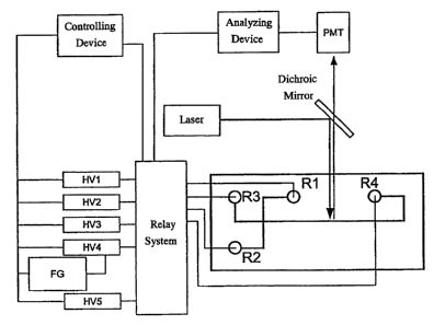
Figure 1 is a conceptual view of a microcapillary electrophoresis device
(hereinafter also referred to as -CE) having a laser-excited fluorescence
detector.
In the figure, FG represents an arbitrary function generator; HV1 to HV5
represent high-voltage power supplies; PMT represents a photomultiplier tube;
and R1 to R4 represent reservoirs. The controlling device may be a general-
purpose computer in which a device-control software (Labview or the like), or
the like is installed. The analyzing device may be a general-purpose computer
in
which a signal analysis software (CIASS-VP or the like), or the like is
installed.
The microchip has flow paths for sample loading (Rl and R2) and flow paths for
sample analysis (R3 and R4), wherein the potential at each reservoir is
controlled
by using high-voltage power supplies HV1 to HV5 and a relay system. Since a
high-speed switching of electric fields is required in the sample injection,
the
sample injection is realized by outputting given voltages from the five power
supplies in advance, and switching the power supplies by the relay system.
Figure 2 is a schematic view showing the migration of a DNA sample
during the sample-loading step of electric field inversion type microchip
electrophoresis. The potentials at each of the reservoirs are: Rl: -0.40 kV,
R2:
GND, R3: -0.55 kV, and R4: -0.95 kV.
Figure 3 is a schematic view showing the migration of a DNA sample
during the sample-injection step of electric field inversion type microchip
electrophoresis. The potentials at each of the reservoirs are: Rl: -0.40 kV,
R2:
-0.4 kV, R3: -0.66 kV, and R4: GND.
Figure 4 is a schematic view showing the migration of a DNA sample
CA 02430312 2003-05-28
7
during the sample-separation step of electric field inversion type microchip
electrophoresis. Pulse electric field (pulse coefficient of 2; 1 Hz) is
applied
between R3 (t0.66 kV) and R4 (GND). R1 and R2 are opened to GND. Each of
the images obtained at an interval of 0.2 seconds is shown in P1 to P8.
Figure 5 is a schematic view showing the potentials at R3 during the
sample-injection step (steady electric field) and the sample-separation step
(electric field inversion). In the figure, Pl to P8 correspond to those in
Figure 4.
Figure 6 is a diagram showing the time evolution of a sample plug after
the injection step in steady electric field for 1 second. The dotted line,
thin solid
line and bold solid line represent the time evolution of the sample plug in
pulse
electric fields at 0.3 Hz, 1 Hz and 10 Hz, respectively.
Figure 7 shows electrophoretograms of a DNA fragment (20-mer) at each
of the frequencies. Panel a) shows the results for 0.3 Hz, Panel b) shows the
results for 1 Hz, and Panel c) shows the results for 10 Hz.
Figure 8 is a schematic view of relative band width with respect to
frequency. The dotted line is a value in steady electric field. The solid line
shows a 20% smooth line.
Figure 9 shows electrophoretograms in steady electric field and pulse
electric field. The samples are each of 20-mer, 40-mer, and 60-mer ssDNA. The
effective length is a distance from the crossing portion of the flow path to
the
sample detection point, and is 6 mm. Panel a) is an electrophoretogram in
steady
electric field (167 V/cm), and Panel b) is an electrophoretogram in a pulse
electric field (10 Hz, pulse coefficient of 2, 167 V/cm).
Figure 10 is a schematic view of non-steady electric field.
Figure 11 is a schematic view of parameters of inverted electric field.
CA 02430312 2003-05-28
8
Figure 12 is a schematic view of the sample-injection member of a
microchip electrophoresis device.
BEST MODE FOR CARRYING OUT THE INVENTION
The present invention is based on the surprising findings of the present
inventors that DNA separation can be achieved by carrying out electrophoresis
under non-steady electric field, especially electric field inversion type (FI)
electrophoresis, in a microchip, even at such short effective lengths which
had
not been able to be separated under ordinary conditions (for instance, steady
electric field and the like).
It has already been reported that the electric field inversion method
improves the resolution during the electrophoresis of long-chain DNA (50 kbp
or
more) in gel, or during the capillary electrophoresis of long-chain DNA in a
polymer solution ["THE FRONTIER ELECTROPHORESIS / THE FRONTIER
CHROMATOGRAPHY," 15, pages 1-9, published on April 2,1994, by ATTO
Corporation, Y. Kim, M. D. Morris, Electrophoresis 17, 152-160 (1996)].
However, the findings of the present inventors that high resolution can be
exhibited by the electric field inversion method even for DNA as short as
about
100 nucleotides, even at a short effective length of 8 cm or less, have been
unexpected.
The electric field inversion type electrophoresis method is an
electrophoresis method employing electric field inversion to periodically
change
the direction of the electric field. Concretely, the method is a method in
which
separation is achieved by setting the period of forward electric field
(electric
field for transferring the molecule to be analyzed from the starting point of
CA 02430312 2003-05-28
9
electrophoresis on gel) and backward electric field (electric field for
returning
the molecule from one point on the gel toward the starting point of
electrophoresis), and the duration time of pulse amplitude or strength of
applied
voltage.
One of the features of the method for analyzing a nucleic acid of the
present invention resides in that the method comprises carrying out
electrophoresis under non-steady electric field, more concretely electric
field
inversion, during electrophoresis on a microchip in microcapillary
electrophoresis. An embodiment of the method for analyzing a nucleic acid of
the present invention includes, for instance, a method for analyzing a nucleic
acid comprising the steps of in microcapillary electrophoresis,
(a) carrying out electric field inversion during electrophoresis on a
microchip,
thereby separating each of the nucleic acids depending on physicochemical
properties thereof, and
(b) detecting the nucleic acid separated by the above step (a).
According to the method for analyzing a nucleic acid of the present
invention, the effective length required for the separation of a nucleic acid,
for
instance, DNA, can be shortened by a combination of electrophoresis method in
microchip with non-steady electric field, concretely with electric field
inversion.
Therefore, there are exhibited some excellent effects such that further
downsizing and higher integration of the microchip can be achieved, that high-
speed analysis of conformation polymorphism of DNA [concretely SSCP
(single-strand conformation polymorphism)] can be performed, and that trace
samples can be analyzed at high sensitivity.
In addition, in the method for analyzing a nucleic acid of the present
CA 02430312 2003-05-28
invention, there are exhibited some excellent effects such that the analysis
can be
performed at an even shorter capillary length because the microcapillary
electrophoresis is applied along with the above-mentioned electric field
inversion
during the separation of the nucleic acid, and that commercially available
electric
5 power supplies that follow electric field inversion at high speeds can be
used
because the voltage applied to both ends of the microcapillary (used maximally
about 10 kV, usually up to about several kV) can be controlled to be low, as
compared to existing capillary electrophoresis devices (concretely, maximally
about 30 kV).
10 The non-steady electric field generically refers to electric field that
changes with time. Concretely, as shown in Figure 10, the non-steady electric
field includes electric fields that change periodically in the form of
rectangular
waves, triangular waves, sine waves and the like, electric fields of which
frequency itself is variable, non-periodical electric fields, and the like.
All these
electric fields can be used for electrophoresis on a microchip in the present
invention.
In the electric field inversion method, the inverted electric field is
generally defined by the parameters shown in Figure 11, concretely, period T,
which is a repeat unit of inversion, time periods T1 and T2 for applying
forward
and backward electric fields, and forward and backward applied electric fields
(voltages) Vl and V2. Although the simplest case of V2 =-V1 will be explained
in Examples in the present specification, the above-mentioned parameters can
be
determined appropriately depending upon the purposes and conditions of the
analysis.
The degree of inversion repeat is hereinbelow expressed by frequency,
CA 02430312 2003-05-28
11
which is a reciprocal of the period, and the degree of inversion is expressed
by a
pulse coefficient, concretely, forward/backward time weight T1/T2.
Hence, it is preferable that the duration time for pulse amplitude
(forward/backward time weight) in electric field inversion is 1/1 to steady
electric field, more preferably 1/1 to 10/1, and still more preferably 1/1 to
5/1,
from the viewpoints of obtaining an appropriate resolution and allowing the
nucleic acid to reach a final migration form. Here, a final migration form of
the
nucleic acid as used herein means a condition in which the bands of individual
nucleic acids are well distinguishable during the separation of the nucleic
acids.
In the method for analyzing a nucleic acid of the present invention, the
frequency in electric field inversion can be determined by, for instance,
electrophoresing a nucleic acid, for instance, a DNA fragment, obtaining a
band
width 0 and a height h of the relative intensity peak ascribed to the DNA
fragment at a migration time t, and finding the frequency at which the value
of
relative band width I' = A/ht reaches its minimum. Also, in the optimization
of
frequency for the separation of a nucleic acid, for instance, DNA, the
movement
of DNA in gel or in a polymer solution responds to the frequency differently
depending upon the length thereof, so that the size of the nucleic acid, for
instance, DNA, to be analyzed can be one of the important parameters.
Concretely, it is preferable that the frequency is at least 10 Hz from the
viewpoint of reduction in dummy peak detection. Concretely, in the case of a
short-chain DNA of about 20-mer, it is desired that the frequency is 10 to 30
Hz,
preferably 10 to 20 Hz.
The frequency can be generated by, for instance, using a commonly used
frequency generator. As a concrete example, in Figure 1, the frequency can be
CA 02430312 2003-05-28
12
generated by controlling the potential at the reservoir (R3) relative to R4
(GND)
using a frequency generator (FG) and the high-voltage power supply (HV4).
The effective length in electrophoresis can be altered appropriately
depending upon the purpose of applications, the above-mentioned frequency,
separation medium, pulse coefficient, voltage, and the like. According to the
method for analyzing a nucleic acid of the present invention, the effective
length
can be made shorter, as compared to the effective length of conventional
capillary electrophoresis systems (shortest effective length: about 75 mm).
Concretely, it is desired that the effective length is 70 mm or less from the
viewpoint of miniaturization of the microchip, 35 mm or less from the
viewpoint
of sufficiently performing high-speed separation, and that the effective
length is
0.5 mm or more, from the viewpoint of preventing the sample leakage from the
crossing portion of the flow path and the backflow of the injected sample into
the
crossing portion, more desirably 1 mm or more, from the practical viewpoint.
The electric field to be applied can be set appropriately according to
conditions of the nucleic acid to be analyzed, the shape, the size and the
effective
length of the flow path, and the separation medium. It is desired that the
above-
mentioned electric field is 1101 V/cm or more, preferably 1501 V/cm or more,
in an
absolute value, from the viewpoint of stable electrophoresis of the sample,
and
11000001 V/cm or less, preferably 1100001 V/cm or less, from the viewpoints of
durability of chips and generation of Joule heat.
The electric field can be generated using, for instance, commonly used
power supply and relay system.
The microchip includes, but is not particularly limited to, concretely, for
instance, as described in Examples and the like set forth below, a chip
CA 02430312 2003-05-28
13
comprising an upper plate and a lower plate, wherein:
(A) the lower plate has thereon two orthogonal channels of 1 to 200 m in
width and 0.5 to 100 m in depth,
(B) the upper plate has four reservoirs of 0.5 to 4 mm in both diameter and
depth, and
(C) any one of the reservoirs as defined in (B) being arranged at a position
corresponding to each end of the channels as defined in (A),
and wherein the reservoir is a reservoir to which electric field can be
applied.
In the method for analyzing a nucleic acid of the present invention, there
can be used, for instance, a microchip comprising a sample-injection member, a
channel for sample analysis and a reservoir for an electrode.
The shapes of the sample-injection member of a microchip electrophoresis
device include, as shown in Figure 11, cross, double-cross, T-shape, double
T-shape, and direct connection-type to the channel for analysis.
The channel in the above-mentioned (A) is a microchannel used for
separation of the nucleic acid. The width of the above-mentioned channel can
be
appropriately set depending upon the size of the microchip, purposes of use
and
the like. Concretely, it is desired that the width of the above-mentioned
channel
is 1 m or more, preferably 10 rn or more, from the viewpoint of sufficiently
obtaining analysis sensitivity of the sample, and that the width is 200 m or
less,
preferably 150 m, from the viewpoint of microfluid dynamics. In addition, the
depth of the above-mentioned channel can be appropriately set depending upon
the size of the microchip, purposes of use and the like. Concretely, it is
desired
that the depth is 0.5 m or more, preferably 5 m or more, from the viewpoint
of
analysis sensitivity of the sample, and that the depth is 100 m or less,
preferably
CA 02430312 2003-05-28
14
50 m, from the viewpoint of microfibricating technique. Furthermore, the
length of the above-mentioned channel for sample separation can be
appropriately set depending upon the size of the microchip, the nucleic acid
to be
separated and the like, ancTit is desired that the length is longer than the
effective
length. The effective length is a distance from the crossing portion of the
channel to a sample detection point. It is desired that the length is 0.5 mm
or
more, preferably 1 mm or more, from the viewpoint of stable electrophoresis of
the sample, and that the length is 70 mm or less, preferably 35 mm or less,
from
the viewpoint of superiority to the existing capillary system.
In addition, the size of the reservoir in the above-mentioned (B) can be
appropriately set depending upon the applied voltage and the applied time.
Concretely, it is desired that the size of the reservoir is 0.5 mm or more,
preferably 1 mm or more, from the viewpoint of maintaining buffer capacity,
and
that the size is 4 mm or less, preferably 3 mm or less, from the viewpoint of
high
integration of the chip.
The separation medium during the electrophoresis includes methyl
cellulose, hydroxypropyl methyl cellulose (HPMC), hydroxyethyl cellulose
(HEC), hydroxypropyl cellulose (HPC), polyethylene glycol (PEG),
polyethylene oxide (PEO), polyacrylamide (PAA), polyvinyl pyrrolidone
(PVP), dextran, agarose and the like. The above-mentioned separation
medium can be used alone or in admixture. Methyl cellulose is desirable,
from the viewpoint of low overlapping concentration. It is desired that the
substance is a solution of polymer not having a cross-linking point, from the
viewpoint of repeated use of the microchip.
In general, in order to separate a nucleic acid, for instance, a polymer
CA 02430312 2003-05-28
compound such as DNA, a separation medium such as gel or a polymer is
required. There has also been reported separation with a free solution not
using a separation medium. In the present invention, the separation may be
also carried out with the free solution not using the separation medium.
5 For instance, when electrophoresis is carried out using the microchip
described in Examples and the like set forth below, it is desired that the
channel of the microchip holds at least one member selected from the group
consisting of methyl cellulose, hydroxypropyl methyl cellulose (HPMC),
hydroxyethyl cellulose (HEC), hydroxypropyl cellulose (HPC), polyethylene
10 glycol (PEG), polyethylene oxide (PEO), polyacrylamide (PAA), polyvinyl
pyrrolidone (PVP), dextran and agarose.
It is desired that pH of the above-mentioned separation medium is 1 to
12, from the viewpoints of stable maintenance of the nucleic acid and
durability of the chip.
15 Concrete example of the above-mentioned separation medium is a
separation medium and the like, of which buffer contains 1% by weight of
methyl cellulose, and of which buffer is at least one member selected from the
group consisting of Tris-borate buffer (pH 8.2), Tris-acetate buffer, TAE
(Tris-
acetate, EDTA) buffer, TBE (Tris-borate, EDTA) buffer, Tris-hydrochloric acid
buffer and phosphate buffer.
The method for analyzing a nucleic acid of the present invention can be
also applied to a microchip electrophoresis device made of silica glass, Pyrex
glass, a resin such as PMMA, silicon, or any other material, and can be also
applied to all the methods for carrying out electrophoresis using a flow path
of a
microscale or nanoscale.
CA 02430312 2003-05-28
16
In the above-mentioned step (a), it is desired that all the procedures are
performed at 0 to 80 C, preferably 10 to 35 C, and more preferably 15 to
35 C, from the viewpoints of prevention of dew condensation on the chip
surface,
suppression of evaporation of the solution from the reservoirs, and influence
on
the resolution.
More concretely, the procedures for step (a) in the method for analyzing a
nucleic acid of the present invention are roughly divided into three steps: a)
a
sample-loading, b) a sample-injection, and c) a sample-separation. Each step
will be explained hereinbelow with reference to the system shown in Figure 1
as
an example.
a) Sample-Loading Step:
Each channel is filled with a 50 mM Tris-borate buffer (pH 8.2)
containing 1% by weight methyl cellulose. A 0.8 l DNA sample
solution is introduced into the reservoir R1, and the reservoirs R2, R3 and
R4 are filled with 1 l of Tris-borate buffer (pH 8.2). The DNA sample is
applied to the channel crossing portion, with keeping the following
potentials for 20 seconds at each of the reservoirs: R1: -0.40 kV, R2:
GND, R3: -0.55 kV, and R4: -0.95 W.
b) Sample-Injection Step:
The potentials are changed for 1 second to Rl: -0.40 kV,
R2: -0.4 kV, R3: -0.66 kV, and R4: GND, thereby injecting a sample plug
to a separation channel.
c) Sample-Separation Step:
Pulse electric field having a pulse coefficient of 2 (forward
time : backward time = 2:1) is applied between electrodes R3 (forward:
CA 02430312 2003-05-28
17
-0.66 kV, backward: 0.66 kV) and R4 (GND). Rl and R2 are opened to
the GND potential. At this time, for instance, when a pulse at a frequency
of 10 Hz is applied, the DNA band migrates linearly at a speed of about
one-third that in steady electric field (-0.66 kV) in the injection step.
In the above-mentioned step (b), the means for detecting a nucleic acid
include laser-excited fluorescence detectors, ultraviolet/visible light
absorption
detection, fluorescence detection, differential refractive index detection,
thermo-
optical detection, circular dichroism detection, electrochemical detection,
electroconductivity detection and the like.
According to the method for analyzing a nucleic acid of the present
invention, since the effective length in electrophoresis can be shortened, the
electrophoresis time can be shortened, so that the method is suitable for high-
speed analysis of a nucleic acid having physicochemical properties such as
molecular weight, nucleic acid conformation polymorphism (especially single-
stranded nucleic acid conformation polymorphism), and higher-order structure.
Also, even when the higher-order structure of DNA is changed due to
modification of a base or the like, the method for analyzing a nucleic acid of
the
present invention can be applied to the detection of the modified bases.
The present invention is hereinafter described in more detail by means of,
but is by no means limited to, the following examples.
Exam , le 1
Three kinds of FITC (fluorescein isothiocyanate)-labeled synthetic single-
CA 02430312 2003-05-28
18
stranded DNA fragments [PAGE (polyacrylamide gel electrophoresis)-purified
grade] having the following sequences:
5'-gttggctctgactgtaccac-3' (SEQ ID NO: 1),
5'-gttggctctgactgtaccaccatccactacaactacatgt-3' (SEQ ID NO: 2), and
5'-gttggctctgactgtaccaccatccactacaactacatgtgtaacagttcctgcatgggc-3'
(SEQ ID NO: 3),
which are parts of the sequence of exon 7 of p53 tumor suppressor gene, were
purchased from KURABO INDUSTRIES LTD. These DNA fragments were
dissolved in TB buffer [50 mM Tris-borate (pH 8.2)]. The DNA-containing
solution obtained was stored at 4 C until use. A 1% by weight methyl cellulose
[4000 cP, 2% by weight solution, manufactured by Sigma Ltd.] was used as a
separation medium.
A schematic view of the microcapillary electrophoresis ( -CE) system is
shown in Figure 1. As the microchip, a microchip manufactured by Shimadzu
Corporation was used. The above-mentioned microchip was composed of two
plates, in which the lower plate has two orthogonal microchannels (width 50
m,
depth 20 m), and the upper plate has four wells (diameter and depth 1 mm) as
reservoirs (Rl to R4). The above-mentioned channels were filled with a TB
buffer containing 1% by weight methyl cellulose. The channel lengths from
each of the reservoirs R1, R2, R3, and R4 to the crossing portion of the
channel
were 7 mm, 7 mm, 7 mm and 32.5 mm, respectively. A platinum (Pt) wire was
inserted into each reservoir. The potentials between the reservoirs were
controlled by Labview (manufactured by National Instrument) using power
supplies (HV1-5) and the relay system. Pulse electric field was generated by
controlling the potential at R3 with respect to R4 (GND) using a high-voltage
CA 02430312 2003-05-28
19
power supply (HV4) and the function generator (FG).
An arbitrary point in the separation flow path from the crossing portion of
the flow path to R4 is referred to as a measurement point, and a distance from
the
crossing portion to a measuring point is referred to as an effective length.
The
measurement point is irradiated with an argon-ion laser through a long focal
lens
of 40-fold magnification, and fluorescence of the FITC-stained sample passing
this point is detected by PMT through a dichroic mirror. The PMT signal is
inputted to the analyzing device to obtain an electrophoretogram.
Prior to these procedures, in order to obtain potential relationship between
the reservoirs according to the conditions of the size and the shape of the
flow
path, and the separation medium, the illumination/signal detection portion of
the
above-mentioned device was modified to illuminate the flow path with a
mercury lamp to a lens of 4-fold magnification, and images of the sample
injected from the crossing portion to the channel for separation were taken by
an
SIT camera. See, for instance, [Ueda, M. et al., Bioimages 1999, 7(4), 157-
161; Ueda, M. et al., Electrophoresis 2000, 21, 176-180].
Example 2: Investigation of Conditions for Electric Field Inversion Type
Macrochip Electrophoresis (R-CE)
1) Procedures
The procedures for sample analysis according to electric field inversion
type microchip electrophoresis ( -CE) are roughly divided into three steps: a)
sample-loading, b) sample-injection, and c) sample-separation. In order to
obtain appropriate relationships in the potentials between the reservoirs in
each
of these steps and an appropriate frequency region in the electric field
inversion
CA 02430312 2003-05-28
process, a sample migrating in the flow path was directly observed, and motion
picture analysis was carried out. On the bases of these data, it was confirmed
that an appropriate pherogram was obtained in the frequency region determined
by the motion picture analysis using the device having the construction shown
in
5 Figure 1. Under these conditions, separation experiments of the samples were
carried out, and it was found that excellent separation was achieved by
electric
field inversion. All the experiments in -CE described below were carried out
at
C.
10 2) Investigation of Electric Field Conditions by Direct Observation Method
a) Sample-Loading:
A 0.8 l DNA sample solution was applied to the reservoir R1, and
the reservoirs R2, R3 and R4 were filled with 1 l of TB buffer. The
DNA sample was loaded to the crossing portion of the flow path by
15 maintaining the following potentials at each of the reservoirs for
20 seconds: R1: -0.4 kV, R2: GND, R3: -0.55 kV, and R4: -0.95 W.
Figure 2 shows a sample-loading step imaged by the SIT camera, and the
directions of migration of negatively charged ions in the flow path are
indicated by arrows. It can be seen from Figure 2 that there are
20 relationships in the potentials such that the sample concentration occurs
in
the crossing portion, and that the sample outflow into R3 and R4 does not
occur.
b) Sample-Injection:
In order to inject a sample plug in the channel for separation, the
25 potentials at each of the reservoirs were switched to and kept for 1 second
CA 02430312 2003-05-28
21
at the following potentials: Rl: -0.40 kV, R2: -0.4 kV, R3: -0.66 kV, and
R4: GND. Figure 3 schematically shows a fluorescence image of the
sample-injection process and the movement of negatively charged ions. It
can be seen from Figure 3 that the sample plug is injected into the channel
for separation in this electric field, and the sample inflow from Rl and R2
does not occur.
c) Sample-Separation (Non-Steady Electric Field Process):
The potentials at R1 and R2 were opened to R4 (GND). A
rectangular wave electric field (forward: -0.66 kV, backward: 0.66 kV)
having a pulse coefficient of 2 (time weight: forward/backward = 2/1) was
supplied between R3 and R4 (GND). Under these conditions, the
frequency of the electric field was changed within the range from 0.1 Hz
to 50 Hz, and the migration behavior of the sample plug was determined
by image analysis. Figure 4 shows a fluorescent image of sample DNA
(20-mer) migrating in the electric field inversion process (pulse
coefficient: 2, frequency: 1 Hz). P1 to P8 in Figure 4 correspond to P1 to
P8 in the schematic view of inverted electric field shown in Figure 5.
Figure 6 shows the time evolution of the position of the center of mass of
the sample plug thus obtained at 0.3 Hz, 1 Hz, and 10 Hz. From the
results, it can be considered that the backward migration by the electric
field inversion is very small at 10 Hz, so that the sample plug as a whole
is migrated by steady electric field in about one-third the strength of the
steady electric field in the injection process (-0.66 kV).
CA 02430312 2003-05-28
22
3) Investigation of Frequency of Inverted Electric Field by Pherogram
The frequency of the electric field investigated in the above-mentioned 2)
was investigated by obtaining a pherogram from the device having the
construction shown in Figure 1.
Electric field inversion type microchip electrophoresis was carried out at
frequency conditions of 0.3 Hz, 1 Hz and 10 Hz, according to the above-
mentioned 2) using the above-mentioned 20-mer ssDNA. The results are shown
in the panels a), b) and c) of Figure 7, respectively.
As shown in the panel a) of Figure 7, it can be seen that the three peaks
indicated by arrows appear at a frequency of 0.3 Hz. In the -CE system used
in
this Example, the size of the laser spot is about 10 m. On the other hand, as
shown in Figure 6, the backward electrophoresis distance is about 500 m.
Therefore, it can be considered that the DNA band crosses over the laser spot
a
number of times to generate dummy peaks.
In the case of 1 Hz, peak occurrence becomes further complicated, as
shown in the panel b) of Figure 7.
At 10 Hz, however, the backward electrophoresis distance is made shorter,
and the DNA band steadily migrated as shown in Figure 6. It can be seen from
the monotony migration pattern that the single peak shown in the panel c) of
Figure 7 is obtained.
Next, with regard to the case of the 20-mer ssDNA, peak width at the
external frequencies of 0.1 Hz or more was examined in order to optimize
external frequency. A band width 0 and a height h of the relative intensity
peak
ascribed to the same DNA fragment at a migration time t were obtained, a
relative band width T= 0/ht was calculated, and a half value of width 0 was
CA 02430312 2003-05-28
23
normalized with the peak height h and the migration time t. The results are
shown in the form of a graph of the relationship between the relative band
width
r and the external frequency in Figure 8. The dotted line in Figure 8
represents a
relative band width in a steady electric field.
As shown in Figure 8, dummy peaks cause apparent band broadening in
the region of less than 10 Hz.
Additionally, in the case of ssDNA of about 20-mer length as in the
present experiment, it is shown that the relative band width has a minimum in
the
range from 10 Hz to 20 Hz, and the band width at the minimum is 1.4 times
greater than that obtained in the steady electric field. The band narrowing
induced by electric field inversion on long-chain DNA having a size of about
40 kbp was not observed under the present experimental conditions. These
tendencies are considered to depend on the length of the DNA to be analyzed,
and the like.
As described above, it is suggested that electric field inversion is effective
for the separation of the DNA fragments at frequencies of 10 Hz or more.
The panels a and b of Figure 9 show electrophoregrams of ssDNA
fragments (20-mer, 40-mer and 60-mer) in each of steady electric field (167
V/cm) and a 10 Hz pulse electric field ( 167 V/cm, pulse coefficient of 2).
The
effective length is 6 mm for both cases. Although a longer separation time was
required in electric field inversion type microchip electrophoresis as
compared to
the migration in the steady electric field, the separation was achieved with
an
effective length of 6 mm. Usually, the mechanism for the separation of long-
chain DNA in electric field inversion type electrophoresis is discussed using
a
reptation model [Kim, Y. et al., Electrophoresis 1996, 17, 152-160; Heller, C.
CA 02430312 2003-05-28
24
et al., Electrophoresis 1995, 16, 1423-1428; Viovy, J. L., Phys. Rev. Lett.,
1988, 60, 855-858]. However, in the present Example, the experiments were
carried out in the Ogston region [Rodbard, D. et al., Proc. Natl. Acad. Sci.
USA,
1970, 65, 970-977]. Although inverted electric fields in the Ogston region
have
no useful effect in commonly used capillary electrophoresis [Kim, Y. et al.,
Electrophoresis 1997, 18, 2901-2908], the electric field inversion type
microchip electrophoresis of the present invention is effective in reducing
the
effective length for the separation even in the Ogston region.
It is suggested that the electric field inversion type microchip
electrophoresis in the Ogston region as described herein has a great
possibility
for application on the detection of SSCP (single-strand conformation
polymorphism), from the viewpoint that the electrophoresis is effective for
separating a single-stranded DNA having a relatively short chain length of
about
100 nucleotides with a shorter effective length.
SEQUENCE FREE TEXT
SEQ ID NO: 1 shows a nucleotide sequence for a synthesized
oligonucleotide (20-mer), which is a part of the sequence of exon 7 of p53
tumor
suppressor gene.
SEQ ID NO: 2 shows a nucleotide sequence for a synthesized
oligonucleotide (40-mer), which is a part of the sequence of exon 7 of p53
tumor
suppressor gene.
SEQ ID NO: 3 shows a nucleotide sequence for a synthesized
oligonucleotide (60-mer), which is a part of the sequence of exon 7 of p53
tumor
suppressor gene.
CA 02430312 2003-05-28
INDUSTRIAL APPLICABILITY
According to the method for analyzing a nucleic acid of the present
invention, a nucleic acid, especially a single-strand conformation
polymorphism,
5 can be analyzed more conveniently in a short time period. Therefore, the
method
for analyzing a nucleic acid of the present invention is useful for the
diagnosis
and treatment of diseases such as detection of gene diseases and application
to
Taylor-made therapy.
CA 02430312 2003-10-28
26
SEQUENCE LISTING
<110> Japan Science and Technology Corporation
<120> Nucleic Acid Analyzing Method
<130> 49427-NP
<140> CA 2,430,312
<141> 2000-11-29
<160> 3
<170> PatentIn Ver. 2.1
<210> 1
<211> 20
<212> DNA
<213> Artificial Sequence
<220>
<223> Description of Artificial Sequence:A sequence for synthesized
oligonucleotide, of which nucleotide sequence is a partial portion of
exon 7of p53 tumor suppressive gene.
<400> 1
gttggctctg actgtaccac 20
<210> 2
<211> 40
<212> DNA
<213> Artificial Sequence
<220>
<223> Description of Artificial Sequence:A sequence for synthesized
oligonucleotide, of which nucleotide sequence is a partial portion of
exon 7of p53 tumor suppressive gene.
<400> 2
CA 02430312 2003-10-28
27
gttggctctg actgtaccac catccactac aactacatgt 40
<210> 3
<211> 60
<212> DNA
<213> Artificial Sequence
<220>
<223> Description of Artificial Sequence:A sequence for synthesized
oligonucleotide, of which nucleotide sequence is a partial portion of
exon 7of p53 tumor suppressive gene.
<400> 3
gttggctctg actgtaccac catccactac aactacatgt gtaacagttc ctgcatgggc 60