Note: Descriptions are shown in the official language in which they were submitted.
CA 02431170 2007-05-22
1
D E S C R I P T I O N
METHOD OF ANALYZING EXPRESSION OF GENE
Technical Field
The present invention relates to a method of
producing an expression profile of gene and a method of
analyzing expression frequency of gene.
Background Art
In 2000, determination of the whole sequence of
the human genome approached completion. The enormous
amount of information obtained as a result of the
determination will be the base for comprehensively
understanding a network including all of the genes and
gene products thereof expressed in specific cells.
Examples of the method employed as a means for
analyzing such a network include: the differential
display method disclosed in USP 5,262,311 and
USP 5,599,672; the serial analysis of gene expression
(which will be referred to as "SAGE" hereinafter)
disclosed in PCT National Publication No. 10-511002;
and the method of using a micro-array and a DNA chip
CA 02431170 2003-06-11
2
disclosed in USP 5,807,522, USP 5,700,637 and
USP 5,744,305.
The differential display method is a method in
which cDNA prepared from a cell is used as a substrate.
By carrying out PCR for the cDNA prepared from a cell,
by using a plurality of types of anchor primer and an
optional primer, various types of gene expression in a
cell can optionally be analyzed. However, according to
this method, only a portion of the whole genes can be
analyzed. Further, use of an anchor primer and an
optional primer results in poor reproducibility, which
is problematic.
On the contrary, SAGE is a method which enables
obtaining an expression profile for all of the genes
expressed in a cell. In SAGE, analysis is carried out
by using cDNA which has been prepared by using mRNA
prepared from a cell. The method includes: a step of
treating the prepared cDNA with a restriction enzyme; a
step of cutting out fragments of approximately 9 to 11
base pairs; a step of ligating the fragments derived
from the obtained cDNA of various types; and effecting
sequencing. However, in SAGE, sequencing has to be
carried out approximately 100,000 times in order to
obtain the information on approximately 50% of all the
types of the expressed genes. In short, SAGE is very
costly. Further, in the case of SAGE, the fragments
derived from cDNA are generally short. In actual
CA 02431170 2007-05-22
3
practice, separation of genes in the form of such short
fragments is often impossible.
The micro-array of USP 5,807,522 and the DNA chip
of USP 5,700,637 and USP 5,744,305 are produced by
fixing a probe of a known gene on a solid phase. In
the methods using such a micro-array and DNA chip, an
expression profile of a gene is obtained by hybridizing
a sample with the probe. In these methods, the
sequence of the gene to be detected must be already
known.
Disclosure of Invention
A first object of the present invention is to
provide a method which enables producing a wide-range
gene expression profile (i.e., an expression profile of
variety of types of genes). A second object of the
present invention is to provide a method of analyzing
expression frequency of genes of a variety of types.
The above-mentioned first object is achieved by a
method of producing a gene expression profile,
comprising:
(a) a step of synthesizing cDNA from mRNA
extracted from a cell, such that a tag substance is
added to the 5' terminal of the cDNA;
(b) a step of cutting the product obtained as a
result of the reaction in step (a) with a first
restriction enzyme X;
(c) a step of connecting, to a fragment obtained
CA 02431170 2003-06-11
4
in step (b), an "X" adaptor having a sequence
complementary to a sequence of a site of the fragment
at which site the incision with the first restriction
enzyme X has been effected;
(d) a step of connecting the fragment obtained in
step (c) to a substance having high affinity with
respect to the tag substance, thereby collecting the
fragment;
(e) a step of cutting the fragment collected in
step (d) with a second restriction enzyme Y and
removing a fragment connected to the tag substance,
thereby obtaining a fragment including the 5' side-
portion of the cut cDNA;
(f) a step of adding, to a fragment obtained in
step (e), a "Y" adaptor having a sequence complementary
to a sequence of a site of the fragment at which site
the incision with the second restriction enzyme Y has
been effected;
(g) a step of carrying out a PCR reaction, for the
fragment obtained in step (f), by using a primer which
has a sequence complementary to the sequence of the "X"
adaptor and an optional two-nucleotide sequence (NN) at
the 3' terminal thereof, and a primer which has a
sequence complementary to the sequence of the "Y"
adaptor and an optional two-nucleotide sequence (NN) at
the 3' terminal thereof; and
(h) a step of subjecting the obtained PCR product
CA 02431170 2003-06-11
to electrophoresis and detecting a migration distance
and a peak, thereby producing gene an expression
profile.
The second object is achieved by a method of
5 analyzing frequency of gene expression, comprising:
(a) a step of producing a gene expression profile,
for each of a control cell and a subject cell, by
employing the above-mentioned method of producing a
gene expression profile; and
(b) a step of analyzing a change in frequency of
gene expression at the subject cell, by comparing the
two profiles of gene expression obtained in step (a).
Other objects and advantages of the present
invention will be described by the description and
examples hereinafter. The present invention will more
clearly be understood with reference to these
description and examples. Further, the objects and
advantages of the present invention will be understood
in detail and achieved by means of the methods and
combination thereof described below.
Brief Description of Drawings
The accompanying drawings, which are incorporated
into the present specification and constitute a portion
thereof, naturally describe the concept of the
preferred embodiment, the aforementioned general
description and the details of the preferred embodiment
described below, of the present invention. The
CA 02431170 2003-06-11
6
drawings are also used for describing the fundamental
idea of the present invention.
FIG. 1 is a view which schematically shows a
method of producing a gene expression profile according
to an embodiment of the present invention.
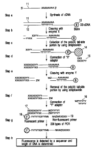
FIG. 2 is a scheme which shows a method of
producing a gene expression profile according to the
embodiment of the present invention.
FIG. 3 is one example of a chart showing a portion
of the gene expression profile obtained according to
the embodiment of the present invention.
FIG. 4 is a table which shows optional
combinations of two nucleotide sequences.
FIG. 5 is a view which shows proportion of the
gene detected by the gene expression profile according
to the embodiment of the present invention.
FIG. 6 is a view which shows preferable examples
of an "X" adaptor and a "Y" adaptor.
FIG. 7 is a view showing the gene sequence of a
portion of human arylamine N-acetyl transferase,
obtained from a database.
FIG. 8 is a view which shows one example of
information on a fragment obtained by the incision with
a restriction enzyme.
FIG. 9 is a view showing a portion of one example
of gene expression profile which represents the
expression of p21.
CA 02431170 2003-06-11
7
FIG. 10 is a view showing a portion of one example
of gene expression profile which represents the
expression of mdm2.
FIG. 11 is a view showing a portion of one example
of gene expression profile which represents the
expression of cyclinG.
FIG. 12 is a view which shows the compositions of
a set of mRNA preparations used in example 2.
FIG. 13 is view which shows a portion of the gene
expression profile obtained in example 2.
FIG. 14 is a view which shows a portion of the
gene expression profile obtained in example 2.
Best Mode for Carrying Out the Invention
1. Summary of the Invention
The inventors of the present invention have
discovered that the degree of complexity of an
operation related to the production of a gene
expression profile, as well as the cost performance,
significantly varies depending on the manner in which a
gene expressed in a specific cell is classified. As a
result of careful study on the basis of this discovery,
the inventors have achieved the present invention.
According to one embodiment of the present
invention, a method is provided which enables
producing, at a time and in a simple and easy manner, a
gene expression profile covering such a wide range as
including substantially all of the genes expressed in a
CA 02431170 2003-06-11
8
specific cell. As substantially all of the expressed
genes can be identified, a remarkably large number of
expressed genes can be identified, as compared with the
conventional method.
Specifically, the one embodiment of the present
invention relates to a gene expression profiling
method, which has been developed on the basis of the
length of the DNA fragment cut with a restriction
enzyme and an application of the polymerase chain
reaction (i.e., PCR). By using such a gene expression
profiling method, almost all of the expressed genes, in
other words, both the known and unknown genes, can
similarly be identified. Further, this method enables
detecting all of the respective genes, without fail,
while identifying each of the genes. Further, it is
possible to determine the expression frequency of the
respective genes.
One essential aspect of the method of producing
gene expression profile according to the present
invention lies in classifying the genes expressed in a
specific cell, as described below. It is assumed that
approximately 20,000 types of mRNA are expressed in a
specific cell. First, cDNA is synthesized from each of
the expressed mRNA preparations. The obtained double-
strand cDNA is cut with two appropriate types of
restriction enzymes, whereby a fragment of the cDNA
having identifiable length is produced for each of the
CA 02431170 2003-06-11
9
expressed genes. Thereafter, the genes are classified
into 256 fractions by identifying the sequence at a
portion of the fragments thereof obtained as described
above. This classification process is carried out by
using the 256 types of primer sets which have been
designed in advance. While the relative amount or
magnitude of expression is still being reflected
therein, the aforementioned fragments are amplified for
each primer set or several primer sets, and then the
fragments are classified. Each of the fractions, e.g.,
256 fractions obtained as a result of classification,
is subjected to electrophoresis, and the components of
each fraction are separated. In this way, the
information of the expressed genes obtained from a cell
is subjected to classification to the analyzable level.
As a result, a gene expression profile which enables
accurately grasping, without fail, the magnitude of
expression of each gene for substantially all of the
expressed genes, can be produced in a simple and easy
manner.
A specific means for classification, e.g.,
classification into 256 fractions, is described by
using FIG. 1. The cDNA group 2 is synthesized from the
group 1 consisting of the expressed mRNA preparations.
Each of the cDNA is cut with two appropriate types of
restriction enzymes, and thereby the cDNA fragment
group 3 is obtained. Each cDNA fragment is classified
CA 02431170 2003-06-11
according to the sequence of the two bases at each end
(i.e., totally four bases) thereof. In other words,
each cDNA fragment is classified according to the type
of the two bases at each end thereof, the type
5 including adenine (which will be referred to as "A"
hereinafter), guanine (which will be referred to as "G"
hereinafter), cytosine (which will be referred to as
"C" hereinafter) and thymine (which will be referred to
as "T" hereinafter). Specifically, the cDNA fragments
10 are first classified into four groups 4 according to
the type of the base at the 5' terminal (which base is
shown in black in FIG. 1), then classified into sixteen
groups 5 according to the type of the next base, then
into sixty-four groups 6 according to the type of the
second base at the 3' terminal, and further into
256 groups 7 according to the type of the first base at
the 3' terminal. Based on the types of mRNA which are
generally expressed, approximately 80 to 100 types of
cDNA are assumed to be included in each of the
256 groups 7 which are obtained eventually. That is,
when the cDNA of each group is subjected to
electrophoresis, it is assumed that approximately 80 to
100 peaks will be detected. Accordingly, all of the
mRNA obtained from a specific cell are expressed in
256 types of chart each exhibiting approximately 80 to
100 peaks. These 256 types of chart constitute a
profile of the expressed gene. FIG. 3 shows one
CA 02431170 2007-05-22
11
example of a chart contained in such a profile. The
chart of FIG. 3 is a chart showing the components
contained in a fraction, which fraction has been
obtained as a result of the fragment-classification,
subsequent PCR amplification and electrophoresis of the
reaction product of each fraction.
Another aspect of the present invention lies in
appropriate cutting of cDNA obtained from the expressed
mRNA with two appropriate types of restriction enzymes,
which are preferably MspI and MseI. Such appropriate
cutting result in successfully carrying out the above-
mentioned classification. The present invention will
be described in more detain hereinafter.
2. Detailed Description of the Embodiments
(1) Gene expression profile
The method of producing a gene expression profile
of the present invention basically includes:
(a) a step of synthesizing cDNA from mRNA
extracted from a cell, such that a tag substance is
added to the 5' terminal of the cDNA;
(b) a step of cutting the product obtained as a
result of the reaction in step (a) with a first
restriction enzyme X;
(c) a step of connecting, to a fragment obtained
in step (b), an "X" adaptor having a sequence
complementary to a sequence of a site of the fragment
at which site the incision with the first restriction
CA 02431170 2003-06-11
12
enzyme X has been effected;
(d) a step of connecting the fragment obtained in
step (c) to a substance having high affinity with
respect to the tag substance, thereby collecting the
fragment;
(e) a step of cutting the fragment collected in
step (d) with a second restriction enzyme Y and
removing a fragment connected to the tag substance,
thereby obtaining a fragment including the 5' side-
portion of the cut cDNA;
(f) a step of adding, to a fragment obtained in
step (e), a "Y" adaptor having a sequence complementary
to a sequence of a site of the fragment at which site
the incision with the second restriction enzyme Y has
been effected;
(g) a step of carrying out a PCR reaction, for the
fragment obtained in step (f), by using a primer which
has a sequence complementary to the sequence of the "X"
adaptor and has an optional two-nucleotide sequence
(NN) at the 3' terminal thereof, and a primer which has
a sequence complementary to the sequence of the "Y"
adaptor and has an optional two-nucleotide sequence
(NN) at the 3' terminal thereof; and
(h) a step of subjecting the obtained PCR product
to electrophoresis and detecting a migration distance
and a peak, thereby producing a gene expression
profile.
CA 02431170 2003-06-11
13
In the present specification, "the 5' side of
double strand DNA" generally represents the 5' side of
a sense strand (a sequence homologous with the mRNA as
a template) and "the 3' side of double strand DNA"
generally represents the 3' side of such a sense
strand.
A specific example of the method of producing gene
expression profile according to the present invention
is described heinafter with reference to FIG. 2. In
FIG. 2, each alphabet letter represents a base which
constitutes a nucleotide sequence. "A" represents
adenine (in other words, adenine will be referred to as
"A" hereinafter), "G" represents guanine (in other
words, guanine will be referred to as "G" hereinafter),
"C" represents cytosine (in other words, cytosine will
be referred to as "C" hereinafter) and "T" represents
thymine (in other words, thymine will be referred to as
"T" hereinafter) . Further, "N" "W" "X" "Y" and "Z"
each represents any suitable or optional base. X and Y
complementarily bind to each other, and W and Z
complementarily bind to each other. Note that
aforementioned steps (a) to (h) each correspond to
steps (a) to (h) of FIG. 2, respectively, exactly in
the alphabetical order.
First, mRNA 11 is extracted from a specific cell
as the test subject.
An oligo dt primer, which is complementary to the
CA 02431170 2003-06-11
14
poly(A) tail at the 3' terminal of the mRNA 11
extracted as described above, is marked with biotin 13.
A cDNA is synthesized by using the marked mRNA as a
primer, and thereby a double strand 12 is obtained
(FIG. 2, step (a)). Here, an example in which biotin
is used as the tag substance is shown.
The double strand 12 is cut by using MspI, which
is a four-base-identifying restriction enzyme, as a
first restriction enzyme X (FIG. 2, step (b)). Here,
an example in which MspI is used as the first
restriction enzyme is shown.
Thereafter, the biotin 13 is captured by using
streptoavidin 14. As a result, the 3'-side portion of
the cut double strand cDNA is captured (FIG. 2, step
(c)). Here, an example, in which streptoavidin is used
as a substance having high affinity with respect to the
tag substance, is shown.
To the 5' side of the double strand cDNA collected
in step (c), an "X" adaptor 15 having a sequence
complementary to the identification-incision site of
the cDNA at which site the incision with the first
restriction enzyme X i.e., MspI, has been effected, is
connected (FIG. 2, step (d)).
The resulting product is cut by using a
restriction enzyme MseI as a second restriction enzyme
Y (FIG. 2, step (e)). Here, an example in which MseI
is used as the second restriction enzyme is shown.
CA 02431170 2007-05-22
Next, a "Y" adaptor 16 having a sequence
complementary to the identification-incision site of
the cDNA at which site incision with the second
restriction enzyme Y i.e., MseI, has been effected, is
5 added or connected (FIG. 2, step (f)). As a result of
the above-mentioned treatments, double strand sequence
17 including known sequences at both ends thereof is
constructed.
Next, a PCR reaction is carried out by using the
10 double strand sequence 17 as a template, and using a
PCR primer 18 at the 5' side of the double strand cDNA
marked with a fluorescent colorant (for the antisense
strand) and a primer 19 at the 3' side of the double
strand cDNA without fluorescent marking (for the sense
15 strand) (FIG. 2, step (g)). Here, the "X" primer 18
and the "Y" primer 19 for the PCR have and utilize
sequences which are :complementary to the sequences of
X adaptor and Y adaptor another sequence including two
bases located next to the one sequence in the direction
of amplification thereof. As each pair of two bases at
the 5' side/the 3' side (i.e., the totally four bases
derived from both terminals) is designed such that each
of the four bases can be any of the four types of bases
A, G, C and T, totally 256 types of primer set can be
obtained. Accordingly, by carrying out PCR for all of
the thus prepared double strand cDNA preparations, it
is possible to classify all of the existing cDNA
CA 02431170 2003-06-11
16
preparations into 256 groups and carrying out PCR
amplification therefor without fluorescent marking.
FIG. 4 shows the combinations of the four bases, in
which the sequence of the four bases are optionally
decided, of the primer set. FIG. 4 discloses the
combinations ranging from AA-AA to TA-GA.
In step (h) of FIG. 1 as the final process, the
PCR products obtained as 256 types of fractions are
subjected to electrophoresis and peaks of each case or
fraction are measured, whereby a gene expression
profile is obtained (FIG. 1, step (h)). FIG. 3 shows a
chart which is an example of the result obtained by
subjecting one of the 256 fractions prepared as
described above to electrophoresis. In FIG. 3, the
Y-axis of the graph indicates the magnitude of
expression, with fluorescent strength being used as the
index, and the X-axis of the graph indicates the
molecular weight, with the migration distance at
electrophoresis being used as the index.
It is acceptable to exchange step (c) and step (d)
in the order. That is, step (d) may be carried out
prior to step (c).
Further, one restriction enzyme which is used as
the first restriction enzyme may be used as the second
restriction enzyme, while another restriction enzyme
which is used as the second restriction enzyme is used
as the first restriction enzyme. As a result, incision
CA 02431170 2003-06-11
17
is made possible for a larger number of genes and
thereby the detection sensitivity is enhanced.
Specifically, the double strand 12 obtained in
step (a) is divided into two groups i.e., cDNA mix A
and cDNA mix B. It is acceptable that the cDNA mix A
is subjected to the treatment of steps (b) to (h) as
described above, and simultaneous with or after the
treatment of the cDNA mix A, the cDNA mix B is
subjected to the following treatment. Specifically,
the cDNA mix B is treated in a manner similar to that
of the above-mentioned method, except that the
restriction enzyme MseI is used as the first
restriction enzyme and the restriction enzyme MspI is
used as the second restriction enzyme. By using the
first restriction enzyme and the second restriction
enzyme in the exchanged manner, the genes which would
not be detected had the restriction enzymes not been
exchanged can also be detected.
More specifically, in the treatment of the cDNA
mix B, the double strand 12 contained in the cDNA mix B
is cut with MseI, which is a four-base-identifying
restriction enzyme. Thereafter, to the identification-
incision site of the cDNA at which site the incision
with the restriction enzyme MseI has been effected, the
MseI adaptor having a sequence complementary to the
identification-incision site is connected or bound.
Then, biotin is captured by using streptoavidin, and
CA 02431170 2003-06-11
18
thereby the 3'-side portion of the cut double strand 12
is collected. Next, the collected 3'-side portion of
the double strand 12 is cut with the restriction enzyme
MspI. Thereafter, to the identification-incision site
of the cDNA at which site the incision with the
restriction enzyme MspI has been effected, the MspI
adaptor having a sequence complementary to the
identification-incision site is bound. As a result of
the above-mentioned treatment, a sequence including the
double strand 12 with known sequences connected to both
terminals thereof is constructed. Next, for the
obtained sequence, a PCR reaction for cDNA is carried
out by using an X primer 18 marked with a fluorescent
colorant and a Y primer 19 without fluorescent marking.
Here, the primer 18 and the primer 19, having sequences
complementary to the X adaptor and the Y adaptor
another sequences of two bases located next to the
sequences in the direction of amplification thereof, is
used. As each pair of two bases at the 5' side/the 3'
side (i.e., the totally four bases derived from both
terminals) is designed so that each of the four bases
can be any of the four types of bases A, G, C and T,
totally 256 types (combinations) of primer set can be
obtained (Refer to the steps (a) to (f) of FIG. 2. The
256 types of the NN-NN nucleotide sequence are
specifically shown in FIG. 4). Accordingly, by
carrying out PCR for all of the cDNA preparations by
CA 02431170 2003-06-11
19
using these primer sets, it is possible to classify all
the existing types of cDNA preparations into 256
groups. The PCR products obtained as 256 types of
fractions are subjected to electrophoresis and
migration distance and peaks of each case or fraction
are measured, whereby a gene expression profile is
obtained.
Regarding the expressed genes which are classified
according to the method of the present invention, in
the case of mouse, for example, approximately 85% of
100 genes of mouse selected at random can be identified
and detected, as shown in FIG. 5. Specifically, when
MspI is used as the first restriction enzyme and MseI
is used as the second restriction enzyme, approximately
66% of the expressed genes goes through incision. When
MseI is used as the first restriction enzyme and MspI
is used as the second restriction enzyme, approximately
19% of the expressed genes goes through incision.
Accordingly, by exchanging the first restriction enzyme
and the second restriction enzyme in the order in use
thereof, approximately 85% of the expressed genes can
be identified and detected, as a whole. Due to this, a
gene profile can be produced more accurately than in
the conventional method. The proportion of genes which
can be identified by the conventional method is
generally 20 to 30%, and 50% at most. Therefore, the
proportion of genes which can be identified by the gene
CA 02431170 2003-06-11
expression profile produced by the method of the
present invention is remarkably higher than the
proportion achieved by the conventional method. It is
concluded that the method of the present invention
5 enables identifying substantially all of the genes
contained in a cell.
The term "gene expression profile" used in the
present specification represents information including
an expression pattern of genes in a specific cell in a
10 given condition, absence/presence of expression of
known and unknown genes, the magnitude of expression of
all the expressed genes, and the like. The gene
expression profile produced by the method of the
present invention can be used as a means for analyzing
15 expression of genes.
The term "poly(A) tail" used in the present
specification represents a sequence at the 3' terminal
of mRNA, which is, in general, also referred to as
"poly(A)". cDNA can be synthesized from mRNA having
20 the aforementioned poly(A) tail by using the "oligo dT
primer" having a sequence complementary to the poly(A)
tail. The "oligo dT primer" used in the present
invention is, in general, also referred to as
"oligo(dT) primer". The synthesis of cDNA by using the
oligo dT primer can be achieved in any suitable
conditions which are generally applied to the
conventional method.
CA 02431170 2003-06-11
21
The "tag substance" and the "substance having high
affinity with respect to the tag substance" used in the
present invention are substances which can specifically
bind to each another with high affinity, thereby
forming a binding pair. Although biotin is used as the
tag substance and streptoavidin is used as the
substance having high affinity with respect to the tag
substance in the example described in the
aforementioned item "(1) Gene expression profile", the
types of the tag substance and the substance having
high affinity with respect to the tag substance are not
limited to these specific examples. Any binding pair
can be used as long as the pair exhibits specific
binding with high affinity therebetween. Examples of
the combination of the tag substance and the substance
having high affinity with respect to the tag substance,
which can be employed in the present invention,
include: biotin and streptoavidin; biotin and avidin;
FITC and FITC antibody; DIG and anti-DIG; protein A and
mouse IgG; latex particles; and the like. However, the
types of the tag substance and the substance having
high affinity with respect to the tag substance are not
limited to the aforementioned examples. Further, in
each of the combinations described above, each of the
two substances can be used as either the tag substance
or the substance having high affinity with respect to
the tag substance.
CA 02431170 2003-06-11
22
The "restriction enzyme" used in the present
invention is an enzyme which is, in general, also
referred to as "restriction endonuclease" and effects
hydrolysis and incision of double strand DNA at a
specific sequence. In the method according to the
present invention, two types of restriction enzymes X
and Y are used in combination, in order to obtain
appropriate fragments. As the restriction enzyme which
can be used in the present invention, an enzyme capable
of cutting the double strand, constituted of cDNA which
has been synthesized from mRNA as the expressed gene,
to a fragment having identifiable length, is
preferable. It is preferable that the enzyme is
capable of cutting as many of the obtained double
strands as possible, and it is more preferable that the
enzyme is capable of cutting substantially all of the
obtained double strands. Table 1 shows examples of
such enzymes. It is acceptable to select any two
enzymes from Table 1 and use these enzymes in
combination. All of the enzymes shown in Table 1 are
four-base-identifying enzymes. Alternatively, four-
base-identifying enzymes of the types other than those
of Table 1 or six-base-identifying enzymes may be used.
In the method according to the present invention, it is
preferable that four-base-identifying enzymes are used,
and it is more preferable that MspI and MseI are used
in combination. In the aforementioned example, MspI
CA 02431170 2003-06-11
23
(or MseI) is used as the restriction enzyme X, and MseI
(or MspI) is used as the restriction enzyme Y.
Table 1
Accil CG/CG HpaII C/CGG
Alal GT/AC Hsp9211 CATG/
Alul AG/CT HspAI G/CGC
AspLEI GCG/C Kzo9I /GATC
BfaI C/TAG MaeI C/TAG
BscFI /GATC MboI /GATC
Bsh1236I CG/CG MseI T/TAA
BshI GG/CC MspI C/CGG
BsiSI C/CGG MvnI CG/CG
Bsp1431 /GATC NdeII /GATC
BstUI CG/CG NlaIII CATG/
BsuRI GG/CC Pall GG/CC
CfoI GCG/C RsaI GT/AC
Csp6I G/TAC Sau3AI /GATC
DpnII /GATC Sse9I /AATT
FnuDII CG/CG TaqI T/CGA
HaeIII GG/CC ThaI CG/CG
HapII C/CGG TrulI T/TAA
HhaI GCG/C Tru9I T/TAA
Hin2I C/CGG Tsp509I /AATT
Hin6I G/CGC TspEI /AATT
HinPiI G/CGC TthHB8I T/CGA
The "adaptor" employed in the present invention is
used for effecting connection of the primers which work
in the final PCR amplification. The adaptor used in
the present invention is designed in accordance with
the restriction enzymes to be used. Specifically, the
"X" adaptor to be connected to the
CA 02431170 2003-06-11
24
identification-incision site at which the incision with
the restriction enzyme X has been effected may include
a sequence complementary to the identification-incision
site (at which the incision with the restriction enzyme
X has been effected) and another optional sequence.
The type of another optional sequence and the base-
length thereof can be designed in consideration of the
factors such as the efficiency of PCR. It is
preferable that the "X" adaptor is designed such that
the "X" adaptor has approximately 15 bases. Such a
structure of the "X" adaptor results in the stable
performance of PCR. The "Y" adaptor to be connected to
the identification-incision site at which the incision
with the restriction enzyme Y has been effected may
include a sequence complementary to the identification-
incision site (at which the incision with the
restriction enzyme Y has been effected) and another
optional sequence. The type of another optional
sequence and the base-length thereof can be designed in
consideration of the factors such as the efficiency of
PCR. It is preferable that the "Y" adaptor is designed
such that the "Y" adaptor has approximately 15 bases.
Such a structure of the "Y" adaptor results in the
stable performance of PCR.
A preferable example of the sequence of the "X"
adaptor in the case in which MspI is used as the
restriction enzyme X is shown in FIG. 6(a). A
CA 02431170 2003-06-11
preferable example of the sequence of the "X" adaptor
in the case in which MseI is used as the restriction
enzyme X is shown in FIG. 6(b). Further, a preferable
example of the sequence of the "Y" adaptor in the case
5 in which MspI is used as the restriction enzyme Y is
shown in FIG. 6(c), and a preferable example of the
sequence of the "Y" adaptor in the case in which MseI
is used as the restriction enzyme Y is shown in
FIG. 6(d). However, the sequence of the "X" adaptor
10 and that of the "Y" adaptor are not limited to the
examples shown in FIGS. 6(a) to 6(d).
The "primer set" used in step (g) includes a pair
of primers, primer "X" and primer "Y", which primers
are used for amplifying by PCR the double strand cDNA
15 obtained in step (f). The details of the primer set
are as described above. The "optional two nucleotide-
sequence (NN)" used in the present invention is a
sequence optionally selected from adenine, thymine,
guanine and cytosine. As described above, in a case in
20 which the optional bases are constituted of two bases
(i.e., NN), a chart obtained as result of PCR of one
sample includes approximately 80 to 100 peaks. In the
method of the present invention, each "optional
sequence" at each side is designed as a two-nucleotide
25 sequence, in consideration of the convenience in
operation and precision in analysis in the method.
Accordingly, in the method according to the present
CA 02431170 2003-06-11
26
invention, the "optional sequence" at each side is
preferably a two-nucleotide sequence (NN) and the
number of the primer set is preferably 256. However,
the type of the "optional sequence" and the number of
the primer set are not limited to the above-mentioned
examples. It is acceptable that the optional two-
nucleotide sequence NN of at least one of the two
primers (i.e., the "X" primer and/or the "Y" primer) is
replaced with a sequence including no less than three
bases. When the number of the bases included in the
"optional sequence" is increased, the number of types
of primers included in the primer set is also
increased. When the optional two-nucleotide sequence
NN of one of the two primers is replaced with a three-
nucleotide sequence, 1024 or 4096 fractions will be
obtained.
Further, in the present invention, it is
preferable that a fluorescent material is bound to one
terminal of one of the primers of each primer set so
that the detection thereof after PCR can be
facilitated. Specifically, it is preferable that a
fluorescent material is bound to the 5' terminal of the
"X" primer having a sequence complementary to the "X"
adaptor. Examples of the fluorescent material which
can be used in the method of the present invention
include 6-carboxyfluorescein (which will be referred to
as "FAM" hereinafter),
CA 02431170 2003-06-11
27
4,7,2',4',5',7'-hexachloro-6-carboxyfluorescein (which
will be referred to as "HEX" hereinafter), NED
(manufactured by Applied Biosystems Japan Co., Ltd.),
6-carboxy-X-rhodamine (which will be referred to as
"Rox" hereinafter) and the like.
The PCR reaction carried out according to the
invention may be carried out in a condition generally
applied to the conventional method. For example, the
PCR reaction can be carried out in the condition of
95 C for 1 minute, (95 C for 20 seconds, 68 C for
30 seconds, 72 C for 1 minute) x 28 times, and 60 C for
30 minutes.
The means for conducting electrophoresis which can
be used in the present invention may be any means for
electrophoresis, in general, as long as the means
enables separation of reagents according to the
molecular weight thereof. Commonly used devices for
electrophoresis can be used, whose examples include a
sequencer, ABI PRISM 3100 (manufactured by Applied
Biosystems Japan Co., Ltd.), ABI PRISM 3700
(manufactured by Applied Biosystems Japan Co., Ltd.),
and MegaBACE 1000 (manufactured by Amersham Pharmacia
Co., Ltd).
(3) Identification of peaks
Further, according to the present invention, it is
possible to identify the gene represented by each peak
of the chart obtained as described above. Due to
CA 02431170 2003-06-11
28
identifying the peak, it is possible to identify the
gene(s) which is/are expressed or whose expression
magnitude is increased/decreased in a specific
environment.
Identification of the gene can be carried out by
collecting the molecule or gene exhibiting a particular
peak in the chart, and determining the sequence thereof
by a laboratory operation including the common method
such as sequencing.
Alternatively, it is possible to theoretically
identify the gene by using a computer, without relying
on a laboratory operation as described above. For
example, it is possible to identify the gene by using a
computer, on the basis of data of the identification
site of the restriction enzyme in use, data of the
molecular weight of the fragment obtained by the
incision with the restriction enzyme, and data which is
available from the free database.
The length of the fragment, observed when a gene
sequence optionally selected from the database is cut
with a specific restriction enzyme, as well as the
details of the identification site of the restriction
enzyme, can easily be determined on a display of a
computer. On the other hand, the length of the
fragment, observed after the incision with the
restriction enzymes used in the method of the present
invention, is clearly known from the result of
CA 02431170 2003-06-11
29
electrophoresis. Accordingly, by further considering
the adaptor sequence in use, it is possible to
determine from which gene the fragment is derived,
without necessitating any laborious analysis by
experients in a laboratory. One example of the method
conducting such theoretical identification by using a
computer will be described in example 1 below.
A computer for common use can be used in the
present invention. For example, a computer device
equipped with an input section including a keyboard, a
mouse and the like, an output section including a
printer, a display and the like, and a computing
section such as CPU, can be used.
Examples of the database from which useful data
can be obtained include public data banks such as
GenBank, EMBL and DDBJ, commercial databases and the
like, with no restriction to these examples.
Further, it is also possible to combine the method
relying on a laboratory operation and the method based
on theoretical computation by a computer, in the
aforementioned gene identification process.
(4) Analysis on gene expression frequency
In the method of producing gene expression profile
according to the present invention, the magnitude of
expression of each gene expressed in the subject cell
is reflected on the magnitude of the peak corresponding
to the gene shown in the chart. Accordingly, by
CA 02431170 2003-06-11
observing the change in the magnitude of the peaks, the
expression frequency of each gene can be analyzed.
For example, it is possible to make comparison,
with regards to the expression frequency of a gene,
5 between a normal cell and an abnormal cell, between a
normal cell and a cancer cell, between cells different
in type, and between cells treated in different
conditions.
Further, if a gene which expresses itself or whose
10 expression magnitude is changed as result of a specific
stimulus is identified by the method of the present
invention in advance, it suffices, in the tests
thereafter, to use only the primers corresponding to
the specific gene and produce the gene expression
15 profile resulted from the primer. The expression
frequency of the targeted gene can be analyzed on the
basis of the gene expression profile obtained in such a
manner.
Example 1
20 Influence of radioactive ray irradiation on the
magnitude of expression of p21, mdm2 and cyclin G
The influence of radioactive ray irradiation on
the magnitude of expression of p21, mdm2 and cyclin G
was studied, as described below. A gene expression
25 profile was produced by using mRNA obtained from a mice
mammary cancer cell stock SR-1 which had been subjected
to radioactive ray irradiation. Another gene
CA 02431170 2003-06-11
31
expression profile was produced by using mRNA obtained
from a mice mammary cancer cell stock SR-1 which had
not been subjected to radioactive ray irradiation. The
two gene expression profiles were compared with each
other.
1. Production of gene expression profiles
The gene expression profiles were actually
produced according to the method of the present
invention.
1-1. Extraction of mRNA and synthesis of cDNA
Mice mammary cancer cell stock SR-1 (donated by
Professor Koyama, Yokohama City University) was
cultured in an aMEM culture medium set in a 75 cm3
flask (manufactured by Falcon Co., Ltd.). Radioactive
rays of 7 Gy were irradiated on the cells, from above,
by using a "Pantac", manufactured by Shimadzu
Corporation, Ltd. The irradiation time was 3 hours.
Mice mammary cancer cell stock SR-1 which had not been
subjected to such irradiation was also prepared as a
control at the same time. 20 gg of mRNA as the whole
weight was extracted from each cell by using a
FastTrack 2.0 kit (manufactured by Invitrogen Co.,
Ltd.).
Each mRNA (20 g) extracted as described above
was mixed with 5'-biotinated oligo dT primer
(100 pmole/0.8 L) (manufactured by BRL Co., Ltd.), and
the mixture was incubated at 65 C for 5 minutes. The
CA 02431170 2003-06-11
32
mixture was then cooled with ice. Thereafter, the
mixture was incubated with MgC12 (the final
concentration thereof was 5 mM), 0.5 mM of dNTP Mix
(manufactured by BRL Co., Ltd.) and 10 mM of DTT
(manufactured by BRL Co., Ltd.), in 20.0 L of a
reverse transcription buffer, at 42 C for 60 minutes.
The resulting product was then incubated with dNTP Mix
(manufactured by BRL Co., Ltd., the final concentration
thereof was 0.27 mM), 1.33 mM of DTT (manufactured by
BRL Co., Ltd.), 20.0 units of E. coli ligase
(manufactured by BRL Co., Ltd.), 40.0 units of E. coli
DNA polymerase (manufactured by BRL Co., Ltd.) and
2.0 units of RNaseH (manufactured by BRL Co., Ltd.), in
150.0 L of a double strand synthesizing buffer, at
first at 16 C for 120 minutes and then 70 C for
15 minutes. Then, the reaction was stopped. The
obtained reaction product was equally divided into two
portions (the reaction product mixture A and the
reaction product mixture B).
1-2. Treatment of the reaction product mixture A
The reaction product mixture A was treated, as
described below. In the present example, MspI was used
as the first restriction enzyme and MseI was used as
the second restriction enzyme.
First, the restriction enzyme MspI (manufactured
by Takara Co., Ltd., the final concentration thereof
being 20 units in 100 L) was reacted with the reaction
CA 02431170 2003-06-11
33
product mixture A containing 10 g of mRNA, at 37 C for
360 minutes. After the reaction, the product was
purified with ethanol (500 gL x 3 times). Thereafter,
the product was subjected to ligation with 5.0 g of
the "X" adaptor having a sequence of GC (i.e., a
sequence complementary to the incision fragment site at
which site the incision with the restriction enzyme
MspI had been effected) (manufactured by BRL Co., Ltd.)
and 10 units of T4 DNA ligase (manufactured by NEB Co.,
Ltd.), in 15 L of the T4 DNA ligase buffer. Then,
magnetic beads having streptoavidin (manufactured by
Dinal Co., Ltd.) fixed thereto were added to the
reaction solution. The biotin included in the double
strand in the reaction solution was bound to
streptoavidin fixed to the magnetic beads, whereby a
ligation product was obtained.
Next, the ligation product was reacted with the
restriction enzyme MseI (manufactured by NEB Co., Ltd.,
the final concentration thereof was 50 units in
200 L), at 37 C for 360 minutes. After the reaction,
the supernatant thereof was transferred to another
tube and was subjected to purification with ethanol
(1000 L x 3 times). Thereafter, the product was
subjected to ligation with 10 pmole of the "Y" adaptor
having a sequence of AT (i.e., a sequence complementary
to the incision fragment site at which site the
incision with the restriction enzyme MseI had been
CA 02431170 2003-06-11
34
effected) (manufactured by BRL Co., Ltd.) and 10 units
of T4 DNA ligase (manufactured by NEB Co., Ltd.), in
L of the T4 DNA ligase buffer.
Next, PCR was carried out with respect to the
5 ligation product obtained as described above. In the
present example, one of the three types of fluorescent
colorants FAM, HEX and NED was bound to the 5' side of
the "X" primer having a sequence complementary to the
"X" adaptor. The "X" primer further includes an
10 optional two-nucleotide sequence (NN), at the 3' side
thereof, next to the sequence complementary to the "X"
adaptor. On the other hand, the "Y" primer having a
sequence complementary to the "Y" adaptor further
includes an optional two-nucleotide sequence (NN), at
the 3' side thereof. The combinations of each
fluorescent colorant and each NN are shown in FIG. 4.
In FIG. 4, the combinations are classified according to
the substance used for marking. That is, the sequences
marked with FAM are shown in row (a), the sequences
marked with HEX are shown in the row b), and the
sequences marked with NED are shown in row (c). The
optional two-nucleotide sequences are expressed as
"(NN) of the X primer"-"(NN) of the Y primer". In the
present example, three types of fluorescent probes were
used in order to enhance the work efficiency. The
method of the present invention can be implemented with
a single fluorescent probe being used, in a manner
CA 02431170 2003-06-11
similar to that of the case in which three types of
fluorescent probes are used.
Specifically, after the ligation was completed,
the reaction solution was diluted to 612 L with Tris-
5 HCl buffer (which buffer will be referred to as "TE"
hereinafter). 1 L of a solution containing the primer
represented by the first sequence of "FAM" row (a) of
FIG. 4 (i.e., "AA-AA"), 1 pL of a solution containing
the primer represented by the first sequence of "HEX"
10 row (b) of FIG. 4 (i.e., "CT-AA"), and 1 L of a
solution containing the primer represented by the first
sequence of "NED" row (c) of FIG. 4 (i.e., "CA-AA"),
were mixed together and then the mixture was mixed with
1 RL of the diluted reaction solution. Similarly, a
15 solution mixture of a set of the three primers,
represented by the sequences derived from rows (a), (b)
and (c) and sharing the same reference number, was
prepared and the solution mixture was mixed with 1 L
of the diluted reaction solution, in a manner similar
20 to that described above. As the primers from No. 81 to
No. 96 in the FAM row do not have corresponding primers
in the HEX and NED rows, the primer solutions of No. 81
to No. 96 in the FAM row were mixed with 1 RL of the
diluted reaction solution, without adding primer
25 solutions of HEX and NED. As a result of the
aforementioned operation, the PCR reaction products
produced from the 256 types of primer sets were
CA 02431170 2003-06-11
36
converted to 96 samples for electrophoresis.
These 96 samples for electrophoresis were then
subjected to electrophoresis. The electrophoresis was
carried out under the condition of a migration voltage
of 15 kV and a migration time of 2000 seconds, with a
capillary sequencer (ABI PRISM 3100, Applied Biosystems
Japan Co., Ltd.). The result of the electrophoresis
was obtained, for each sample, as a chart in which the
X-axis represents the molecular weight shown according
to the migration distance, which migration distance
being used as an index, and the Y-axis represents the
magnitude of gene expression, shown according to the
fluorescent intensity, which fluorescent intensity
being used as an index. One sample includes the PCR
15. products marked with the three different types of
fluorescent materials. However, these PCR products (or
the three different types of fluorescent materials) can
be identified by changing the wavelength to be applied.
1-3. Treatment of the reaction product mixture B
The reaction product mixture B was subjected to a
treatment in a manner similar to that in the treatment
of the reaction product mixture A, except that the MseI
was used as the first restriction enzyme and MspI was
used as the second restriction enzyme. Thereafter,
96 samples obtained from the reaction product mixture B
were subjected to electrophoresis in a manner similar
to that in the reaction product mixture A, to obtain
CA 02431170 2003-06-11
37
charts.
According to the method similar to that described
in the aforementioned 1-1 and 1-2, the gene expression
profile was obtained as the charts representing the
results of the electrophoresis.
1-4. Analysis of the gene database
With regard to the peaks detected in the gene
expression profile obtained as described above,
information on the incision site at which the incision
with the restriction enzyme in use had been effected
and information on the fragments produced as a result
of the incision were obtained, by using data in the
database, in order to identify the genes represented by
these peaks.
First, the data from the gene database of GenBank,
on the genes the length of whose mRNA had been
revealed, and the data from EST, on the genes only a
portion of whose sequence had been registered, were all
accumulated, and the data derived from each gene was
classified as one group, according to the type thereof.
A consensus sequence was obtained from the accumulated
data. FIG. 7 shows the consensus sequence and a
portion of the sequence used for arranging the
consensus sequence (refer to FIG. 7). The sequence
indicated at the top of FIG. 7 is the consensus
sequence. The term "consensus sequence" used in the
present specification represents a sequence of one type
CA 02431170 2003-06-11
38
of gene, obtained by determining for each portion of
the sequence a base which appears at the highest rate
among all of the plural sequences which have been
determined with regards to the gene. FIG. 6 shows, as
one example, a portion of gene sequence of human
arylamine N-acetyl transferase.
Next, with respect to the consensus sequence and
all the data used for determining the consensus
sequence, the identification sequence of the
restriction enzyme X located closest to the 3' terminal
was detected. Thereafter, the identification sequence
of the restriction enzyme Y located closest, in the 3'
direction, to the identification site of the
restriction enzyme X was detected. The identification
sequence of MspI is C/CGG, and the incision is effected
at the site of "/". The identification sequence of
MseI is T/TAA. Further, the number of the bases of
DNA, which can be assumed on the basis of the incision
fragments of the restriction enzyme X and the
restriction enzyme Y obtained as described above, was
theoretically calculated. One example of the data
obtained as described above is shown in FIG. 8 (refer
to FIG. 8).
In FIG. 8, it is understood from the data from the
database of GenBank and a number of registered data of
EST that an incision fragment of 104 bp is obtained
(refer to the "length" column of FIG. 8). However, the
CA 02431170 2003-06-11
39
data of FIG. 8 also indicates a possibility that some
data include mutation or errors in sequence reading,
and thereby an incision fragment of 23 bp is also
obtained (refer to the "length" column of FIG. 8).
Further, with regards to p21, mdm2, cyclinG and
gadd45 among the above-mentioned gene data, which are
the genes whose expression is known to be increased as
a result of radioactive ray irradiation, the length of
the incision fragment and the sequence of the two bases
located on the inner side of the restriction enzyme
identification site were analyzed.
1-5. Identification of genes
By studying the data obtained from the above-
mentioned 1-2 and 1-3, together with the data obtained
from the above-mentioned 1-4, with comparing the sets
of data with each other, the genes represented by the
peaks detected at the gene expression profile of the
present invention were identified.
On the basis of the length of the incision
fragments of p21, mdm2, cyclicG and gadd45 and the
sequence of the two bases on the inner side of the
restriction enzyme identification site obtained from
the databases, it was determined that, from which
primer set, i.e., from which set of X primer and Y
primer among the 512 types of primers used in the
method of the present invention, p21, mdm2, cyclicG and
gadd45 were each detected.
CA 02431170 2003-06-11
Further, the length of DNA, detected in a manner
similar to that described above, was revealed. On the
basis of the length of DNA, a peak corresponding to the
molecular weight matching the revealed DNA length was
5 separated from the peaks representing the molecules
separated by the electrophoresis. The nucleotide
sequence of the DNA represented by the peak separated
as described above was analyzed by sequencing, whereby
it was confirmed that the genes were the targeted
10 genes, i.e., p21, mdm2, cyclicG and gadd45.
1-6. Influence of radioactive ray irradiation on the
expression magnitude of p21, mdm2, cyclicG and gadd45
The peaks of the respective genes of p21, mdm2 and
cyclicG, obtained according to the method described
15 above, are shown in FIGS. 9, 10 and 11. The upper
chart of each of FIGS. 9, 10 and 11 shows a portion of
the gene expression profile derived from mRNA obtained
from a cell which was not subjected to radioactive ray
irradiation. The lower chart of each of FIGS. 9, 10
20 and 11 shows a portion of the gene expression profile
derived from mRNA obtained from a cell which was
subjected to 7 Gy radioactive-ray irradiation for
3 hours. The peak of the targeted gene is shown with
an arrow.
25 As shown in FIGS. 9, 10 and 11, it has been
confirmed that the expression magnitude is increased by
radioactive ray irradiation in each of p21, mdm2 and
CA 02431170 2003-06-11
41
cyclicG. Further, a similar result was obtained for
gadd45, although the data thereof is now shown.
Example 2
Analysis of gene expression frequency
Further, the gene expression frequency was
analyzed. Fission yeast (which will be referred to as
"S. p." hereinafter) and budding yeast (which will be
referred to as "S. c." hereinafter) were used as the
cells. For each type of cell, mRNA was extracted in a
manner similar to that of example 1. The extracted
mRNA of each type of cell was mixed with each other
such that the whole amount of mRNA derived from S. p.
was varied in a range of 0, 0.02, 0.2, 1, 2 and 2 ( g),
while the whole amount of mRNA derived from S. c. was
varied in a range of 2, 2, 2, 2, 2 and 0 ( g), as shown
in FIG. 12.
For each of the six types of mRNA preparations
prepared as described above, a gene expression profile
was prepared in a manner similar to that of example 1.
FIG. 13 shows charts representing a portion of the
gene expression profile obtained as described above.
The composition of the mRNA preparations from which
each chart is derived is shown at the left-hand side of
each chart. The uppermost chart shows a portion of the
gene expression profile of S. p. The lowermost chart
indicates a portion of the gene expression profile of
S. c.
CA 02431170 2003-06-11
42
FIG. 14 is a view in which the peaks derived from
S. p. in the charts of FIG. 13 are linked with vertical
dotted lines. As shown in FIG. 14, the magnitude of
the peaks is changed depending on the amount of mRNA of
S. p. contained in the mRNA preparations.
From the results of examples 1 and 2, it has been
confirmed that a gene expression profile is produced by
the method of the present invention and the magnitude
of expression of a gene, shown in the obtained gene
expression profile, sufficiently reflects the amount of
mRNA present in the sample. Accordingly, it is
possible to analyze the frequency of gene expression by
the method of the present invention.
According to the method of the present invention,
a gene expression profile regarding genes expressed in
a wide range can be produced in a simple and easy
manner. Further, by using such a gene expression
profile, it is possible to identity a far more number
of expressed genes, i.e., substantially all of the
expressed genes, as compared with the conventional
method. Yet further, in the gene expression profile
according to the present invention, identification of
genes can be carried out for each gene. Yet further,
as the gene expression profile of the present invention
reflects the expression magnitude of genes, the
frequency of gene expression can also be analyzed.
Specifically, the expression frequency of an unknown
CA 02431170 2003-06-11
43
gene can also be analyzed as is the case with the
expression frequency of known genes.
Further advantages and modifications will easily
be noticed by one skilled in the art. Therefore, in
terms of the aspects of such a wide range of further
advantages and modifications, the present invention is
not limited to the detailed description and the
representative embodiment described above. In other
words, various changes may be applied to the present
invention within the sprit or scope of the general idea
of the invention, which is clearly shown by the
accompanying claims and equivalents thereof.
CA 02431170 2003-06-11
1/9
SEQUENCE LISTING
<110> AISIN SEIKI KABUSHIKI KAISHA
National Institute of Radiological Sciences
Maze, Inc.
ABE, Masumi
SAITO, Toshiyuki
HATTORI, Atsushi
SATO, Shinji
KASAMA, Koji
<120> Method for analysing of gene expression
<130> 01S1459P
<150> JP 2000-377887
<151> 2000-12-12
<160> 24
<170> Patentln version 3.1
<210> 1
<211> 22
<212> DNA
<213> Artificial
<400> 1
cgggtcgtat cagacttgca ca 22
CA 02431170 2003-06-11
2/9
<210> 2
<211> 20
<212> DNA
<213> Artificial
<400> 2
tgtgcaagtc tgatacgacc 20
<210> 3
<211> 22
<212> DNA
<213> Artificial
<400> 3
tacatcaggt gtccgatgat tc 22
<210> 4
<211> 20
<212> DNA
<213> Artificial
<400> 4
gaatcatcgg acacctgatg 20
<210> 5
<211> 22
<212> DNA
<213> Artificial
CA 02431170 2003-06-11
3/9
<400> 5
cgagtcgtat cagacttgca ca 22
<210> 6
<211> 20
<212> DNA
<213> Artificial
<400> 6
tgtgcaagtc tgatacgact 20
<210> 7
<211> 22
<212> DNA
<213> Artificial
<400> 7
tacttggact acagtcgtga ca 22
<210> 8
<211> 20
<212> DNA
<213> Artificial
<400> 8
tgtcacgact gtagtccaag 20
<210> 9
CA 02431170 2003-06-11
4/9
<211> 116
<212> DNA
<213> Homo sapiens
<400> 9
gaaaccgggg tgggtggtgt ctccaggtca atcaacttct gtactgggct ctgaccacaa 60
tcggttttca gaccacaatg ttaggagggt atttttacat ccctccagtt aacaaa 116
<210> 10
<211> 116
<212> DNA
<213> Homo sapiens
<400> 10
gaaaccgggg tgggtggtgt ctccaggtca atcaacttct gtactgggct ctgaccacaa 60
tcggttttca gaccacaatg ttaggagggt atttttacat ccctccagtt aacaaa 116
<210> 11
<211> 116
<212> DNA
<213> Homo sapiens
<400> 11
gaaaccgggg tgggtggtgt ctccaggtca atcaacttct gtactgggct ctgaccacaa 60
tcggttttca gaccacaatg ttaggagggt atttttacat ccctccagtt aacaaa 116
CA 02431170 2003-06-11
5/9
<210> 12
<211> 116
<212> DNA
<213> Homo sapiens
<400> 12
gaaaccgggg tgggtggtgt ctccaggtca atcaacttct gtactgggct ctgaccacaa 60
tcggttttca gaccacaatg ttaggagggt atttttacat ccctccagtt aacaaa 116
<210> 13
<211> 116
<212> DNA
<213> Homo sapiens
<400> 13
gaaaccgggg tgggtggtgt ctccaggtca atcaacttct gtactgggct ctgaccacaa 60
tcggttttca gaccacaatg ttaggagggt atttttacat ccctccagtt aacaaa 116
<210> 14
<211> 116
<212> DNA
<213> Homo sapiens
<400> 14
gaaaccgggg tgggtggtgt ctccaggtca atcaacttct gtactgggct ctgaccacaa 60
tcggttttca gaccacaatg ttaggagggt atttttacat ccctccagtt aacaaa 116
CA 02431170 2003-06-11
6/9
<210> 15
<211> 116
<212> DNA
<213> Homo sapiens
<400> 15
gaaaccgggg tgggtggtgt ctccaggtca atcaacttct gtactgggct ctgaccacaa 60
tcggttttca gaccacaatg ttaggagggt atttttacat ccctccagtt aacaaa 116
<210> 16
<211> 116
<212> DNA
<213> Homo sapiens
<400> 16
gaaaccgggg tgggtggtgt ctccaggtca atcaacttct gtactgggct ctgaccacaa 60
tcggttttca gaccacaatg ttaggagggt atttttatat ccctccagtt aacaaa 116
<210> 17
<211> 116
<212> DNA
<213> Homo sapiens
<400> 17
gaaaccgggg tgggtggtgt ctccaggtca atcaacttct gtactgggct ctgaccacaa 60
CA 02431170 2003-06-11
7/9
tcggttttca gaccacaatg ttaggagggt atttttatat ccctccagtt aacaaa 116
<210> 18
<211> 116
<212> DNA
<213> Homo sapiens
<400> 18
gaaaccgggg tgggtggtgt ctccaggtca atcaacttct gtactgggct ctgaccacaa 60
tcggttttca gaccacaatg ttaggagggt atttttacat ccctccagtt aacaaa 116
<210> 19
<211> 116
<212> DNA
<213> Homo sapiens
<400> 19
gaaaccaggg tgggtggtgt ctccaggtca atcaacttct gtactgggct ctgaccacaa 60
tcggttttca gaccacaatg ttaggagggt atttttacat ccctccagtt aacaaa 116
<210> 20
<211> 116
<212> DNA
<213> Homo sapiens
<400> 20
gaaaccgggg tgggtggtgt ctccaggtca atcaacttct gtactgggct ctgaccacaa 60
CA 02431170 2003-06-11
8/9
tcggttttca gaccacaatg ttaggagggt atttttacat ccctccagtt aacaaa 116
<210> 21
<211> 116
<212> DNA
<213> Homo sapiens
<400> 21
gaaaccgggg tgggtggtgt ctccaggtca atcaacttct gtactgggct ctgaccacaa 60
tcggttttca gaccacaatg ttaggagggt atttttatat ccctccagtt aacaaa 116
<210> 22
<211> 116
<212> DNA
<213> Homo sapiens
<400> 22
gaaaccgggg tgggtggtgt ctccaggtca atcaacttct gtactgggct ctgaccacaa 60
tcggttttca gaccacaatg ttaggagggt atttttacat ccctccagtt aacaaa 116
<210> 23
<211> 116
<212> DNA
<213> Homo sapiens
<400> 23
CA 02431170 2003-06-11
9/9
gaaaccgggg tgggtggtgt ctccaggtca atcaacttct gtactgggct ctgaccacaa 60
tcggttttca gaccacaatg ttaggagggt atttttacat ccctccagtt aacaaa 116
<210> 24
<211> 116
<212> DNA
<213> Homo sapiens
<400> 24
gaaaccaggg tgggtggtgt ctccaggtca atcaacttct gtactgggct ctgaccacaa 60
tcggttttca gaccacaatg ttaggagggt atttttatat ccctccagtt aacaaa 116