Note: Descriptions are shown in the official language in which they were submitted.
CA 02431903 2003-06-12
WO 02/059586 PCT/US01/49823
-1-
APPARATUS AND METHODS FOR ANALYZING AND IlVIPROVING
AGRICULTURAL PRODUCTS
FIELD OF THE INVENTION
The present invention relates to devices and methods for analyzing
agricultural products. More particularly, the present invention relates to
devices
and methods for rapid, non-destructive analysis of the physical and chemical
characteristics of one or more seeds, and methods for breeding plants to
produce
new plants with desirable physical and chemical characteristics.
BACKGROUND OF THE INVENTION
Breeding for compositionally enhanced agricultural products can require the
analysis of a large number of seed samples from plants to identify those
plants with
the desired compositional and agronomic properties for use or advancement to
the
next generation. Analysis of bulk seed batches for certain traits, such as
high oil or
protein content, on a single plant or ear, in conjunction with an appropriate
breeding
methodology such as recurrent selection, often allows for the selection and
introduction of such traits into a commercial population. Although the
analysis of
these seed batches can be performed by various techniques, methods that are
rapid,
low cost, and non-destructive are the most desirable.
Magnetic resonance imaging (MRI) is based on a non-invasive spectroscopic
technique known as nuclear magnetic resonance (N1VIlZ). NMR requires the
sample
under investigation to contain atoms that exhibit nuclear spin, an intrinsic
quality
makes the atomic nuclei magnetic. The most common atom with nuclear spin is
hydrogen, whose nucleus is a proton with spin'/2. A typical proton NMR (1H
NMR) experiment involves placing a sample to be studied in a strong
homogeneous
magnetic field. The strong magnetic field causes preferential alignment of the
protons in the sample with the magnetic field, a phenomenon that is analogous
to a
magnetic compass needle aligning with the earth's magnetic field. In a simple
modern "pulsed-NMR" experiment, another magnetic field (the radiofrequency, or
CA 02431903 2003-06-12
WO 02/059586 PCT/US01/49823
-2-
RF field) is transiently applied to the sample, which has the effect of
rotating the
aligned protons to a higher energy state, which is perpendicular to the strong
magnetic field (this is called a 90 pulse). The protons precess at
characteristic rates
as they realign with the strong field, thus inducing a current in a pair of
coils that
serve as the detector. The current is measured in the detection coils as a
function of
time, and from this the rates of precession of the protons in the sample are
inferred.
The rates of precession are determined primarily by the strength of the strong
magnetic field (with stronger fields leading to greater precession rates), but
the
unique molecular environment experienced by each of the protons also has an
effect
on the rate of precession. It is the unique molecular environment that is the
object
of study in NMR experiments.
In a slightly more complicated modern pulsed NNM experiment, the
complexity of the signals arising from multiple molecular environments can be
eliminated, leaving only the signals from protons in a selected subset of
molecular
environments. This approach, called the "spin-echo" experiment, can be used,
for
example, on a sample comprising a mixture of oil and water to eliminate the
signal
arising from the protons in water molecules, and to leave only signals arising
from
protons in oil molecules. This can be accomplished by using a series of RF
pulses of
90 and 180 , and commercial instruments typically come with software that is
useful for programming the instrument to properly execute the pulse sequence.
Low-field pulsed NMR methods rely on differentiation of oil from other
components in the seed based on inherent differences in longitudinal and spin-
spin
nuclear relaxation rates between oil and other protonated species. NNM methods
used to measure oil have been standardized, they are non-destructive, robust,
and
they yield both accurate and reproducible results. The MRI method for
measuring
oil content is based on the same physical principles described in pulsed NNM
studies. The difference is that 1VIRI provides spatially encoded NNM signals,
providing displays of data in an image format rather than a conventional NNM
spectrum.
Another similarity exists between conventional NNM studies and the MRI
method for measuring oil levels in single seeds. Both methods provide a
relative oil
CA 02431903 2003-06-12
WO 02/059586 PCT/US01/49823
-3-
content for seeds on a percent basis (wt/wt) by comparing the experimental
results
with oil calibration standards, which is the generally accepted and useful
method for
comparing oil levels in single seeds. See Tiwari et al., "Rapid and
nondestructive
determination of seed oil by pulsed nuclear magnetic resonance technique," J.
Am.
Oil Chem. Soc. 51: 104-109 (1974). Relative numbers are obtained because a
small
portion of the NMR signal intensity is lost during the timing delays
incorporated
into the data acquisition schemes. The relative numbers can be corrected to
absolute numbers by normalizing the data using an independent oil measurement
for
a given population of seeds, e.g., the average of the measured oil value for
the
population is set to the oil value determined in a bulk measurement and all
individual
seeds are adjusted accordingly. Absolute oil numbers also can be obtained
using
NMR spectroscopy and MRI methods by using seed standards in the analysis. In
this case, the oil content of a seed is determined absolutely since the seed
standard
more closely matches the nuclear relaxation and wax content of the
experimental
seed. However, absolute oil numbers have been shown to be less precise for
comparing relative oil levels between seeds. See G. Rubel, "Simultaneous
determination of oil and water contents in different oilseeds by pulsed
nuclear
magnetic resonance," J. Am. Oil Chem. Soc. 71: 1057-1062 (1994).
While the best NMR instruments require extremely homogenous magnetic
fields, MRI machines purposely induce field gradients (variations in magnetic
field
strength with respect to position) using three gradient magnets. The gradient
magnets are much weaker than the strong magnet, but they are sufficient to
cause
distinguishable proton precession rates at different parts of the sample.
Modern
MRI machines pulse not only the RF field, but also the gradient fields, in
order to
selectively rotate and selectively detect protons in particular regions of the
sample.
Measurement of the signal strength of the various frequency components
(precession rates) indicates the density and relaxation times (the time it
takes
particular protons to relax back to their original low-energy state and re-
align with
the strong magnetic field, the longitudinal relaxation time, and to precess
out of
phase, or dephase due to spin-spin relaxation, with respect to the other
protons) of
protons at various locations in the sample.
CA 02431903 2003-06-12
WO 02/059586 PCT/US01/49823
-4-
MRI methods provide direct measurements of characteristics such as oil
levels, thus providing a primary assay independent of a chemometric model.
Thus a
three dimensional image can be constructed where the intensities at various
points in
the image relate to the densities and relaxation times of protons at those
points in
the sample. Furthermore, because different molecular environments result in
different precession frequencies, the molecular environment at each point in
the
sample can be determined. Such an approach is called "chemical shift imaging"
(CSI).
An alternative to CSI is "spin echo imaging" (SEI). In this experiment, one
class of protons can be singled out (based on their dephasing rates), and the
signals
from all other protons can be suppressed. For example, if a sample contains
spatially separated oil and water, and a SEI experiment is directed to
detecting the
oil, then the resulting image will show only those regions of the sample in
which oil
is found. 15 MRI is a well-known non-invasive radiological technique commonly
used in
the medical sciences. The long-wavelength (radio wave) radiation is
universally
regarded as less harmful than the forms of radiation used in other types of
non-invasive radiological techniques such as X-ray CAT (computed axial
tomography) scans. As early as 1988, MRI techniques were starting to be
applied
to the study of plants. See Introduction section of Lakshminarayana et al.,
"Spatial
distribution of oil in groundnut and sunflower seeds by nuclear magnetic
resonance
imaging," J. Biosci. 17(1): 87-93 (March 1992) (hereinafter Lakshminarayana et
al.)
(describing a history up to 1992 of the use of MRI in the study of plants,
seeds, and
plant tissue). Lakshminarayana et al. describe an experiment in which MRI was
used on single seeds to determine the spatial distribution of oil and water in
single
seed samples. They used a spin echo pulse sequence to selectively detect only
the
protons that were part of oil molecules. MRI was also used to study water
uptake
in dry kidney beans by Heil et al., "Magnetic resonance imaging and modeling
of
water up-take into dry beans," Lebensm.-Wiss. u.-Technol. 25:280 (1992).
Both SEI and CSI MRI were used to image lipids in pecan embryos by
Halloin et al., "Proton magnetic resonance imaging of lipid in pecan embryos,"
J.
CA 02431903 2003-06-12
WO 02/059586 PCT/US01/49823
-5-
Am. Oil Chemists' Soc. 70:1259 (1993). These experiments studied the
differences
in the images of pecan embryos that were normal, infected by fungus, and
damaged
by insects. The CSI MRI experiment showed the distribution of lipids and water
within the pecan embryos.
Other MRI techniques are known to those skilled in the art. One example is
relaxography, or relaxation time mapping. In this technique, different regions
of a
sample being imaged are distinguished based on the differences in relaxation
times
of the protons in the different regions.
MRI experiments on seeds have traditionally been conducted using research
grade MRI instruments, for example, the Bruker AMX-400 9.4 Tesla instrument
with an 8.9 cm diameter bore (Bruker Instruments Inc., Billerica, MA) or the
GE
Omega system 7.1 Tesla instrument with a 15 cm diameter bore (General
Electric,
Milwaukee, WI). These instruments typically have a bore size of a few
centimeters,
and consequently can only be used to study samples that are smaller than the
bore
size. The advantage of the small scale of these instruments is that the
detection coils
are close to the sample under investigation, and therefore their sensitivity
is very
high. Larger research grade MRI instruments also exist with bore sizes and
detection coil diameters of 20-50 cm, for example, the Bruker Biospec 114.7
Tesla,
40 cm diameter bore (Bruker Instruments, Inc., Billerica, MA). These
instruments
are useful for imaging of mice and other small animals. Clinical MRI
instruments,
on the other hand, must have a bore size and detection coils that are large
enough to
accommodate a human body. For example, Siemens (Siemens AG, Erlangen,
Germany) and GE Medical Systems (Milwaukee, WI) manufacture a wide range of
clinical instruments that can accommodate objects with 50 cm diameters and
larger.
Some newer systems with permanent magnets with magnetic fields as low as 0.2
Tesla are not limited by bore size, but by coil diameter. But the larger
diameter
detection coils in traditional superconducting instruments, as well as newer
permanent magnet systems, make these clinical instruments ill-suited for
imaging
small samples such as seeds. The large distance of the detection coils from a
small
sample and the inherently weak signal emanating from a small sample conspire
to
make conventional approaches to quantitative imaging of small samples using
CA 02431903 2003-06-12
WO 02/059586 PCT/US01/49823
-6-
clinical MRI instruments impractical. But the small volumes of small bore size
research MRI instruments do not allow the simultaneous imaging of as many
seeds
as a clinical MRI instrument would allow if the larger MRI instruments were
amenable to detecting signals from small samples.
Time-intensive techniques for finding desirable characteristics are especially
disadvantageous to selective plant breeding programs, where many single seeds
need to be screened rapidly in order to allow seed selection before the next
planting
generation. Delays in providing the breeder with the analytical results can
cause the
loss of an entire breeding cycle.
Non-imaging techniques such as IR spectroscopy suffer from the further
disadvantage of collecting information from only a subset of a total sample by
spot
sampling only portions of only a few seeds out of the hundreds of seeds in the
bulk
sample. Furthermore, since spot sampling interrogates arbitrary portions of
the
seed, different tissues of the seeds in the samples can be misrepresented by
the
analytical data. Since qualities like oil content are often present in
different amounts
in different tissues, non-imaging techniques can fail to accurately assess the
desired
quality. Non-imaging techniques disregard spatial information, and thus
provide no
information to a plant breeder about the size, shape, mechanical damage,
insect
infestation, or fungal damage.
Conventional seed analysis techniques also fail to provide an efficient
method for single seed analysis, which can greatly accelerate the rate of
varietal
development. Single seed analysis is necessary to differentiate and select
individual
seeds from the heterogeneous population of seeds often encountered in breeding
populations. Single seed analysis can reduce the number of generations
required for
the production of a plant with the desired trait. Single seed selection also
reduces
the number of individual plants required. In corn, for example, the ability to
identify
the individual seeds with the desired trait at the single seed level rather
than at the
whole ear level can reduce the nursery requirement by 100 fold. This makes it
possible to conduct a far greater number of breeding projects with the same
resources.
Other conventional analytical techniques, such as gas chromatography, also
CA 02431903 2003-06-12
WO 02/059586 PCT/US01/49823
-7-
often fail to provide an efficient method for single seed analysis. For
example, the
conventional method for single seed analysis of canola requires manual
excision of
one half of each seed for fatty acid analysis by gas chromatography, while the
other
half is planted. Because of the manual sample preparation and the low
throughput
of this analytical technique, only a small number of samples can be run per
hour
using this process. Furthermore, this technique allows for the possibility of
destroying analyzed seeds' potentials to grow into mature, seed bearing
plants.
Although single seed analysis is desirable, conventional approaches and
sampling methods do not allow for efficient processing of single seeds.
Conventional techniques require extensive manual input, which limits the rate
of
development of plants with improved characteristics.
Conventional spectroscopic analysis techniques do not allow for the
localization of chemical component levels within different tissues of seeds.
Conventional approaches, such as manual dissection of the seed followed by
chemical analysis by traditional analytical techniques, are not only laborious
and
destructive, they also result in poor resolution of the components and poor
quantitation, since the sample size resulting from dissection of individual
seeds is
below the sample size at which most traditional techniques produce reliable
results.
Needed in the art are devices and methods for rapid analysis of bulk and
single seeds that can efficiently and non-destructively analyze the
morphological
and/or chemical characteristics of individual seeds, and that can be
integrated into an
agricultural processing machine. The present invention provides such devices
and
methods.
SUMMARY OF THE INVENTION
This invention provides devices and methods for the rapid, non-destructive
analysis of any sample in order to determine the presence or absence of a
trait of a
specimen within that sample by using magnetic resonance imaging on either a
single
specimen, or on a plurality of specimens, and for using the information gained
by the
MRI analysis to select individuals exhibiting the characteristic from within a
group
of candidates potentially exhibiting the characteristic.
CA 02431903 2003-06-12
WO 02/059586 PCT/US01/49823
-8-
This invention further provides devices and methods for rapid,
non-destructive analysis of the physical and chemical characteristics of one
or more
seeds or plant tissues and for using this analysis to selectively breed plants
with one
or more desired characteristics. The analysis is carried out on a sample of
one or
more seeds using MRI to measure one or more characteristics of the sample.
Seeds
exhibiting the desired characteristics can be selected to be grown from among
many
seeds analyzed. The invention is further useful for observing insect or fungal
infestation, shapes of seeds, and damage to seeds.
The present invention includes and provides a method for determining
whether a seed exhibits a trait, comprising: (A) providing the seed in a
sampling
device; (B) generating a magnetic resonance image of the seed; (C) analyzing
the
magnetic resonance image for the trait; and (D) determining whether the seed
exhibits the trait based on the analysis, wherein the magnetic resonance image
is
obtained using a magnetic resonance imaging instrument with a bore size
greater
than about 20 cm.
The present invention includes and provides a method for determining
whether any seeds within a sample comprising a plurality of seeds exhibit a
trait,
comprising: (A) providing the sample in a sampling device; (B) generating a
magnetic resonance image of the sample; (C) analyzing the magnetic resonance
image for seeds exhibiting the trait; and (D) determining whether seeds in the
sample exhibit the trait based on the analysis, wherein the determining step
comprises associating the seeds with corresponding image volume elements, and
wherein the magnetic resonance image is obtained using a magnetic resonance
imaging instrument with a bore size greater than 20 cm.
The present invention includes and provides methods for selectively breeding
plants comprising imaging one or more seeds using a magnetic resonance imaging
instrument, analyzing the image of the one or more seeds in order to determine
whether any of the one or more seeds exhibits a trait or traits, selecting one
or more
seeds based on whether they exhibit the trait or traits, planting one or more
seeds
that exhibit the trait or traits, growing the resulting plants to maturity,
and cross
fertilizing the plants with each other or with other plants.
CA 02431903 2003-06-12
WO 02/059586 PCT/US01/49823
-9-
The present invention includes and provides a method for optimizing the
field-of-view parameters and gradients in a magnetic resonance imaging
instrument
used to measure characteristics of a sample comprising seeds, comprising: (A)
selecting a field of view that covers the entire sample; (B) selecting a
number of
image layers; (C) selecting an image layer thickness; (D) selecting a number
of
image pixels within each image layer, wherein the number of image layers
multiplied
by the image layer thickness is greater than about the height of the sample to
be
imaged, the image layer thickness is less than about the thickness of the
seeds, and
the number of image pixels is sufficient to prevent inter-pixel crosstalk.
The present invention includes and provides a sampling device for magnetic
resonance imaging experiments comprising a spacer and a plurality of plates,
wherein each plate is comprised of a plurality of wells and is slidably
stacked and
held by the spacer, and wherein the spacer and the plurality of plates consist
of
materials that are amenable to magnetic resonance experiments.
The present invention includes and provides a device for measuring
properties of agricultural products, comprising: a sampling device for
providing the
sample, and a magnetic resonance imaging instrument for imaging the sample,
wherein the magnetic resonance imaging instrument has a bore size greater than
about 20 cm, wherein the sample exhibits an inductance that is substantially
less
than that of an approximately equivalent volume of water, and wherein the
detection
and RF coils of the magnetic resonance imaging instrument are loaded or
retuned to
be sensitive to the sample.
The present invention includes and provides a device for measuring
properties of agricultural products, comprising: a processing device for
producing a
sample; a sampling device for providing a sample, wherein the sampling device
is
disposed to receive the sample from the processing device; and a magnetic
resonance imaging system, wherein the system is disposed to analyze the sample
in
the sampling device.
The present invention includes and provides a device for measuring
properties of agricultural products, comprising: a sampling device for
providing a
sample; a magnetic resonance imaging system, wherein the system is disposed to
CA 02431903 2003-06-12
WO 02/059586 PCT/US01/49823
-10-
analyze the sample in the sampling device; and a sorting device for sorting
the
sample into two or more different groups, wherein the sorting device is
disposed to
receive the sample from the sampling device.
The present invention includes and provides a device for measuring
properties of agricultural products, comprising: a processing device for
producing a
sample; a sampling device for providing a sample, wherein the sampling device
is
disposed to receive the sample from the processing device; a magnetic
resonance
imaging system, wherein the system is disposed to analyze the sample in the
sampling device; and a sorting device for sorting the sample into two or more
different groups, wherein the sorting device is disposed to receive the sample
from
the sampling device.
DESCRIPTIONS OF THE DRAWINGS
Figure 1 shows high resolution MRI images of four different corn kernels
with different levels of oil content. These images indicate that kernel A
(18.4% oil
by weight) has a greater oil content than kernel B (7.8% oil by weight), which
has a
greater oil content than kernel C (5.7% oil by weight), which has a greater
oil
content than kernel D (2.8% oil by weight). These images used the spin-echo
pulse
sequence that imaged only protons that were part of oil molecules. Thus the
darker
regions indicate a higher oil content, while the lighter regions indicate a
lower oil
content.
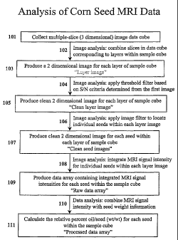
Figure 2 is a block diagram detailing the steps from the initial data
collection
from a sample cube containing multiple layers of seeds to the production of an
array
detailing the percent oil content by weight of each of the seeds within the
sample.
This block diagram shows the steps that can be applied to the raw data
obtained
from an MRI experiment to produce the final data array for the seeds in the
form of
percent oil content by weight for each seed within the sample cube. First, a
three
dimensional data set is collected from the sample cube to produce a three
dimensional image data cube 101. (See Fig. 3 for an example of an image data
cube.) Image analysis software such as IDL (interactive data language) is used
to
combine slices 102 comprising the data cube into a set of two dimensional
images
CA 02431903 2003-06-12
WO 02/059586 PCT/US01/49823
-11-
103, where each of the resulting two dimensional images corresponds to a layer
of
the sample cube. (See Fig. 4a for a visual representation of one such layer
image.)
Other image analysis software can be used, and many examples of such software
are
known to those skilled in the art of image analysis. The image analysis
software is
used to apply a threshold filter 104 to each of the layer images based on the
signal-to-noise ratio calculated using the first layer image to produce
a"clean" layer
image. (See Fig. 4b for a representation of a layer image resulting from the
application of the threshold filter.) Other filters useful for this step are
known to
those skilled in the art of image analysis. The image analysis software is
next used
to locate the boundaries of individual seeds within each image layer 106,
using an
edge detection filter or another filter known to those skilled in the art of
image
analysis for locating objects within an image, to produce clean two-
dimensional
images of each seed within each layer of the sample cube 107. Next, the image
analysis software is used to integrate the MRI image intensity for each seed
within
each layer of the sample cube 108. The integrated intensities are stored as a
raw
data array where each element of the raw data array is the integrated
intensity
corresponding to each seed in the sample cube 109. Finally, the information
stored
in the raw data array is combined with information about the weights of each
seed
within the sample cube 110 to calculate the relative percent oil by weight in
each
seed within the sample cube. The results of this calculation are stored in a
processed data array where each element of the processed data array
corresponds to
the relative percent oil by weight in each seed within the sample array 111,
which
has not been corrected for imperfections in the main magnetic field, pulsed-
field
gradient, and inhomogeneous RF detection coil, collectively refered to as
field
defects. This data is further processed by incorporating the measured MRI
intensity
of a seed standard with known oil content in order to correct for field
defects and
produce an array of data containing the relative oil content of each seed
within the
sample cube.
Figure 3 shows the raw data from a 12 layer sample cube of corn seeds
where each layer is comprised of 12 plates, and each plate has 24 wells, for a
total
of 3456 seeds.
CA 02431903 2003-06-12
WO 02/059586 PCT/US01/49823
-12-
Figure 4a is the combined data of several slices of the data cube
corresponding to one layer of the sample cube and showing the data for 288
seeds.
Figure 4b is a representation of the data from figure 4a after application of
a
"threshold" filter.
Figure 5 shows the steps required to set up the MRI screening experiment of
the invention. The fundamental steps of sample pre-analysis sample preparation
for
a high-resolution MRI experiment are shown. First, individual multi-well
plates are
loaded with the seeds of interest. In the two examples shown, corn kernels are
loaded into a 48 well plate, and soybeans are loaded onto a 96 well plate.
Smaller
specimens can be loaded onto much denser well plates, for example, canola
seeds
can be loaded onto 960 well plates. Next, the plates are stacked in layers to
make a
sample cube. In this figure, there are two sets of four layers, for a total of
8 well
plates. Much larger sample cubes are possible (for example, 12 or more layers
are
possible), but the size of the detection coil is a limiting factor in this
particular
example. The sample cube is packed into the RF coil, which is then loaded into
the
MRI magnet. The experiment is then ready to run.
Figure 6 is a comparison of the high resolution MRI screening experiment of
the invention (figure 6a) and the high throughput MRI screening experiment of
the
invention (figure 6b). This clearly illustrates how the clinical MRI
instrument is
more amenable to larger sample cubes than a research MRI instrument. In Fig.
6a,
the RF coil of the research instrument limits the size of the sample cube, in
this case
to 2 stacks of 5 layers of 48-well plates, for a total of 480 seeds. The
higher
magnetic field strength of the research magnet (4.7 T) allows for higher
resolution
images, but these high resolution images also require longer acquisition times
(3
hours) and smaller sample sizes. Clinical instruments such as that shown in
Fig. 6b,
on the other hand, can accommodate much larger sample cubes. In this example
there are 12 stacks of 12 layers of 24-well plates, for a total of 3456 seeds.
While
this clinical instrument is not the best system for obtaining the higher
resolution
imaging compared to the research magnet, it is better suited for high
throughput
imaging. Not only are there more than seven times the number of seeds imaged
simultaneously when compared to the research instrument, the lower resolution
CA 02431903 2003-06-12
WO 02/059586 PCT/US01/49823
-13-
imaging experiment takes only 18 minutes, one-tenth the amount of time
required by
the higher resolution imaging experiment. Thus, in this example, more than 70
times the number of seeds that can be analyzed in a given amount of time using
the
research instrument can be analyzed using the clinical instrument. By altering
the
data acquisition parameters, high resolution images can be otained using the
clinical
instrument. This change will considerably increase the amount of time it takes
to
acquire the data.
Figure 7 shows two embodiments of the sampling device of the invention.
These embodiments are just two of several possible different designs for the
higher
quantity sample cubes, each of which is preferably used in a clinical MRI
instrument.
Figure 8 is a graph showing the correlation between measurements of oil
content using MRI and using infrared (IR) spectroscopy.
Figure 9 is a graph showing comparisons between the results of 1VIRI
analyses of the oil content of the same seeds oil standards at two different
times on
the same day.
Figure 10 is a graph showing comparisons between the results of MRI
analyses of the oil content of the same seeds oil standards on two different
days.
Figure 11 is a graph showing comparisons between the results of MRI
analyses of the oil content of the same seeds oil standards using two
different MRI
instruments.
Figure 12 is a map of the percent standard deviation relative to the mean for
each well position in each of nine layers of a calibration standard cube.
DETAILED DESCRIPTION OF THE INVENTION
The present invention is an improvement over existing MRI techniques for
measuring characteristics of seeds, and it further provides methods for
selectively
breeding plants in order to improve properties such as seed oil content. The
invention provides a method for using clinical (or other large bore) MRI
instruments
to analyze small samples such as seeds. Smaller research grade MRi magnets
have
bore sizes and RF and detection coil diameters that are significantly smaller
than
CA 02431903 2003-06-12
WO 02/059586 PCT/US01/49823
-14-
those of clinical grade and larger research grade MRI magnets. This difference
presents both advantages and disadvantages for determining characteristics of
small
specimens such as seeds. One advantage of research grade instruments is that
the
detection coils are closer to the specimen under investigation, so the
sensitivity of
the instrument is greater, and consequently the signal-to-noise ratio is
greater.
However, a significant disadvantage is that the active volume is inherently
smaller in
small bore research grade instruments. The active volume as used herein is
defined
as that volume within the instrument wherein the sample under investigation
can be
placed such that an image can be obtained from the sample without significant
distortions. The imaging volume is limited by, for example, homogeneity of the
strong magnetic field, linearity of the field gradients, and the homogeneity
of the RF
detection coil. Significant strong magnetic field inhomogeneities, field
gradient
nonlinearities, and RF detection coil inhomogeneities (collectively referred
to herein
as "field defects") in any regions inside the instrument cause distortions in
the
images of the sample obtained from those regions. Small distortions of these
types
can be compensated by, for example, correcting the field defects or by post-
image
acquisition data correction, but there will invariably be regions in the
instrument
where the distortions are too substantial to correct, and data from these
regions is
unusable.
Clinical grade instruments have larger active volumes than research grade
instruments. For example, a typical small-bore research grade instrument
(with,
e.g., a 9 cm bore size) may have an active volume defined by a cylinder of,
say, 4 cm
diameter and 4 cm in length, whereas a clinical instrument, designed to
accommodate substantial parts of a human body, may have an active volume
defined
by a cylinder of, say, 25 cm in diameter and 30 cm in length. The larger
active
volume of clinical grade instruments can therefore accommodate much larger
sampling devices. For example, Fig. 6a shows a sample cube designed for a
medium-bore research grade instrument. Its dimensions are approximately 15 cm
X
20 cm X 12 cm, and it holds 480 seeds. Fig. 6b shows a sample cube designed
for a
clinical grade instrument. Its dimensions are approximately 30 cm X 35 cm X 30
cm, and it holds 3456 seeds. All other differences between the instruments
aside,
CA 02431903 2003-06-12
WO 02/059586 PCT/US01/49823
-15-
the active volume alone gives more than a seven-fold improvement in the number
of
seeds that can be analyzed simultaneously in the clinical grade instrument.
There are several obstacles that must be overcome in order to benefit from
the larger active volume of clinical grade instruments to analyze small
specimens
such as seeds. One such obstacle is overcome by recognizing that the detection
and
RF coils must be loaded, or the detection and RF circuits must be re-tuned to
account for the difference in the inductance of the detection coils between
when the
seed sample (giving a smaller inductance) and the human sample (giving a
greater
inductance) is in the coil.
There are three related issues that can present difficulties when magnetically
imaging samples comprising specimens such as seeds. The first issue is that
the
magnetic susceptibility of organic material is drastically different than that
of air.
Thus the air gaps between seeds in a sample of many seeds and air gaps between
the
seeds and the intervening plastic material of the sampling device cause
distortions in
the otherwise uniform magnetic field as a result of the abrupt spatial
variations in
the magnetic field lines. These distortions in the strong magnetic field cause
variations in the magnetic field strength experienced by different regions of
a
sample. Because different regions of the sample experience different magnetic
fields, otherwise equivalent protons in those different regions precess at
different
rates. This causes a decrease in the dephasing times, which results in a more
rapid
loss of signal strength than in the absence of the susceptibility differences.
The second issue that can present difficulties when magnetically imaging
samples such as seeds is nonlinearities in the field gradients. Field gradient
nonlinearities create difficulties with mapping actual regions in the sample
to volume
elements within an image of the sample. In extreme cases, such nonlinearities
can
make the image data intractable. Gradient nonlinearities are of particular
concern
with larger samples because linear gradients are more difficult to maintain
over
longer distances.
The third issue that can present difficulties when magnetically imaging
samples such as seeds is inhomogeneities in the RF dection coils. RF coil
inhomogeneities create difficulties with quantitating actual regions in the
sample to
CA 02431903 2003-06-12
WO 02/059586 PCT/US01/49823
-16-
levels of the detect material in the sample. In extreme cases, such
inhomogeneiteis
can make the image data intractable. RF detector inhomogeneities are of
particular
concern with larger samples because homogeneous fields are more difficult to
maintain over longer distances.
Methods for analyzing seeds using MRI
It is an aspect of the present invention to provide a method for determining
whether a seed exhibits a trait or whether any seeds within a sample of seeds
exhibit
a trait. In this aspect of the invention, a sampling device is used to provide
the seed
or the sample of seeds for analysis by an MRI instrument. The MRI instrument
is
used to provide an image of the seed or of the sample of seeds. The resulting
image
is analyzed, and the presence or absence of the trait is determined on the
basis of the
analysis.
Seeds
Any seed can be utilized in a method or device of the present invention. In a
preferred embodiment, the seed is selected from the group consisting of
alfalfa seed,
apple seed, banana seed, barley seed, bean seed, broccoli seed, castorbean
seed,
citrus seed, clover seed, coconut seed, coffee seed, corn seed, cotton seed,
cucumber seed, Douglas fir seed, Eucalyptus seed, Loblolly pine seed, linseed
seed,
melon seed, oat seed, olive seed, palm seed, pea seed, peanut seed, pepper
seed,
poplar seed, Radiata pine seed, rapeseed seed, rice seed, rye seed, sorghum
seed,
Southern pine seed, soybean seed, strawberry seed, sugarbeet seed, sugarcane
seed,
sunflower seed, sweetgum seed, tea seed, tobacco seed, tomato seed, turf,
wheat
seed, and Arabidopsis thaliana seed. In a more preferred embodiment, the seed
is
selected from the group consisting of cotton seed, corn seed, soybean seed,
rapeseed seed, rice seed and wheat seed. In an even more preferred embodiment,
the seed is a corn seed.
Samples
Individual seeds or batches of seeds can be utilized with the methods and
devices of the present invention. A "sample" of seeds is any number of seeds,
or a
single seed. In a preferred embodiment, a sample of seeds is greater than 10
seeds,
more preferably greater than 20, 50, 500, 1,000 or 10,000 seeds. In another
CA 02431903 2003-06-12
WO 02/059586 PCT/US01/49823
-17-
embodiment the sample of the seeds may be classified by its origin, such as
seeds
that are derived from a single ear, single plant or single plant cross. As
used herein,
"sample" is an object or collection of objects that are to be studied using an
analytical technique such as MRI. A sample comprises one or more "specimens",
which are the objects of study within the sample. The preferred specimens of
the
invention are seeds.
The individual seeds in a sample can be simultaneously analyzed with a
method of the present invention. As used herein, "simultaneously" means any
set of
data that derives from a single analysis. A single analysis can be a single
MRI
experiments, or the average of multiple MRI experiments. Such simultaneous
analysis can be the simultaneous analysis of a batch of seeds for one or more
traits.
Such simultaneous analysis can also be the simultaneous analysis of a seed for
multiple traits. In one embodiment, more than one trait can be analyzed
simultaneously, for example, both water and oil content can be analyzed
simultaneously. In an alternative embodiment, more than 3, 4, 5, or 6 traits
can be
analyzed simultaneously. In other alternative embodiments, between 5 and 10 or
between 10 and 20 traits can be analyzed simultaneously. In the preferred
embodiment, the 1VIRI experiment is directed to determining the oil content of
seeds.
Traits
The methods of the present invention can be used to detect any trait that can
be measured by magnetic resonance. In one preferred embodiment, the trait is a
biochemical trait. As used herein, a biochemical trait is any trait that
affects the
chemical composition of the agricultural sample. In one embodiment the
biochemical trait is selected from the group consisting of oil content,
protein
content, carbohydrate content, starch content, fiber content and water
content. As
used herein content refers to the amount of a component, e.g. 5 milligrams
(mg) of
protein per seed or 5 mg of protein per 10 grams of dry weight of tissue. The
preferred traits are relative oil content and relative water content. The most
preferred trait is relative oil content.
Damage to kernels caused during harvesting, drying, elevating, and moving
grain through commercial channels can be determined with the methods of the
CA 02431903 2003-06-12
WO 02/059586 PCT/US01/49823
-18-
present invention. Use of modern farming techniques, such as the use of field
picker-sheller harvesters, has led to a much higher kernel moisture content in
samples than if the samples were allowed to dry on the ear. High moisture
content
requires the use of artificial drying at temperatures in excess of 80 C,
which can
lead to stress cracks and kernel breakage. Kernel breakage indicators can
include,
but are not limited to, the ratio of vitreous to non-vitreous endosperm,
kernel
density, average kernel weight, pericarp quantity and quality, and kernel size
and
shape. The methods of the present invention can be used in the identification
of
breakage and breakage susceptibility, and in the identification of chemical
and
physical traits that can minimize these problems.
In any of the single seed and multiple seed analysis embodiments given
above, the seed can be analyzed for more than one trait at a time. For
example,
traits corresponding to different chemical shifts or dephasing rates, or
ranges of
chemical shifts and dephasing rates and traits that have a cumulative effect
within
the same range can be simultaneously investigated. Also, different tissues of
an
individual seed can be analyzed separately. Using spectral modeling to
differentiate
between the two tissues, regions of contiguous image volume elements can be
associated with any portion of a seed or plant tissue, such as, for example,
the germ
and the endosperm. The spectral data for the different portions can then be
used to
differentially analyze the different tissues of the seed. In the preferred
embodiment,
seeds are analyzed for relative oil content.
Sampling devices
Sampling devices are used for providing samples. As used herein,
"providing" means any method used to place a sample in a MRI instrument or any
method used to hold a sample while it is in a MRI instrument.
In one embodiment, the seeds from a single source are provided together in
the sampling device. The single source can be any source that provides seeds
having a common genetic background, such as an ear of corn, a single plant, or
the
product of a single cross. Using this method, seeds from the batch are
provided as a
randomly provided group in the sampling device. As used herein, "randomly
providing" a batch of seeds in a sampling device is a particular way of
providing the
CA 02431903 2003-06-12
WO 02/059586 PCT/US01/49823
-19-
seedsby disposing them in the sampling device without regard to orientation or
separation of seeds at a later time. For example, a batch of 100 seeds that is
poured
into a large, single plexiglass plate for analysis is said to be "randomly
provided."
Any sampling device can be used if that sampling device does not
significantly interfere with magnetic resonance measurements. Sampling devices
include, but are not limited to, devices such as containers made from
plexiglass
having, for example, 12, 24, 96, or 384 wells into which seed samples can be
loaded
for analysis. Other materials that may be used for construction of sampling
devices
are known to those skilled in the art of magnetic resonance imaging.
Particularly
preferred sampling devices are plates containing multiple wells that are
stacked in
multiple layers to provide a three-dimensional array of specimens to be
analyzed in
order to maximize the number of specimens analyzed in one imaging experiment.
In a preferred embodiment, seeds in a batch are provided in a sampling
device that is capable of maintaining each seed in its own individual
compartment.
An "individual compartment" as used herein can be anything that can position
each
seed so that the seed can be identified as corresponding to a particular
volume
element within the image as measured by MRI. In one embodiment the sampling
device comprises a flat surface and is disposed horizontally, and the
individual
compartments ("wells") are designated portions of the flat surface. In another
embodiment, the sampling device comprises individual compartments having a
floor
and four walls arranged in a square pattern into which individual seeds can be
provided. In yet another embodiment, the sampling device is a flat surface
upon
which is removably positioned individual compartments having only four walls.
In
this embodiment, either the flat surface or the individual compartments can be
removed to allow sorting of the seeds. In a preferred embodiment, the sampling
device comprises removably stacked multiple well plates with high well density
to
provide a greater number of identifiable positions in a smaller volume.
Data acquisition and analysis
A sample containing one or many seeds is placed in an 1VIRI instrument, and
an imaging experiment is performed using standard data acquisition techniques.
Any magnetic resonance imaging instrument may be used in this invention, and
any
CA 02431903 2003-06-12
WO 02/059586 PCT/US01/49823
-20-
MRI experiment that is sensitive to the trait or characteristic of interest;
such as
CSI, SEI, or relaxometry may be used. Preferred NM instruments are those that
have a strong, homogenous magnetic fields greater than about 0.2 Tesla. Also
particularly preferred are clinical MRI instruments with the detection coil
built into
the gradient insert such that the detection coil is much larger than in
research grade
instruments, and hence can accommodate much larger sample volumes.
Particularly
preferred MRI instruments are those that have a strong magnetic field greater
than
about 1 Tesla.
By using an MRI system with a large-bore magnet, e.g., a clinical or larger
research MRI instrument, spatial discrimination of N1VIR signals for
individual seeds
is possible. This approach permits large numbers of seeds to be analyzed in a
single
NM experiment. Specifically, the procedures described herein enable 3456 corn
kernels to be measured simultaneously in less than 30 minutes. Preferred oil
seeds
such as soybean and canola can also be examined using similar high-throughput
MRI methods. In any of the embodiments above for sample analysis, the time to
perform the method for the entire batch can be less than 30 minutes,
preferably less
than 20 minutes. This short sampling time results in rapid throughput of
samples
relative to the prior art, and allows greater screening of crop samples within
one
breeding cycle. Preferred bore sizes are greater than 20 cm, 30 cm, 40 cm, or
50
cm.
In general, the sample size (or seed number per sample cube) for MRI
analysis is dictated by physical limitations and performance characteristics
of the
NM scanner and the MRI detection device (RF coil). The following factors
control
sensitivity in the experiment and consequently impact the sample throughput:
(a) the
main magnetic field strength, homogeneity, and bore size, (b) the RF NM signal
detector quality, size, and response characteristics, and (c) the imaging
gradient
strength, homogeneity, and linearity.
In a preferred embodiment, the pulse sequence for the NM experiment is a
spin-echo sequence that allows preferential detection of protons on
hydrocarbon
(oil) molecules. In this embodiment, signal intensity is related to quantity
of oil.
Thus a particularly strong image of a seed is indicative of a high oil content
in that
CA 02431903 2003-06-12
WO 02/059586 PCT/US01/49823
-21-
seed, while a weaker image is indicative of lower oil content. In an
alternative
embodiment, the pulse sequence for the MRI experiment is a chemical shift
imaging
sequence that allows detection of the full NMR spectrum of protons on
hydrocarbon (oil) molecules. In this embodiment, the signal spectrum allows
inference as to the type of oil. Thus, the presence of particular types of oil
such as
poly-unsaturated oils can be determined.
An image of the sample is constructed from the data using available
software, such as Varian ImageBrowserTM (Varian, Inc., Palo Alto, CA)
software.
Such an image contains MRI signal intensities for each of the individual seeds
(in an
experiment studying multiple seeds). Image data can then be further processed
in
order to quantify the trait or traits being studied using data analysis
techniques
known to those skilled in the art. Results of all the analysis can be
displayed in a
user-friendly manner, showing the quantification analysis of the trait or
traits being
studied for each individual seed in a batch of seeds being studied.
Devices for analyzing seeds
It is an aspect of the present invention to provide a method for using larger
bore MRI instruments in the analysis of low-inductance samples, such as
samples
comprising seeds. As detailed above, clinical grade MRI instruments have
detection
and RF coil circuits that are tuned to samples having larger inductances than
samples of seeds. Consequently, the coils must be loaded, or the circuits must
be
retuned to match the smaller inductances of seed samples. The loading step,
can be
as simple as placing a phantom sample within the detection coils, but outside
of the
volume that is imaged. Such a phantom sample can be, for example, a volume of
water to simulate the human tissue that the clinical instrument is designed to
image.
Phantom samples are available from manufacturers of clinical MRI instruments
(e.g., Siemens AG, Erlangen, Germany; GE Medical Systems, Milwaukee, WI).
The alternative to loading the coil is to retune the instrument's detection
and RF
circuits so that the coils are made to be sensitive to the sample given its
smaller
inductance. If the coils are not loaded, or alternatively, if the circuit is
not retuned,
then there will be a dramatic loss of signal caused both by inefficient
magnetization
rotation by the RF coil (i.e., a putative 90 pulse will rotate the sample
CA 02431903 2003-06-12
WO 02/059586 PCT/US01/49823
-22-
magnetization by less than 90 ), and by inefficient detection. If the
difference
between the inductance the clinical instrument is tuned to and the actual
inductance
given a sample such as several seeds is great enough, no signal will be
observable.
It is an aspect of the present invention to overcome the difficulties
associated with imaging samples such as collections of seeds using larger bore
research and clinical grade NIRI instruments. In this aspect of the invention,
the
gradient field strength is set so that: (1) the field of view (the active
volume) covers
the entire sample to be imaged; (2) the number of two-dimensional (horizontal)
image layers is set such that the number of image layers multiplied by the
(vertical)
thickness of the sample slices is approximately equal to the (vertical) height
of the
sample; (3) the number of pixels within each image layer is sufficient to
resolve the
seeds by enough pixels to avoid inter-pixel crosstalk. These three criteria
are
determinative of the field gradient to be used for an arbitrary sample within
an
arbitrary MRI instrument. However, these must be particularly chosen for
larger
bore research and clinical MRI instruments, whose field gradients are not
appropriately set to image samples comprising specimens such as seeds. Each of
these three criteria are discussed in more detail below.
A first criterion for determining the appropriate field gradients is that the
instrumental field of view should cover the entire sample to be imaged. The
purpose of this criterion is simply to ensure that all of the seeds in the
sample are
imaged. It also determines the dimensions over which the field gradients
should be
substantially linear.
A second criterion for determining the appropriate field gradients is that the
number of two-dimensional image layers is set such that the number of image
layers
multiplied by the thickness of the sample slices is approximately equal to the
height
of the sample. This ensures that the entire height of the sample is imaged.
Surprisingly, the optimal thickness of the image layers does not generally
correspond to the thickness of the seeds to be imaged. Fewer slices
(corresponding
to greater thicknesses) require less imaging time and less data processing
time for an
equivalent amount of information. The greatest practical thickness should be
the
thickness of an individual specimen to be imaged, i.e., the thickness of a
seed. But
CA 02431903 2003-06-12
WO 02/059586 PCT/US01/49823
-23-
this is not the case; instead, the optimal thickness of the image layers is
less than the
thickness of a single seed. When the thickness of an image layer is set to be
equal to
the thickness of a single seed, the results of the imaging are frequently
anomalous
and unpredictable. In one example, a sample of seeds with equivalent oil
contents
gave results ranging from no signal at all from some of the seeds, and maximum
detectable signal intensity from others. An unexpected solution to this
problem was
found by increasing the field gradient strength, thus causing a decrease in
the slice
thickness and an increase in the number of slices required to image the entire
height
of the sample. This solution was counterintuitive because: (1) it is common
knowledge that imaging a smaller volume (as is done by imaging a thinner
slice)
results in weaker signals because there is less material contributing to the
signal, and
(2) imaging thinner slices requires imaging more slices in order to obtain
image data
from the same volume, and thus the experiment could take longer.
The optimal image slice thickness can be determined quickly and easily by
one of ordinary skill in the art by following these steps: (1) obtaining a
sample of
seeds with known oil content; (2) selecting a starting image slice thickness;
(3)
obtaining MRI data for the sample of seeds with that image slice thickness;
(4)
comparing the relative oil content as measured by MRI to the known relative
oil
content of the seeds; and (5) if the measured relative seed oil content does
not
match the known relative seed oil content, reducing the image slice thickness
and
repeating steps (3) - (5) until the relative oil content as measured by MRI
matches
the known relative oil content of the seeds.
An example of how the image slice thickness is set to less than the seed
thickness is shown in figure 3. In that figure, subsequent image slices are
shown.
The fourth, fifth, and sixth frames of the top row show data acquired from a
single
layer of seeds. In the final analysis, these frames are combined into a single
image
(see Fig. 4a). The data analyzed from the combined image accurately reflects
the
relative oil content of the seeds therein, whereas an analogous experiment
that
collects the data from the entire layer of seeds in one image does not. In
Fig. 3, the
slice thickness was set to be approximately 5 mm. While the optimal slice
thickness
generally depends on the thickness of the seeds being imaged, it is generally
CA 02431903 2003-06-12
WO 02/059586 PCT/US01/49823
-24-
contemplated that the optimal slice thickness is less than about 1 cm,
preferably
about 5 mm. The optimal slice thickness is less than about 95% of the
thickness of
the seeds to be imaged, preferably less than about 80% of the seed thickness,
and
most preferably about 75% of the seed thickness. The slice thickness would
have to
be independently determined based on the sizes of the seeds to be imaged. A
lower
limit could be determined wherein a further reduction is slice thickness would
not be
necessary.
A third criterion for determining the appropriate field gradients is that the
number of pixels within each image layer should be sufficient to resolve the
seeds by
enough pixels to avoid inter-pixel crosstalk. Inter-pixel crosstalk occurs
when an
image pixel contains data derived from signals from more than one seed. The
competing goals for this criterion are to minimize the number of pixels
required to
image one layer, thus minimizing the amount of time required to acquire data
from
one layer, but to have a sufficient number of pixels to avoid inter-pixel
crosstalk. If
the number of pixels is insufficient to avoid inter-pixel crosstalk, then the
data for
any particular seed will be contaminated with data corresponding to adjacent
seeds,
and thus accuracy is reduced for measurements of characteristics such as
relative oil
content. The optimal number of pixels required to image one layer depends on
the
density of seeds in the layer and the size of the seeds, with greater
densities and
smaller seeds requiring more pixels, and on a related parameter, the seed
spacing. If
the distance between seeds is approximately equal to the size of the seeds,
then an
optimal number of pixels would provide approximately four pixels between the
seeds, and thus two pixels to represent one dimension of the seed in the image
plane
(for a total of two pixels for each dimension, or four pixels). The number of
pixels
per image layer should be sufficient to represent a single seed by between 1
and 40
pixels, preferably between 1 and 20 pixels, and most preferably between 4 and
10
pixels.
Methods for Analyzing and Sorting Seeds
The seed analysis described above can be coordinated with a sorting device
that sorts and/or weighs each seed individually. In this embodiment, the
sampling
device has partitions to separate the individual seeds into cells. The bottom
of the
CA 02431903 2003-06-12
WO 02/059586 PCT/US01/49823
-25-
sampling device comprises doors that can be differentially opened to release
any or
all of the seeds, depending upon the programmed selection criteria.
Alternatively,
the sorting device can comprise differentially controllable movable vanes for
each
cell in the sampling device, thereby allowing the simultaneous discharge of
the seeds
into the sorting device.
Methods for Analyzing Multiple Traits
Quantitative chemical information for the sample can be extracted from the
spectral data collected in an MRI experiment. It is well known that the
protons in
polyatomic organic molecules contained in biological materials exhibit
characteristic
chemical shifts and splittings based on their local functional groups and on
nearby
protons. By using chemical shift imaging, a full NMR spectrum can be measured
for each volume element within the sample. Such measurements provide
tremendous quantities of information about every seed within the sample. When
used as a spectral imaging system, the present invention provides many
advantages.
Because spectral data can be collected from every unit of the sample, more
accurate
data can be obtained for bulk grain samples.
While the illustrated embodiments of the present invention includes magnetic
resonance imaging in the radiofrequency spectral region, other spectral
regions
could be used, such as the microwave regions, for example, in an electron spin
resonance experiment. Furthermore, as is known to those of skill in the art,
other
nuclei besides protons can be used in magnetic resonance experiments including
magnetic resonance imaging experiments. Such nuclei include 13C, 14N, 15N,
170, 19F, 31P, and 35Cl.
In an alternative embodiment of the invention, multiple pulse imaging
techniques or back projection methods known to those skilled in the art of
solid-state magnetic resonance can be used in an MRI experiment to quantify
characteristics other than oil or water content. In this embodiment,
components of
plant tissues including, but not limited to, proteins, carbohydrates, lignin,
cellulose,
acid detergent fiber, neutral detergent fiber, and hydrated starch can be
measured.
The methods of the present invention can be used to detect any trait that can
be measured by magnetic resonance. In one preferred embodiment, the trait is a
CA 02431903 2003-06-12
WO 02/059586 PCT/US01/49823
-26-
biochemical trait. As used herein, a biochemical trait is any trait that
affects the
chemical composition of the agricultural sample. In one embodiment the
biochemical trait is selected from the group consisting of oil content,
protein
content, carbohydrate content, starch content, fiber content and water
content. As
used herein content refers to the amount of a component, e.g. 5 milligrams
(mg) per
seed of protein or 5 mg protein per 10 grams of dry weight of tissue. In
another
preferred embodiment the biochemical trait is selected from the group
consisting of
oil composition, protein composition, carbohydrate composition, and fiber
composition. As used herein, composition refers to biochemical constituents of
an
agricultural sample, for example, the ratio of high molecular weight proteins
to low
molecular weight proteins or the ratio of saturated oils to non-saturated
oils.
Specific characteristics such as oil composition can be determined in the
present
invention by use of the chemical shift imaging (CSI) experiment, wherein each
voxel
of the three dimensional image contains a nuclear magnetic resonance spectrum
of
the matter in the region of the sample corresponding to that voxel. Thus,
based on
the NMR spectrum, the particular type of oil can be determined. The most
preferred trait to be analyzed by the methods of the invention is the relative
oil
content of seeds.
In one embodiment, the methods of the present invention are used to
differentiate starch samples with desirable phenotypes. Starch from normal
dent or
flint corn is composed of about 73% amylopectin (the starch fraction with
branched
molecules) and 27% amylose (the fraction with linear molecules). Waxy corn
(having the wx gene) was first found in China, but waxy mutations have also
been
found in American dent strains. Starch from this mutant is 100% amylopectin.
The
endosperm mutant amylose-extender (ae) increases the amylose fraction of
starch to
50% and above. The kernel of this corn is characterized by a tarnished,
translucent,
and partially full appearance. Several other mutant genes, either alone or in
combination, affect starch composition by changing the amylose-amylopectin
ratio.
The characteristic firm opaque starch gel produced by common corn is
attributed to
the amylose fraction. Properties of the waxy corn starch are the result of the
amylopectin sols produced having a characteristic soft translucent paste form.
CA 02431903 2003-06-12
WO 02/059586 PCT/US01/49823
-27-
These differences in the native starch gel characteristics carry on through
the starch
modification processes and are desirable in certain applications. The methods
of the
present invention can readily discern the different mutant types and can be
used as a
high throughput, non-destructive screening technique for them.
In another embodiment, for example, the methods of the present invention
are used to identify samples having desired endosperm traits. For example,
several
endosperm mutants that alter the balance of amino acids have been identified.
It has
been shown that the mutant lines opaque-2 (o2), floury-2 (fl2), and opaque-7
(o7)
have reduced zeins (the protein in corn that lacks essential amino acids such
as
lysine and tryptophan) in the endosperm and increased lysine. Kernels with the
opaque-2 gene are characterized by a soft, chalky, non-transparent appearance,
with
very little hard vitreous endosperm. The methods of the present invention may
be
used to discern the different mutant types and levels of lysine, and therefore
can be
used as a high through-put, non-destructive screening technique for this
trait.
In another embodiment the trait is a morphological trait. As used herein, a
morphological trait is any structural trait. Preferred morphological traits
are
endosperm size, germ size, seed shape, seed size, seed color, seed surface
texture,
seed weight, seed density, and seed integrity. Seed integrity can be
correlated with
disease resistance or susceptibility. The presence of holes within a seed coat
is often
indicative of insect infection.
The correlation of a disease state with a structural change such as holes can
be established by challenging samples of the seed to be tested with the
organism. As
used herein, a"sample" refers to any plant material that is being interrogated
by a
method of the present invention. A sample can be, for example, a fraction of a
seed,
a whole seed, more than one seed, and other plant tissues, among others.
Controls
can include seeds known to be susceptible and resistant. The correlation of
the
disease to a particular structural change can be established by an appropriate
statistical analysis. It is understood that controls need not be run against a
particular seed or seed batch once a correlation has been established.
One or more of the traits described above or any other trait that is
detectable
using magnetic resonance imaging can be simultaneously analyzed using the
CA 02431903 2003-06-12
WO 02/059586 PCT/US01/49823
-28-
methods of the present invention. In one embodiment, two or more of the above
traits or other traits are simultaneously analyzed. In another embodiment,
three,
four, five or more of the above traits or other traits measurable by MRI are
simultaneously analyzed.
Analytical Systems
The present invention provides a device for measuring properties of
agricultural products, comprising: a processing device for producing a sample;
a
sampling device for providing a sample, wherein the sampling device is
disposed to
receive the sample from the processing device; and, a magnetic resonance
imaging
system, wherein the system is disposed to analyze the sample in the sampling
device.
The present invention also provides a device for measuring properties of
agricultural
products, comprising: a sampling device for providing a sample; a magnetic
resonance imaging system, wherein the system is disposed to analyze the sample
in
the sampling device; and, a sorting device for sorting the sample into two or
more
different groups, wherein the sorting device is disposed to receive the sample
from
the sampling device. The present invention further provides a device for
measuring
properties of agricultural products, comprising: a processing device for
producing a
sample; a sampling device for providing a sample, wherein the sampling device
is
disposed to receive the sample from the processing device; a magnetic
resonance
imaging system, wherein the system is disposed to analyze the sample in the
sampling device; and, a sorting device for sorting the sample into two or more
different groups, wherein the sorting device is disposed to receive the sample
from
the sampling device.
A sample can be prepared for analysis with a spectroscopic imaging system
with a processing device. As used herein, a "processing device" is any device
that is
capable of separating the desired portion of a plant from the rest of the
plant or
plant part. In a preferred embodiment, the processing device is a sheller, a
thresher,
or a combine. The sheller can be, for example, an Almaco modified single ear
corn
sheller (Almaco, 99 M Avenue, P. O. Box 296, Nevada, IA 50201). As used
herein, "producing a sample" refers to any method a processing device might
use to
separate the desired portion of a plant from the rest of the plant or plant
part.
CA 02431903 2003-06-12
WO 02/059586 PCT/US01/49823
-29-
Other plant tissues or agricultural samples can be substituted for seeds. As
used herein, plant tissues include, but are not limited to, any plant part
such as leaf,
flower, root, and petal. As used herein, agricultural samples include, but are
not
limited to, plant tissues such as seeds, but also include non-plant based
material such
as non-organic matter or non-plant based matter that occur in an agricultural
context. Fungal samples are an example of an agricultural sample.
Other specimens besides plant tissues or agricultural samples can be
substituted for seeds. The invention is generally useful for analyzing any
type of
specimen that would not normally be amenable to analysis using larger bore
magnetic resonance instruments. Furthermore, the invention is generally useful
for
high-throughput analysis of samples comprising any type of specimen, whether
or
not that type of specimen would normally be amenable to analysis using larger
bore
magnetic resonance instruments. In one embodiment, specimens have volumes
equivalent to a sphere with a diameter less than about 50 cm. In an
alternative
embodiment, specimens have volumes equivalent to a sphere with a diameter less
than about 25 cm, more preferably less than about 10 cm, and most preferably
less
than about 1 cm.
After the sample is prepared with the processing device, it is analyzed by
magnetic resonance imaging. After MRI analysis, the sample can be
automatically
sorted with a sorting device.
As used herein, a "sorting device" is any device that is capable of separating
the sample into at least two different bins depending upon the results of the
analysis.
A sorting device can be, for example, a single movable vane that directs the
sample
in one of two directions. In a preferred embodiment, a sorting device is
capable of
independently sorting 10, 20, 50, or 100 individual seeds. As used herein, a
"bin" is
any device that can hold a portion of a sample separate from other portions.
In a preferred embodiment, the sorting device is capable of sorting a single
batch of seeds into one of a multiple of bins. This type of sorting is most
useful if
more than a single trait is being examined during analysis.
In a preferred embodiment, a processing device and a sorting device are
coupled to a sampling device and a spectroscopic imaging system to provide a
CA 02431903 2003-06-12
WO 02/059586 PCT/US01/49823
-30-
device for automatically providing a sample, analyzing a sample, and sorting a
sample. In a preferred embodiment, a sample can be provided, analyzed, and
sorted
faster than once every 5 minutes, more preferably faster than once every
minute.
Selective Breeding Methods Based on Analyzed Traits
The present invention provides analytical methods for analyzing seeds
having a desired trait. In an aspect of the invention, the analytical methods
allow
discrete portions or attributes of a single seed to be analyzed. Moreover, in
another
aspect of the present invention, the analytical methods allow individual seeds
to be
analyzed that are present in a batch or a bulk sample such that a distribution
of a
characteristic can be determined. Of particular interest is use of the present
invention to analyze the oil content of seeds.
Methods and devices of the present invention can be used in a breeding
program to select plants or seeds having a desired trait. In one aspect, the
present
invention provides a method for selecting a seed having a trait, comprising:
(A)
providing the seed in a sampling device; (B) obtaining a magnetic resonance
image
of the seed; (C) analyzing the data within the image; and (D) determining
whether
the seed exhibits each of the traits based on the analyzed data; (E) growing a
fertile
plant from the seed; and (F) utilizing the fertile plant as either a female
parent or a
male parent in a cross with a second plant.
Another aspect of the present invention provides a method of introgressing a
trait into a plant, comprising: (A) providing the seed in a sampling device;
(B)
obtaining a magnetic resonance image of the seed; (C) analyzing the data
within the
image; (D) selecting the seed having the trait based on the analyzed data; (E)
growing a fertile plant from the seed; and (F) utilizing the fertile plant as
either a
female parent or a male parent in a cross with a second plant.
The methods of introgression and selection of the present invention can be
used in combination with any breeding methodology and can be used to select a
single generation or to select multiple generations. The choice of breeding
method
depends on the mode of plant reproduction, the heritability of the trait(s)
being
improved, and the type of cultivar used commercially (e.g., Fl hybrid
cultivar,
pureline cultivar, etc). Selected, non-limiting approaches, for breeding the
plants of
CA 02431903 2003-06-12
WO 02/059586 PCT/US01/49823
-31-
the present invention are set forth below. It is further understood that any
commercial and non-commercial cultivars can be utilized in a breeding program.
Factors such as, for example, emergence vigor, vegetative vigor, stress
tolerance,
disease resistance, branching, flowering, seed set, seed size, seed density,
standability, and threshability etc. will generally dictate the choice.
For highly heritable traits, a choice of superior individual plants evaluated
at a single
location will be effective, whereas for traits with low heritability,
selection should be
based on mean values obtained from replicated evaluations of families of
related
plants. Popular selection methods commonly include, but are not limited to,
pedigree selection, modified pedigree selection, mass selection, and recurrent
selection. In a preferred embodiment a backcross or recurrent breeding program
is
undertaken.
The complexity of inheritance influences choice of the breeding method.
Backcross breeding can be used to transfer one or a few favorable genes for a
highly
heritable trait into a desirable cultivar. This approach has been used
extensively for
breeding disease-resistant cultivars. Various recurrent selection techniques
are used
to improve quantitatively inherited traits controlled by numerous genes. The
use of
recurrent selection in self-pollinating crops depends on the ease of
pollination, the
frequency of successful hybrids from each pollination, and the number of
hybrid
offspring from each successful cross.
Breeding lines can be tested and compared to appropriate standards in
environments representative of the commercial target area(s) for two or more
generations. The best lines are candidates for new commercial cultivars; those
still
deficient in traits may be used as parents to produce new populations for
further
selection.
One method of identifying a superior plant is to observe its performance
relative to other experimental plants and to a widely grown standard cultivar.
If a
single observation is inconclusive, replicated observations can provide a
better
estimate of its genetic worth. A breeder can select and cross two or more
parental
lines, followed by repeated selfing and selection, producing many new genetic
combinations.
CA 02431903 2003-06-12
WO 02/059586 PCT/US01/49823
-32-
The development of new soybean cultivars entails the development and
selection of soybean varieties, the crossing of these varieties and selection
of
superior hybrid crosses. The hybrid seed can be produced by manual crosses
between selected male-fertile parents or by using male sterility systems.
Hybrids are
selected for certain single gene traits such as pod color, flower color, seed
yield,
pubescence color or herbicide resistance which indicate that the seed is truly
a
hybrid. Additional data on parental lines, as well as the phenotype of the
hybrid,
influence the breeder's decision whether to continue with the specific hybrid
cross.
Pedigree breeding and recurrent selection breeding methods can be used to
develop cultivars from breeding populations. Breeding programs combine
desirable
traits from two or more cultivars or various broad-based sources into breeding
pools from which cultivars are developed by selfing and selection of desired
phenotypes. New cultivars can be evaluated to determine which have commercial
potential.
Pedigree breeding is used commonly for the improvement of self-pollinating
crops. Two parents who possess favorable, complementary traits are crossed to
produce an Fl. An F2 population is produced by selfing one or several Fl's.
Selection of the best individuals in the best families is selected. Replicated
testing of
families can begin in the F4 generation to improve the effectiveness of
selection for
traits with low heritability. At an advanced stage of inbreeding (i.e., F6 and
F7), the
best lines or mixtures of phenotypically similar lines are tested for
potential release
as new cultivars.
Backcross breeding has been used to transfer genes for a simply inherited,
highly heritable trait into a desirable homozygous cultivar or inbred line,
which is the
recurrent parent. The source of the trait to be transferred is called the
donor parent.
The resulting plant is expected to have the attributes of the recurrent parent
(e.g.,
cultivar) and the desirable trait transferred from the donor parent. After the
initial
cross, individuals possessing the phenotype of the donor parent are selected
and
repeatedly crossed (backcrossed) to the recurrent parent. The resulting parent
is
expected to have the attributes of the recurrent parent (e.g., cultivar) and
the
desirable trait transferred from the donor parent.
CA 02431903 2003-06-12
WO 02/059586 PCT/US01/49823
-33-
The single-seed descent procedure refers to planting a segregating
population, harvesting a sample of one seed per plant, and using the one-seed
sample to plant the next generation. When the population has been advanced
from
the F2 to the desired level of inbreeding, the plants from which lines are
derived will
each trace to different F2 individuals. The number of plants in a population
declines each generation due to failure of some seeds to germinate or some
plants to
produce at least one seed. As a result, not all of the F2 plants originally
sampled in
the population will be represented by a progeny when generation advance is
completed.
In a multiple-seed procedure, for example, soybean breeders commonly
harvest one or more pods from each plant in a population and thresh them
together
to form a bulk. Part of the bulk is used to plant the next generation and part
is put
in reserve. The procedure has been referred to as modified single-seed descent
or
the pod-bulk technique.
The multiple-seed procedure has been used to save labor at harvest. It is
considerably faster to thresh pods with a machine than to remove one seed from
each by hand for the single-seed procedure. The multiple-seed procedure also
makes it possible to plant the same number of seed of a population each
generation
of inbreeding.
In one embodiment the present invention is used to differentiate haploid
samples from non-haploid samples. The use of haploid plants increases the
efficiency of recurrent selection. in plant breeding programs by providing a
way to
produce completely homozygous lines of doubled haploid lines. Because haploids
are hemizygous, i.e., only one allele per locus, they are useful for mutation
studies
and selecting against undesirable recessive alleles. Methods of the present
invention
can be used to discriminate haploid samples from other types of samples, such
as
diploids. Any haploid trait that produces a phenotype that produces reflected
or
transmitted light that has a different spectral composition than a non-haploid
sample
can be determined with the methods of the present invention. For example, some
parental lines carry marker genes such as R-nj, which enable haploids to be
identified at the stage of mature seeds by the anthocyainin coloration of the
top of
CA 02431903 2003-06-12
WO 02/059586 PCT/US01/49823
-34-
the endosperm and the lack of coloration in the embryo. Methods of the present
invention can readily discern the presence or absence of these phenotypes at
the
required location on the seed.
Descriptions of other breeding methods that are commonly used for different
traits and crops can be found in one of several reference books. See, e.g.,
Fehr,
Principles of Cultivar Development Vol. 1, pp. 2-3 (1987).
In a preferred embodiment, the breeding trait of interest is oil levels or
composition. In an even more preferred embodiment, the plant that is subject
to a
breeding program is a corn plant and the trait of interest is oil levels.
The following examples are illustrative only. It is not intended that the
present invention be limited to the illustrative embodiments.
EXAMPLE 1
Images of corn kernels are obtained using a 1.5 tesla MagnetomTM Vision
MR scanner (Siemens, Erlangen) using the standard whole-body resonator. The
gradient strength is set to 25 mT/m on all axes. With the gradient booster,
the full
strength ramp time was 312 ms. The full gradient capabilities of the gradient
set are
not used in the seed imaging sequence. A spin-echo sequence is used (tr/te =
1920/17 ms) with an excitation flip angle of 90 and refocussing flip angle of
180 .
The receiver bandwidth is 130 Hz/pixel. The field-of-view is set to 450 x 450
mm.
The data matrix obtained is 256 x 256. The images obtained from each sample
cube
consist of 50 contiguous coronal slices, acquired using the interleaved
option,
positioned so that there is at least one non-sample-bearing slice between each
layer
of samples in the cube. For the corn oil calibration standards, two scans are
obtained, with the receiver gain set to 97.98 dB, and FFT scale factor set to
0.1.
For the corn-seed samples, four scans are obtained, with the receiver gain set
to
116.98 dB and the FFT scale factor set to 0.007114.
The sample cubes for the corn seed MRI analysis in these experiments
contain 108 24-well plates. These, are arranged in 9 layers of 12 plates each.
This
arrangement provides a total analytic capability of 2592 single corn seeds per
sample cube.
Error analysis
CA 02431903 2003-06-12
WO 02/059586 PCT/US01/49823
-35-
The accuracy and precision for the high-throughput analysis of oil content
using MRI are summarized and can be tracked using the graphs of Figs. 8-12. In
general, the MRI methods are most useful for oilseeds containing greater than
1%
oil by weight, although these methods are also useful for oilseeds containing
less
than 1% oil by weight. Seeds with lower oil levels generate lower signal
intensities.
The sensitivity of the technique can be improved to detect lower oil contents
by, for
example, lowering the threshold limit, increasing the data sampling times (or
sensitivity), or otherwise improving the performance of the MRI scanner.
Comparison to bulk measurements. The average of the oil content on a
large group of seeds determined using MRI should correlate with the bulk oil
content for the seeds measured using an alternate technique, e.g., IR
spectroscopy.
This correlation is illustrated in Fig. 8, which shows a strong agreement
between the
two methods: y= 1.0322x + 1.9726; R2 = 0.9232. Because MRI oil results are
reported as relative numbers, these values are lower (by a constant offset)
than the
results obtained from the bulk oil measurement. The lower relative MRI numbers
are expected due to signal loss attributed to relaxation processes that is
known to
occur in the MRI experiment.
Intra-day and inter-day comparisons in MRI oil measurements. The data
obtained using MRI methods should correlate for different runs of the same
sample,
whether the data are acquired on the same day or on different days. These same
day
and different day comparisons for a corn oil calibration standard are shown in
Figs.
9 and 10, respectively. These data show good correlation for multiple runs of
the
same sample in both of these cases (same day: y = 1.0007x, R2 = 0.9267;
different
day: y = 0.9992x; R2 = 0.8435).
Variation in the MRI measurement using different instrumentation. The
MRI signal RF detection coil is the major component of the hardware that
affects
the sensitivity or the signal-to-noise ratio in the MRI experiment. Each RF
coil
possesses a performance profile that is characteristic of its construction and
physical
geometry. Results obtained on two different devices can be compared to confirm
the reproducibility between two hardware arrangements. Such a comparison is
shown in Fig. 11 for a Varian (Varian, Inc., Palo Alto, CA) and a Monsanto
CA 02431903 2003-06-12
WO 02/059586 PCT/US01/49823
-36-
(Monsanto, St. Louis, MO) MRI detection coil using a standard oil sample. Both
detectors yield essentially identical results for the integrated MRI signal
intensity (y
= 1.0413x - 0.1725; R2 = 0.9452) on a measured amount of corn oil in the
standard
sample.
Coefficient of variation for MRI methods. The coefficient of variation for
the MRI data measured for each layer in a sample cube provides a reasonable
estimate of the error in the MRI measurement. A map for each of the nine
layers in
one of the calibration standard cubes is shown in Fig. 12, which plots the
percent
standard deviation (N = 8) relative to the mean for every well position in the
cube.
Roughly 90% of the well positions in most layers show less than a 5% error,
and the
remaining positions show 5-10%. A couple of positions, e.g., the upper left
corner
in layer 1, show an error greater than 10%. Larger errors can be expected near
the
corners of the cube relative to the center of the cube, where the magnetic
field
homogeneity and the MRI signal response is degraded.
EXAMPLE 2
48-well sample trays are loaded with oil seeds including corn, soybean, and
canola. Two stacks of 5 plates containing individual seeds are placed in
plexiglass
sample holders to make an array comprising a total of 480 seeds. An array of
seeds
is inserted into a commercially available MRI detection coil that is large
enough to
accommodate the sample holder. An MRI coil containing the seeds is inserted
into
the MRI magnet for data collection using standard, non-proprietary data
acquisition
techniques. MRI data is sent to a computer, where it is processed using a
custom
macro in Varian ImageBrowserTM (Varian, Inc., Palo Alto, CA) software to
obtain
MRI signal intensities for each of the individual corn kernels in the sample.
The
resulting data is used to calculate the percentage of oil by weight in each of
the 480
seeds within the sample.
EXAMPLE 3
A 1.5 tesla clinical grade MRI scanner is used to analyze a sample of 3456
seeds organized in 12 layers of 12 24-well trays. The experiment is conducted
according to the protocol set=forth in Example 2, above.
EXAAlVIl'LE 4
CA 02431903 2008-11-13
-3 7-
The oil content of corn kernels as determined by the protocol described
above is used to breed corn plants with higher oil content. LH185(2) x BHO is
the
source material for generation 1. This seed was planted and the resulting
plant is
used as a female in the breeding program. This plant was crossed by haploid
induction stocks including Stock 6 and KHI1. The seed produced is harvested
and
dried. With the aid of a marker system to identify putative haploid kernels
(R-Navajo system), haploid seeds are isolated from the seed produced. This
seed is
then analyzed using the method set forth above in Example 1 to identify the
seeds
with the greatest oil content. Only the seeds with the greatest oil content
are
advanced to the next generation. In generation 2, these kernels are planted
and
treated with a chromosome doubling agent to produce dihaploid inbred lines.
EXAMPLE 5
The seeds from the plants resulting from Example 4 are planted and the
resulting plants were pollinated with KHI1. After harvest and drying, putative
haploid kernels are selected using the R-Navajo selectable marker system.
These
kernels are placed in 24 well plantes and analyzed using MRI as in Example 1
to
determine oil content. The kernels with the greatest oil content are selected
and
planted. The resulting plants are treated with a doubling agent and self
pollinated to
create dihaploid inbred lines.