Note: Descriptions are shown in the official language in which they were submitted.
CA 02432574 2003-06-19
WO 02/051985 PCT/GBO1/05606
I
CULTURING TISSUE USING MAGNETICALLY GENERATED
MECHANICAL STRESSES
The present invention relates to a method of culturing cells and relates more
particularly (but not exclusively) to the culturing of cells to form
replacement human
or animal tissue. The invention relates even more particularly, but again not
exclusively, to the culturing of mechano-responsive tissue.
The in vitro cultivation of replacement tissue for humans and animals is an
important development allowing the tissue to be grown from cells taken from
the
patient so that the replacement tissue does not cause rejection problems.
Examples of
replacement tissues that may be produced for such replacement therapies
include
connective tissue, bone, cartilage, tendon and pancreas.
The replacement tissue must not only be comprised of the same type of cells
as the tissue it is intended to replace but must also have the required,
possibly
complex, 3-dimensional shape. As such, the replacement tissue is generally
grown on
or within a suitably shaped scaffold immersed in a culture medium in a
bioreactor.
The scaffold is a cell growth substrate which is shaped to provide growth of
the tissue
into the required 3-dimensional form. Within the bioreactor there is a
(generally
constant) flow of culture medium ensuring that the tissue-forming cells on or
within
the scaffold continuously receive a supply of nutrients and that the metabolic
waste
products of the cells are removed. The typically increased volumes of culture
medium that can be used in bioreactors, compared to static culture flasks,
allow
immersion of scaffolds of a range of sizes suitable fox production of a number
of
different tissue types. The perfusion of culture medium throughout the
scaffold
allows all cells to benefit from viable conditions for growth throughout the
structure
[1].
In addition, and particularly in the case of mechano-responsive tissue, it may
be necessary to subject the tissue forming cells to mechanical stresses during
their
culture to produce fully functional tissue. Thus, for example, some types of
CA 02432574 2003-06-19
WO 02/051985 PCT/GBO1/05606
2
connective tissue such as bone, cartilage, ligament and tendons need to be
subjected
to mechanical stress during culturing thereof to give the required mechanical
properties [2].
The degree of stress needed varies according to the cell types used and tissue
types required. A number of ways of producing such stresses are known in the
art,
including direct mechanical stimulation of the cells, and hydrodynamic
compression
systems. The former method uses rollers or the like to compress the cells,
whereas
the latter utilises pulses of increased pressure in the culture medium
supplying the
bioreactor to mechanically stimulate the cells. None of the known methods of
mechanically stimulating cells in order to produce functional tissue are,
however,
entirely satisfactory for many types of tissue such as bone, tendon and
ligaments.
Direct mechanical methods are cumbersome, and produce difficulties in
maintaining
the aseptic conditions required for culture. Hydrodynamic compression methods
are
generally ineffective. Moreover, alI previously known methods suffer from the
disadvantages that only one magnitude of stress can be applied across the
range of
cultured cells at any one time (generally a much higher stress than is
required at a
cellular level), and that the scaffold upon which the cells are grown must
itself have
considerable mechanical resilience to withstand the stresses applied to it.
It is therefore an obj ect of the present invention to obviate or mitigate the
above mentioned disadvantages.
According to a first aspect of the present invention there is provided a
method
of culturing tissue comprising growing tissue forming cells while subjecting
the cells
to mechanical stresses characterised in that the mechanical stresses are
generated
magnetically.
Thus in accordance with the invention, magnetically generated stresses are
applied to the tissue forming cells so as to ensure that fully functional
tissue is
produced.
CA 02432574 2003-06-19
WO 02/051985 PCT/GBO1/05606
3
The method of the invention may be applied in vitro for the growth of tissue
to
be implanted in a patient. If effected ih vitYO it is preferred that the
tissue forming
cells are cultured on or in a 3-dimensional scaffold and preferably also in a
bioreactor
through which flows a tissue culture medium. Other types of tissue culture
vessels
may however be used. It is also possible for the method of the invention to be
applied
ih vivo to grow new tissue in situ within the body of the a patient.
The stresses may be generated by a magnetic material capable of generating' a
force in response to a magnetic field applied within the bioreactor and
transmitting
that force to the tissue forming cells being cultured so as to apply the
required stresses
thereto. In preferred embodiments of the invention, the magnetic material is
attached
to the tissue forming cells and, for preference, takes the form of micro- or
nano-
particles, preferably coated magnetic micro- or nano-particles. Alternatively
the
magnetic material may be a ferrofluid which is inserted into the culture
medium. A
further possibility is the combined use of magnetic material attached to the
cells and a
ferrofluid.
Irrespective of the particular magnetic material used, the use of a time-
varying
magnetic gradient or homogeneous field modulates the movement of the magnetic
material and consequently allows stresses to be repeatedly applied to the
tissue
forming cells. Such stresses may be accurately varied both in their magnitude
and
direction of application so that the tissue forming cells may be subjected to
the
required stress forming regime to ensure that fully functional tissue is
produced. This
can be accomplished by varying the magnetic properties of particles attached
to
different cells in different regions of the same scaffold (or different
scaffolds) or
through the use of the spatial variation of field strength in the gradient
field.
The magnetic f eld may be varied at a frequency of, for example, 0.1 to 10 Hz.
But, frequencies outside this range can also be used. The magnetic field will
typically
have a flux density on the order of (but not limited to) 10 mT to 1400 mT.
CA 02432574 2003-06-19
WO 02/051985 PCT/GBO1/05606
4
The magnitude of the stresses applied to the cells will be generally on the
order of (but not limited to) 0.1 to 100 piconewtons (pI~ and the direction in
which
the stress is applied may result from a linear, transitional motion (due to
the gradient,
particle does not need to be magnetically blocked) or rotational motion (due
to the
angle of the particle's magnetization vector with the applied field, must be a
magnetically blocked particle) of the magnetic material in the applied
magnetic field.
Significant advantages of the present invention are that (as indicated) it is
easy
to control the direction and magnitude of the applied stresses whilst
maintaining
aseptic conditions in vitro e.g. in a bioreactor or in vivo since variation of
the
magnetic field may be controlled remotely. Moreover the stresses that are
generated at ,
the cellular level are generally small (e.g. a few piconewtons) [3] and as
such any
scaffolds (on or in which the tissue forming cells are grown) do not need
enhanced
mechanical properties.
The method of the invention can be used to produce a variety of tissue types
in
both bioreactors and ih vivo which require mechancal loading or activation of
mechanosensitive ion channels. These include (but are not limited to)
connective
tissue such as bone, cartilage, ligament and tendons. Biopsies of the cells to
be
cultured may be obtained by standardized procedure [4].
It is also.possible for the method of the invention to be applied to tissue
constructs
comprised of tissues of at least two different types, e.g. bone and cartilage.
It is also
possible to use human mesenchymal stem cells as a source which are
differentiated
into chondrocytes or bone cells in situ on or within scaffolds.
As mentioned above, a preferred embodiment of the invention involves
attachment of magnetic micro- or nano- particles to the tissue forming cells
for the
purposes of applying the required stresses thereto. The magnetic micro- and
nano-
particles may be functionalised and attached to the tissue forming cells prior
to
seeding of the latter onto a scaffold on or in which the tissue is to be
grown. Thus, for
CA 02432574 2003-06-19
WO 02/051985 PCT/GBO1/05606
example, the micro- and nano- particles may be coated with adhesion molecules,
e.g.
fibronectin and RGD molecules for attachment to the cells.
The micro- and nano- particles (intended to be attached to the cells) will
generally be spherical or elliptical and have a diameter in the range l Onm to
10~,m.
The particles for attachment to the cells may be coated or uncoated and single
or mufti-domain. Examples of suitable particles include, but are not limited
to:
(i) Coated magnetic rnicrospheres (d = 4 pm) available from Spherotech, Inc.
These microspheres consist of a magnetically blocked core -coated by a
polymer.
(ii) Single-domain, ferrite-doped silica nanoparticles with tunable size (d =
50-
300 nm) and narrow size distribution [5].
It is not however essential for the magnetic material to be particulate nor
that it
be attached to the cells. It is possible, for example, for the profusion
medium in a
bioreactor or ih vivo to contain a ferrofluid which is used to generate
forces, from an
applied magnetic gradient field, to the cells being cultured. The ferrofluid
may for
example be a PVA/magnetite nanoparticle-based ferrofluid (d = 4-lOnm) [6]. It
is
possible also to use particles attached to the cells in combination with a
ferrofluid.
The bioreactor may for example be a modification of an existing bioreactor
such as profusion, spinner flask, hydrodynamic compression and rotating vessel
systems.
Conveniently the magnetic field is generated outside the tissue culture vessel
(if the method is applied ih vitro), or outside the body for the case of in
vivo
applications, and may be provided by a permanent magnet or an electromagnet.
In
order to generate variable fields, a permanent magnet may be moved relative to
the
cells being cultured. Thus, in the case of a bioreactor, such movement may for
CA 02432574 2003-06-19
WO 02/051985 PCT/GBO1/05606
6
example be longitudinally along the reactor, towards-and-away form the reactor
or
around the reactor. Any combination of these movements may also be used. In
the
case of an electromagnet, varying magnetic fields may be generated by
provision of
appropriate electric current levels to the electromagnet optionally in
combination with
movement of the electromagnet in the same manner as described for the
permanent
magnet.
Examples of commercially available magnets that may be used include
Neodymium-Iron-Boron and Samarium-Cobalt permanent magnets that are capable of
generating the required field gradients and flux densities. They can be
geometrically
tailored and magnetized to a variety of required specifications and produce
flux
densities at the surface ,in excess of 1 T (10,000 Gauss). Examples of
electromagnets
that may be used include cryo-cooled, superconducting magnetic coils capable
of
producing fields of several tesla.
The forces applied to the cells will generally be from 0.1 to lOpN (as
previously indicated), such forces being capable of opening transmembrane ion
channels. The magnetic fields and field gradients required to generate these
forces
vary depending on the magnetic, volumetric and shape properties of the
particles and
the distance between the tissue construct and the magnet. These parameters are
governed by the equation:
ag - (x2 - x1 )V ~o B(vB)
where x2 is the volume magnetic susceptibility of the magnetic particle, x1 is
the volume magnetic susceptibility of the surrounding medium (i.e.
tissue/bone), ~,o is
the magnetic permeability of free space, B is the magnetic flux density in
Tesla (T).
Though this assumes spherical particles and no magnetic dipole interactions,
it should
give a good approximation of the field and gradient required for the system.
CA 02432574 2003-06-19
WO 02/051985 PCT/GBO1/05606
7
As the value of x1 for human tissue is very small and negative in comparison
with the magnetic susceptibility of magnetite (or other magnetic material
which will
be used in the ferrofluids, and nanoparticles), x1 is negligible for this
calculation and
the expression (xa - xi) can be reduced to x~. Also, as we are interested in
the
translational motion of the magnetite particle/fluid/material in an applied
field along
the z-axis (vertical) and, assuming a relative permeability of l, the force
expression
can be reduced to:
Fag -(xz)VB ~B
for particles close to the magnetic field source.
It can be seen from these equations that the compressional (translational)
force
experienced by the tissue constructs in the presence of ferrofluids and
magnetic
particles is dependent on the strength of the field, the field gradient and
the volumetric
and magnetic properties of the particles. One of these parameters will have a
strong
spatial variation - the field strength/gradient product. This will enable the
application
of differential forces in three dimensions. In addition, by seeding different
regions of
the scaffold with particles, ferrofluids and magnetic materials of differing
magnetic
and volumetric properties, the three-dimensional variation in applied force
can be
enhanced. This facilitates the growth of complex tissue structures via the
spatial
variation of applied forces inside the bioreactor.
A number of different scaffold types (essentially 3-dimensional porous blocks
which can be varied in dimension) can be used. One of the advantages of the
present
invention is that the scaffolds do not need enhanced mechanical properties as
the
stresses involved are generally small. It is possible, for example, to use a
biodegradable, porous polylactic acid (PLA) based scaffold. Alternative
scaffolds
include PGA (poly glycolic acid) materials which are rapidly degrading and are
less
mechanically strong and collagen scaffolds which are natural materials [7].
The
CA 02432574 2003-06-19
WO 02/051985 PCT/GBO1/05606
8
scaffold may be coated with collagen type 1 or other adhesion molecules (such
as
RGD or non-RGD based molecules) to improve cell adhesion.
The invention will be illustrated, by way of example only, with reference to
the accompanying drawings, in which:
Fig 1 illustrates a first embodiment of the invention;
Fig 2 illustrates a second embodiment of the invention; and
Fig 3 illustrates activation of a mechano-sensitive transmembrane ion channel
by the use of a magnetic field.
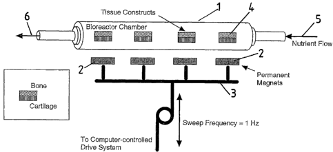
Referring to Fig 1, there is illustrated a tubular bioreactor 1 associated
with
permanent magnets 2 which are positioned externally of the reactor 1 and which
are
moiuzted on a Garner arrangement 3 connected to a computer-controlled (or
other
time-varying) drive system not illustrated in detail in the drawings.
Within the bioreactor 1 are a number of longitudinally spaced tissue
constructs
4 each depicted as being comprised on bone tissue on one side (the side remote
from
the magnets 3) and cartilage tissue on the other side (other tissue types may
also be
used). The tissue cells have magnetic beads (not shown) attached thereto and
are
seeded on 3-D scaffolds (again not shown). Nutrients are supplied to the
bioreactor as
depicted by arrow 5 and exit therefrom as depicted by arrow 6.
There are a total of four magnets 2 (though this number can vary to match the
number of tissue constructs) which are external of the bioreactor and
longitudinally
spaced therealong. The positioning of the magnets is such that there is one
magnet
associated with each of the tissue constructs, the magnets being provided on
the
cartilage sides thereof.
CA 02432574 2003-06-19
WO 02/051985 PCT/GBO1/05606
9
In use of the apparatus, the earner is driven so as to oscillate the magnets
transversely towards and away from the bioreactor. The oscillation frequency
at
which the magnets are driven will usually be varied and generally be in the
range of
0.1 to 10 Hz although values outside this range may be used.
The oscillation of the magnets stimulates a compression/relaxation cycle
which is applied to the tissue constructs, the frequency of which can also be
varied by
mechanical drivers (not shown) attached to the magnets. The magnet field
gradient
(spatially varying magnetic field strength) ensures that the cartilage
experiences
slightly higher flux densities than the bone cells.
Strong magnetic field gradients will produce a translational motion on the
nanoparticles directed towards the magnets, compressing the cells and scaffold
inside
the bioreactor. This compression will simulate mechanical loading without
requiring
direct access to the cells inside the bioreactor. Loads can be easily varied
by changing
the magnetic field strength and gradient, magnet position and/or the physical
properties of the nanoparticles compressing the tissue constructs.
If desired, the magnetic particles associated with the bone cells may have
different magnetic properties from those associated with the cartilage so that
different
mechanically stresses are applied to the two different types of cells.
Fig 2 illustrates a modification of the apparatus shown in Fig 1. In the
modification of Fig 2, the (permanent) magnets are oscillated parallel to the
longitudinal axis of the bioreactor (rather then transversely to the axis in
the case of
Fig 1).
A number of modifications may be made to the illustrated embodiments.
Thus, for example, the magnets may be swept relatively around the bioreactor.
This may most conveniently, but not necessarily, be achieved by keeping the
magnets
fixed and rotating the bioreactor around its longitudinal axis.
CA 02432574 2003-06-19
WO 02/051985 PCT/GBO1/05606
Alternatively or additionally the permanent magnets illustrated in Figs 1 and
2
may be replaced by electromagnets. A fuxther possibility is for the
nanoparticles
attached to the cells to be replaced by ferrofluids. If desired, a combination
of
attached nanoparticles and ferrofluids may also be used.
A further possibility is to use magnetic/metal plates or other structures
which
could be attracted to the magnets in order to deform the entire scaffold.
Reference is now made to Fig 3 which illustrates an alternative method of
activation of mechano-sensitive transmembrane ion channels by the use of a
magnetic
field so as to simulate ,period mechanical loading of a tissue construct.
More particularly, Fig 3 illustrates a cell 10 having a membrane 11 enclosing
the cell's cytoplasm 12. Within membrane 11 is a mechanosensitive ion channel
13.
A functionalized magnetically blocked particle 14 (such as Sphereotech's
coated
ferromagnetic particles, d=4.Spm) is rigidly attached to the cell membrane 11
either
directly or indirectly via cytoskeletal coupling [8, 9].
In the condition shown in Fig 3(a), no magnetic field is applied to the cell
10
and the ion channel 13 is closed.
By oscillating a magnetic field source (not shown) the magnetic particle 14
attached to the cell 10 can be twisted, exerting a mechanical stress on the
cell
membrane 11 and activating the mechano-sensitive in channel 13 (Figure 3b).
This
ion channel activation initiates biochemical reaction pathways in the cells
being
cultured and simulates periodic mechanical loading of tissue constructs inside
the
bioreactor.
CA 02432574 2003-06-19
WO 02/051985 PCT/GBO1/05606
11
REFERENCES:
1. Ying, Y., Peak, M., Magnay, J., and El Haj, A.J. (2000) Dynamic cell
scaffold interactions: implications for tissue engineering. Proceedings of
the seco~ad Smith and Nephew international symposium oh tissue
engineering, York, UK.
2. El Haj, AJ, LM Walker, MR Preston, SJ Publicover (1999)
Mechanotransduction pathways in bone: calcium fluxes and the role of
voltage-operated calcium channels. Med. Biol. Eng. Comp., 37: 403-409.
3. Howard J and AJ Hudspeth (1989) Compliance of the hair bundle
associated with the gating of mechanoelectrical transduction channels in
the bullfrog's saccular hair cell. Neuron 1: 189-199.
4. Walker et al J Cell Biochem 2000.
5. Pardoe, H, W Chua-anusorn, TG St. Pierre, J Dobson (2000) Structural
and magnetic properties of nanoscale magnetic particles synthesised by
coprecipitation of iron oxide in the presence of dextran or polyvinyl
alcohol. J. Magh. Mag. Materials. In Press.
6. Tan, W, S Santra, Z Peng, R Tapec and J Dobson (2000) Coated
nanoparticles. US Patent Pefzding (Filed May 17, 2000).
7. Sittinger et al 1996.
8. Kirschvink, JL (1992) Comments on "Constraints on biological effects of
weak extremely-low-frequency electromagnetic fields". Phys. Rev. A. 46:
2178-2184.
9. Dobson, J and TG St. Pierre (1996) Application of the Ferromagnetic
Transduction Model to D.C. and Pulsed Magnetic Fields: Effects on
Epileptogenic Tissue and Implications fox Cellular Phone Safety. Biochem.
Biophys. Res. Commun., 227:718-723.