Note: Descriptions are shown in the official language in which they were submitted.
CA 02433361 2003-06-27
WO 02/053174 PCT/USO1/50355
Controlled Release Systems for Polymers
Bacli~round of the Invention
With the advent of genetic engineering, the large-scale availability of many
bioactive polymers, such as proteins, carbohydrates and nucleic acids, has
been
achieved. However, the administration of these recombinantly produced peptides
and proteins presents a unique set of problems. In many cases the maintenance
of the
biological effect of these proteins requires long-term administration. Daily
administration of these agents in aqueous vehicles is inconvenient and costly;
sustained or prolonged release is preferred. In addition, proteins are highly
unstable
to in an aqueous environment most suitable for administration.
Moreover, successful treatment of a variety of conditions is limited by the
fact that agents lalown to effectively treat these conditions may have severe
side
effects, requiring low dosages to minimize these side effects. In other
instances, the
therapeutic agents may be very labile, or have very short half lives requiring
repeated administration. In still other instances, the long term
administration of a
pharmaceutical agent may be desired.
In all these cases, the ability to deliver a controlled dosage in a sustained
fashion over a period of time may provide a solution.
Summary of the Invention
2o One aspect of the present invention relates to controlled release delivery
of
biologically active molecules from a solid composition prepared by exposure of
the
molecules to an organic compound. For instance, the organic compound is an
organic solvent, such as an alcohol (e.g., preferably a lower alcohol, such as
methanol, ethanol, isopropanol, n-propanol, n-butanol, isobutanol, t-butanol,
etc.),
a mixture of alcohols, an aldehyde, a lcetone, a hydrocarbon (saturated or
unsaturated), or an aromatic hydrocarbon. The solvent can be a mixture of
different
organic solvents, or the resulting formulation can be a mixture of, e.g.,
different
lyophilized preparations, such as may be used to control the release profile
of the
resulting admixture.
The subject molecule to be formulated for controlled release can be axl
organic compounds. In certain embodiments, it is a polymer, preferably a
1
CA 02433361 2003-06-27
WO 02/053174 PCT/USO1/50355
biopolymer such as a protein, a peptide, a nucleic acid, an oligonucelotide, a
carbohydrate, a ganglioside, or a glycan. The subject molecule can be a lipid,
a sterol
or other lipophilic moiety. The subject controlled delivery system can be used
to
deliver the controlled release of small molecules (e.g., organic compounds).
In certain embodiments,° the subject preparations are prepared by
precipitation and/or lyophilization.
Brief Description of Drawings
Figures 1-5. Graphs showing various release profiles for BSA preparations.
Figures 6A-D. Effect salt concentration of formulation on release of HSA
1o and IFN-a012. Solution I consisted of 9.0 mg of HSA (Immuno-U.S.) and 10
~,g of
IFN-a012 in 40% (w/w) n-propanol (0.364 g n-propanol) in HZO for a total
weight
of 0.91 g. The various Solution II compositions consisted of various
quantities of
sodium acetate (1 M, pH 6.3) and deionized water and 0.040 g n-propanol to
malce
solutions of 40% n-propanol and 250, 450, and 600 mM final sodium acetate
concentrations with a total volmne of 0.10 g. Solution II (0.10 g) was added
to
Solution I (0.91 g) with stirring to yield a fnal 1.01 g of each formulation.
The final
1.01 g formulations containing 40% n-propanol and 25, 45, and 60 mM
concentrations of sodium acetate were stirred in 2 ml glass vials for 6 hr at
24°C and
passed through 25G syringe needles just prior to separating supernatants from
2o precipitates. The quantity of HSA and IFN-a012 in washed precipitates was
determined as described in Materials and Methods. Release was performed in
PBS/0.01% thimerosal. A & B. Absolute (mg) and percent release of precipitated
HSA, respectively. C & D. Absolute (ng) and percent release of precipitated
IFN-
x012, respectively.
Figures 7A-B. Effect of cation species in formulation on release of HSA.
Solution I consisted of 8.1 mg of HSA (Immtmo-U.S.) in 40% (w/w) n-propanol in
deionized water in a total volume of 0.91 ml. The various Solution II
compositions
consisted of adding none or 0.025 ml of various salt stocks (each at 1 M
cation
concentration, pH 6.3) to deionized water followed by n-propanol to make
solutions
40% (w/w) n-propanol and 250 mM final cation concentration in a total volume
of
0.10 ml. Solution II (0.10 ml) was added to 0.91 ml of Solution I with
stirring to
2
CA 02433361 2003-06-27
WO 02/053174 PCT/USO1/50355
give a final 1.01 ml formulation having 40% (w/w) n-propanol. The final 1.01
ml
formulations containing 40% n-propanol and no or 25 mM concentrations of
potassium, sodium or magnesium acetate were stirred in 2 ml glass vials for 6
hr at
24°C prior to separating supernatants from precipitates. The quantity
of HSA in
washed precipitates was determined as described in Materials and Methods.
Release
was performed in PBS/0.01% thimerosal. A & B. Absolute (mg) and percent
release of precipitated HSA, respectively. Salts were sodium, potassium, a~.zd
magnesium acetate (indicated by NaOAc, KOAc, and Mg(OAc)2, respectively).
' Figure 8A-B. Effect of canon species in formulation on release of IFN
l0 x012. Solution I consisted of 45 mg of HSA (Immuno-U.S.) and 5.44 ~.g IFN
a,012 in 40% (w/w) n-propanol in deionized water in a total volume of 4.55 ml.
The
various Solution II compositions consisted of adding 36 ~.1 of 0.1 M acetic
acid (to
compensate for the buffer capacity of the HSA solution) and 0.250 g of
potassium,
sodium or magnesium acetate solution (each at pH 6.3) to 0.314 g of deionized
water
and 0.400 g of n-propanol to make solutions of 40% (w/w) n-propanol and 250 mM
final acetate concentration in a total weight of 1 g. The potassium acetate
solution
was made with 0.980 g potassium acetate, 10.061g water and 0.274 ml 1 M acetic
acid. The sodium acetate solution was made with 0.823 g sodium acetate, 10.056
g
water and 0.245 ml 1 M acetic acid. The magnesium acetate solution was made
with
2.144 g magnesium acetate, 10 g water and 0.200 ml 1 M acetic acid. Solution
II
(0.50 ml) was added to 4.55 ml of Solution I with stirring to give a final
5.05 ml
formulation having 40% (w/w) n-propanol. The final formulations were stirred
in 50
ml conical tubes for 6 hr at 24°C, the precipitates washed with 5 ml of
PBS10.01%
thimerosal, then suspended in 5 ml PBS/0.01% thimerosal, then split into two
individual 2.5 ml samples prior to separating supernatants from precipitates.
Release data is from the precipitates from one 2.5 ml portion of the
formulation.
The amount of IFN-oc012 in washed precipitates was determined as described in
Materials and Methods. Release was performed in PBS/0.01% thimerosal. A & B.
Absolute (ng) and percent release of precipitated IFN-o~012, respectively.
Salts were
3o sodium, potassium, and magnesium acetate (indicated by 21 mM NaOAc, 20 mM
KOAc, and 18 mM Mg(OAc)2, respectively).
3
CA 02433361 2003-06-27
WO 02/053174 PCT/USO1/50355
Figures 9A-B. Effect of aqueous solution pH of formulation on release of
IFN-cc012. Acetic acid (0.1 M) was used to adjust 5% HSA (Alpha Therapeutic)
stoclc solutions to pH 5.0 or pH 7Ø Solution I consisted of 10 mg of HSA
from
either pH 5.0 or pH 7.0 HSA stock solutions, 6.83 ~g IFN-x,012 and additional
water to a total weight of 0.6 g. The final formulations were prepared by
adding 0.4
g of n-propanol to Solution I with stirring to yield a concentration of 40%
(w/w) n-
propanol. Final 1 g formulations v,~ere stirred in 2 ml glass vials for 24 hr
at 24°C
prior to separating supernatants from precipitates. The quantity of IFN-x012
in
washed precipitates was determined as described in Materials and Methods.
Release
to was performed in PBS/0.01% thimerosal. A & B. Absolute (ng) and percent
release
of precipitated IFN-x,012, respectively.
Figure l0A-B. Effect of aqueous solution pH of formulation on release of
HSA and IFN-x012. Solution I consisted of 45 mg of HSA (Immuno-U.S.) and 5.44
~.g IFN-x012 in 40% (w/w) n-propanol in deionized water in a total volume of
4.55
ml. Solution II compositions were prepared as follows. Solution IIa: 1.55 ml
of 1 M
acetic acid was added to 0.82 g aWydrous sodium acetate and 10 g deionized
water
to adjust pH of this Solution A to 5.52; then 0.036 ml of 0.1 M acetic acid
was added
to 0.250 g of Solution A to compensate for the buffer capacity of the HSA
solution;
deionized water was then added to bring the total weight to 0.600 g; then
0.400 g of
2o n-propanol was added to make a final solution of 40% (w/w) n-propanol in a
total
weight of 1.00 g. Solution IIb: 0.40 ml of 1 M acetic acid was added to 0.82 g
anhydrous sodium acetate and 10 g of deionized water to adjust pH of this
Solution
B to 6.13; then 0.036 ml of 0.1 M acetic acid was added to 0.250 g of Solution
B to
compensate for the buffer capacity of the HSA solution; deionized water was
then
added to bring the total weight to 0.600 g; then 0.400 g of n-propanol was
added to
make a final solution of 40% (w/w) n-propanol in a total weight of 1.00 g.
Solution
IIc: 0.245 ml of 1 M acetic acid was added to 0.823 g anhydrous sodium acetate
and
10.056 g deionized water to adjust pH of this Solution C to 6.31; then 0.036
ml of
0.1 M acetic acid was added to 0.250 g of Solution C to compensate for the
buffer
capacity of the HSA solution; deionized water was then added to bring the
total
weight to 0.600 g; then 0.400 g of n-propanol was added to make a final
solution of
4
CA 02433361 2003-06-27
WO 02/053174 PCT/USO1/50355
40% (w/w) n-propanol in a total weight of 1.00 g. To prepare the final
fomnulations,
0.50 ml from Solutions IIa, IIb, or IIc was added to 4.55 ml of Solution I
with
stirring to yield three 5.05 ml formulations having 40% (w/w) n-propanol and
pH
5.52, pH 6.13 or pH 6.31, respectively. Final formulations were stirred in 50
ml
conical tubes for 6 hr at 24°C, then split into two individual 2.52 ml
samples prior to
separating supernatants from precipitates. Release data is from one 2.52 ml
portion
of the formulation. The amount of IFN-x,012 in washed precipitates was
determined
as described in Materials and Methods. Release was performed in PBS/0.01%
thimerosal. A & B. Absolute (mg) and percent release of precipitated HSA,
to respectively. C & D. Absolute and percent release of precipitated IFN-
oc012,
respectively.
Figure 11A-B. Effect of acid conceiltration of formulation on release of
HSA and IFN-x001 from precipitates formed in the presence of 25 mM sodium
acetate. Solution I consisted of 8.1 mg of HSA (Immuno-U.S.) and 0.92 ~,g IFN-
x001 in 40% (w/w) n-propanol in deionized water in a total volume of 0.9 ml.
Several Solution II formulations, IIa, IIb, IIc and IId, were prepared
consisting of
0.004, 0.010, 0.015 and 0.025 ml of 0.1 M acetic acid, respectively, in 40%
(w/w) n-
propanol in deionized water. Solution III consisted of 1 M sodium acetate and
40%
(w/w) n-propanol in deionized water in a total volume of 0.025 ml. Several
Solution
2o IV formulations, IVa, IVb, IVc and IVd, were prepared consisting of 0.071,
0.065,
0.060 and 0.050 ml of 40% (w/w) n-propanol, respectively, in deionized water.
In
preparing the final formulations, Solutions IIa, IIb, IIc and IId were matched
with
Solutions IVa, IVb, IVc and IVd, respectively. Solutions II, III and IV were
mixed
together then Solution I added rapidly to the mixture to give a final 1 ml
formulation. This yielded a formulation having a final concentration of 25 mM
sodium acetate, 40% (w/w) n-propanol and the final acetic acid concentrations
indicated on the Figure. Formulations were stirred in 2 ml glass vials for 6
hr at
24°C prior to separating supernatants from precipitates. After washing,
precipitates
were lyophilized 4 hr at <400 mTorr. The amount of HSA in washed precipitates
3o was determined as described in Materials and Methods. Release was performed
in
PBS/0.01% thimerosal. A & B. Absolute (mg) and percent release of precipitated
5
CA 02433361 2003-06-27
WO 02/053174 PCT/USO1/50355
HSA, respectively. C & D. Absolute (ng) and percent release of precipitated
IFN-
oc012, respectively.
Figure 12A-D. Effect of salt concentration of formulation on release of
HSA and IFN-x001 from precipitates formed in the presence of 1.5 mM acetic
acid.
Solution I consisted of 8.1 mg of HSA (Immuno-U.S.) and 0.92 ~.g IFN-x001 in
40% (w/w) n-propanol in deionized water in a total volume of 0.9 ml. Solution
II
consisted of 0.1 M acetic acid and 40% (w/w) n-propanol in deionized water in
a
total volume of 0.015 ml. Several Solution III formulations, IIIa, IIIb, IIIc
and IIId,
were prepared consisting of 0, 0.015, 0.025 and 0.035 ~nl of 1 M sodium
acetate,
to respectively, in 40% (w/w) n-propanol in deionized water. Several Solution
IV
formulations, IVa, IVb, IVc and IVd, were prepared consisting of 0.085, 0.070,
0.060 and 0.050 ml of 40% (w/w) n-propanol, respectively, in deionized water.
In
preparing the final formulations, Solutions IIIa, IIIb, IIIc and IIId were
matched with
Solutions IVa, IVb, IVc and IVd, respectively. Solutions II, III and IV were
mixed
together then Solution I added rapidly to the mixture to give a final 1 ml
formulation. This yielded a final concentration of 1.5 mM acetic acid, 40%
(w/w) n-
propanol (w/w) and the final sodium concentrations indicated on the Figure.
Formulations were stirred in 2 ml glass vials for 6 hr at 24°C prior to
separating
supernatants from precipitates. After washing, precipitates were lyophilized 4
hr at
2o <400 mTorr. The amounts of HSA and IFN-x,001 in washed precipitates were
determined as described in Materials and Methods. Release was performed in
PBS/0.01% thimerosal. A & B. Absolute (mg) and percent release of precipitated
HSA, respectively. C & D. Absolute (ng) and percent release of precipitated
IFN-
a,001, respectively.
25' Figure 13A-B. Effect of salt concentration and pH of formulation on
release
of HSA with tertiary butanol precipitates. Acetic acid (0.1 M) was used to
adjust 5%
HSA stoclc solutions (Alpha Therapeutic) to pH 5.35 or 7Ø Solution I
consisted of
18.0 mg of HSA from the pH 5.35 or pH 7.0 5% stock solution, 1.0 ~,g IFN-x,012
and deionized water bringing the total solution weight to. 0.375 g. To prepare
3o Solutions IIa and IIb with NaCI concentrations of 0.02 M and 0.1 M,
respectively,
sufficient deionized water was added to 0.021 and 0.0043 ml of a 3.75 M NaCl
6
CA 02433361 2003-06-27
WO 02/053174 PCT/USO1/50355
solution to bring the total weight of each solution to 0.425 g. Both pH 5.35
and pH
7.0 variants of Solution I (0.375 g) were added to Solutions IIa and IIb to
yield 0.80
g of the various combinations of pH and NaCI concentration as shown in the
Figure
prior to the addition of 0.31 or 0.47 g of tert-butyl alcohol to yield 28.1%
and 36.9%
s (w/w) tert-butyl alcohol (see summary of the chart legends). Final 1.1l-1.27
g
formulations were stirred in 2 ml glass vials for 24 hr at 24°C prior
to separating
supernatants from precipitates. The amount of HSA in washed precipitates was
determined as described in Materials and Methods. Release was performed in
PBS/0.01% thimerosal. A & B. Absolute (mg) and percent release of precipitated
to HSA, respectively.
Figure 14. Effect of pH and salt concentration of formulation on tlueshold
of precipitation of HSA by n-propanol. An 11 % (w/w) HSA (USB) was dialyzed 3
times for 6 hr each time against 2 L deionized H20 in a Pierce Slide alyzer
(15 ml
capacity, No. 66410, lot # BJ44820B). The final concentration was analyzed by
15 spectrophotometiy at 280 mn to be 8.28% (w/w). This solution was diluted to
4%
(w/w) with deionized water. Amounts (0.9 g) of 4% HSA were weighed into 2 ml
glass vials. Sodium acetate (1 M), acetic acid (1 M), sodiLUn hydroxide (1 M),
and
water were added in various combinations in a total weight of 0.1 g to yield
the final
sodium concentrations and pH values measured in 1 g formulations as shown in
the
20 Figure. Subsequently, n-propanol was added in about 50 ~.l increments with
stirring,
and the point at which intial precipitates were stable (did not re-dissolve
with
stirring within 5 minutes) was recorded. Connected data points indicate
equivalent
sodium concentrations at various pH and n-propanol (w/w) concentrations.
BEST MODE FOR CARRYING OUT THE INVENTION
25 Description of the Invention
I. Overview
The present invention relates to a controlled release delivery system and is
based on the discovery that treatment of proteins and other molecules such as
carbohydrates, nucleic acids, and other substances with organic compounds can
3o modify their solubility in aqueous media. For example, in one embodiment
the
exposure of the proteins to the organic solvent (such as an alcohol) replaces
the
7
CA 02433361 2003-06-27
WO 02/053174 PCT/USO1/50355
water molecules and other associated moieties with organic residues. In
certain
embodiments, the subject preparations are solids, e.g., powders or crystals
formed by
lyophilization, precipitation or the Iilce.
The resulting preparations can provide prolonged release formulations of the
s proteins, e.g., suitable for sustained biological effects when used as
pharmaceuticals
or in other aqueous uses. The examples given refer to protein, but the
principle can
apply to other water soluble biopolymers as well such as peptides,
carbohydrates,
nucleic acids, oligonucleotides, lipids, glycans, gangliosides and other
biopolymers.
Small organic molecules and some inorganic molecules that are solvated with
l0 attached water residues can be treated in an analogous way to provide
controlled
delivery of the specific molecules.
Furthermore, solubility of proteins is also modulated by porttranslational
modifications that can change the solubility of the proteins. The methods
described
can alter the solubility of the proteins with and without the post-
translational
15 modifications.
In certain embodiment, the biomolecules are precipitated from the aqueous
solution by addition of organic solvents and then lyophilized. In alternative
procedures, the solution can be lyophilized directly from solution containing
organic
solvents to provide for the dried material to be formulated into a controlled
release
2o system; the precipitated protein washed with aqueous solution and then
formulated
directly without lyophilization; or the dry protein treated with organic
solvent, then
formulated after removal of the solvent.
In certain preferred embodiments, the solvent is a an inert solvent, and even
more preferably an anhydrous organic solvent. The solvent should not
irreversibly
25 denature the polymer, e.g., the tirnescale for renaturation, if any is
requireed, should
not be signiificantly longer than the rehydration process.
Formulation and size of the material can be controlled by the timing and
method of precipitation and lyophilization conditions. Upon precipitation of
the
molecules, the precipitate is lyoplulized to remove excess water and prevent
water
3o from immediately replacing the organic solvents. Colloidal suspensions
without
direct precipitation can be used to substitute for precipitation. The
colloidal
s
CA 02433361 2003-06-27
WO 02/053174 PCT/USO1/50355
suspensions can be used to generate particles of small size. Furthermore, the
mixtures can be lyophilized directly without precipitation or colloid
formation to
provide particles of different sizes dependent on the concentration of the
molecules
in the organic-aqueous media, the method of precipitation and the
concentration of
the protein solution. In some instances, inorganic molecules that can replace
the
water molecules on the molecules to be released slowly can be used in a total
aqueous system to provide the same results. after lyophilization. The release
is
affected by the specific organic solvent used, the buffer used, and the
particle size of
the precipitated and/or lyophilized protein.
to In addition, the method of invention permits greater tailoring of release
profiles. The subj ect preparations can be made to exhibit short-term or long-
term
release l~inetics, thereby providing either rapid or sustained release of
macromolecules. In any event, the subject preparations leave, relative to
preparations
of the polymer lyophilized from aqueous solutions, a reduced solubility in
serum or
other biological fluid, e.g., the solubility rate over a period of at least
24, 48, or even
168 hours (7 days) is at least 2 fold less than preparations of the polymer
lyophilized
from aqueous solution, and more preferably at least 10, 25, 50 or even 100
fold less.
In certain preferred embodiments, the subject compositions permit the
release of biologically active compound at a rate which provides an average
steady
2o state dosage of at least the EDSO for the active compound for a period of
at least 2
days, and more preferably at least 7, 14, 21, 50, or even 100 days.
In certain preferred embodiments, the solvents) are chosen such that, when
administered to a patient (particularly a human), the solvent released from
the
formulation is done so at a rate which remains below the ICSO for deleterious
side
effects, if any, of the solvent, and more preferably at least l, 2 or even 3
orders of
magnitude below such ICso concentrations.
In certain embodiments, the organic agent is a polar erotic solvent, such as
for example, aliphatic alcohols,glycols, glycol ethers, and mixtures thereof.
In
certain preferred embodiments, the organic agent is a water-miscible polar
erotic
solvent.
9
CA 02433361 2003-06-27
WO 02/053174 PCT/USO1/50355
Biodegradable or non-biodegradable materials lalov~m in the aut in the form
of gels, microspheres, wafers or inplants can be mixed with the subject
modified
molecules.
These subject formulations can be used in parenteral, oral, intramuscular,
subcutaneous, dermal, intravenous, intrarterial, intralesional, intrathecal or
other
sites of delivery for the treatment, prevention and diagnosis of many
diseases.
Still another aspect of the invention relates to a method for doing business,
e.g., for the preparation of pharmaceutical formulations for the treatment of
humans
or other animals. In an exemplary embodiment of such methods, there is
provided a
to lyophilization facility for generating the lyophilized preparations
described herein.
The lyophilized preparations are packaged as e.g., pills, tablets, patches,
injectables
and the lilce, preferably at a govermnent approved facility, e.g., an FDA-
approved
facility. In preferred embodiments, the lyophilized preparation is provided in
single
dosage form, even if packaged in larger lots.
II. Definitions
"Bioerodible" signifies that the material may be dissolved or digested into
component molecules by the action of the environment or particularly by the
action
by living organisms, and optionally metabolized or digested into simpler
constituents
without poisoning or distressing the environment or the organism.
"Administered to a mammal" means that the composition containing an
active ingredient is administered orally, parenterally, enterically,
gastrically,
topically, transdermally, subcutaneously, locally or systemically. The
composition
may optionally be administered together with a suitable pharmaceutical
excipient,
which may be a saline solution, ethyl cellulose, acetotephtalates, mannitol,
lactose,
starch, magnesium stearate, sodimn saccharin, talcum, glucose, sucrose,
carbonate,
and the like.
"Sustained delivery" or "sustained time release" denotes that the active
ingredient is released from the delivezy vehicle at an ascertainable and
manipulatable
rate over a period of minutes, hours, days, weeks or months, ranging from
about
thirty minutes to about two months or longer.
CA 02433361 2003-06-27
WO 02/053174 PCT/USO1/50355
Abbreviations
HSA Human serum albumin
HOAc Acetic acid
NaOAc Sodium acetate
s I~OAc Potassium acetate
Mg(OAc)2 Magnesium acetate
IFN-x001 Interferon a-001
IFN-oc012 Interferon a,-012
PBS Phosphate-buffered saline
1o III. Exemplary Biopolymers
The biopolymers which may be used in the present invention include
proteins, carbohydrates, nucleic acids and combinations thereof.
Advantageously, according to the present invention, the subject method can
be used to formulate a protein which is pharmaceutically valuable or of value
in the
15 agri-foodstuffs industry. Proteins of interest include cytolcines, growth
factors,
somatotropin, growth hormones, colony stimulating factors, , erythropoietin,
plasminogen activators, enzymes, T-cell receptors, surface membrane proteins,
lipoproteins, clotting factors, anticlotting factors, tumor necrosis factors,
transport
proteins, homing receptors, addressins, etc. Examples of mammalian
polypeptides
2o include molecules such as renin, a growth hormone, including human growth
hormone; bovine growth hormone; growth hormone releasing factor; parathyroid
hormone; thyroid stimulating hormone; lipoproteins; a-1-antitrypsin; insulin;
proinsulin; follicle stimulating hormone; calcitonin; luteinizing hormone;
glucagon;
clotting factors such as factor VIIIC, factor IX, tissue factor, and von
Willebrands
25 factor; anti-clotting factors such as Protein C; atrial natriuretic factor;
lung
surfactant; a plasminogen activator, such as urolcinase or human urine or
tissue-type
plasminogen activator (t-PA); bombesin; thrombin; hemopoietic growth factor;
tiunor necrosis factor-a, and -(i; enlcephalinase; RANTES (regulated on
activation
normally T-cell expressed and secreted); human macrophage inflammatory protein
30 (MIP-1-oc); a serwn albumin such as htunan serum albumin; mullerian-
inhibiting
substance; relaxin A-chain; relaxin B-chain; prorelaxin; mouse gonadotropin-
11
CA 02433361 2003-06-27
WO 02/053174 PCT/USO1/50355
associated peptide; a microbial protein, such as beta-lactamase; DNase;
inhibin;
activin; vascular endothelial growth factor (VEGF); receptors for hormones or
growth factors; integrin; protein A or D; rheumatoid factors; a neurotrophic
factor
such as bone-derived neurotrophic factor (BDNF), neurotrophin-3, -4, -5, or -6
(NT-
3, NT-4, NT-5, or NT-6), or a nerve growth factor such as NGF-Vii; platelet-
derived
growth factor (PDGF); fibroblast growth factor such as aFGF and bFGF;
epidermal
growth factor (EGF); transforming growth factors (TGF) such as TGF-a, TGF-(3
and
BMPs; insulin-lilce growth factor-I and -II (IGF-I and IGF-II); des(1-3)-IGF-I
(brain
IGF-I), insulin-like growth factor binding proteins; CD proteins such as CD-3,
CD-4,
l0 CD-8, and CD-19; erythropoietin; osteoinductive factors; immunotoxins; a
bone
morphogenetic protein (BMP); an interferon such as interferon-a, -(3, and -y;
colony
stimulating factors (CSFs), e.g., M-CSF, GM-CSF, and G-CSF; interleulcins
(ILs),
e.g., IL-1 to IL-10; superoxide dismutase; T-cell receptors; surface membrane
proteins; decay accelerating factor; antigens (e.g., bacterial and viral
antigens);
transport proteins; homing receptors; addressins; regulatory proteins;
immunoglobulin-life proteins; antibodies; nucleases; and fragments of any of
the
above-listed polypeptides.
Other examples of suitable therapeutic and/or prophylactic biologically
active agents include nucleic acids, such as antisense molecules; and small
z0 molecules, such as antibiotics, steroids, decongestants, neuroactive
agents,
anesthetics, sedatives, cardiovascular agents, anti-tumor agents,
antineoplastics,
antihistamines, hormones (e.g., thyroxine) and vitamins.
IV. Exemplary Methods
The rate of controlled release of the protein can be modified by many
variables. The variables include rate of addition of organic solvent, time of
protein
(or other molecule) in organic solvent (time of exposure of protein to organic
solvent), concentration of organic solvents for precipitation of the protein,
concentration of the organic solvents prior to precipitation, concentration of
the
organic solvents prior to lyophilization from solution directly, organic and
non
organic composition of media, temperature, concentration of cations,
concentration
of anions, rate of precipitation, pH, mixtures of organic solvents, stirring,
agitation,
12
CA 02433361 2003-06-27
WO 02/053174 PCT/USO1/50355
presence of other proteins as carriers, presence of other proteins for
controlled
release of multiple proteins, protein stabilizers, dissolved gasses, reducing
agents,
oxidizing agents, mass to surface area of the particles, washing of samples
prior to
preparation for release, salt concentration, length of time exposed to
modifier agents,
concentration of the proteins or other polymer, inorganic compounds, type of
orga~zic
compounds, for example. Inorganic cations can be monovalent, divalent,
trivalent,
tetravalent or pentavalent; inorganic anions can be monovalent, divalent,
trivalent,
tetravalent or pentavalent. In some embodients, lyophilization can be omitted.
For
example, the precipitate can be washed with a nonpolar solvent such as ~-
hexane to
l0 remove the organic solvent without affecting the protein; or the
precipatate can be
washed with an aqueous medium to remove the organic solvent removing the
excess
organic solvent fiom the protein mass. Furthermore, the precipitate can be
washed
and/or preincubated to remove soluble protein and eliminate the higher initial
release
r ate.
Organic compound does not need to be solvent, just constituent in the
mixture. .
In addition, the protein precipitates can be placed into a variety of
biodegradable or non-biodegradable materials known in the art in the form of
gels,
microspheres, wafers or implants. In these cases, the release is controlled by
both
2o the intrinsic protein release rate and the rate of release controlled by
the gels,
microspheres, wafers or implants. These formulations can be used in
parenteral,
oral, intramuscular, subcutaneous, dermal, intravenous, intraxterial,
intralesional,
intrathecal or other sites of delivery for the treatment, prevention and
diagnosis of
many diseases.
During equilibration of the protein with the solvent, the organic solvent used
is attached to the protein in the precipitates. The organic solvent can be
replaced
partially or completely with other organic compounds soluble in the solution.
The
organic compounds can be active pharmaceuticals such as antibiotics,
antimicrobial
agents, aminoglycosides, chloramphenicol, macrolides, antifimgals,
cephalosporins,
3o 3,4-dihydroxyphenylalanine (DOPA), adrenergic agonists, adrenergic
antagonists,
cholinergic agonists, cholinergic antagonists, muscarinic agonists, muscarinic
13
CA 02433361 2003-06-27
WO 02/053174 PCT/USO1/50355
antagonists, antiviral agents, sympathomimetics, sympatholytics, serotonin
agonists,
serotonin antagonists, antihypertensive agents, monoamiye oxidase iWibitors,
diuretics, antianhytlunic drugs, phosphodiesterase izW ibitors, digitalis
glycosides,
calcium antagonists, vasodilators, prostaglandins, autacoids, lipid lowering
drugs,
anticoagulants, fibrinolytics, platelet aggregation inhibitors,
antidepressants,
benzodiazepines, antiepileptics, antiparl~inson agents, analgesics, opioids,
opioid
peptides, opiates, peptides, antiinflarrunatory drugs (NSAIDs, acetaminophen),
barbiturates, peptide hormones, steroids, glucocorticoids, mineralocorticoids,
estrogens, progestins, androgens, antiandrogens, thyroxine, triiodothyronine,
1o cyclooxygenase inhibitors, growth hormone releasing hormone (GHRH),
antineoplastic drugs, and antihistamines. The attached organic compounds (as
drugs) lined to bovine or human serum albumin or other proteins such as
immunoglobulins can then be delivered as the protein is released and
dissolved. The
proteins with attached organic solvents are thus able to be used as effective
delivery
systems. Furthermore, with the use of immunoglobulins and other proteins that
can
target to specific tissues or cells, the attached molecules can then be
delivered to the
tissues or cells.
Preparations made by the subject process can be either homogeneous or
heterogeneous mixtures of active agents, or of preparations of active agents
prepared
2o under different conditions (e.g., using different solvents, etc).
The amount of a biologically active agent, which is contained in a specific
preparation, is a therapeutically, prophylactically or diagnostically
effective amount,
which can be determined by a person of ordinary skill in the art taking into
consideration factors such as body weight, condition to be treated, type of
polymer
used, and release rate from the preparation.
The biologically active agent can also be mixed with other excipients, such
as stabilizers, surfactants, solubility agents and bullring agents.
Stabilizers are added
to maintain the potency of the agent over the duration of the agent's release.
Suitable
stabilizers include, for example, carbohydrates, amino acids, fatty acids and
3o surfactants and are lcnown to those spilled in the art. Solubility agents
are added to
modify the solubility of the agent in aqueous solution or, as the case may be,
in
14
CA 02433361 2003-06-27
WO 02/053174 PCT/USO1/50355
organic solvents. Suitable solubility agents include complexing agents, such
as
albmnin and protamine, which can be used to control the release rate of the
agent.
Bulking agents typically comprise inert materials.
In another embodiment, a biologically active agent can be lyophilized with a
metal cation component, to further stabilize the agent and control the release
rate of
the biologically active agent.
The subject formulations, if used a therapeutics, may be achninistered to a
human or animal by oral or parenteral administration, including intravenous,
subcutaneous or intramuscular injection; administration by inhalation;
intraarticular
l0 administration; mucosal administration; ophthalmic administration; and
topical
administration. Intravenous administration includes catheterization or
angioplasty.
In other embodiments, the subject preparations can be used in non-
tlaerapeutic aqueous environments, such as for the release of agents (such as
enzymes) into a water supply or water treatment facility.
In addition to the active agent, the formulation can include other suitable
polymers, e.g., to permit the resulting formulation to be used to form a
microparticle.
In a preferred embodiment, a polymer used in this method is biocompatible. A
polymer is biocompatible if the polymer, and any degradation products of the
polymer, such as metabolic products, are non-toxic to humans or animals, to
whom
the polymer was administered, and also present no significant deleterious or
untoward effects on the recipient's body, such as an immunological reaction at
the
injection site. Biocompatible polymers can be biodegradable polymers, non-
biodegradable polymers, a blend thereof or copolymers thereof.
Suitable biocompatible, non-biodegradable polymers include, for instance,
polyaciylates, polymers of ethylene-vinyl acetates and other acyl substituted
cellulose acetates, non-degradable polyurethanes, polystyrenes, polyvinyl
chloride,
polyvinyl fluoride, polyvinyl imidazole), chlorosulphonate polyolefins,
polyethylene oxide, blends and copolymers thereof.
Suitable biocompatible, biodegradable polymers include, for example,
poly(lactide)s, poly(glycolide)s, poly(lactide-co-glycolide)s, poly(lactic
acids,
poly(glycolic acids, polycarbonates, polyesteramides, polyanhydrides,
poly(amino
CA 02433361 2003-06-27
WO 02/053174 PCT/USO1/50355
acids), polyoi-thoesters, polyacetals, polycyanoacrylates, polyetheresters,
polycaprolactone, poly(dioxanone)s, poly(allcylene allcylate)s, polymethanes,
blends
and copolymers thereof. Polymers comprising poly(lactides), copolymers of
lactides
and glycolides, blends thereof, or mixtures thereof are more preferred. Said
polymers
can be formed from monomers of a single isomeric type or a mixture of isomers.
A polymer used in this method can be blocked, unblocked or a blend of
blocked and unblocked polyners. An unblocked polymer is as classically defined
in
the art, specifically having free carboxyl end groups. A blocked polymer is
also as
classically defined in the art, specifically having blocked carboxyl end
groups.
1o Generally, the blocking group is derived from the initiator of the
polymerization
reaction and is typically an alkyl radical.
In certain embodiments, the subject formulations are prepared by
lyophilization. The simplest form of iyophilizer would consist of a vacuum
chamber
into which wet sample material could be placed, together with a means of
removing
water vapor so as to freeze the sample by evaporative cooling and freezing and
then
maintain the water-vapor pressure below the triple-point pressure.
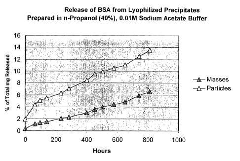
Example 1
Release of bovine serum albumin (BSA) was measured up to 811 hours from
samples of lyophilized protein precipitated from an alcohol/aqueous solution.
This
2o example briefly describes sample preparation and analytical methodology and
presents results showing controlled release of BSA. The release is affected by
the
specific alcohol used, the buffer used, and the particle size of the
precipitated and
lyophilized protein.
Solutions of BSA (USB, Amersham Life Sciences, Cat. No. 10868) at 5
(w/w) were prepared in 0.01 M acetate buffer using an equivalent volume of
0.005
M sodium acetate and 0.005 M acetic acid. The pH was approximately 5. The
alcohol n-propanol was added to a concentration of 40% (v/v). After overnight
equilibration at room temperature, the supernatant was removed and the
precipitate
frozen at -20 C and brought to -70 C before lyophilization. The surface upon
which
3o the vials were placed and the lyophilizer chamber was precooled to maintain
the
samples frozen during the lyophilization procedure. The sample was lyophilized
for
16
CA 02433361 2003-06-27
WO 02/053174 PCT/USO1/50355
hours. The time of lyophilizatioii can be longer or shorter depending on the
voltune to be lyophilized. The lyophilized sample was divided into several
pieces
with a spatula. The pieces were divided into small pauticles by crushing the
pieces
against the wall and bottom of the glass vial. The larger masses and small
crushed
5 particles were weighed so that 5 to 10 mg of the masses and the crushed
particles
were placed into separate 1.5 ml conical polypropylene tubes, then 1 ml of
phosphate
buffered saline was added. The masses or particles were disbursed into the
liquid.
One hour after disbursing the samples, the contents of the tubes were mixed
again
and then the tubes centrifuged for 5 minutes at 5,000 rpm (Eppendorf
Centrifuge,
to Model No. 5415). A sample of 0.1 ml was removed for assay and replaced with
0.1
ml of PBS. This procedure was repeated to take samples at 65 hours. At 98
hours
aazd each time point thereafter, the full volume of release medium was removed
and
replaced with a fresh 1 ml of PBS.
Samples were analyzed for protein content with the microassay procedure for
microtiter plates (Bio-Rad protein assay, based on the method of Bradford;
Coomassie Brilliant Blue Dye, Cat. No. 500-0006) with 96 well microtiter
plates.
Standards contained 5 to 60 ~g/ml of BSA. Standards and samples were added to
the wells in a volume of 0.16 ml first, then 40 ~,1 of dye was added to each
well with
mixing before reading the absorbance at 630 mn. Standard curves were
constructed
from absorbances corrected for the blank values in the absence of added
protein
(BSA). Protein concentrations of the samples were calculated from the standard
curve that was based on the same lot of BSA and prepared on the basis of
weight of
BSA to total volume (w/v). The values for the protein released at various
times were
adjusted by determining differences in the protein concentration of the
lyophilized
BSA that was weighed and placed in solution from the BSA taken directly from
the
bottle of the commercial supplier (USB, Amersham Life Sciences, Cat. No.
10868)
and placed in solution.
The results of the controlled release are shown in Fig. 1 [nP, represents ~z
propanol]. As can be seen, there is little or no burst effect and the release
is
3o essentially linear. The smaller particles with a large surface area to mass
ratio
release at a faster rate. There appears to be a slightly faster rate of
release during the
17
acids), polyoi-thoesters, polyace
CA 02433361 2003-06-27
WO 02/053174 PCT/USO1/50355
first hours of release (Fig. 1). This faster release rate can be eliminated by
preincubating the samples in medium prior to use.
Example 2
Release of BSA was measured up to 811 hours from samples of lyophilized
protein precipitated from alcohol/aqueous solution. This example briefly
describes
sample preparation and analytical methodology and presents results showing
controlled release of BSA. The release is affected by the specific alcohol
used, the
buffer used, and the particle size of the precipitated and lyophilized
protein.
Solutions of BSA (USB, Amersham Life Sciences, Cat. No. 10868) at 5
1o (w/w) were prepared in 0.1 M acetate buffer using an equivalent volume of
0.05 M
sodium acetate and 0.05 M acetic acid. The pH was approximately 5. The alcohol
~z-propanol was added to a concentration of 50% (v/v). After overnight
equilibration
at room temperature, the supernatant was removed and the precipitate frozen at
-20
C and brought to -70 C before lyophilization. The surface upon which the vials
were
placed and the lyophilizer chamber was precooled to maintain the samples
frozen
during the lyophilization procedure. The sample was lyophilized for 5 hours.
The
time of lyophilization can be longer or shorter depending on the volume to be
lyophilized. The lyophilized sample was divided into several pieces with a
spatula.
The pieces were divided iilto small particles by crushing the pieces against
the wall
2o and bottom of the glass vial. The larger masses and small crushed pay-
ticles were
weighed so that 5 to 10 mg of the masses and the crushed particles were placed
into
separate 1.5 ml conical polypropylene tubes, then 1 ml of phosphate buffered
saline
was added. The masses or particles were disbursed into the liquid. One hour
after
disbursing the samples, the contents of the tubes were mixed again and then
the
tubes centrifuged for .5 minutes at 5,000 rpm (Eppendorf Centrifuge, Model No.
5415). A sample of 0.1 ml was removed for assay and replaced with 0.1 ml of
PBS.
This procedure was repeated to take samples at 65 hours. At 98 hours and each
time
point thereafter, the full volume of release medium was removed and replaced
with a
fresh 1 ml of PBS.
3o Samples were analyzed for protein content with the microassay procedure for
microtiter plates (Bio-Rad protein assay, based on the method of Bradford;
is
CA 02433361 2003-06-27
WO 02/053174 PCT/USO1/50355
Coomassie Brilliant Blue Dye, Cat. No. 500-0006) with 96 well microtiter
plates.
Standards contained 5 to 60 ~g/ml of BSA. Standards and samples were added to
the wells in a volume of 0.16 ml first, then 40 y1 of dye was added to each
well with
mixing before reading the absorbance at 630 rm. Standard curves were
constructed
from absorbances corrected for the blank values in the absence of added
protein
(BSA). Protein concentrations of tile samples were calculated from the
standard
curve that was based on the same lot of BSA and prepared on the basis of
weight of
BSA to total volume (w/v). The values for the protein released at various
times were
adjusted by determining differences in the protein concentration of the
lyophilized
to BSA that was weighed and placed in solution from the BSA taken directly
from the
bottle of the commercial supplier (USB, Amersham Life Sciences, Cat. No.
10868)
and placed in solution.
The results of the controlled release are shown in Fig. 2 [nP, represents ~z
propanolj. As can be seen, there is no burst effect and the release is
essentially
linear. The smaller particles with a large surface area to mass ratio release
at a faster
rate. There appears to be a slightly faster rate of release during the first
hours of
release (Fig. 2). This faster release rate can be eliminated by preincubating
the
samples in rneditun prior to use.
Example 3
2o Release of BSA was measured up to 811 hours from samples of lyophilized
protein precipitated from alcohollaqueous solution. This example briefly
describes
sample preparation and analytical methodology and presents results showing
controlled release of BSA. The release is affected by the specific alcohol
used, the
buffer used, and the particle size of the precipitated and lyophilized
protein.
Solutions of BSA (USB, Amersham Life Sciences, Cat. No. 10868) at 5
(w/w) were prepared in 0.01 M acetate buffer using an equivalent volume of
0.005
M sodium acetate and 0.005 M acetic acid. The pH was approximately 5. The
t-butyl alcohol was added to a concentration of 40% (v/v). After overnight
equilibration at room temperature, the supernatant was removed and the
precipitate
3o frozen at -20 C and brought to -70 C before lyophilization. The surface
upon which
the vials were placed and the lyophilizer chamber was precooled to maintain
the
19
CA 02433361 2003-06-27
WO 02/053174 PCT/USO1/50355
samples frozen during the lyophilization procedure. The sample was lyophilized
for
hours. The time of lyophilization can be longer or shorter depending on the
volume to be lyophilized. The lyophilized sample was divided into several
pieces
with a spatula. The pieces were divided into small particles by crushing the
pieces
5 against the wall and bottom of the glass vial. The larger masses and small
crushed
particles were weighed so that 5 to 10 mg of the masses and the crushed
particles
were placed into separate 1.5 ml conical polypropylene tubes, then 1 ml of
phosphate
buffered saline was added. The masses or particles were disbursed into the
liquid.
One hour after disbursing the samples, the contents of the tubes were mixed
again
l0 and then the tubes centrifuged for 5 minutes at 5,000 rpm (Eppendorf
Centrifuge,
Model No. 5415). A sample of 0.1 ml was removed for assay and replaced with
0.1
ml of PBS. This procedure was repeated to take samples at 65 hours. At 98
hours
and each time point thereafter, the full volume of release medium was removed
and
replaced with a fresh 1 ml of PBS.
Samples were analyzed for protein content with the microassay procedure for
microtiter plates (Bio-Rad protein assay, based on the method of Bradford;
Coomassie Brilliant Blue Dye, Cat. No. 500-0006) with 96 well microtiter
plates.
Standards contained 5 to 60 ~g/ml of BSA. Standards and samples were added to
the wells in a volume of 0.16 ml first, then 40 ~,l of dye was added to each
well with
2o mixing before reading the absorbance at 630 nm. Standard curves were
constructed
from absorbances corrected for the blanlc values in the absence of added
protein
(BSA). Protein concentrations of the samples were calculated from the standard
cua-ve that was based on the same lot of BSA and prepared on the basis of
weight of
BSA to total volume (w/v). The values for the protein released at various
times were
adjusted by determining differences in the protein concentration of the
lyophilized
BSA that was weighed and placed in solution from the BSA tal~en directly from
the
bottle of the commercial supplier (USB, Amersham Life Sciences, Cat. No.
10868)
and placed in solution.
The results of the controlled release are shown in Fig. 3 [tBA, represents t
3o butyl alcohol]. As can be seen, there is no major burst effect and the
release is
essentially linear after the first hours. The smaller particles with a large
sL~rface area
CA 02433361 2003-06-27
WO 02/053174 PCT/USO1/50355
to mass ratio release at a faster rate. There appears to be a slightly faster
rate of
release during the first hours of release (Fig. 3). This faster release rate
can be
eliminated by preincubating the samples in medium prior to use.
Example 4
Release of BSA was measured up to 811 hours from samples of lyophilized
protein precipitated from alcohol/aqueous solution. This example briefly
describes
sample preparation and analytical methodology and presents results showing
controlled release of BSA. The release is affected by the specific alcohol
used, the
buffer used, and the particle size of the precipitated and lyophilized
protein.
to Solutions of BSA (USB, Amersham Life Sciences, Cat. No. 10868) at 5
(w/w) were prepared in 0.1 M acetate buffer using an equivalent volume of 0.05
M
sodium acetate and 0.05 M acetic acid. The pH was approximately 5. The alcohol
t-butyl alcohol was added to a concentration of 40% (v/v). After overnight
equilibration at room temperature, the supernatant was removed and the
precipitate
fiozen at -20 C and brought to -70 C before lyophilization. The surface upon
which
the vials were placed and the lyophilizer chamber was precooled to maintain
the
samples frozen during the lyophilization procedure. The sample was lyophilized
for
5 hours. The time of lyophilization can be longer or shorter depending on the
volume to be lyophilized. The lyophilized sample was divided into several
pieces
2o with a spatula. The pieces were divided into small particles by crushing
the pieces
against the wall and bottom of the glass vial. The larger masses and small
crushed
particles were weighed so that 5 to 10 mg of the masses and the crushed
pauticles
were placed into separate 1.5 ml conical polypropylene tubes, then 1 ml of
phosphate
buffered saline was added. The masses or particles were disbursed into the
liquid.
One hour after disbursing the samples, the contents of the tubes were mixed
again
and then the tubes centrifuged for 5 minutes at 5,000 rpm (Eppendorf
Centrifuge,
Model No. 5415). A sample of 0.1 ml was removed for assay and replaced with
0.1
ml of PBS. This procedure was repeated to take samples at 65 hours. At 98
hours
and each time point thereafter, the full volume of release medium was removed
and
3o replaced with a flesh 1 ml of PBS.
21
CA 02433361 2003-06-27
WO 02/053174 PCT/USO1/50355
Samples were analyzed for protein content with the microassay procedure for
microtiter plates (Bio-Rad protein assay, based on the method of Bradford;
Coomassie Brilliant Blue Dye,Cat. No. 500-0006) with 96 well microtiter
plates.
Standards contained 5 to 60 ~.g/ml of BSA. Standards and samples were added to
the wells in a volume of 0.16 ml first, then 40 ~1 of dye was added to each
well with
mixing before reading the absorbance at 630 run. Standard curves were
constructed
from absorbances corrected for the blank values in the absence of added
protein
(BSA). Protein concentrations of the samples were calculated from the standard
curve that was based on the same lot of BSA and prepared on the basis of
weight of
to BSA to total volume (w/v). The values for the protein released at various
times were
adjusted by determining differences in the protein concentration of the
lyophilized
BSA that was weighed and placed in solution from the BSA taken directly from
the
bottle of the commercial supplier (USB, Amersham Life Sciences, Cat. No.
10868)
and placed in solution.
The results of the controlled release are shown in Fig. 4 [tBA, represents t
butyl alcohol]. As can be seen, there is no major burst effect and the release
is
essentially linear after the first hours. The smaller particles with a large
surface area
to mass ratio release at a faster rate. There appears to be a slightly faster
rate of
release during the first hours of release (Fig. 4). This faster release rate
can be
2o eliminated by preincubating the samples in medium prior to use.
Comparison of the Release Data. A comparison of the release kinetics for
all the samples are shown together on a single chart (Fig. 5). It can be seen
that the
various samples have release kinetics that will last for a wide variety of
periods:
from 500 hrs (21 days) to about 10,000 hrs (over 1 year). Combinations of the
samples can produce release lcinetics with a variety of release rates at
different times.
The small particles exhibited faster release rates except for the most rapidly
releasing
preparation (Fig. 5; Fig. 4; 0.1 M acetate; t-butyl alcohol, 40%). The results
demonstrate that salt concentrations and the type of alcohol can modify the
release
rates extensively.
22
CA 02433361 2003-06-27
WO 02/053174 PCT/USO1/50355
General Materials and Methods for Examples 5-13
(i) Materials
~ Bovine Sermn Albumin (Cat. #10868, lot # 107331, USB)
~ Human Serum Albumin (Cat. #10878, lot # 103077, USB)
~ Albumin (Human) 25% Solution: Immuno-U.S., Inc. (NDC 64193-228-05,
lot # 628808)
~ Albumin (Human) 25% Solution: Alpha Therapeutic (Cat # 521302, lot #
NG9856A)
~ Interferon-x,001 (PBL) 0.94 mg/ml in Tris Buffer [see also U. S. Patents
to 5,789,551, 5,869,293, 6,001,589, 6,299,870, 6,300,474]
~ Interferon-oc012 (PBL) 1.38 mg/ml in Tris Buffer
~ Tris Buffer (20 mm Tris, 200 mm NaCI, 6% glycerol, pH 7-8)
~ Interferon ELISA (PBL product #41110)
~ PBS (Dulbecco's Phosphate Buffered Saline, Cat. #8537, Sigma Chemical
Co., or Cat. #14198-144, Gibco-BRL)
(ii) Methods
Protein precipitation. Proteins were precipitated at ambient temperature
(about 24°C) by one of two basic procedures: the organic addition
method or the acid
addition method. With the organic addition method, the protein solution was
2o prepared in aqueous solution and an organic component added to precipitate
the
protein. (Alternatively, an aqueous solution containing protein can be added
to the
organic solution.) For the acid addition method, a portion of the organic
component
was added to the protein solution under conditions that do not precipitate the
protein.
Precipitation was initiated by adding an acidified solution concurrent with or
after
addition of organic components to the protein solution. Unless otherwise
stated in
the legends, deionized water was used to dilute formulation reagents. HSA
stoclc
solutions were made by diluting 25% source material to 1% final concentration,
and
data presented were obtained using Immuno-U.S. Human Serum Albumin.
Adjustment of pH. Because organic solvent hinders the ability to accurately
3o measure pH, the pH specified for any formulation refers to the pH of the
(aqueous)
solution prior to addition of the organic component. In the case of the
organic
23
CA 02433361 2003-06-27
WO 02/053174 PCT/USO1/50355
addition method, the pH of an aqueous protein solution was adjusted to the
desired
pH just prior to adding the organic component. To make the same formulation by
the acid addition method, an equivalent amount of acid was added in the final
step
rather than prior to addition of the organic solvent.
Maturation procedures. The maturation period began after addition of the
final formulation component to initiate precipitation and ended when
centrifugation
was initiated to separate precipitate from supernatant. The release properties
of the
precipitate depend on the maturation time as well as the conditions of the
formulation during this period. Temperature was ambient, about 24°C
unless
to otherwise noted. Formulations were mixed by vessel rotation, stirred in
tubes or in
vials containing a magnetic stir-bar, or mixed initially and left undisturbed.
In
addition, during the matl~ration period some formulations were drawn through a
syringe needle one to three times toward the end of the maturation period'.
Wash procedures. The first steps in washing precipitates were to 1) separate
the precipitate from supernatant by centrifugation, 2) remove as much
supernatant as
possible without distl~rbing the precipitate, and 3) re-suspend the
precipitate in
PBS/0.01 % thimerosal. Precipitates were harvested and washed (PBS/0.01
thimerosal) once or twice by centrifugation for 2-5 min at 3,000 to 15,000 rpm
in a
Becl~nan or Eppendorf microcentrifuge, A sample of the harvested supernatant
was
2o diluted 10-fold in PBS/0.01% thimerosal to prevent (through dilution of
organic and
acid) further precipitation of protein in the diluted supernatant. If the
release
experiment was to begin immediately, the last harvested wash sample was
labeled as
the zero time sample and the resuspended preparations placed in an incubator
at 37
°C at which temperature release was measured for all samples.
Alternatively, the
sample could be lyophilized without resuspension after initial harvest or
after wash
cycles.
Lyophilization. Precipitates to be lyophilized without washing were cooled
to 0-4°C, then sequentially at -20 °C, -70 °C, and -135
°C, at least 15 min at each
temperature. Precipitates to be lyophilized after washing with PBS/0.01%
3o thimerosal were fiozen only at -20 °C. Formulations were lyophilized
in a Virtis
Freezemobile 6 equipped with a Unitop 100 SM Bull~lStoppering Chamber. The
24
CA 02433361 2003-06-27
WO 02/053174 PCT/USO1/50355
lyophilizes shelf was pre-cooled with diy ice before transferring vials from
the
freezer to the shelf. Vials were lyophilized for 2-5 1u at <400 mTorr.
Release measurements. Sufficient PBS to make a total volume of 1 ml of
release medium (PBS/0.01% thimerosal) was added to the washed and/or
lyophilized
precipitates. Each precipitate was suspended in release medium (PBS/0.01%
thimerosal) before placing the release sample in a 37°C incubator to
begin measuring
release of the proteins. At selected time intervals, tubes containing the
samples with
the release medium were removed from the incubator and centrifuged for 2-5 min
at
3,000 to 15,000 rpm. The majority of the medium containing the released
protein in
l0 tile supernatant, usually about 0.9 ml, was removed and replaced with an
equal
volume of fresh PBS/0.01% thimerosal.
Sample anal~is. Albumin samples were assayed as is or diluted with
PBS/0.01% thimerosal to the range of the Bio-Rad Protein Assay (Bio-Rad Labs).
Stock solutions diluted from the source albumin raw material in the
formulations
were used as assay standards. Interferon samples, as is or diluted with
PBS/0.01
thimerosal, were assayed by ELISA (PBL Biomedical Laboratories, product #
41110).
Calculations. The cumulative quantity of analyte released at each sample
time was calculated by adding the amount released in the ntl' sample to the
sum of
2o the quantities released in the previous samples. The quantity released in
the nt~'
sample was corrected for the residual quantity left in the tube from the
previous
sample since typically 0.9 ml of the total voltune of 1.0 ml was collected at
each
sample interval. Cumulative quantities released were plotted as the mass
released or
as a percentage of the calculated total analyte present in the precipitate at
the start of
incubation at 37 °C (start of the release). The total analyte present
in the precipitates
at the start of the release was calculated by subtracting the quantity of
analyte
recovered in the supernatant and wash samples from the original amoLmt of
analyte
added to the formulation.
Example 5
3o As an embodiment of the sustained release, the release of HSA and human
IFN-x,012 as a function of sodium acetate concentration was evaluated as shown
in
CA 02433361 2003-06-27
WO 02/053174 PCT/USO1/50355
Fig. 6. Solution I consisted of 9.0 mg of HSA (Inununo-U.S.) and 10 ~,g of IFN-
cc012 in 40% (w/w) ~-propanol (0.364 g n-propanol) in H20 for a total weight
of
0.91 g. The various Solution II compositions consisted of various quantities
of
sodium acetate (1 M, pH 6.3) and deionized water and 0.040 g n-propanol to
make
solutions of 40% 3Z-propanol and 250, 450, and 600 mM final sodium acetate
concentrations with a total volume of 0.10 g. Solution II (0.10 g) was added
to
Solution I (0.91 g) with stirring to yield a final 1.01 g of each formulation.
The final
1.01 g formulations containing 40% n-propanol and 25, 45, and 60 mM
concentrations of sodimn acetate were stirred in 2 ml glass vials for 6 hr at
24°C and
to passed through 25G syringe needles just prior to separating supernatants
from
precipitates. The quantity of HSA and IFN-x012 in washed precipitates was
determined as described in Materials and Methods. Release was performed in
PBS/0.01% thimerosal. As can be seen the early burst phase of the sustained
release
and the rate of release of HSA and human IFN-oc012 can be altered by the
sodium
acetate concentration. Higher sodium acetate concentration decreased the burst
rate
(0 - 24 hour period) extensively and decrease the rate of release of the HSA
and
human IFN-oc012 (Fig. 6A-D). Release continued after analysis period of about
7
days. The burst phase for release of human IFN-oc012 was especially sensitive
to the
sodium acetate concentration. The release was monitored for about 160 lus
(over six
2o days). a
Example 6
Effect of cation species in formulation on release of HSA is shown in Fig.
7A,B. Solution I consisted of 8.1 mg of HSA (Immuno-U.S.) in 40% (w/w) r~
. propanol in deionized water in a total vohune of 0.91 ml. The various
Solution II
2s compositions consisted of adding none or 0.025 ml of various salt stocks
(each at 1
M cation concentration, pH 6.3) to deionized water followed by n-propanol to
make
solutions 40% (w/w) v~-propanol and 250 mM final cation concentration in a
total
volume of 0.10 ml. Solution II (0.10 ml) was added to 0.91 ml of Solution I
with
stirring to give a final 1.01 ml formulation having 40% (w/w) n-propanol. The
final
30 1.01 ml formulations containing 40% n-propanol and no or 25 mM
concentrations of
potassium, sodium or magnesium acetate were stirred in 2 ml glass vials for 6
hr at
26
CA 02433361 2003-06-27
WO 02/053174 PCT/USO1/50355
24°C prior to separating supernatants from precipitates. The quantity
of HSA in
washed precipitates was determined as described in Materials and Methods.
Release
was performed in PBS/0.01 % thimerosal. The burst rate in the first 24 hoL~rs
was
reduced substantially by sodium and even further by magnesium in the
formulation.
Furthermore, the release rate can be increased or reduced by use of the
various
acetates. Extended release rates of over 25 days (over 600 hrs) were achieved
with
all these formulations. Release was projected to continue beyond the time
measured
by the graphs (Fig. 7A,B). The release was monitored for over 600 hrs or 25
days.
Example 7
l0 Effect of canon species in formulation on release of human IFN-x012 is
shov~m in Fig. 8A,B. Solution I consisted of 45 mg of HSA (hnmuno-U.S.) and
5.44
~.g IFN-x012 in 40% (w/w) h-propanol in deionized water in a total volume of
4.55
ml. The various Solution II compositions consisted of adding 36 p,1 of 0.1 M
acetic
acid (to compensate for the buffer capacity of the HSA solution) and 0.250 g
of
potassium, sodium or magnesium acetate solution (each at pH 6.3) to 0.314 g of
deionized water and 0.400 g of ~-propanol to male solutions of 40% (w/w) fz-
propanol and 250 mM final acetate concentration in a total weight of 1 g. The
potassium acetate solution was made with 0.980 g potassium acetate, 10.061g
water
and 0.274 ml 1 M acetic acid. The sodium acetate solution was made with 0.823
g
2o sodium acetate, 10.056 g water and 0.245 ml 1 M acetic acid. The magnesium
acetate solution was made with 2.144 g magnesium acetate, 10 g water and 0.200
ml
1 M acetic acid. Solution II (0.50 ml) was added to 4.55 ml of Solution I with
stirring to give a final 5.05 ml formulation having 40% (w/w) n-propanol. The
final
formulations were stirred in 50 ml conical tubes for 6 hr at 24°C, the
precipitates
2s washed with 5 ml of PBS/0.01% thimerosal, then suspended in 5 rnl PBS/0.01%
thimerosal, then split into two individual 2.5 ml samples prior to separating
supernatants from precipitates. Release data are from the precipitates from
one 2.5
ml portion of the formulation. The salt concentrations in the formulations
were 21
mM NaOAc, 20 mM I~OAc and 18 mM Mg(OAc)a in the respective solutions. The
3o quantity of IFN-x012 in washed precipitates was determined as described in
Materials and Methods. Release was performed in PBS/0.01% thimerosal. The
27
CA 02433361 2003-06-27
WO 02/053174 PCT/USO1/50355
burst rate can be reduced extensively from potassium to sodium and to
magnesium
acetate in that order (Fig. 8). In addition, the overall rate of release can
be
modulated by these salts: the rate of release of IFN-oc012 is fastest with
potassium
acetate, less with sodium acetate and slowest with magnesium acetate (Fig. 8).
The
release was monitored for about 170 hrs or seven days.
Example 8
Effect of pH of formulation on release of human IFN-x012 is shown in Fig.
9. Acetic acid (0.1 M) was used to adjust 5% HSA stoclc solutions to pH 5.0 or
pH
7Ø Solution I consisted of 10 mg of HSA (Alpha Therapeutic) from either pH
5.0
to or pH 7.0 HSA stock solutions, 6.83 ~,g IFN-x012 and additional water to a
total
weight of 0.6 g. The final formulations were prepared by adding 0.4 g of ~z-
propanol
to Solution I with stirring to yield a concentration of 40% (w/w) v~-propanol.
Final 1
g formulations were stirred in 2 ml glass vials for 24 hr at 24°C prior
to separating
supernatants from precipitates. The quantity of IFN-x012 in washed
precipitates
was determined as described in Materials and Methods. Release was performed in
PBS/0.01% thimerosal. The burst was modest at both pH 5.0 and pH 7.0 and was
remarkably approaching linearity at both pH values (Fig. 9). The lower pH
increased the rate of release extensively. Relatively little or no overall
burst effect
was evident. The release was monitored for about 240 lus or ten days.
2o Example 9
Effect of pH of formulation on release of HSA and human IFN-oc012 is
shown (Fig. l0A-D). Solution I consisted of 45 mg of HSA (Immuno-U.S.) and
5.44 ~.g IFN-x012 in 40% (w/w) n-propanol in deionized water in a total volume
of
4.55 ml. Solution II compositions were prepaxed as follows. Solution IIa: 1.55
ml
of 1 M acetic acid was added to 0.82 g anhydrous sodium acetate and 10 g
deionized
water to adjust pH of this Solution A to 5.52; then 0.036 ml of 0.1 M acetic
acid was
added to 0.250 g of Solution A to compensate for the buffer capacity of the
HSA
solution; deionized water was then added to bring the total weight to 0.600 g;
then
0.400 g of f2-propanol was added to make a final solution of 40% (w/w) ~-
propanol
3o in a total weight of 1.00 g. Solution IIb: 0.40 ml of 1 M acetic acid was
added to
0.82 g anhydrous sodium acetate and 10 g of deionized water to adjust pH of
this
2s
CA 02433361 2003-06-27
WO 02/053174 PCT/USO1/50355
Solution B to 6.13; then 0.036 ml of 0.1 M acetic acid was added to 0.250 g of
Solution B to compensate for the buffer capacity of the HSA solution;
deionized
water was then added to bring the total weight to 0.600 g; then 0.400 g of fZ-
propanol
was added to malce a final solution of 40% (w/w) iZ-propanol in a total weight
of
1.00 g. Solution IIc: 0.245 ml of 1 M acetic acid was added to 0.823 g
anhydrous
sodium acetate and 10.056 g deionized water to adjust pH of this Solution C to
6.31;
then 0.036 ml of 0.1 M acetic acid was added to 0.250 g of Solution C to
compensate for the buffer capacity of the HSA solution; deionized water was
then
added to bring the total weight to 0.600 g; then 0.400 g of n-propanol was
added to
1o make a final solution of 40% (w/w) iZ-propanol in a total weight of 1.00 g.
To
prepare the final formulations, 0.50 ml from Solutions IIa, IIb, or IIc was
added to
4.55 ml of Solution I with stirring to yield three 5.05 ml formulations having
40%
(w/w) ~-propanol and pH 5.52, pH 6.13 or pH 6.31, respectively. Final
formulations
were stirred in 50 ml conical tubes for 6 hr at 24°C, then split into
two individual
2.52 ml samples prior to separating supernatants from precipitates. Release
data is
from one 2.52 ml portion of the formulation. The amount of IFN-x012 in washed
precipitates was determined as described in Materials and Methods. Release was
performed in PBS/0.01% thimerosal. The overall burst was minimal at all pH
values
(pH 5.52, pH 6.13 and pH 6.31 (Fig. lOA,B) for HSA, but slightly greater for
human
2o IFN-x012 (Fig. l OC,D). The rate of release of both HSA and human IFN-oc012
was
increased by lowering the pH in all cases (Fig. l0A-D) as also shown in Fig.
9. Of
note is that small changes in the pH can modulate the rate of release and that
overall
changes in release are the same for HSA and IFN-x012.
Example 10
Effect of acid concentration of formulation on release of HSA and human
IFN-x,001 from precipitates formed in the presence of 25 mM sodium acetate is
shovm in Fig. 11. Solution I consisted of 8.1 mg of HSA (Immlmo-U.S.) and 0.92
~.g IFN-x001 in 40% (w/w) n-propanol in deionized water in a total volume of
0.9
ml. Several Solution II formulations, IIa, IIb, IIc and IId, were prepared
consisting of
0.004, 0.010, 0.015 and 0.025 ml of 0.1 M acetic acid, respectively, in 40%
(w/w) fz-
propanol in deionized water. Solution III consisted of 1 M sodium acetate and
40%
29
CA 02433361 2003-06-27
WO 02/053174 PCT/USO1/50355
(w/w) h-propanol in deionized water in a total volume of 0.025 ml. Several
Solution
IV formulations, IVa, IVb, IVc and IVd, were prepared consisting of 0.071,
0.065,
0.060 and 0.050 ml of 40% (w/w) n-propanol, respectively, in deionized water.
In
preparing the final formulations, Solutions IIa, IIb, IIc and IId were matched
with
Solutions IVa, IVb, IVc and IVd, respectively. Solutions II, III and IV were
mixed
together then Solution I added rapidly to the mixture to give a final 1 ml
formulation. This yielded a formulation having a final concentration of 25 mM
sodium acetate, 40% (wlw) h-propanol and the final acetic acid concentrations
indicated on the Figure. Formulations were stiiTed in 2 ml glass vials for 6
hr at
24°C prior to separating supernatants from precipitates. After washing,
precipitates
were lyophilized 4 hr at <400 mTorr. The amount of HSA in washed precipitates
was determined as described in Materials and Methods. Release was performed in
PBS10.01% thimerosal. The burst is increased by increased quantity of acetic
acid
comparable to the increase in burst on decrease of pH as seen in Figs. 9 and
10.
Furthermore, the rate of release increases with the quantity of acid also
comparable
to the increase in rate of release with decrease in pH as seen in Figs. 9 and
10. Tlae
release of HSA and hmnan IFN-oc001 was monitored for about 90 hrs (Fig. 11A-
D).
Example 11
Effect of salt concentration of formulation on release of HSA and human
2o IFN-x,001 from precipitates formed in the presence of 1.5 mM acetic acid is
shown
in Fig. 12. Solution I consisted of 8.1 mg of HSA (Immuno-U.S.) and 0.92 ~g
IFN-
a,001 in 40% (w/w) h-propanol in deionized water in a total volume of 0.9 m1.
Solution II consisted of 0.1 M acetic acid and 40% (w/w) n-propanol in
deionized
water in a total volume of 0.015 ml. Several Solution III formulations, IIIa,
IIIb, IIIc
and IIId, were prepared consisting of 0, 0.015, 0.025 and 0.035 ml of 1 M
sodium
acetate, respectively, in 40% (w/w) h-propanol in deionized water. Several
Solution
IV formulations, IVa, IVb, IVc and IVd, were prepared consisting of 0.085,
0.070,
0.060 and 0.050 ml of 40% (w/w) i~-propanol, respectively, in deionized water.
In
preparing the final formulations, Solutions IIIa, IIIb, IIIc and IIId were
matched with
Solutions IVa, IVb, IVc and IVd, respectively. Solutions II, III and IV were
mixed
together then Solution I added rapidly to the mixture to give a final 1 ml
CA 02433361 2003-06-27
WO 02/053174 PCT/USO1/50355
formulation. This yielded a final concentration of 1.5 mM acetic acid, 40%
(w/w) u-
propanol (w/w) and the final sodium concentrations indicated on the Figure.
Formulations were stirred in 2 ml glass vials for 6 hr at 24°C prior to
separating
supernatants from precipitates. After washing, precipitates were lyophilized 4
1u at
<400 mTorr. The amounts of HSA and IFN-oc001 in washed precipitates were
determined as described in Materials a~ld Methods. Release was performed in
PBS/0.01% thimerosal. Increased salt concentration minimizes the burst and
reduces the rate of release of HSA (Fig. 12A, B) and IFN-x.001 (Fig. 12C, D).
Much
of the burst can be eliminated by sodium acetate concentrations above 15 mM.
The
l0 release was monitored for about 90 hrs.
Example 12
Effect of salt concentration and pH of formulation on release of HSA with
tertiary butanol precipitates is shown in Fig 13. Acetic acid (0.1 M) was used
to
adjust 5% HSA stock solutions to pH 5.35 or 7Ø Solution I consisted of 18.0
mg of
1s HSA (Alpha Therapeutic) from the pH 5.35 or pH 7.0 5% stoclc solution, 1.0
~,g
IFN-x,012 and deionized water bringing the total solution weight to 0.375 g.
To
prepare Solutions IIa and IIb with NaCI concentrations of 0.02 M and 0.1 M,
respectively, sufficient deionized water was added to 0.021 and 0.0043 ml of a
3.75
M NaCl solution to bring the total weight of each solution to 0.425 g. Both pH
5.35
2o and pH 7.0 variants of Solution I (0.375 g) were added to Solutions IIa and
IIb to
yield 0.80 g of the various combinations of pH and NaCI concentration as shown
in
the Figure prior to the addition of 0.31 or 0.47 g of test-butyl alcohol to
yield 28.1
and 36.9% (w/w) test-butyl alcohol (see summary of the chart legends). Final
1.11-
1.27 g formulations were stirred in 2 ml glass vials for 24 hr at 24°C
prior to
25 separating supernatants from precipitates. The amount of HSA in washed
precipitates was determined as described in Materials and Methods. Release was
performed in PBS/0.01% thimerosal. The pH had very little effect on the burst
in
the formulations with tertiary butyl alcohol (Fig. 13A and B). Furthermore,
the rate
of release of HSA was decreased by decrease in pH in contrast to the
formulations
30 with n-propanol (Figs. 9 and 10). Nevertheless, the overall rate of release
of HSA
over the 350 hrs of monitoring (Fig. 13). The release rates were more near
linearity
31
CA 02433361 2003-06-27
WO 02/053174 PCT/USO1/50355
at pH 7.0 than at pH 5.3 5.
Example 13
Effect of pH and salt concentration of formulation on threshold of
precipitation of HSA by fZ-propanol is shown in Fig. 14. An 11% (w/w) HSA
(USB)
solution was dialyzed 3 times for 6 hr each time against 2 L deionized H20 in
a
Pierce Slide alyzer (15 ml capacity, No. 66410, lot # BJ44820B). The final
concentration was analyzed by spectrophotometry at 280 inn to be 8.28% (wlw).
This solution was diluted to 4% (w/w) with deionized water. Amounts (0.9 g) of
4%
HSA were weighed into 2 ml glass vials. Sodium acetate (1 M), acetic acid (1
M),
to sodium hydroxide (1 M), and water were added in various combinations in a
total
weight of 0.1 g to yield the final sodium concentrations and pH values
measured in 1
g formulations as shown in the Figure. Subsequently, ~c-propanol was added in
about 50 ~,l increments with stirring, and the point at which initial
precipitates were
stable (did not re-dissolve with stiiTing within 5 minutes) was recorded.
Connected
is data points indicate equivalent sodium concentrations at various pH and J~-
propanol
(w/w) concentrations. The threshold of precipitation of HSA can be modified
greatly by the sodium acetate concentration. At low sodium acetate
concentrations,
the least level of n-propanol is required to initiate precipitation of the
HSA. These
data (Fig. 14) provide general approaches to modulate the formulations.
32