Note: Descriptions are shown in the official language in which they were submitted.
CA 02433396 2003-06-30
WO 02/058545 PCT/US02/00317
SURGICAL INSTRUMENT WITH OFF-AXIS ELECTRODE
Background of the Invention
Field of the Invention
This invention is directed to electrodes used in electrosurgical procedures.
Descriation of Related Art
Numerous patents and patent applications exist in the field of electrosurgical
methods and apparatuses that describe electrode shapes used to modify tissue
i~ vivo.
Early devices, including the design of their electrodes, were crude, and
advances in
surgical techniques over the years, especially the development of surgery
using
endoscopes (arthroscopic surgery), have continually led to new designs of
electrodes
as new uses call for the design of an apparatus specifically designed for that
new use.
Arthroscopic surgery is becoming increasingly popular, because it generally
does less
damage than open procedures, produces less scarring in and around joints, and
results
in faster healing and return of the patient to full productivity.
Nevertheless, arthroscopic surgery has its limitations. The surgeon must
operate through a narrow tube formed in the body on which surgery is being
carried
out, which is awkward. Only one probe can be used at a time for many
operations.
Often the viewing camera is positioned at an angle different from the
surgeon's
normal gaze. This contrasts with "open surgery" where the surgeon has relative
ease
of viewing the surgical site and can freely move both hands, even utilizing
the hands
of colleagues.
In view of such difficulties of arthroscopic surgery, it is understandable
that
radiofrequency (RF) probes which simultaneously cut and coagulate are
preferred.
However, current RF probes are poorly adapted to certain activities, such as
smoothing surfaces located at an angle to the axis of entry of an arthroscopic
probe.
Current probes have convex, pointed and/or flat tips and are generally
oriented
so that the ablation process occurs substantially along the axis of the
elongated probe
being used for the operation. U.S. Patent No. 5,30,311 (issued May 3, 1994 to
Eggers and Shaw) is exemplary in that it has a pointed tip with a convex side.
With
current probes, the surgeon has poor ability to ablate tissue in directions
off the axis of
-1-
CA 02433396 2003-06-30
WO 02/058545 PCT/US02/00317
insertion of the probe and little control when attempting to ablate a tough
substrate,
such as cartilage. Thus, there are certain procedures that surgeons still
prefer to
perform in the "open." Unfortunately, this often results in bigger scars,
longer
convalescence, and more irritation of an already irritated joint.
What is needed is a probe that can direct ablation of tissue at an angle to
the
principal axis of the probe, as well as a technique for easy manufacture of
such an
apparatus. Some procedures which have been considered too awkward or difficult
to
perform by arthroscopy can then be performed efficiently by arthroscopy.
Surnmaxy of the Invention
It is an object of the invention to provide an electrode for an
electrosurgical
apparatus capable of superior performance in ablating collagen, including
collagen
present in cartilage, relative to existing electrodes.
It is a further object of the invention to provide an electrode for an
electrosurgical apparatus that can ablate tissue at an angle to the principal
axis of the
probe on which it is used without requiring a bend in the probe itself.
It is another object of the invention to provide an electrode for an
electrosurgical apparatus that can be adapted to multiple end uses by a single
selection
of a mechanical operation on different locations on the electrode surface
during
manufacture of the electrode.
These and other objects of the invention as will hereinafter become more
readily apparent have been accomplished by providing an electrosurgical
electrode,
comprising a metal conductor having a first external surface area and having a
convex
body, a flat face on the body, and a connector for attaching the body to an
electrosurgical probe handle, and an insulating layer covering the first
external surface
of the metal conductor except at a selected second area of one of the convex
body and
the flat face, the second area being positioned on the metal conductor so that
a line
from a geometric center of the second area and substantially perpendicular to
the
second area intersects at an angle an axis formed by a probe handle and the
metal
conductor upon attachment of a probe handle to the connector, wherein the
second
area is less than 30% of the first area.
CA 02433396 2003-06-30
WO 02/058545 PCT/US02/00317
The electrode is used in preferred embodiments in an electrosurgical probe,
comprising a handle, an elongated probe neck connected to the handle and
having a
terminus distal to the handle, and the electrode of the invention located at
the terminus
of the elongated probe neck. The electrosurgical probe so formed is preferably
used as
part of an electrosurgical system, comprising an electrical power supply, a
first
electrode adapted to contact and electrically ground a living body, the first
electrode
being electrically connected to the power supply, and an electrosurgical
probe, the
probe comprising a handle, an elongated probe neck connected to the handle and
having a terminus at a distal end from the handle, and a second electrode, the
second
electrode being the electrode of the invention and being located at the
terminus of the
elongated probe neck, the probe being adapted to contact the body and complete
an
electrical circuit, the second electrode being electrically connected to the
power
supply.
Also provided is a general method of manufacturing off axis electrosurgical
electrodes, by preparing a metal conductor having a first external surface
area and
having a first body shape and a connector for attaching the body to an
electrosurgical
probe handle, applying an insulating layer to cover all of the first external
surface of
the metal conductor, and removing a portion of the insulating layer at a
selected
second area of the body shape, the second area being positioned on the metal
conductor so that a line from a geometric center of the second area and
substantially
perpendicular to the second area intersects the principal axis of the probe at
an angle,
the axis being defined by the probe handle and the metal conductor upon
attachment
of a probe handle to the connector, generally through an elongated linear
neck. Any
electrode so formed is also part of the present invention.
In another embodiment of the invention a surgical instrument is disclosed. The
surgical instrument includes a handle, an elongated probe and at least one
electrode.
The elongated probe is connected to the handle. The elongated probe has a
terminus
distal to the handle. The electrode includes an electrode surface positioned
on the
terminus so that a line from a geometric center of the electrode surface and
substantially perpendicular to the electrode surface intersects at an angle a
longitudinal axis formed by the elongated probe. The conductor attaches the
electrode
to the handle.
-3-
CA 02433396 2003-06-30
WO 02/058545 PCT/US02/00317
In a further embodiment of the invention an apparatus for electrosurgical
treatment of a body is disclosed. The apparatus comprises an elongated probe
member having proximal and distal extremities, a handle connected to the
proximal
extremity and an electrode carried by the distal extremity, the probe member
including a shaft having proximal and distal ends and a distal opening, the
electrode
including a flat plate and a cap, the plate being fixed to the distal end of
the shaft and
at least partially covering the distal opening, the a cap enclosing the plate
and a
portion of the distal end of the shaft.
Brief Description of the Drawings
The present invention now being generally described, the same will become
better understood by reference to the drawings that form part of this
specification,
wherein:
FIGS. lA-C show alternate views of an embodiment of an off axis RF tool tip.
FIGS. 2A-C show another embodiment of an off axis tool tip for RF surgical
instruments.
FIGS. 3A-B show the tool tip of FIG. 2A-C in a surgical instrument.
FIGS, 4A-E show a surgical instrument with an integral off axis tip.
FIGS. SA-B show cross-sectional views of the surgical tool and tips shown in
FIGS. 4A-E.
FIGS. 6 and 7A-B show alternate embodiments of the surgical tool with an
integral off axis tip.
FIGS. 8A-C show a further embodiment of the surgical tool with an off axis
tip.
Description of the Preferred Embodiments
The invention comprises improved electrodes used for electrosurgical
operations, any apparatus incorporating such electrodes, and a general method
for
making an off axis electrode useful for arthroscopic surgery. The
electrosurgical
electrode, in its preferred embodiments, comprises a metal conductor having a
first
external surface area and having a convex body, a flat face on the body, and a
connector for attaching the body to an electrosurgical probe handle. This
preferred
-4-
CA 02433396 2003-06-30
WO 02/058545 PCT/US02/00317
electrode shape will be used to describe preparation of an electrode of the
invention,
but those skilled in the arts of making and manipulating solid metal bodies
will
recognize that other shapes can be manufactured in a similar manner. The
preferred
electrode bodies can readily be formed from a spherical metal body blank by
grinding
one region with a flat grinding element to produce a flat face, before or
after drilling
(or otherwise providing) a location to attach the electrode to an elongated
probe.
Instead of providing a pencil-eraser-like electrode at the terminus of the
probe,
so that cutting or ablation operations occur primarily at the tip of and
collinearly with
the principal axis of the probe, an insulating layer covering the first
external surface of
the metal conductor is provided except at a selected second area of one of the
convex
body and the flat face. The area is positioned on the metal conductor so that
a line
from the geometric center of the second area and substantially perpendicular
to the
second area intersects at an angle an axis defined by a line between a probe
handle
and the metal conductor upon attachment of a probe handle to the connector. In
preferred embodiments, the angle is greater than 60°; in more preferred
embodiments,
greater than 80°. The second area is sufficiently small so that none of
the second axea
is intersected by the principal axis of the probe. Typically, the second area
is less than
30% of the first area, preferably less than 20%.
Selection of materials to use in manufacturing an electrode of the invention
(which are primarily the metal used in the body of the electrode and the
insulator used
to cover the metal) can be made from any of the materials normally used in the
art of
electrosurgical electrode manufacture. Biocompatibility and stability in the
presence
of heat are primary factors in the choice of both metals and insulating
layers. For
preferred embodiments of the invention, metals include stainless steel, gold,
silver,
and platinum; insulating materials include polytetrafluoroethylene (e.g.,
Teflon) and
nylon. A typical electrode will be prepared from a metal electrode having a
tensile
strength of 25 to 400 ksi, a thermal conductivity of 0.025 to 1.0
cal/cm2/cm/s/°C, a
resistivity of 80 to 1500 nom, and an EMF of -0.44 to +1.5 V.
For indications in which high power output through the electrode is desired,
such as in the ablation of cartilage, small active electrode areas are desired
in order to
have high current densities. For such uses, the exposed electrode area
typically has a
surface area of from 0.005 to 0.150 square inches, preferably from 0.010 to
0.080
-5-
CA 02433396 2003-06-30
WO 02/058545 PCT/US02/00317
square inches for treatment of chondromalacia, and from 0.015 to 0.020 square
inches
for ablation. For exposed electrode areas of these sizes, a 50-watt RF power
supply
provides satisfactory cartilage ablation.
Probes containing electrodes of the invention can have any of the features
present in other probes, such as thermocouples or other sensing devices for
use in
feedback control of the power supply. Electrodes in which the body has a
hollow
interior (to allow room for such thermocouples, for example) are preferred
when
appropriate for the intended end use of the electrode.
An electrosurgical probe of the invention will have a handle, an elongated
probe neck connected to the handle and having a terminus distal to the handle,
and the
electrode of the invention located at the terminus of the elongated probe
neck. Any
other device incorporating an electrode of the invention falls within the
scope of the
invention. For example, an electrosurgical system of the invention will have
an
electrical power supply, a first electrode adapted to contact and electrically
ground a
living body, the first electrode being electrically connected to the power
supply, and
an electrosurgical probe of the invention, which will in turn incorporate the
electrode
of the invention, that electrode being electrically connected to the power
supply to
complete the circuit. Preferred axe radio frequency energy power supplies,
although
the electrodes of the invention can be used with other power supplies, such as
microwave power supplies. Systems in which the power supply is operably
connected
to a temperature-sensitive feedback monitor located in the probe are
preferred, such as
those described in U.S. applications serial Nos. 0/637,095 and 0~/714,9~7.
These
applications also contain many details related to other components that can be
used
with the electrodes of the present invention.
In all of these operations, current and voltage are used to calculate
impedance.
An operator-set level of power and for temperature may be determined, and this
level
can be maintained manually or automatically if desired. The amount of RF
energy
delivered controls the amount of power. Feedback can be the measurement of
impedance or temperature and occurs either at the controller or at the RF
source if it
incorporates a controller. Impedance measurement can be achieved by supplying
a
small amount of nontherapeutic RF energy. Voltage and current are then
measured to
confirm electrical contact. Accordingly, it is well within the skill of the
art to
-6-
CA 02433396 2003-06-30
WO 02/058545 PCT/US02/00317
determine satisfactory optimum operating conditions for electrodes of the
invention
having different active electrode areas from those exemplified herein.
Circuitry,
software and feedback to a controller result in full process control and are
used to
change (i) power (modulate) - including RF, incoherent light, microwave,
ultrasound
and the like, (ii) the duty cycle (on-off and wattage), (iii) monopolar or
bipolar energy
delivery, (iv) fluid (electrolytic solution delivery, flow rate and pressure
and (v)
determine when ablation is completed through time, temperature and/or
impedance.
The present invention provides a general method of manufacturing off axis
electrosurgical electrodes, by preparing a metal conductor having a first
external
surface area and having a first body shape and a connector for attaching the
body to an
electrosurgical probe handle, applying an insulating layer to cover all of the
first
external surface of the metal conductor, and removing a portion of the
insulating layer
at a selected second axea of body shape, the second area being positioned on
the metal
conductor so that a line from a geometric center of the second area and
substantially
perpendicular to the second area intersects the principal axis of the probe at
an angle,
the axis being defined by the probe handle and the metal conductor upon
attachment
of a probe handle to the connector, generally through an elongated linear
neck. Since
the body of the electrode is formed from metal that is harder than the
insulators
commonly used in such electrodes, a grinding process can be used to remove a
selected portion of an initially applied layer that covers the entire external
surface
of the electrode body. Care may need to be taken with softer metals if their
original
shape is to be maintained, but selection of grinding conditions based on the
harness of
the material being removed axe well known in the grinding art. In an
embodiment of
the invention insulating material on a flat surface is readily removed using a
grinding
disk; if desired a flat face can be formed on the electrode at the same time
by using a
grinding material harder than both the insulator and the metal used in the
disk. Such a
technique is particularly useful with softer metals, such as gold. Insulating
material on
a convex surface can be removed by a wire brush or a specially shaped grinding
wheel. In an another embodiment the probe surface can be masked in the tip
region.
Insulation material can then be applied to the probe. The mask is then removed
exposing the conductive tip. Any electrode formed by the manufacturing process
described here is also part of the present invention.
CA 02433396 2003-06-30
WO 02/058545 PCT/US02/00317
Turning now to the drawings, FIGS. lA-C, 2A-C show alternate embodiments
of a detachable tip with an off axis electrode. In the embodiments shown in
FIGS.
lA-C the detachable tip may be made from a generally insulating material. In
the
embodiment shown in FIGS. 2A-C the detachable tip may be made from a generally
conductive material. FIGS. lA-C show, respectively, a cross-sectional
elevation, an
end view and an exterior view of the detachable tip. As shown in FIG. 1A, the
detachable tip 100 includes a tapered shaft 108A connected to an arcuate
extension
104A. A flat electrode surface 120A is defined at the terminus of the arcuate
extension. The electrode's surface is located about a normal axis which is
orthogonal
to the axis of the tapered shaft 108A. As shown in FIG. 1A, an annular cavity
102A is
defined by both the tapered shaft and arcuate extension. The flat surface 120A
defines
a through hole 114 which connects to the annular cavity 102A. A conductive
material
106 fills the through hole. In an embodiment of the invention the conductive
material
comprises silver solder, or conductive powdered metal. RF power is provided to
the
tip through a wire 110B which is joined at 110A to the conductive material
106. To
provide feedback for control of RF power, a thermal couple 112A is also bonded
to
the conductive material. Lead wires 112B extend from the thermal couple to an
exit
point at a proximal end of the tapered shaft.
FIGS. 2A-C show an alternate embodiment of the detachable tip to that
discussed above in connection with FIGS. lA-C. In this embodiment the entire
detachable tip 200 may be comprised of a conductive material. An annular
cavity
102B is defined within both the tapered shaft 108B and the arcuate extension
104B of
the detachable tip. At a terminus of the arcuate extension a flat surface 120B
is
defined. Portions of the arcuate extension are covered with insulator 260 so
as to
localize the RF generation to flat surface 120B. Within the annular cavity
102B of the
generally conducting detachable tip 200, both the RF and thermal couple
connections
are made. Because the detachable tip is generally conducting, no through hold
to the
flat surface 120B is required. Instead, thermal couple 112A is bonded to an
interior
surface of the annular cavity and wires 112B to that thermal couple extend
from the
distal end of the tapered shaft 108B. The RF wire 11 OB terminates in a bond
11 OA
to the interior surface of the annular cavity.
_g_
CA 02433396 2003-06-30
WO 02/058545 PCT/US02/00317
In the embodiments shown the electrode portion of the detachable tip provides
monopolar RF delivery which induces tissue heating by a combination of
molecular
friction and conduction. A complete electrical circuit for monopolar RF
delivery
includes a return current pad in electrically conductive contact with the
patient's body.
The pad in turn is connected to the RF generator to complete an electrical
circuit from
the RF delivery 110 within the detachable tip through the conductive tissue to
the
return pad. This will be obvious to those skilled in the art. Bipolar delivery
can be
implemented using the teachings of the current invention by providing at least
two
distinct electrodes on the tip, each connected to outgoing and return
electrical paths
from the RF power supply. Monopolar heating has the advantage that tissue,
rather
than the surgical instrument itself, is heated. In bipolar delivery, energy
follows the
path of least resistance through conductive irrigating solution in the body
tissue,
causing superficial surface heating with minimal tissue penetration.
FIGS. 3A-B show the detachable tip assembled with a surgical instrument
350. FIG. 3A shows the surgical instrument to include a handle 354, an
extended
probe or shaft 352 and the detachable tip 200 at a distal end of the probe
352. The
handle is attached to the proximal end~of the probe. FIG. 3B shows the
detachable tip
200 frictionally affixed within the distal end of probe 352. Probe 352 is
tubular in
cross-section and has an interior annular surface dimensioned to press fit
with the
exterior surface of tapered shaft 108B. Thus, the detachable tip is fastened
to the distal
end of probe 352. In alternate embodiments the tip can be fastened to the
shaft by
press fit, by mechanical fastener, by an interlocking action, by an adhesive
compound,
a bonding compound, by braising or by welding, for example. Electrical
connections
to both the RF and thermal couple connections discussed above in connection
with
FIGS. 2A-C extend the length of the probe to power and control connections
within
the handle 354.
FIGS. 4A-E show an alternate embodiment for the off axis RF tip of the
current invention. The tip in these embodiments is integrated with the probe
352. The
probe has a distal end 400 on which various embodiments of arcuate extensions
404B-E are shown in, respectively, FIGS. 4B-E. These arcuate extensions can be
formed on the distal end of the probe through fabrication steps such as
swaying,
rotoforming, bending, etc. The probe may be solid or tubular in cross-section.
In
-9-
CA 02433396 2003-06-30
WO 02/058545 PCT/US02/00317
embodiments where the probe is solid, it may be made of a conductive material
coated
with an insulator 460B-E. At the terminus of the probe the insulating covering
420B-E ceases and an exposed portion of the probe forms a conductive electrode
on
the tip. Planar electrode surfaces 420B-E are shown in, respectively, FIGS.
4BE. RF
connection can be made to the probe within the handle. The electric current
will be
carried the length of the conductive probe and will radiate from the flat
surfaces 420
at the exposed probe tip 406B-E also shown respectively in FIGS. 4B-E. In an
alternate embodiment of the current invention the probe is annular in cross-
section
and may be made from an insulating or conductive material. In the event the
probe is
made from an insulating material, the probe tip 406B-E shown in, respectively,
FIGS.
4B-E may be comprised of a conductive material such as silver solder or
conductive
metallic powder. RF and thermal couple connections may be made to this
conductive
material 406 through wires extending from the handle through the annular
opening
within the probe 352 to the conductive tip material 406. The flat surfaces
420B-E may
be formed on the tip by grinding and allow radiation of RF energy from an
electrode
surface whose normal axis is off the longitudinal axis about which the probe
3S2 is
defined.
FIGS: SA-B show cross-sectional views of the integrated off axis tip discussed
above in connection with FIGS. 4A-E. FIG. 5A shows an embodiment in which the
probe 352A is fabricated from a conductive material. FIG. 5B shows an
embodiment
in which probe 352B may be fabricated from an insulating material. The
conductive
probe 562A shown in FIG. 5A is covered with an insulating material 560. This
material covers all portions of the probe with the exception of the distal
end. The
probe has an arcuate extension 404E. The probe may have an annular cavity 564
in
cross-section. In alternate embodiments the probe may be solid in cross-
section. At
the distal end of the probe, a conductive material 406E fills the annular
opening and
defines a flat electrode surface 420E. A normal to this surface is in the
embodiment
shown off axis or in the embodiment shown orthogonal to the longitudinal axis
of the
probe 352A. RF power is supplied to the conductive material 406E via the
conductive
probe 562A from an RF attachment in the handle 354. An RF junction SlOA to an
RF
delivery wire S lOB is made to the proximal end of the probe where it joins to
handle
354.
-10-
CA 02433396 2003-06-30
WO 02/058545 PCT/US02/00317
In FIG. 5B the probe 562B defines an annular cavity 564 extending from the
proximal to the distal end of the probe. At the distal end of the probe an
arcuate
extension 404E is defined. At the terminus of the probe a conductive material
406E
fills the annular opening and defines a flat electrode surface 420E. Because
the probe
is generally insulating, a connection is made between RF delivery wire S l OB,
which
extends the length of the annular cavity of the probe and forms a junction S
10A with
the conductive material 406E. Either embodiment shown in FIG. 5A or SB can
additionally include a thermal couple to provide temperature feedback to an RF
power
source.
Although, each of the above mentioned embodiments disclose an electrode
surface which is flat it will be obvious to those skilled in the art that
other surface
profiles including concave and convex may also be utilized for the off axis
electrodes.
Choice of surface profile will depend on the surgical environment. For
example, in
joints a flat electrode surface allows a probe with a low form factor.
Additionally, a
flat surface allows a larger contact area between the electrode and the
surgical site. A
concave surface may have the further advantage of isolating the surgical site
from
surrounding saline solution. The isolation of the concave design allows better
thermal
conductivity and therefore reduced thermal fluctuation.
Embodiments intended for use in the treatment of chondromalacia preferably
have a convex surface with a radius of curvature of from 0.010 to 0.25 inches,
preferably from 0.040 to 0.060 inches. For this indication, an electrode
formed from a
metal body so that the first external surface area described above is
essentially the
surface of a sphere with one flat face is preferred.
FIGS. 6 and 7A-B show an alternate embodiment to the probes shown in
FIGS. 1-5. In these embodiments, the tip itself is formed from the distal end
of the
probe via machining operations such as swaying, thermal forming, bending, etc.
No
conductive material is required and no attachable/detachable tip is required.
Instead,
the terminus of the probe is formed into an off axis tip. In the embodiment
shown in
FIG. 6, the probe 652 is made from a conductive material 662 about which an
insulating shell 660 is formed. The insulating shell can be formed in a
variety of
fashions. In one embodiment the insulating shell can be formed by heat shrink
tubing
which is slipped over the conductive probe and to which heat is applied to
cause it to
-11-
CA 02433396 2003-06-30
WO 02/058545 PCT/US02/00317
conform to the exterior of the probe. In an alternate embodiment, the probe
itself after
being formed can be dipped in an insulating solution. In still another
embodiment, the
probe can be coated with a powdered insulator which is activated by
temperature to
conform to the exterior surface of the probe. (In one such embodiment, Corvel~
nylon
coating may be used and is available from Morton Powder Coatings, P.O. Box
15240,
Reading, PA 19612-5240, (800) 367-3318). In FIG. 6 arcuate surfaces 604B-C are
formed on opposing sides of the distal end of the probe. As shown in cross-
sectional
view, A-A an arcuate extension 604A is also formed at the terminus of the
probe,
thereby positioning the tip of the probe 620 so as to form a surface the
normal to
which is off the longitudinal axis about which the probe itself is defined.
The tip itself
is further formed to pinch or close the opening of annular cavity 664. This
has the
advantage of forming a longitudinal electrode surface 620 which may, with
appropriate shaping operations such as grinding, offer a cutting surface or
scraping
surface which can be utilized in conjunction with the cutting or cauterizing
capability
of RF alone.
FIG. 7A shows an alternate embodiment to that shown in FIG. 6 in which a
distal end of a probe is formed into an off axis electrode 720 having a
longitudinal
electrode surface the normal to which is orthogonal to a longitudinal axis
about wluch
the probe is defined. Additionally, in contrast to the embodiment shown in
FIG. 6, the
electrode surface 720 is rectangular in cross-section and the longitudinal
axis of that
surface is also orthogonal to the longitudinal probe axis. Appropriate shaping
of this
surface allows chiseling or scraping of a mechanical nature to complement the
RF
surgical process. All portions of the probe except for the exposed tip are
wrapped in
an insulating shell 760. The probe 752 is fabricated with opposing arcuate
surfaces
704B-C and an arcuate extension 704A which positions the electrode tip 720 in
the
manner described and discussed above. The probe may be annular or solid in
cross
section.
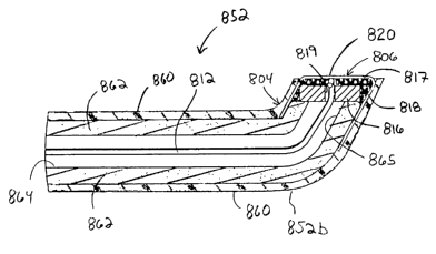
In another embodiment shown in FIGS. 8A-8C, probe 852 includes a modified
distal end but is otherwise similar to the off axis electrode embodiments
shown in
FIGS. 4A-E and SA-B. Probe 852 has a proximal end 852a attached to a handle
854
and a distal end 852b having an arcuate extension 804 which terminates and
forms an
off axis electrode surface 820. Handle 854 includes a suitable electrical
connector for
-12-
CA 02433396 2003-06-30
WO 02/058545 PCT/US02/00317
connection to a radio frequency or RF power supply. Probe 852 includes a
hollow
elongate probe member 862 which is made from a conductive material. An
insulating
material or shell 860 is provided around elongate probe member 862 in the
manner
described and discussed above. Radio frequency power is selectively supplied
to the
flat electrode surface 820 via elongate probe member 862 when the radio
frequency
power source is operably connected to handle 854.
An inner surface of elongate probe member 862 forms a cavity or lumen 864
which extends from the proximal end 852a to the distal end 852b of probe 852
and
forms an opening 865 as shown in FIG. 8C. Although the illustrated embodiment
includes an annular cavity terminating in an annular opening, one should
appreciate
that the cavity and opening may have other cross-sectional shapes, for
example,
elliptical, triangular, square, or any other polygonal shape. Alternatively,
one should
appreciate that the probe may instead be solid in cross-section.
A conductive extremity 806 is provided at the distal end of probe 852 and at
least partially covers annular opening 865 to define the flat electrode
surface 820.
Conductive extremity 806 includes a perforated plate or washer 816 and a thin
walled
cap 817, each made from any suitable conductive material. Washer 816 is
affixed to
the distal extremity of arcuate extension 804 with a suitable adhesive such as
epoxy
818, which is preferably conductive. One should appreciate that other suitable
materials and/or means may be used to affix washer 116 and cap 117 to the
distal end
of probe 852. Washer 816 substantially encloses or covers annular opening 865
of
arcuate extension 804 and is preferably provided with a small central aperture
819. A
thermal couple 812 extends through probe lumen 864 and aperture 819 and
terminates
adjacent an inner surface of cap 817. Thermal couple 812 provides temperature
feedback to the radio frequency power source.
Flat electrode surface 820 of the embodiment shown in FIGS. 8A-C is off axis
in a similar manner as described above in that the longitudinal axis of probe
852 is not
normal to flat electrode surface 820. Instead and as shown in FIGS. 8B and 8C,
a
normal axis extending from flat electrode surface 820 is substantially
orthogonal to
the longitudinal axis of the probe 852.
Advantageously, probe 852 includes a reinforced construction which is
particularly suited for the significant temperatures obtained during an
electrosurgical
-13-
CA 02433396 2003-06-30
WO 02/058545 PCT/US02/00317
procedure. The reinforced configuration of plate 816 and cap 8I7 of probe 852
minimizes any tendency of flat electrode surface 820 from popping or blowing
off of
the distal extremity 852b of probe 852.
All publications and patent applications mentioned in this specification are
herein incorporated by reference to the same extent as if each individual
publication
or patent application was specifically and individually indicated to be
incorporated by
reference.
The invention now being fully described, it will be apparent to one of
ordinary
skill in the art that many changes and modifications can be made thereto
without
departing from the spirit or scope of the appended claims.
-14-