Note: Descriptions are shown in the official language in which they were submitted.
CA 02433406 2003-06-30
WO 02/056784 PCT/US02/01312
Steerable Sphincterotome and
Methods for Cannulation, Papillotomy and Sphincter otomy
The present invention is an improvement of the devices and methods disclosed
in
U.S. Patent No. 5,547,469, U.S. Patent No. 5,868,698 and U.S. Patent No.
5,683,362 and in
U.S. Patent Application serial no. 09/154,834 in the name of Rowland, et al.,
all owned by
the owner of the present application, and incorporated in their entirety.
BACKGROUND
1. Field of the Invention
This invention generally relates to apparatus that is useful in performing
diagnostic
and therapeutic modalities in the biliary tree and more particularly to
apparatus that is
adapted for facilitating the diagnosis of gallstones in the bile duct and
other portions of the
biliary tree and the removal of such gallstones.
2. Description of Related Art
According to the present state of the art, endoscopic cannulation of the
common bile
duct and papillotomy and/or sphincterotomy of the Papilla of Vater and/or the
Sphincter of
Oddi is accomplished by advancing a sphincterotome (or papillotome or
cannulotome) into
an endoscope/duodenoscope so that the distal tip of the sphincterotome exits
the endoscope
adjacent the sphincter muscles at the Papilla of Vater. The endoscope
mechanisms are then
manipulated to orient the distal tip of the sphincterotome to the desired
position for proper
cannulation of the duct. Due to inconsistencies in the sphincterotome,
anatomy, and
endoscope manipulation, it is difficult to accurately and consistently
position the
sphincterotome for proper cannulation.
Historically the migration of gallstones into an individual's common bile duct
was
corrected by general surgical procedures. A surgeon would incise the bile duct
and remove
the gallstones and normally remove the gallbladder. In recent years less
invasive treatment
CA 02433406 2003-06-30
WO 02/056784 PCT/US02/01312
modalities have replaced these general surgical procedures and reduced patient
trauma, long
hospital stays and recovery periods.
For example, U.S. Pat. No. 4,696,668 and U.S. Pat. No. 4,781,677, both to
Wilcox,
disclose a heatment modality involving the administration of a dissolution
agent in the bile
S duct to essentially dissolve any gallstones. More specifically, a catheter
contains several
lumens for inflating and deflating each of two balloons, venting bile, and
infusing and
aspirating the dissolution agent. Inflating the balloons occludes the bile
duct at two spaced
sites and creates a sealed spaced that receives the dissolution agent. As the
space is sealed
from the remaining biliary tree, the dissolution agent finds access to the
gallbladder and any
gallstones therein through the cystic duct with the exclusion of bile from the
gallbladder
fundus. The dissolution agent also will be confined in high concentration
around bile duct
gallstones. After the gallstones dissolve the balloons are deflated and the
catheter can be
withdrawn. In this particular approach, the catheter is directed into the
biliary tree using a
standard duodenoscope that passes through the alimentary tract. Although this
and analogous
1S approaches have the potential of minimizing patient trauma, such treatments
require
extended placement of the duodenoscope in the patient, exhibit low efficacy
and introduce
a potential for adverse reactions to the dissolution agents.
In an alternative approach, a surgeon directs a surgical extractor into the
biliary tree
through at least an incision in the bile duct. For example, in U.S. Pat. No.
3,108,593 to
Glassman a surgeon incises both the bile duct and duodenum. Then the surgeon
directs an
extractor through the bile duct incision, biliaiy tree, sphincter of Oddi and
duodenum to exit
through the duodenum incision. This extractor includes a series of
longitudinally spaced
cages for trapping any gallstones in the bile duct and removing them through
either of the
incisions.
2S U.S. Pat. No. 4,627,837 to Gonzalo discloses a catheter device with a pair
of
inflatable balloons at its distal end. This catheter is Ied through an
incision in the bile duct
toward the duodenum. After the distal balloon passes through the sphincter of
Oddi, both
-2-
CA 02433406 2003-06-30
WO 02/056784 PCT/US02/01312
balloons are expanded to anchor the catheter in place. This enables the
catheter to be used
for irrigating and flushing through other lumens in order to capture any
gallstone in the
second balloon for removal through the incised bile duct.
In accordance with still another modality as for the treatment of strictures,
a surgeon
may insert a catheter device through the bile duct or duodenum for the purpose
of dilating
or enlarging the sphincter of Oddi. For example, U.S. Pat. No. 4,705,041 to
Kim discloses
a dilator that is directed through an incision in the bile duct and the
sphincter of Oddi. An
expandable tip dilates the sphincter of Oddi. U.S. Pat. No. 5,035,696 to
Rydell discloses an
electrosurgical instrument that is directed through the duodenum and to the
sphincter of Oddi
for performing a sphincterotomy. This apparatus contains a cutting wire that
is heated to cut
the sphincter muscle. U.S. Pat. No. 5,024,617 to Kayiel, discloses a similar
device that can
be directed through a duodenoscope. U.S. Pat. No. 5,152,772 to Sewell, Jr.
discloses a device
for performing a sphincterotomy that is directed through an incision in the
bile duct and
includes a knife for cutting the sphincter muscle.
The use of the duodenoscope and sphincterotomy devices, such as shown in the
Rydell and Karpiel patents, enables an internist to diagnose and treat
problems in the biliary
tree with minimal patient invasion. For example, modalities as described in
these patents
eliminates the surgery needed for incising the bile duct. Consequently, these
modalities can
be performed as outpatient or day surgical procedures. These procedures
greatly reduce
patient trauma, the length of a hospital stay and recovery times. For example,
if an internist
determines that gallstones are present in the biliary tree, particularly the
common bile duct,
the internist can insert a duodenoscope into the duodenum to view the
sphincter of Oddi.
Then a first catheter can be advanced through the worl~ing channel of the
duodenoscope with
or without a guidewire and directed through the sphincter of Oddi into the
biliary tree.
Contrast agent inj ected through the catheter enables fluoroscopy or other
imaging procedures
to confirm the presence of gallstones within the biliary tree. Next the
internist exchanges the
first catheter for a second catheter for performing a sphincterotomy such as
the types
-3-
CA 02433406 2003-06-30
WO 02/056784 PCT/US02/01312
disclosed in the above-identified Rydell and Karpiel patents. The second
catheter is then
exchanged for a third catheter such as shown in the Glassman patent or some
other
equivalent retrieval catheter for drawings gallstones through the enlarged
sphincter of Oddi.
Thereafter the retrieval catheter is manipulated to release the gallstone into
the duodenum.
The catheter, any guidewire and the duodenoscope can then be removed to
complete the
procedure.
This procedure is significantly less traumatic to the patient than other prior
art
procedures because the only incision occurs during the sphincterotomy.
However, this
procedure, as described above, requires three separate catheters and two
catheter exchanges.
These exchanges are required because the first, second and third catheters
function solely to
inj ect contrast agent to perform the sphincterotomy and to dislodge
gallstones, respectively.
The time required for performing each catheter exchange can increase patient
trauma and
increase the duration of the procedure and reduce efficiency. Moreover, each
such procedure
requires the use of two or three separate catheter devices.
Mufti-lumen catheters are available which typically reduce the number of
catheters
and catheter exchanges used during a procedure and thereby reduce both the
time required
and the patient's trauma while increase efficiency. The use of mufti-lumen
devices also
eliminates the need for the repositioning of subsequent catheters because the
original catheter
was withdrawn. While the mufti-lumen device may have to be repositioned, the
repositioning is considerable less then when a single lumen catheter is used.
While precision
positioning of the mufti-lumen device is essential for safe and effective
results, accurate
positioning of the mufti-lumen device is difficult to achieve. State of the
art mufti-lumen
devices are typically positioned by torque transmission from the handle to the
distal tip
approximately 6 feet away. Additionally, when an incision is made, proper
lcnife depth is
difficult to maintain because of the connection between the knife lumen and
the knife shaft.
When pressure is applied to the knife lumen an undesirable movement of the
needle knife
tip may occur because of this imprecise connection.
-4-
CA 02433406 2003-06-30
WO 02/056784 PCT/US02/01312
A need exists for an apparatus and a methodology of accurate placement of
catheters,
multi-lmnen devices and needle knives. A further need exists for an apparatus
for and a
methodology of an accurate depth control for needle lcnives and other cutting
instruments.
SUMMARY
Therefore, this invention provides an apparatus for, and a methodology of,
accurate
placement of the catheter, papillotome, sphincterotome, and/or needle knife.
This invention
further provides an apparatus for, and a methodology of, accurate control of
the depth of the
needle knife and the resulting incision and an apparatus which can allow
accurate control of
the depth of the needle knife while allowing the user to accurately place the
needle knife
within the patient.
The invention discloses an endoscopic catheter which has a distally located
tissue
cutting device in a first lumen, and includes a second lumen which has 1) a
reciprocating
cable inside and 2) includes a fixed member which is used to impart rotary
motion to the
cable inside of it where the reciprocation of the cable causes a rotation of
at least the distal
portion of the catheter to orient the cutting device. The cable may have
spiral threads on its
outer circumference and the fixed member may have spiral threads on its inner
circumference
which mate with the threads on the cable. The cutting device may be a
sphincterotorne, a
papillotome or a needle knife with a curved distal portion and the cutting
device may operate
in response to energy from an rf heating source.
In another embodiment a sliding member rnay be included which is attached to
the
distal end of the cable and is located distal from the fixed member. The cross
section of the
lumen containing the sliding member as well as the cross section of the
sliding member may
be non-round or even square.
In another embodiment of the invention, an endoscopic catheter has a cable
actuated
needle lcnife within a lumen where the needle knife is deployable from a
distal end of the
catheter. In this embodiment the invention substantially prevents movement of
the needle
-5-
CA 02433406 2003-06-30
WO 02/056784 PCT/US02/01312
k~zife after deployment and includes a distally positioned fixed stabilizing
element in the
lumen which internally engages the needle knife cable to prevent such motion.
The cable
attached to the needle knife may have spiral threads on its outer
circumference and the fixed
stabilizing element may have spiral threads on its inner circumference which
mate with the
threads on the cable. The needle knife may have a curved distal portion and
the cutting
device may operate in response to energy from an rf heating source. A Apivot
element may
be included, preferably proximal to the stabilizing element, to prevent
torsion build up
within the cable.
In another embodiment, the invention includes an endoscopic catheter having a
cable
actuated needle knife within a first lumen deployable from a distal end of the
catheter and
the cutting device may be substantially prevented from movement after
deployment. In this
embodiment a second lumen containing a reciprocating cable and a fixed member
imparts
rotary motion to the cable when reciprocated. Reciprocation of the cable
causes rotation of
at least a distal portion of the catheter to orient the cutting device and a
distally positioned
fixed stabilizing element in the first lumen internally engages the needle
knife cable to
substantially prevent movement. The cable may have spiral threads on its outer
circumference and the fixed member may have spiral threads on its inner
circumference
which mate with the threads on the cable. The cutting device may be a needle
knife with a
curved distal portion and the cutting device may operate in response to energy
from an rf
heating source. A pivot element and/or a sliding member may be included.
BRIEF DESCRIPTION OF THE DRAWINGS
The various objects, advantages and novel features of this invention will be
more
fully apparent from a reading of the following detailed description in
conjunction with the
accompanying drawings in which like reference numerals refer to lilce parts,
and in which:
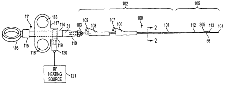
FIG. 1 is a plan view of one embodiment of apparatus constructed in accordance
with
this invention;
-6-
CA 02433406 2003-06-30
WO 02/056784 PCT/US02/01312
FIG. 2 is a cross-section talcen along lines 2--2 in FIG. 1;
FIG. 3 is a cross-section talcen along lines 3-3 in FIG. 2;
FIG. 4 depicts the apparatus of FIG. 1 positioned through a duodenoscope for
inj ecting contrast agent into the biliary tree;
FIG. 5 is an enlarged view that depicts the orientation of the apparatus in
FIG. 1 for
performing a sphincterotomy;
FIG. 6 depicts the apparatus of FIG. 1 positioned through a duodenoscope for
dislodging material within the common bile duct;
FIG. 7 is a cross-section of a~1 alternative embodiment of the apparatus as
viewed
generally along lines 7-7 in FIG. 2.;
FIG. 8 is a cross-section of still another embodiment of the apparatus taken
along
lines 7-7 in FIG. 2;
FIG. 9 is a partial cross-section of the invention highlighting the
positioning device;
FIG. 10 is a cutaway view of an alternative embodiment of the present
invention used
to support the extension of the needle knife;
FIGS. 11A and 11B are enlarged views of the a pivot element and a stabilizing
element from FIG. 10; and
FIG. 12 is a plan view of an alternate embodiment of the present invention
which
combines a positioning device and a support for the needle knife.
DESCRIPTION OF ILLUSTRATED EMBODIMENTS
FIG. 1 depicts catheter apparatus 100 that has the capability of injecting a
contrast
agent into the biliary tree, accurately positioning a cutting wire, of
performing a
sphincterotomy amd of dislodging a gallstone into the duodenum. Apparatus 100
includes a
catheter 101 which, forpurposes ofdefmition, includes proximal portion 102
extending from
proximal end 103 and distal end 104 with distal portion 1 OS extending a short
distance from
distal end 104. In a typical application, the catheter will have a working
length of 200 cm
and distal portion 105 will have a length of 6 cm to 9 cm. Normally distal
portion 105 will
CA 02433406 2003-06-30
WO 02/056784 PCT/US02/01312
have a diameter that is smaller than the diameter of proximal portion 102 to
increase the
flexibility of distal portion 105. The reduction in diameter also makes distal
end 104 less
traumatic and allows distal portion 105 to reach smaller passages while
allowing the larger
proximal portion 102 to provide necessary hoop strength and rigidity,
particularly where
proximal portion 102 is coextensive with the working channel of a
duodenoscope. For
example, the proximal and distal portions might have diameters corresponding
to 7 Fr and
5.5 Fr catheter sizes (i.e., 0.09"and 0.07"respectively).
As shown particularly in FIG. 2, catheter 101 has three lumens. First lumen
201 has
a diameter that is greater than either second lumen 202 or third lumen 203. In
one particular
embodiment first lumen 201 is square shaped with each side approximately
0.040" in
proximal portion 102 that reduces to about 0.037" in distal portion 105 to
receive a standard
0.035" guidewire. In addition first lumen 201 may be, a~zd as shown in FIG. 2,
is offset from
the center of the catheter 101.
The cross section of both second lumen 202 and third lumen 203 are each
smaller
than the cross section of first lumen 201 and are radially offset from the
centerline of catheter
101, from each other and from first lumen 201. In one particular embodiment
the cross
section of third lumen 203 has a diameter of 0.028" in proximal portion 102
that reduces to
about 0.020" in distal portion 105 and second lumen 202 has an internal
diameter of 0.028"
in proximal portion 102 that reduces to about 0.020" in distal portion 105. As
described later,
this third lumen 203 carries a cutting wire for performing a sphincterotomy
and for allowing
the infusion of a contrast agent at reasonable rates. The cutting wire can
also be positioned,
as described later, as desired. While the description contained herein
describes the first
lumen 201 having a square cross section shape, it would be apparent to one of
ordinary skill
in the art that the invention may be practiced in any of the lumens by
changing the cross
section of the lumen to a shape other than a circle. The angular spacing
between second
hunen 202 and third lumen 203 is about 45 degrees and the angular spacing
between first
lumen 201 and each of lumens 202 and 203 each is about 157.5 degrees. In this
configuration
_g_
CA 02433406 2003-06-30
WO 02/056784 PCT/US02/01312
and with these dimensions proximal portion 102 readily passes through the
working channel
of any duodenoscope. These angular relationships have been used in the past to
position the
device. While the invention may be used with these angular relationships, the
invention
itself allows the device to be positioned which reduces the necessity of
strict adherence to
the previously used angular relationships.
Referring again to FIGS. 1 and 2, each of lumens 201, 202 and 203 includes an
entry
port in proximal portion 102 and an exit port in distal portion 105.
Generally, and as
described in more detail later, first lumen 201 has an exit port through
distal end 104 while
the exit ports for lumens 202 and 203 can be sited at different locations in
distal portion 105
depending upon a particular application.
In FIG. 1, the entry ports in proximal portion 102 adjacent proximal end 103
include
an entry port 106 that provides access to first lumen 201 and includes an
optional Leur lock
fitting 107. Proximally positioned entry port 108 provides access to second
lumen 202 and
includes optional Leur lock fitting 109. Proximal entry port 110 for third
lumen 203 is
located coextensively with a portion of handle 111 attached to proximal end
103. One of
ordinary skill in the art would understand that this specific configuration is
given as an
example and not meant to limit the invention. Various other configurations
would be
apparent to one of ordinary skill in the art to practice the invention
described herein.
Referring to the distal portion 105, catheter 101 in this particular
embodiment carries
expansible balloon 112 proximally of the excursion of cutting wire 113
externally of catheter
101. As described in U.S. Patent Application serial no. 09/154,834 in the name
of Rowland,
et al., and owned by the owner of the present application and already
incorporated herein by
reference in its entirety, second lumen 202 emerges at a distal exit port
through the side of
catheter 101 with the interior of expansible balloon 112. An extension of
second lumen 202
?5 beyond the distal port is sealed by known methods of manufacture.
Consequently, fluid
forced through entrance port 108, as by a syringe (not shown) attached to Leur
lock fitting
_g_
CA 02433406 2003-06-30
WO 02/056784 PCT/US02/01312
109, expands balloon 112 into an occluding orientation with an inflated
diameter in the range
up to 20 mm.
First lumen 201 extends through catheter 101 and terminates with an exit port
in
distal end 104. Thus first lumen 201 is adapted for receiving a guidewire
through the entry
port 106 that will extend through catheter 1 O 1 and exit distal end 104 and
allow the catheter
to slide over that guidewire.
Referring to FIG. 3, distal end 301 of cutting wire 113 attaches to a clamp
302
formed at the distal end of third lumen 203. Spaced skived ports 303 and 304
allow active
portion 305 ofthe cutting wire 113 to emerge from catheter 101 through skived
aperture 303,
parallel the catheter 101 exteriorly thereof and return into third lumen 203
through port 304
and reinforcing sleeve 306. Cutting wir a 113 then extends through third lumen
203 to handle
111 shown in FIG. 1 where it emerges as proximal end portion 114.
Handle 111, as shown in FIG. l, includes central member 115 terminating with
thumb ring 116. The central member 115 extends through and slides with respect
to body
section 117 having opposed finger rings 118. The central member 115 also
attaches to
catheter 101, and is therefore an extension of catheter 101. Member 117
additionally includes
internal connector 119 for clamping proximal end 114 of cutting wire 113.
Thus, when body
117 is at its distal position as shown in FIG. 1, distal portion 105 of
catheter 101 is in
essentially straight line as shown in FIG. 1 with active portion 305 of
cutting wire 113 being
closely adj acent catheter 101. Retracting body portion 117, causes cutting
wire 113 to bend
distal end 104 upwardly as shown in FIG. 3 to a position that is essentially
at right angles to
the main axis of the catheter, as will be shown later.
Connector block 119 and cutting wire 113 are generally conductive members that
attach through RF connector 120 to RF heating source 121. The use of such RF
heating
sources 121 for energizing cutting wire 113 thereby to cut the sphincter
muscle is well
known in the art and represents one possible sphincterotomy procedure that can
be adapted
for the apparatus of this invention and is not described fiuther.
-10-
CA 02433406 2003-06-30
WO 02/056784 PCT/US02/01312
With this description of the apparatus structure, it will now be possible to
understand
its use in a particular application. FIG. 4 discloses, in a partially brolcen
and schematic view,
the positioning of duodenoscope 401 in duodenum 402 adjacent sphincter of Oddi
403.
Catheter 101 such as constructed in FIG. 1 passes through sphincter of Oddi
403 into the
common bile duct 404, bypassing pancreatic duct 405. Distal end 104 does not
extend to
gallbladder 406.
Fluoroscopy allows the appropriate positioning by utilizing a series of radio-
opaque
markers 406 at distal portion 105 that may include clamp 302 and reinforcing
sleeve 306 in
FIG. 3. Catheter 1 O1 can be positioned with or without the presence of
guidewire 408 in first
lumen 201 shown in FIGS. 2, and 3. For purposes of injecting the contrast
agent, any
guidewire 408 can be withdrawn to allow the contrast agent to be injected
through first
lumen 201 for purposes of fluoroscopic examination to confirm the presence of
one or more
gallstones .409. It is also possible during the operation to expand balloon
112 to occlude
common bile duct 404 and block any migration of contrast agent into duodenum
402 or
1 S pancreatic duct 405.
FIG. 5 is an enlarged view showing duodenum 402, sphincter of Oddi 403,
portions
ofpancreatic duct 405 and common bile duct 404. In FIG. 5 catheter I OI has
been positioned
relative to the duodenoscope 401 through the opening of sphincter of Oddi 403.
The handle
111 in FIG. 1 has been drawn proximally to deflect distal portion 105 into
essentially a right
angle configuration such that cutting wire 113 abuts a portion of sphincter of
Oddi 403. The
application of RF heating to cutting wire I 13 then will cut sphincter of Oddi
403 and enlarge
the opening therethrough. As will be apparent, the sphincterotomy is performed
with direct
visualization of the sphincter of Oddi through the duodenoscope.
Moreover, as has been observed by others, catheters having guidewire and
cutting
wire lumens tend to assume a particular angular orientation when distal
portion 105 emerges
from the duodenoscope. This orientation is essentially independent of the
angular position
of the catheter when it is inserted into the duodenoscope. The offset nature
of lumen 203 as
-11-
CA 02433406 2003-06-30
WO 02/056784 PCT/US02/01312
shown in FIG. 2, improves the location of cutting wire 113 as distal portion
105 passes
through sphincter of Oddi 403. Specifically the angularly offset brings
cutting wire 113 into
better alignment with common bile duct 404 and displaces the cutting wire from
pancreatic
duct 405.
FIG. 6 depicts the catheter after the sphincterotomy and after catheter 101 is
advanced over guidewire 408, if used. FIG. 6 also discloses catheter 101 after
balloon 112
has been moved beyond gallstone 409 in bile duct 404. Balloon 112 is expanded
so that upon
withdrawal of catheter 101 balloon 112 will dislodge gallstones 409 and sweep
them through
sphincter of Oddi 403 into duodenum 402.
As will now be apparent from the description ofthe particular catheter
apparatus 100
shown in FIG. 1 and its use as discussed with respect to FIGS. 4, 5, and 6,
the single catheter
apparatus is capable of providing diagnostic contrast agent injection, of
performing a
sphincterotomy and of dislodging gallstones in the common bile duct or other
portions of the
biliary tree without having to exchange a catheter. Moreover, positioning and
sizing of the
lumens enables these functions to be performed with a catheter apparatus that
is readily
adapted for use in the working channels of standard duodenoscopes.
Consequently the
gallstones can be removed from the biliary tree without bile duct incisions
and
accompanying surgical procedures, as duodenoscope can be introduced through
the
alimentary tract. Consequently the entire procedure is adapted for being
performed more
rapidly than prior art procedures and with fewer components. The net effect is
to reduce
patient trauma and the overall time and cost of conducting the procedure.
In FIG. 1 balloon 112 is located proximally of cutting wire 113. FIG. 7
discloses an
alternative embodiment in which balloon 701 is located distally of cutting
wire 113. More
specifically, the distal end of lumen 202A, corresponding to second lumen 202
in FIG. 3
is sealed. Side facing exit port 702 skived or otherwise formed in catheter
101 opens into
chamber 703 formed by balloon 701. First sealing portion 704 and a sealing
portion 705 of
_12_
CA 02433406 2003-06-30
WO 02/056784 PCT/US02/01312
balloon 701 connect proximally and distally of aperture 702 respectively arid
seal chamber
703.
Introduction of a balloon inflation fluid through lumen 202A expands balloon
701
into an occluding orientation corresponding to the orientation of balloon 701.
Retraction of
catheter 1 O1 with distal balloon 701 inflated enables withdrawal of a
gallstone from the bile
duct. This particular embodiment is particularly adapted when it is determined
that a
gallstone is located high in the biliary tree to minimize the incursion of
distal portion 105
through the biliary tree beyond the,gallstone or in ably application in which
the internist
desires to minimize the length of distal portion 105 that extends beyond the
occluding
balloon.
FIG. 8 discloses another embodiment for enlarging the sphincter of Oddi and
performing another procedure, such as injecting a contrast agent into the
biliary tree, as
might be used in the diagnosis and treatment of a stricture in the biliary
tree. In this particular
embodiment exit port 801 from second lumen 202B is located in distal end 104
of distal
portion 1 O5. First lumen 201 then can be used for a guidewire and lumen 202B,
for inj ecting
the contrast agent directly into the biliary tree while the guidewire remains
in place. The
apparatus would then be positioned to perform a spluncterotomy without having
to exchange
a catheter should the procedure be warranted.
As still another alternative, the internist could utilize a conventional
catheter for
purposes of injecting the contrast agent to determine the need for gallstone
removal. If
treatment were indicated, the internist could then utilize apparatus as shown
in FIG. 1 with
a single exchange over the guidewire that would pass through lumen 201 as
previously
described.
As can be seen from the above description one of the steps in the treatment of
obstructive disease is normally the practice of tissue incision which is
achieved by advancing
a cutting wire endoscopically to the target site. As explained above, once the
catheter tip is
in position, the catheter tip is bowed (FIG. 5) to expose cutting wire 113 to
tissue.
-13-
CA 02433406 2003-06-30
WO 02/056784 PCT/US02/01312
Diathermic current is passed through cutting wire 113 from RF Heating Source
121 (FIG.
1) which allows the endoscopist to incise and cauterize the tissue at the
target site. Safe and
effective results are only obtained through precision positioning of cutting
wire 113.
FIG. 9 depicts a section of a positioning device 900 residing within lumen 201
of a
rnulti-lumen catheter 101. As shown in FIG. 2 lumen 201 has an internal shape,
in this case
a square, which allows the positioning device 900 to transfer torque to distal
end 104. The
internal shape of lumen 201 in FIG. 2 is depicted to be a square, but one of
ordinary skill in
the art would understand that any internal shape which allows the torque
transfer may be
used and is within the disclosed invention. Referring back to FIG. 9,
positioning device 900
consists of cable assembly 901, which is substantially encircled by torque
transmission
element 902 and cog 903. Proximate end 904 of positioning device is attached
to handle 111
(not shown) while distal end 905 of positioning device 900 is located in the
distal portion
105 (not shown). While FIGURE 9 illustrates a torque transmission element 902
which
completely encircles cable assembly 901, it would be apparent to one of
ordinary skill in the
art that torque transmission element 902 need not entirely encircle cable
assembly 901 and
any configuration between torque transmission element 902 and cable assembly
901 which
allows the translation of reciprocal movement to rotational motion is within
the scope of the
invention. The cog 903 can also be referred to as a sliding member.
Cable assembly 901 is connected at its proximal end (not shown) to the distal
end of
handle 11 l, traverses through lumen 203 and torque transmission element 902
with the distal
end of cable assembly 901 fixed to cog 903. Reciprocal motion of the handle
111 attached
to the cable assembly 901 introduces reciprocal motion in the proximal portion
of the cable
assembly 901 between the handle 111 and torque transmission element 902. The
outer
circumference 906 of the cable assembly 901 includes a helical or advancing
spiral thread.
The torque transmission element 902 is located and fixed within lumen 201 a
short
distance from distal end 104 (FIG. 1) and proximal to cog 903. The internal
portion of
torque transmission element 902, or the portion which comes into contact with
the cable
- 14-
CA 02433406 2003-06-30
WO 02/056784 PCT/US02/01312
assembly 901, contains a helical or advancing spiral thread which interacts
and mates with
the helical or advancing spiral thread of cable assembly 901. The external
portion of torque
transmission element 902, or the portion which comes into contact with lumen
201, is shaped
to interact with and mate with the interior surface of lumen 201 and is fixed
to inner lumen
201. The purpose of torque transmission element 902 is to change the
reciprocal cable
movement received from the reciprocal movement of the attached handle 111 to
rotational
cable movement in direction 907. The torque transmission element 902 may be
molded as
part of or attached to lumen 201.
The cog 903 is located between torque transmission element 902 and distal end
104
and at a distance from each so as to aid in creating effective rotation of the
catheter distal end
104. This rotation, in the direction of 907, is the result of the torque
transmission element
902 translation of the reciprocal movement received from handle 111 into
rotational cable
movement. As torque transmission element 902 receives reciprocal movement
from.cable
assembly 901, torque transmission element 902 cannot moved because it is fixed
to lumen
201 and the internal helical or advancing spiral inside of torque transmission
element 902
imparts a rotational affect on cable assembly 901 in a similar mamler to the
spin a bullet
receives from the rifling inside of a rifle's barrel. Cog 903 is not fixed to
lumen 201 and is
capable of reciprocal movement within lumen 201 as the cable assembly 901
advances and
retracts. The purpose of cog 903 is to transfer the torque received from cable
attached to
torque transmission element 902 to the distal segment of the catheter and this
is achieved
when cog 903 is fixed to cable assembly 901. While the invention is shown with
cog 903,
one of ordinary skill in the art would understand that the cog 903 is not
absolutely necessary
to the invention, but instead aids in the transmission of the torque created
by torque
transmission element 902. Cog 903, while included in the preferred embodiment,
may be
eliminated and the distal tip 104 would still be capable of being positioned.
The resistance
between the torque transmission element 902 and the cable assembly 901 may be
varied to
adjust the proportion of the reciprocal movement which is translated into
rotational motion.
-15-
CA 02433406 2003-06-30
WO 02/056784 PCT/US02/01312
Cog 903 may also be shaped to increase the efficiency ofthe transfer
ofrotational movement
from the distal end of the cable to the distal segment of the catheter. For
example, where the
cross-section of lumen 201 is in the shape of a square the cross section of
cog 903 would also
be a square.
In operation distal end 104 of the cutting device is advanced through the
lumen 203
of the endoscope to the target area. Cutting wire 113 is retracted to bow the
tip exposing the
cutting wire (FIG. 5). The distal end of device 900 is advanced through lumen
201 until
torque transmission element 902 is in, or near the distal portion 1 O5. The
handle 111, which
is comzected to cable 901, is reciprocated, causing the proximal end 904 of
cable assembly
901 to be reciprocated. When the proximal end of cable assembly 901 is
reciprocated, torque
transmission element 902 translates this movement into rotation which is
transferred from
cog 903 to the catheter distal end 104. As catheter distal end 104 is rotated
so is cutting wire
113 which resides in lumen 203. After the incision is made in the target area,
cutting wire
113 is advanced relieving the bow. The catheter assembly can then be removed
from the
body. Overall, the effect of the positioning system 900 is to translate the
reciprocal
movement in the handle 111 into rotational movement at the distal end 104 of
the catheter.
FIG.10 depicts a mufti-lumen catheter 1000 which includes an alternate
embodiment
of the present invention for the precise positioning of a needle knife. Within
catheter 101
of mufti-lumen catheter 1000 is lumen 1001 which is used for needle knife
1002. Within
lumen 1001 resides needle knife wire 1003. Needle knife wire 1003 is attached
at the
proximal end to a sliding mechanism of handle 1004 and on the distal end to
needle knife
1002. Needle knife 1002 is capable of extending beyond distal end 104 of
catheter 101.
Circumference 1005 of needle knife wire 1003 includes helical or advancing
spiral 1006.
FIG. 10 shows helical or advancing spiral 1006 along the entire length of
needle knife wire
1003, but helical or advancing spiral 1006 portion of needle knife wire 1003
need not be
included along the entire length and may be limited to inclusion over a short
distance of
YIPPI~~P ltn;fe wire 1003 close to the distal end 104. Helical or advancing
spiral 1006 is
-16-
CA 02433406 2003-06-30
WO 02/056784 PCT/US02/01312
required where pivot element 1007 and stabilizing element 1008 attach and
along needle
knife wire 1003 where pivot element 1007 and stabilizing element 1008 may
travel. In one
embodiment the helical or advancing spiral 1006 may be located between 6 and
10 cm from
the distal end.
Pivot element 1007 and stabilizing element 1008 are attached to needle knife
wire
1003. Pivot element 1007 is located proximal to stabilizing element 1008 and
is used to
prevent torsion build up. While the preferred embodiment includes pivot
element 1007, the
invention can be practiced without the inclusion of pivot element 1007.
Stabilizing element
1008 is located a short distance from distal end 104 and may be molded as part
of the lumen
or attached to the Lumen. In one embodiment the stabilizing element 1008 was
located 6 to
1'0 cm from the distal tip. The purpose of the stabilizing element is to
prevent needle knife
1002 from being pushed back into lumen 1001 when pressure is applied to the
needle knife
1002, for example when an incision is made. The stabilizing element 1008 is
part of or fixed
to the lumen wall and uses this attachment to prevent the needle knife 1002
from being
pushed back into the lumen. Stabilizing element 1008 may have a helical or
advancing spiral
1109 (FIGURE 11B) along its inner circumference which mates with the helical
or
advancing spiral 1006 of needle knife wire 1003.
When the sliding mechanism of handle 1004 which is attached to needle knife
wire
1003 reciprocates, needle lmife wire 1003 also reciprocates. As needle knife
wire 1003
reciprocates, threaded needle knife wire 1003 rotates through stabilizing
element 1008 and
needle lcnife 1002 advances out of or retracts into lumen 1001. The sliding
mechanism of
handle 1004 can be locked when needle knife 1002 is deployed to its desired
length. As
pressure is applied to needle knife 1002 during incision any forward or
backward movement
of needle knife 1002 is negated by stabilizing element 1008 which acts, to
resist movement
of the needle Icnife back into the Lumen 1001. This resistance is created by
the interaction
of the matched helical windings of the stabilizing element 1008 and the needle
knife wire
-17-
CA 02433406 2003-06-30
WO 02/056784 PCT/US02/01312
1003. Stabilizing element 1008 allows needle knife 1002 to be loclced
regardless of the
overall catheter length.
In operation distal end 104 of device 100 is advanced through the endoscope to
the
target area. Needle knife wire 1003 is advanced via the sliding mechanism of
handle 1004
to expose needle kW fe 1002 to the desired length. The sliding mechanism of
handle 1004
is then loclced into position. As pressure is applied to needle knife 1002
during the incision,
stabilizing element 1008 ensures the integrity of the depth of cut of needle
knife 1002. Once
the incision is made in the target area, needle knife 1002 is retracted and
the catheter
assembly. is removed from the body.
FIGURES 11 A and 11 B show a blown up diagram of the pivot element 1007 and
the
stabilization elements 1008 respectively. The pivot element shown in FIG. 1
1A, consists of
three parts, a proximal element 1101, a distal element 1102 and an enclosure
element 1103.
When the needle knife wire 1003 is pushed towards the distal end 104,
proximate element
1101 makes contact with distal element 1102 and ensures the entire needle
lmife wire
progresses within the lumen toward the distal end 104. When the needle knife
wire 1003 is
retracted, proximal element 1101 contacts the proximal portion 1104 of the
enclosure
element 1103 and the distal portion 1105 of the enclosure element 1103
contacts the distal
element 1102 and ensures the entire needle knife wire is retracted. Within the
pivot element
1007 is a space 1106 between the proximal element 1101 and the distal element
1102 and
the sides of the enclosure element 1103. There is also a space 1107 between
the needle knife
wire 1003 and the entrance 1107 and exit 1108 of the enclosure element 1103.
The
stabilizing element 1008 is shown in FIGURE 11B which highlights the mating
1104
between the helical windings on the needle knife wire 1003 and the stabilizing
element 1008.
FIGURE 12 is a perspective view of a device which incorporates both a stable
needle
knife to maintain the blade depth and which allows the user to position the
blade in a desired
direction. Within catheter 101 of multi-lumen catheter 1200 is lumen 1001
which is used
for needle knife 1002. Within lumen 1001 resides needle lmife wire 1003.
Needle knife
-18-
CA 02433406 2003-06-30
WO 02/056784 PCT/US02/01312
wire 1003 is attached at the proximal end to a sliding mechausm of handle 1004
and on the
distal end to needle knife 1002. Needle knife 1002 is capable of extending
beyond distal end
104 of catheter 101. The circumference 1005 of needle knife wire 1003 includes
helical or
advancing spiral 1006. In one embodiment the helical or advancing spiral 1006,
6 cm of the
needle knife wire includes the helical or advancing spiral feature which is
located at a
distance of 12 cm from the distal tip. Needle knife (stabilizing) threaded
element 1008 is
also included in the device 1200 to prevent the blade 1002 from being pushed
back into the
lumen when pressure is applied to the needle knife blade 1002. The mechanism
of FIGURE
12 which prevents the blade of the needle knife 1002 from being pushed back
into the lumen
, has been described with respect to FIGURE 10. The needle knife (stabilizing)
threaded
element may be molded as part of or attached to the lumen. A pivot element
1007 (FIGURE
10) may be attached to the needle knife wire 1003 if desired.
FIGURE 12 also includes a mechanism for steering the position of the cutting
wire
113. Steering wire 1201 is connected at its proximal end to the distal end of
handle 1202,
traverses through lumen 1203 and steering wire threaded element 1204. The
internal surface
of steering wire threaded element 1204 matches and mates with the external
circumference
of steering wire 1201. As the steering wire 1201 is reciprocated via a sliding
mechanism on
handle 1202, the threaded steering wire 1201 rotates through the steering wire
threaded
element 1204. This rotation causes the distal section of the catheter to
rotate. The sliding
mechanism can be locked when the desired position is achieved. In one
embodiment the
distal end of the steering wire 1201 was threaded for 10 cm at a distance of 8
cm from the
distal tip. The major advantage of device 1200 is the ability to rotate the
distal tip either
clockwise or counterclockwise as the sliding mechanism is reciprocated and to
ensure the
blade of the needle knife does not retract into the lumen when used. The
steering wire
threaded element may be molded as part of or attached to the lumen. A cog 903
(FIGURE
9) may also be included for a more effective transfer of rotational position
to the distal end
104.
-19-
CA 02433406 2003-06-30
WO 02/056784 PCT/US02/01312
FIGURE 12 also shows a bowing wire 1205 included in device 1200. The inclusion
of the bowing wire 1205 allows the distal tip of the catheter to be turned up
to 90 degrees
from the longitudinal axis of the catheter body 101.
In operation the distal tip of the device is advanced through lumen.1001 of
the
endoscope to the target area. Once the distal tip reaches the target area, the
tip of the catheter
is bowed to the desired angle. The steering device is then advanced through
lumen 1203
until the steering element is in, or near, the distal portion 1 O5. The tip of
the catheter is than
rotated to the desired position and locked when the desired position is
obtained. The needle
knife is than advanced to expose the needle knife to the desired length. Once
the exposed
length is attained, the handle is locked to ensure the needle knife blade is
not pushed back
into the lumen when pressure is applied to it. The incision is then made in
the target area and
the needle knife is retracted, the bow is released and the catheter assembly
is removed from
the body.
Therefore, it will now be apparent that apparatus constructed in accordance
with this
invention attains the several objects and the advantages of this invention.
More particularly,
catheter apparatus constructed in accordance with this invention allows the
injection of a
contrast agent, the performance of a sphincterotomy and dislodging gallstones
from the
common bile duct through the enlarged sphincter of Oddi into the duodenum all
without
requiring any catheter exchanges. Moreover, this apparatus allows such a
procedure to occur
through a duodenoscope to minimize patient trauma. The use of a single
catheter with an
elimination of catheter exchanges further reduces the time and costs
associated with the use
of multiple, single-function catheter devices.
As will be apparent from the foregoing description, many alterations can be
made to
the specifically disclosed embodiments. Different balloon 'structures can be
used and located
at alternative positions. Different cutting wire embodiments and orientations
can be used.
Thus, although this invention has been disclosed in terms of certain
embodiments, it will be
apparent that many modifications can be made to the disclosed apparatus
without departing
-20-
CA 02433406 2003-06-30
WO 02/056784 PCT/US02/01312
from the invention. In particular, it is considered that all of the foregoing
embodiments may
be used in conjunction with a handle fixed to the cutting wire but rotatable
relative to the
catheter. Therefore, it is the intent of the appended claims to cover all such
variations and
modifications as come within the true spirit and scope of this invention.
-21 -