Note: Descriptions are shown in the official language in which they were submitted.
CA 02433936 2003-07-04
WO 02/064157 PCT/US02/03118
LOCALIZED MYOCARDIAL INJECTION METHOD
FOR TREATING ISCHEMIC MYOCARDIUM
RELATED APPLICATIONS
This application claims the benefit of U.S. Provisional Application Serial No.
60/263,468, 'filed January 23, 2001, the entire contents of which are
incorporated herein.
FIELD OF THE INVENTION
This invention relates to a method of treating ischemic or diseased myocardium
by
injecting a therapeutic agent, such as a gene, protein, cell or drug, into
normal myocardium,
preferably adjacent to an ischemic zone in the heart of a subject. The method
is useful for
inducing angiogenesis and collateral blood vessel formation to improve cardiac
function in
subjects with ischemic heart disease. The method can also be used to promote
tissue
regeneration in such subjects.
BACKGROUND OF THE INVENTION
Cardiovascular diseases are generally characterized by an impaired supply of
blood to
the heart or other target organs. When the blood supply to the heart is
compromised, cells
respond by generating compounds that induce the growth of new vessels to
increase the
supply of blood to the heart. The process by which these new blood vessels,
termed collateral
blood vessels, are induced to grow out of the existing vasculature is termed
angiogenesis, and
the substances that are produced by cells to induce angiogenesis are termed
angiogenic
factors. As the body's natural angiogenic response is often inadequate, the
use of
exogenously supplied angiogenic factors is currently being explored as a means
to treat
cardiovascular disease.
Myocardial gene therapy can be used for the treatment of a number of
cardiovascular
diseases, including ischemic cardiomyopathies, congestive heart failure, and
malignant
arrhythmias (Nabel (1995) Circulation 91:541-548). Gene therapy to treat
cardiac disease
requires that gene therapy agents be delivered to the heart in a manner that
will produce a
favorable response. Intracoronary delivery of angiogenic growth factors and
gene therapy
vectors is possible, but this approach may result in dilution of the
therapeutic agent due
dispersal of the agent in the systemic circulation. Furthermore, such delivery
methods may
result in undesired side effects due to potential systemic distribution of
such angiogenic
agents, including vascularization of tumors and retinopathy. Intramyocardial
injection
-1-
CA 02433936 2003-07-04
WO 02/064157 PCT/US02/03118
provides a means to deliver angiogenic agents that avoids these pitfalls.
Kornowski et al. (J.
Ar~2. Coll. Cardiol. 35:1031-9, 2000) teaches the delivery of an angiogenic
gene therapy
vector directly to ischemic tissue using catheter-based and surgical
techniques. Post et al.
(Card. afzd vast. RegefxeratioTZ 2:106-113) discloses the transfection
efficiency of
transendocardial and direct epicardial injection of an angiogenic gene therapy
vector.
It is currently unknown whether precise localization of intramyocardial
injections is
necessary. At present, most studies have targeted injections into the ischemic
or diseased
portion of the myocardium. However, injections could be placed, for example,
in an
ischemic area, in the zone bordering an ischemic area, or in normal
myocardium.
In accordance with the present invention, it has surprisingly been found that
a
favorable functional response occurs when the angiogenic agent is injected
into the normal
myocardium, and more particularly into the normal myocardium adjacent to an
ischemic
zone.
SUMMARY OF THE INVENTION
The present method of delivering a therapeutic agent to normal myocardium or
normal myocardial tissue adjacent to a site of ischemia in an ischemic or
diseased heart can
be used to induce angiogenesis, to increase contractile function in the heart,
to increase blood
flow within the heart, to stimulate collateral vessel development in the
heart, to promote
tissue regeneration and to treat myocardial ischemia, particularly in a human
patient.
In one aspect of the present invention, the invention provides a method for
delivering
a therapeutic agent to an ischemic or diseased heart by delivering a
therapeutically-effective
amount of the therapeutically effective agent to normal tissue in the ischemic
or diseased
heart. In accordance with the present invention, the therapeutic agent can be
a transgene
encoding an angiogenic protein or peptide that is delivered into the
myocardium of the
subject by intramyocardial injection of a gene therapy vector comprising that
transgene. The
vector is injected into normal tissue in the heart, , and preferably into the
non-ischemic or
non-diseased myocardium adjacent to an ischemic or diseased zone in the heart.
The gene
therapy vector may be a plasmid or a viral vector, such as an adenoviral
vector or
recombinant adenoviral vector, or an adeno-associated vector or recombinant
adeno-
associated vector. The plasmid or viral vector may be delivered naked or in a
liposome.
Alternatively the therapeutic agent can be an angiogenic protein or peptide, a
cell or cells,
one or more drugs, an antisense DNA or RNA, or any other therapeutic agent
useful to induce
_2_
CA 02433936 2003-07-04
WO 02/064157 PCT/US02/03118
angzogenesis, increase contractile function in the heart, increase blood flow
within the heart,
stimulate collateral vessel development in the heart, promote tissue
regeneration, improve
exercise tolerance, or treat myocardial ischemia.
In another aspect of the present invention, the invention provides a method
for
stimulating collateral blood vessel formation in the myocardium, by
intramyocardially
delivering a sufficient amount of an angiogenic factor to normal tissue in an
ischemic heart of
a subject to stimulate collateral blood vessel formation. The angiogenic
factor may be
delivered, for example, by an adenovirus vector or an adeno-associated virus
vector that
comprises a coding sequence operatively linked to a promoter which induces
expression of
the coding sequence in a cardiac cell. The invention also provides methods for
inducing
collateral vessel formation in myocardium, inducing angiogenesis in
myocardium, and
improving contractile function of the heart. In these methods, an angiogenic
factor, or cells
capable of producing an angiogenic factor, is delivered intramyocardially to
normal tissue of
the diseased or damaged heart.
Still another aspect provides a method for promoting tissue regeneration in an
ischemic or diseased heart of a subject by delivering a therapeutic agent, or
cells capable of
producing a therapeutic agent, to normal tissue in an ischemic or diseased
heart of a subject
in an amount sufficient to stimulate tissue regeneration in the heart. The
therapeutic agent
can be a protein or nucleic acid encoding, for example, a ligand for stem or
progenitor cells,
or any other agent which stimulates tissue regeneration.
In another aspect of the present invention, the invention provides a method
for
treating myocardial ischemia by delivering a therapeutic agent to normal
myocardial tissue in
an amount sufficient to ameliorate the symptoms of myocardial ischemia. In
this aspect of
the invention, amelioration of ischemia can include induction of angiogenesis,
stimulation of
collateral vessel development in the heart, tissue regeneration, improvement
of contractile
function in the heart, increased blood flow within the heart, increased
tolerance to exercise,
decreased angina pectoris =and relief of other symptoms and conditions
associated with
myocardial ischemia. The therapeutic agent can be delivered to multiple sites
throughout the
normal myocardium, or to a site or sites bordering the ischemic zone. Suitable
therapeutic
agents for use in this aspect of the invention include angiogenic proteins or
peptides,
transgenes encoding angiogenic proteins or peptides, a cell or cells, one or
more drugs,
antisense RNA or DNA, or other therapeutic agents.
-3-
CA 02433936 2003-07-04
WO 02/064157 PCT/US02/03118
BRIEF DESCRIPTION OF THE DRAWINGS
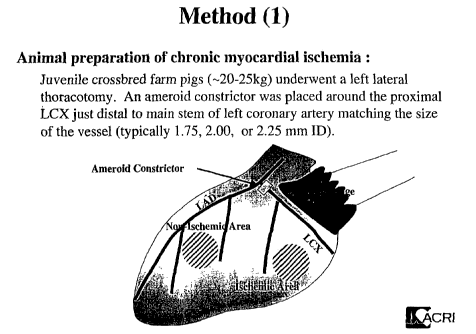
Fig. 1 provides a schematic illustration of a porcine heart with placement of
an
ameroid constrictor for inducing chronic myocardial ischemia. The ischemic and
non-
ischemic areas are noted.
Fig. 2 shows anterior (top left), lateral (bottom left) and posterior (bottom
right) views
of a porcine heart and indicates the ischemic risk areas induced by an ameroid
constrictor.
Fig. 3 is a bar graph depicting myocardial blood flow in pigs injected in an
ischemic
zone with the Ad-(3Ga1 construct (group 3 animals, Example 1). Blood flow (in
ml/min/mg
tissue) at rest (open box) and with pacing (shaded box): top left panel, in
ischemic
endocardial zone; top right panel, in ischemic epicardial zone; bottom left
panel, in non-
ischemic endocardial zone; and bottom right panel, in non-ischemic epicardial
zone.
Fig. 4 is a bar graph depicting myocardial blood flow in pigs injected in an
ischemic
zone with the Ad-VEGFI6s construct (group 1 animals, Example 1). Blood flow
(in
mllmin/mg tissue) at rest (open box) and with pacing (shaded box): top left
panel, in
ischemic endocardial zone; top right panel, in ischemic epicardial zone;
bottom left panel, in
non-ischemic endocardial zone; and bottom right panel, in non-ischemic
epicardial zone.
Fig. 5 is a bar graph depicting myocardial blood flow in pigs injected in a
non-
ischemic zone with the Ad-VEGFI6s construct (group 2 animals, Example 1).
Blood flow (in
ml/min/mg tissue) at rest (open box) and with pacing (shaded box): top left
panel, in
ischemic endocardial zone; top right panel, in ischemic epicardial zone;
bottom left panel, in
non-ischemic endocardial zone; and bottom right panel, in non-ischemic
epicardial zone.
Fig. 6 is a bar graph depicting transmural myocardial blood flow in pigs
injected with:
top left panel, the Ad-(3Ga1 construct in an ischemic zone (group 3 animals,
Example 1); top
right panel, PBS in an ischemic zone (group 4 animals, Example 1); bottom left
panel, the
Ad-VEGFISS construct in an ischemic zone (group 1 animals, Example 1); and the
Ad-
VEGFI6s construct in a non-ischemic zone (group 2 animals, Example 1). Blood
flow (in
ml/min/mg tissue) is shown at rest (open box) and with pacing (shaded box).
Fig. 7 is a bar graph depicting regional wall motion scores on dobutamine
stress
echocardiography. Wall motion scores are 1 = normal, 2 = hypokinesis, 3 =
akinesis, and 4 =
dyskinesis, pre-stress (open box), at low dose dobutamine (light shaded box),
and high dose
dobutamine (dark shaded box). Top left panel = the Ad-(3Ga1 construct in an
ischemic zone
-4-
CA 02433936 2003-07-04
WO 02/064157 PCT/US02/03118
(group 1 animals, Example 2), top right panel, the VEGFI6s construct in an
ischemic zone
(group 2 animals, Example 2), bottom left panel, the VEGFI6s construct in a
normal zone
(group 3 animals, Example 2), bottom right panel, the VEGFI~s construct in
normal and
ischemic zones (group 4 animals, Example 2).
Fig. ~ is a bar graph depicting myocardial blood flow in the ischemic zone of
pigs
injected with: Ad-(3Gal into an ischemic zone (top left), Ad-VEGFI6s into an
ischemic zone
(top right), Ad-VEGFI6s into a normal zone (bottom left) or Ad-VEGFISS into
both ischemic
and noxmal zone (bottom right). Blood flow (in ml/min/mg tissue) at rest (open
box) and
with pacing (shaded box) at baseline and after treatment.
Fig. 9 is a bar graph depicting capillary density in pigs (number/mm2)
injected with
Ad-[3Gal into an ischemic zone, Ad-VEGFI~s into an ischemic zone, Ad-VEGFI6s
into a
normal zone, and Ad-VEGFISS into both ischemic and normal zone.
DETAILED DESCRIPTION OF THE INVENTION
The invention provides a method of treating ischemic heart disease by
injecting a
therapeutic agent into normal myocardium in an amount sufficient to induce
angiogenesis,
stimulate collateral blood vessel formation, improve contractile function or
promote tissue
regeneration. The therapeutic agent can be injected into normal myocardium
adjacent to a
zone of ischemic myocardium or ischemic myocardial tissue of an animal, or the
therapeutic
agent can be injected into multiple sites distributed throughout the normal
myocardium. In a
preferred embodiment the therapeutic agent is a gene therapy vector encoding
at least one
nucleic acid, i.e., the transgene, encoding an angiogenic factor; and
expressing that factor in
an amount effective to treat the ischemic heart disease or to stimulate
collateral blood vessel
formation, to treat or ameliorate the cardiovascular condition or to promote
tissue
regeneration. The invention also contemplates methods to induce angiogenesis,
to increase
contractile function in the heart, to increase blood flow within the heart, to
stimulate
collateral vessel development in the heart, to promote tissue regeneration and
to treat
myocardial ischemia, preferably in a human patient using the therapeutic
agents of the
invention.
In some embodiments, a therapeutic agent is delivered to an ischemic or
diseased
heart by intramyocardially delivering a therapeutically effective amount of a
therapeutic
agent to normal tissue in the heart. The invention also provides methods for
stimulating
collateral blood vessel formation in the myocardium, for inducing angiogenesis
in the
-5-
CA 02433936 2003-07-04
WO 02/064157 PCT/US02/03118
myocardium, and for improving contractile function of the heart by delivering
an angiogenic
factor or cells capable of producing an angiogenic factor to normal tissue in
an ischemic or
diseased heart. The angiogenic factor is preferably delivered by an adenovirus
vector or an
adeno-associated vector which comprises a coding sequence encoding an
angiogenic factor,
wherein the coding sequence is operatively linked to a promoter which can
direct expression
of the angiogenic factor in a cardiac cell. Preferred vectors for use with the
invention include
replication-defective adenoviruses, serotype 5 adenoviruses, and adenoviruses
lacking the
early gene region El, the early gene region E3, or both.
The invention also provides a method for ameliorating the symptoms associated
with
myocardial ischemia, which comprises delivering a therapeutic agent to normal
myocardial
tissue in an amount sufficient to ameliorate one or more of the symptoms of
ischemia.
Amelioration of symptoms includes, for example, increased tolerance to
exercise, decreased
chest pain, and decreased shortness of breath.
The therapeutic agent can be a gene therapy vector, protein, peptide,
antisense DNA
or RNA, drug, cells, cells which express a therapeutic agent, whole bone
marrow and or any
other therapeutic agent capable of or useful to induce angiogenesis, increase
contractile
function in the heart, increase blood flow within the heart, stimulate
collateral vessel
development in the heart, treat myocardial ischemia, or promote tissue
regeneration.
Any suitable gene therapy vector can be used to supply the transgene. For
example,
the gene therapy vector can be a replication-deficient adenovirus, a
recombinant adeno-
associated virus vector (rAAV), a retroviral vector, a plasmid, or any other
vector useful in
cardiac gene therapy. Non-limiting examples of recombinant adenoviral vectors
suitable for
use in the invention include the recombinant adenoviruses described in Graham
et al.
(Vif-ology 163:614-617, 1988), as well as those in Graham F. et al. (Methods
an Molecular
Biology 7: 109-128, Murray, E., ed. Humana Press, Clifton, N.J, 1991), Curiel,
et al. (Proc.
Natl. Acad. Sci. USA 88:8850-8854, 1991), Miller et al. (FASEB J. 9:190-199,
1995), and
Curiel (Anna. NYAcad. Sci. 886:158-171). Adenovirus vectors suitable for use
with the
invention also include adenoviruses of adenovirus serotype 5, and adenoviruses
lacking the
early gene E1 region, lacking the early gene E3 region, or lacking both. Adeno-
associated
vectors are described in, for example, Smith-Arica et al. (Curr. Cardi.ol.
Rep. 3:43-49, 2001),
Philips (Expert Opinion. Biol. Tlzer. 1:655-662, 2001), Rabinowitz, et al. (J.
Virol 76:791-
801). Other viral vectors suitable for use in the invention include retroviral
vectors, corona
virus based vectors, and vaccinia-based vectors: Plasmid and other non-viral
vectors such as
-6-
CA 02433936 2003-07-04
WO 02/064157 PCT/US02/03118
plasmid/liposome vectors, virus/liposome vectors, oligonucleotides, and others
are described
in, for example, ~VIcKay, et al. (Cariovasc. Drug. Rev. 19: 245-62, 2001) and
Rosenzweig
(Vectors for Gene Therapy. In: Current Protocols in Hu~za~z GesZetics.
Dracopoli, et al. eds.
New York, NY: Joh~.i Wiley and Sons, Inc., 1999).
Gene therapy vectors useful in the present invention can be any vector with
one or
more transgenes (or nucleic acids of interest) inserted therein in a manner
allowing
expression of the transgene under control of appropriate regulatory elements
such as
promoters, enhancers, transcription terminators and the like. Gene therapy
vectors are well
known in the art and can be prepared by standard methodology known to those of
ordinary
skill in the art.
Further, the nucleic acid is operably linked to a control region, e.g.,
promoters,
enhancers, termination signals and the like, to permit expression of the
molecule. When
more than one nucleic acid is present on the vector, each can be controlled
separately by
individual control regions or, any group of them, or all of them, can be
controlled in an
operon, i.e., with one control region driving expression of multiple genes on
a single
transcr ipt.
A "transgene" or "nucleic acid of interest" or the "nucleic acid encoded in
the vector"
as used herein refers to any nucleotide sequence which encodes a
therapeutically-effective
molecule to induce angiogenesis, to stimulate collateral blood vessel
formation, or to increase
myocardial blood flow in ischemic areas of the heart. These transgenes can
encode the
proteins and angiogenic factors of the invention described herein. The
transgenes can be
foreign to the animal being treated, or can be genes normally found, in the
animal being
treated, but for which altered expression, is desired. Expression can be
altered by changing
the amount of expression, or temporal or spatial pattern of expression.
As used herein, a "control region" or "regulatory element" refers to
polyadenylation
signals, upstream regulatory domains, promoters, enhancers, transcription
termination
sequences and the like which regulate the transcription and translation of a
nucleic acid
sequence.
The term "operably linked" refers to an arrangement of elements wherein the
components are arranged so as to perform their usual function. Thus, control
regions or
regulatory elements operably linked to a coding sequence are capable of
effecting the
expression of the coding sequence. The control elements need not be contiguous
with the
coding sequence, so long as they function to direct the expression thereof.
Thus, for
CA 02433936 2003-07-04
WO 02/064157 PCT/US02/03118
example, intervening untranslated yet transcribed sequences can be present
between a
promoter sequence and the coding sequence and the promoter sequence can still
be
considered "operably linked" to the coding sequence.
The regulatory elements of the invention can be derived from any source, e.g.,
viruses, mammals, insects or even synthetic, provided that they function after
injection into
the heart. For example, any promoter can used to control expression of the
transgene. Such
promoters can be promiscuous, i.e., active in many cell types, such as the
SV40 early
promoter, the mouse mammary tumor virus LTR promoter, the adenovirus major
late
promoter (Ad MLP), a herpes simplex promoter, a CMV promoter such as the CMV
immediate early promoter, or a rous sarcoma virus (RSV) promoter.
Alternatively the
promoter can be tissue-specific for expression in cardiac cells such as
cardiomyocytes. Non-
limiting examples of tissue specific promoters are known in the art (see,
e.g., Lee, et al.
(1992) J. Biol. CheJn. 267:15875-15885; Jeyaseelan et al. (1997) Pr~oc. Natl.
Acad. Sci. USA
272:22800-22808; Condorelli, et al. (2001) Pf~oc. Natl. Acad. Sci. USA 98:9977-
9982)
include the left ventricular myosin light chain-2 (MLC2v) promoter, myosin
heavy chain
(MHC) promoters such as the a-MHC and (3-MHC, natriuretic peptide precursor A
promoter
(NppA), the promoter of the cardiac adriamycin responsive protein (CARP), the
promoter of
the cTNC gene, and others.
Proteins that can be administered (encoded in gene therapy vectors or
directly)
include proteins or peptides competent to induce angiogenesis, e.g.,
angiogenesis factors. A
protein or peptide competent to induce angiogenesis or an "angiogenesis
factor" as used
herein is a protein or substance that causes proliferation of new blood
vessels and includes
fibroblast growth factors, endothelial cell growth factors or other proteins
with such
biological activity. Angiogenic factors, and particular proteins known to
induce
angiogenesis, include but are not limited to, FGF-l, FGF-2, FGF-5, VEGF and
active
fragments thereof such as VEGFI6s, HIF-1 PDGF-1, PDGF-2, DELI, angiopoietins,
HGF,
MCP-1, eNOS and iNOS. Other angiogenic factors suitable for use in the
invention are
growth factors, including endothelial growth factors, vascular smooth muscle
growth factors,
and FGF-1, FGF-2, FGF-5, PDGF-1, and PDGF-2. The abbreviations are as follows:
FGF,
fibroblast growth factor; VEGF, vascular endothelial growth factor; HIF,
hypoxia inducible
factor; PDGF, platelet-derived growth factor; DEL, developmental embryonic
locus: HGF,
hepatocyte growth factor; MCP, monocyte chemoattractant protein; eNOS,
endothelial
nitrous oxide synthase; and iNOS, inducible nitrate oxide synthase.
_g_
CA 02433936 2003-07-04
WO 02/064157 PCT/US02/03118
Other proteins or transgenes are also suitable for use in the invention, for
example
factors involved in myocardial preservation or reperfusion injury, such as
heme oxygenase,
hlcis, AKT, PR39, and (3arkCT, can be used in the methods of the invention.
Tissue
regeneration factors, including, but not limited to, ligands for progenitor or
stem cells, such as
c-kit ligand, CD34 ligand, and other factors are also suitable for use in the
methods of the
invention.
Cells that can be administered by the present method include, but are not
limited to,
endothelial progenitor cells (angioblasts), cardiac myoblasts, mononuclear
cells, bone
marrow stromal cells and stem cells. "Stem cells" as used herein refers to
mononuclear cells
from placental or umbilical cord blood. The cells described herein can be
administered as
primary cells, i.e., without transformation or other ex vivo manipulation.
Alternatively, any
of these cells, or other appropriate cell types, can be manipulated or
expanded ex vivo, or
genetically engineered ex vivo or selected to produce an angiogenic factor
using methods
known in the art. Typically, cells are engineered to produce an angiogenic
factor are
engineered to secrete the desired angiogenic factor. Additionally, filtered
whole bone
marrow is know to be angiogenic and such a preparation can be administered in
accordance
with the invention.
The therapeutic agents described herein can be administered singly or in
combination.
In one non-limiting example, a therapeutic agent according to the invention
may comprise a
viral vector delivered in combination with angiogenic cells. In another non-
limiting example,
a therapeutic agent according to the invention may comprise a viral vector
delivered in
combination with an angiogenic protein. The therapeutic agents can also be
delivered in
combination with other active agents, such as anti-apoptotic agents.
Pharmaceutical formulations of the therapeutic agents of the invention are
prepared
for storage by mixing those entities having the desired degree of purity with
optional
physiologically acceptable carriers, excipients or stabilizers (Remington's
Pharmaceutical
Sciences 16th edition, Osol, A. Ed. (1980)), in the form of lyophilized
formulations or
aqueous solutions. Acceptable carriers, excipients, or stabilizers are
nontoxic to recipients at
the dosages and concentrations employed, and include buffers such as
phosphate, citrate, and
other organic acids; antioxidants including ascorbic acid and methionine;
preservatives (such
as octadecyldimethylbenzyl ammonium chloride; hexamethonium chloride;
benzalkonium
chloride, benzethonium chloride; phenol, butyl or benzyl alcohol; alkyl
parabens such as
methyl or propyl paraben; catechol; resorcinol; cyclohexanol; 3-pentanol; and
m-cresol); low
-9-
CA 02433936 2003-07-04
WO 02/064157 PCT/US02/03118
molecular weight (less than about 10 residues) polypeptide; proteins, such as
serum albumin,
gelatin, or immunoglobulins; hydrophilic polymers such as
polyvinylpyrrolidone; amino
acids such as glycine, glutamine, asparagine, histidine, arginine, or lysine;
monosaccharides,
disaccharides, and other carbohydrates including glucose, mannose, or
dextrins; chelating
agents such as EDTA; sugars such as sucrose, mannitol, trehalose or sorbitol;
salt-forming
counter-ions such as sodium; metal complexes (e.g., Zn-protein complexes);
and/or non-ionic
surfactants such as TWEENTM, PLURONICSTM or polyethylene glycol (PEG).
The formulation herein may also contain more than one active compound as
necessary
for the particular indication being treated, preferably those with
complementary,activities that
do not adversely affect each other. Such molecules are suitably present in
combination in
amounts that are effective for the purpose intended.
The active ingredients may also be entrapped in microcapsule prepared, for
example,
by coacervation techniques or by interfacial polymerization, for example,
hydroxymethylcellulose or gelatin-microcapsule and poly-(methylmethacylate)
microcapsule,
respectively, in colloidal drug delivery systems (for example, liposomes,
albumin
microspheres, microemulsions, nano-particles and nanocapsules) or in
macroemulsions. Such
techniques are disclosed in Remington's Pharmaceutical Sciences 16th edition,
Osol, A. Ed.
( 1980).
The formulations to be used for in vivo administration must be sterile. This
is readily
accomplished by filtration through sterile filtration membranes.
Sustained-release preparations may be prepared. Suitable examples of sustained-
release preparations include semipermeable matrices of solid hydrophobic
polymers
containing the polypeptide variant, which matrices are in the form of shaped
articles, e.g.,
films, or microcapsule. Examples of sustained-release matrices include
polyesters, hydrogels
(for example, poly(2-hydrbxyethyl-methacrylate), or poly(vinylalcohol)),
polylactides (U.S.
Pat. No. 3,773,919), copolymers of L-glutamic acid and y ethyl-L-glutamate,
non-degradable
ethylene-vinyl acetate, degradable lactic acid-glycolic acid copolymers such
as the LUPRON
DEPOTTM (injectable microspheres composed of lactic acid-glycolic acid
copolymer and
leuprolide acetate), and poly-D-(-)-3-hydroxybutyric acid. While polymers such
as ethylene-
vinyl acetate and lactic acid-glycolic acid enable release of molecules for
over 100 days,
certain hydrogels release proteins for shorter time periods. When encapsulated
antibodies
remain in the body for a long time, they may denature or aggregate as a result
of exposure to
moisture at 37°C, resulting in a loss of biological activity and
possible changes in
- 10-
CA 02433936 2003-07-04
WO 02/064157 PCT/US02/03118
immunogenicity. Rational strategies can be devised for stabilization depending
on the
mechanism involved. For example, if the aggregation mechanism is discovered to
be
intermolecular S-S bond formation through thio-disulfide interchange,
stabilization may be
achieved by modifying sulfhydryl residues, lyophilizing from acidic solutions,
controlling
moisture content, using appropriate additives, and developing specific polymer
matrix
compositions.
Those of skill in the art can readily determine the amounts of the therapeutic
agents to
be included in any pharmaceutical composition and the appropriate dosages for
the
contemplated use.
The method of the present invention can be used with any animal, including but
not
limited to, mammals such as rodents, dogs, cats, cattle, primates and humans.
Preferably the
method is used for gene therapy to treat human ischemic cardiac conditions or
diseases.
The amount of the therapeutic agent injected into the animal is proportional
to the
body weight of the animal and also depends on the selected agent. Those of
skill in the art
can readily determine the appropriate dosage for the selected agent. By way of
example,
when the agent is a gene therapy vector such as a replication-defective
adenovirus, the dosage
can range from about 106 to about 1012 plaque-forming units (pfu), and is
preferably between
about 108 to about 101° pfu. For stable and efficient transduction
using rAAV, the dosage can
be from about 1 x 10⁵ IU (infectious units) of AAV per gram body weight to
about 1 x 10⁹ IU
AAV per gram body weight, and preferably from about 1 X 10⁶ IU AAV per gram
body
weight to about 1 x 10⁷ IU AAV per gram body weight. When the agent is a
protein, the
dosage can range from as little as about 1 picograms to several hundred
micrograms, but in
any event can be readily determined by those of skill in the art.
Methods for measuring cardiac function are well known in the art. See, e.g.,
Simons
et al. (2000) Circulation 102:e732-e86, "Clinical Trails in Coronary
Angiogenesis: Issues,
Problems and Consensus." For example, blood flow to ischemic myocardium can be
measured using various non-invasive imaging techniques, including single
photon emission
computed tomography (SPECT), position emission tomography (PET), magnetic
resonance
imaging (MRI), and injection of fluorescent microspheres. Coronary angiography
can be
used to measure disease progression and to document the appearance of new
vessels.
Echocardiography can be used to assess cardiac wall motion at rest and under
stress, such as
dobutamine-induced stress. Exercise tolerance testing such as treadmill
testing can provide
another means for assessing cardiac function .
11-
CA 02433936 2003-07-04
WO 02/064157 PCT/US02/03118
Delivery to myocardium can be accomplished using a catheter (e.g. infusion
catheter,
diagnostic catheter, etc) stiletto catheter, needle or needles, needle-free
injector, balloon
catheter, channeling device, or other appropriate medical device for
introduction into the
myocardium. In a preferred delivery method, an endocardial injection catheter,
such as a
Stiletto catheter (Boston Scientific, Natick, Massachusetts) is used to
deliver the therapeutic
agent without requiring open chest surgery Catheter injections can be guided
by fluoroscopy,
echocardiography, MRI, or electromechanical mapping. The catheter is used to
deliver the
therapeutic agent to non-ischemic tissue in the myocardium by transendocardial
injection.
Appropriate devices and methods for catheter injection are described in U.S.
Patent No.
6,238, 406. Alternatively, a transepicardial surgical approach may be
necessary for delivery
to myocardium, either via open chest or via thoracoscopy.
Throughout this application, various publications, patents, and patent
applications
have been referred to. The teachings and disclosures of these publications,
patents, and
patent applications in their entireties are hereby incorporated by reference
into this
application to more fully describe the state of the art to which the present
invention pertains.
It is to be understood and expected that variations in the principles of
invention herein
disclosed in an exemplary embodiment may be made by one skilled in the art and
it is
intended that such modifications, changes, and substitutions are to be
included within the
scope of the present invention.
2p EXAMPLE 1
Induction of Chronic Myocardial Ischemia: Juvenile cross bred pigs (~20-25 kg)
underwent left lateral thoracotomy. An ameroid constrictor was placed around
the proximal
LCX just distal to the main stem of the left coronary artery using an ameroid
constrictor
matching the size of the vessel, typically 1.75, 2.00 or 2.25 mm inner
diameter (ID). Fig. 1
illustrates placement of the ameroid constrictor.
Assessment of Cardiac Function and Myocardial Injections: Baseline
measurements
of cardiac function were obtained four weeks after placement of the ameroid
constrictor. The
measurements included coronary angiography, dobutamine stress
echocardiography, blood
flow measurements by injection of microspheres at rest and at atrial pacing of
180 beats per
minute (bpm).
After baseline measurements were completed, vectors or saline were introduced
into
the heart in the indicated zones by intramyocardial injection as described in
Kornowski et al.
- 12-
CA 02433936 2003-07-04
WO 02/064157 PCT/US02/03118
(2000) J. Am. Coll. Card. 35:1031-1039. This method allows direct injection
into normal or
ischemic myocardium during open-heart surgery with a magnetic guidance
catheter-based
navigational system. The injections consisted of 10 injections of 20 uL of 5 x
109 pfu/mL of
Ad-VEGFI6s or Ad-(3Ga1 or 10 injections of 20 uL of phosphate-buffered saline
(PBS).
Four weeks after the injections, i.e., eight weeks after implantation of the
ameroid
constrictor, the baseline measurements were repeated. Additionally, ischemic
and adjacent
normal areas were harvested post-mortem for regional myocardial blood flow
measurement,
histopathologic analysis and morphometric analysis.
Treatment Groups: The animals were divided into four groups and received
injection
of (1) Ad-VEGFISS into the ischemic zone (n=9); (2) Ad-VEGFI6s into the normal
zone
(n=8); (3) Ad-(3Ga1 into the ischemic zone (n=8); or (4) PBS into the ischemic
zone (n=7).
Results: The blood flow data indicate that when injections are targeted to the
ischemic zone, modest improvements in perfusion occur at rest. However, when
injections
are made in to the normal zone of the myocardium, significant improvements are
observed in
blood perfusion at both rest and stress. Further more, transmural blood flow
reaches a much
higher level of 0.815 (normal zone injections) versus 0.351 (ischemic zone
injections) under
stress.
EXAMPLE 2
Induction of Chronic Myocardial Ischemia: Ameroid constrictors were placed
around
the proximal LCX of juvenile pigs via left lateral thoracotomy as described in
Example 1.
Assessment of Cardiac Function and Myocardial Injections: Baseline
measurements
of cardiac function were obtained four weeks after placement of the ameroid
constrictor. The
measurements included coronary angiography, dobutamine stress
echocardiography, blood
flow measurements by injection of fluorescent microspheres at rest and at
atrial pacing of 180
beats per minute (bpm).
After baseline measurements were completed, vectors or saline were introduced
into
the heart by Stiletto injection catheter. Each animal received 10 injections,
each 20 p.L, of 5 x
109 pfulmL of Ad-VEGFI6s or Ad- (3Gal.
Four weeks after the injections, i.e., eight weeks after implantation of the
ameroid
constrictor, the baseline measurements were repeated. Additionally, ischemic
and adjacent
-13-
CA 02433936 2003-07-04
WO 02/064157 PCT/US02/03118
normal areas were harvested post-mortem for regional myocardial blood flow
measurement,
histopathologic analysis, and morphometric analysis.
Treatment Groups: The animals were divided into four groups and received
injection
of (1) Ad- (3Gal into the ischemic zone ( n = 7); (2) Ad-VEGFISS into the
ischemic zone
(n = 7); (3) AdVEGFI6s into the normal tissue adjacent to the ischemic zone (n
= 7); and (4)
AdVEGFl6s throughout the left ventricular free wall in both normal and
ischemic tissue (n =
8).
Results: Under resting conditions, animals that received injections of Ad-
(3Ga1 into
the ischemic zone did not show significant improvement in blood flow at rest,
but did show
improvement in blood flow with pacing. Trends toward improvement in blood flow
were not
seen in animals that received injections of AdVEGFISS into the ischemic region-
. Animals
that received injections of Ad-VEGFISS into the normal zone showed trends
toward
improvement both at rest and with pacing. Animals that received injections
throughout the
left ventricular flee wall in both ischemic and normal zone also showed trends
toward
improvement both at rest and with pacing.
Dobutamine stress echocardiography indicated trends toward improvement in wall
motion in all animals that received the Ad-VEGFI6s construct. In contrast,
animals that
received the Ad-(3Ga1 construct showed decrements in wall motion.
Animals that received injections of Ad-VEGFI6s in the ischemic zone had lower
capillary density than animals that received Ad-(3Gal in the ischemic zone.
Animals that
received injections of Ad-VEGFI6s in the normal zone had higher capillary
density than
animals that received injections of Ad-(3Ga1 in the ischemic zone, and animals
that received
injections of Ad-VEGFI6s in both the normal and ischemic zones had capillary
density
similar those that received injections of Ad-(3Gal in the ischemic zone.
- 14-