Note: Descriptions are shown in the official language in which they were submitted.
CA 02435694 2003-07-22
WO 03/009744 PCT/US02/03450
APPARATUS FOR IMPLANTATION INTO BONE
Related Applications
This application is a continuation-in-part of
co-pending U.S. patent application Serial No.
09/708,940, filed November 8, 2000, and co-pending
U.S. patent application Serial No. 09/708,292, also
filed on November 8, 2000. The entire subject matter
of the aforementioned two co-pending applications is
incorporated herein by reference.
Technical Field
The present invention is directed to an apparatus
for implantation into a bone in a patient's spine or
pelvis, and is particularly directed to an apparatus
that, when implanted, is resistant to toggling in the
bone and to being pulled from the bone. The present
invention is also directed to an apparatus for
attaching and stabilizing adjacent vertebral bodies
while the vertebral bodies fuse together.
CA 02435694 2003-07-22
WO 03/009744 PCT/US02/03450
-2-
Background of the Invention
Bone screws are used in the medical field for a
variety of purposes. Typical uses for bone screws,
also referred as bone anchors, include treating a bone
fracture, attaching a corrective device to parts of a
fractured bone in an area adjacent to the fracture, and
attaching a corrective device to a group of bones, such
as vertebrae of a spinal column.
Most known bone screws use a conventional screw
design, i.e. a solid shank, with one or more external
thread convolutions. The solid shank and external
threads of the conventional bone screws can cause the
bone screws to displace an undesirably large amount of
bone when implanted. Further, such conventional bone
screws require a large amount of torque to implant the
scredr into a vertebral body.
It is also known to use a corkscrew-style helical
spike as a tissue anchor. The known corkscrew-style
tissue anchors, when implanted, displace less bone than
the conventional bone screws, but are generally not
able to withstand high. tensile loads without structural
failure. European,Patent No. 0 374 088 A1 discloses a
bone screw having a twin-corkscrew design. In this
twin-corkscrew design, which is formed by drilling a
CA 02435694 2003-07-22
WO 03/009744 PCT/US02/03450
-3-
passage up through a screw having a solid shank and
then machining out the material between the two
corkscrews, the junction of the corkscrews with the
shank is unlikely to be capable of structurally
withstanding high tensile loads and repetitive fatigue
loads. This structural weakness in the design of the
screw in the EP 0 374 088 document is further
compounded by the corkscrews having a larger overall
diameter than the head of the screw where torque is
applied.
One of the more challenging applications of a bone
screw is implantation of the screw into the cancellous
bone of a patient's spine or pelvis. For example, bone
screws are frequently implanted into the cancellous
bone of a patient's lumbar vertebrae during a spinal
fixation procedure to correct scoliosis. Once
implanted, the bone screws are used to mount suitable
spinal fixation instrumentation, such as clamps, rods,
and plates. Unfortunately, many of the known bone
~0 screws, such as those described above, can be
susceptible to toggling in the vertebral body and can
also pull out of the vertebral body due to the
substantial forces on the screws from human body
movement and muscle memory. In order to achieve a high
CA 02435694 2003-07-22
WO 03/009744 PCT/US02/03450
-4-
pull-out resistance, it is known to thread a bone screw
all of the way through a vertebrae and place a nut on
the opposite side. However, use of such a nut
increases the complexity of the surgical procedure.
Hence, it is desirable to provide an apparatus for
implantation into a bone in a patient's spine or pelvis
in a minimally invasive endoscopic procedure with a
reduced amount of insertion torque required. The
desirable apparatus would provide a platform for
connecting spinal fixation instrumentation and, when
implanted, be highly resistant to toggling in the bone
and to being pulled out of the bone despite the
substantial forces on the apparatus from human body
movement and muscle memory.
Another application for an anchor or
fastening-type apparatus in the field of spine surgery
is the stabilization of adjacent vertebrae. Each
adjacent pair of vertebrae in the human spinal column
are separated by an intervertebral disc that makes
relative movement of the vertebrae possible. Problems,
however, can develop with one or more of the discs,
causing severe back pain. In some cases, it is
necessary to remove a problematic disc and to fuse the
adjacent vertebrae together in order to relieve pain.
CA 02435694 2003-07-22
WO 03/009744 PCT/US02/03450
-5-
One known method for fusing an adjacent pair of
vertebrae following removal of a disc is to implant a
device, commonly referred to as a fusion cage, into the.
interbody space where the disc was removed. The fusion
cage facilitates fusion of the vertebrae. Typically,
procedures such as reaming and/or tapping of adjacent
vertebrae are required to prepare the adjacent
vertebrae to receive the fusion cage. Such procedures
normally involve substantial cutting of the hard
cortical bone of the end plates of the adjacent
vertebrae, which can weaken the end plates and lead to
collapse of the vertebrae. The fusion cage is then
positioned in the interbody space and into engagement
with the adjacent vertebrae. At least one known fusion
cage has relatively movable parts that enable the
fusion cage to be expanded after the fusion cage is
positioned in the interbody space between adjacent
vertebrae. The design of this expandable fusion cage
is, however, relatively complex.
Typically, a fusion cage includes an internal
cavity that is filled with bone graft material. The
fusion cage and the bone graft material promote bone
growth that slowly unites the adjacent vertebrae. The
typical fusion cage, while in engagement with the
CA 02435694 2003-07-22
WO 03/009744 PCT/US02/03450
-6-
adjacent vertebrae, does not attach to the vertebrae
and thus does not resist relative movement of the
vertebrae, through bending or rotation, along any one
of the three planes of motion (sagittal, coronal, or
horizontal). Rather, the typical fusion cage relies on
the viscoelasticity of the surrounding ligaments to
stabilize the adjacent vertebrae.
It is desirable to provide an apparatus for
implantation into an adjacent pair of vertebral bodies
that attaches to and thus fastens the vertebral bodies
while they fuse together despite the forces on the
apparatus from human body movement and muscle memory.
It is further desirable to provide an apparatus which
has a reduced insertion torque requirement, a simple
one-piece construction, and which may be implanted into
an adjacent pair of vertebrae without having to prepare
the adjacent vertebrae to accept the apparatus by
substantial cutting of the cortical bone.
Summary of the In~sention
The present invention is an apparatus for
implantation into a bone in a patient's spine or
pelvis. The apparatus, when implanted, is resistant to
toggling in the bone and to being pulled from the bone.
The apparatus comprises a platform having a first
CA 02435694 2003-07-22
WO 03/009744 PCT/US02/03450
surface for facing a bone in a patient's spine or
pelvis. The platform includes structure for connection
to a spinal fixation implant. The apparatus further
comprises at least one helical spike for embedding into
the bone upon rotation of the platform. The at least
one helical spike projects tangentially from the
platform and extends around a longitudinal axis. The
at least one helical spike has a tip portion at a
distal end which penetrates into the bone as the
platform is rotated. The at least one helical spike
further has a connecting portion at a proximal end
connected to the platform and an intermediate portion
extending between the connecting portion and the tip
portion. At least one of the intermediate portion and
the connecting portion of the at least one helical
spike has a tubular cross-section defined by an outer
diameter and an inner diameter of the at least one
helical spike.
In accordance with another embodiment, the present
invention is an apparatus comprising at least one
anchor for implantation into a bone. The anchor, when
implanted, is resistant to toggling in the bone and to
being pulled from the bone. The apparatus further
comprises a spinal fixation implant for extending
CA 02435694 2003-07-22
WO 03/009744 PCT/US02/03450
_g_
between and connecting a plurality of bones. The
anchor includes a platform having a first surface for
facing the bone. The platform further has structure
for connection with the spinal fixation implant. The
anchor further includes at least two helical spikes for
embedding into the bone upon rotation of the platform.
The at least two helical spikes are spaced apart and
project tangentially from the first surface on the
platform. The at least two helical spikes extend
around a longitudinal axis. Each of the at least two
helical spikes has a tip portion at a distal end which
penetrates into the bone as the platform is rotated.
Each of the at least two helical spikes further has a
connecting portion at a proximal end that is connected
to the platform, and an intermediate portion extending
between the connecting portion and the tip portion. At
least one of the intermediate portion and the
connecting portion of each of the at least two helical
spikes has a tubular cross-section defined by an outer
diameter and an inner diameter of the at least two
helical spikes.
In accordance with yet another embodiment, the
present invention comprises an apparatus for
implantation into an adjacent pair of vertebral bodies
CA 02435694 2003-07-22
WO 03/009744 PCT/US02/03450
-9-
having first and second surfaces that oppose each
other. The apparatus, when implanted, is attached to
the adjacent pair of vertebral bodies and stabilizes
the vertebral bodies while the vertebral bodies fuse
together. The apparatus comprises a platform having a
third surface extending transverse to the first and
second surfaces. The apparatus further comprises at
least one helical spike for embedding into each of the
adjacent pair of vertebral bodies upon rotation of the
platform to attach the at least one helical spike to
each of the vertebral bodies and thus fasten (pin) the
vertebral bodies together. The at least one helical
spike projects from the platform and extends around a
longitudinal axis. The at least one helical spike has
a tip portion at a distal end for penetrating the first
and second surfaces and for screwing into the adjacent
pair of vertebral bodies as the platform is rotated.
At least a portion of the at least one spike has a
tubular cross-section defined by an outer diameter and
an inner diameter. The at least one helical spike at
least partially defines an internal cavity for
receiving material that promotes fusion of the
vertebral bodies.
CA 02435694 2003-07-22
WO 03/009744 PCT/US02/03450
-10-
Brief Description of the Drawings
The foregoing and other features of the present
invention will become apparent to those skilled in the
art to which the present invention relates upon reading
the following description with reference to the
accompanying drawings, in which:
Fig. 1 is a schematic anterior view of an
apparatus constructed in accordance with the present
invention implanted in a vertebral body;
Fig. 2 is a schematic anterior view of several
vertebral bodies implanted with the apparatus of Fig. 1
and connected by a spinal fixation implant in
accordance with the present invention;
Fig. 3 is a side view of the apparatus of Fig. 1;
Fig. 4 is a sectional view taken along 4-4 in
Fig. 3;
Fig. 5 is a sectional view taken along 5-5 in
Fig. 3;
Fig. 6 is a sectional view taken along 6-6 in
Fig. 3;
Fig. 7 illustrates an alternate configuration for
an end portion of the apparatus of Fig. 1;
CA 02435694 2003-07-22
WO 03/009744 PCT/US02/03450
-11-
Fig. 8 is a side view illustrating a second
embodiment of an apparatus in accordance with the
present invention;
Fig. 9 is a sectional view taken along line 9-9 in
Fig. 8;
Fig. 10 is a sectional view taken along 10-10 in
Fig. 8;
Fig. 11 is a sectional view taken along 11-11 in
Fig. 8;
Fig. 12 is a sectional view taken along 12-12 in
Fig. 8;
Fig. 13 is a schematic view, partially in section,
of a third embodiment of the present invention;
Fig. 14 is an exploded perspective view of the
apparatus of Fig. 13;
Fig. 15 is a sectional~view taken along line 15-15
in Fig. 13;
Fig. 16 is a sectional view taken along line 16-16
in Fig. 13;
Fig. 17 is a schematic anterior view of an
apparatus implanted in an adjacent pair of vertebral
bodies in accordance with a fourth embodiment of the
present invention;
CA 02435694 2003-07-22
WO 03/009744 PCT/US02/03450
-12-
Fig. 18 is an end view taken along line 18-18 in
Fig. 17;
Fig. 19 is a side view of the apparatus of
Fig. 17;
Fig. 20 is a sectional view taken along 20-20 in
Fig. 17;
Fig. 21 is a sectional view taken along 21-21 in
Fig. 19;
Fig. 22 is a sectional view taken along 22-22 in
Fig. 19;
Fig. 23 illustrates an alternate configuration for
an end portion of the apparatus of Fig. 19;
Fig. 24 is a schematic anterior view illustrating
a fifth embodiment of the present invention;
Fig. 25 is a side view illustrating a sixth
embodiment of an apparatus for implanting in an
adjacent pair of vertebral bodies in accordance with
the present invention;
Fig. 26 is a sectional view taken along line 26-26
in Fig. 25;
Fig. 27 is a sectional view taken along 27-27 in
Fig. 25;
Fig. 28 is a sectional view taken along 28-28 in
Fig. 25;
CA 02435694 2003-07-22
WO 03/009744 PCT/US02/03450
-13-
Fig. 29 is a sectional view taken along 29-29 in
Fig. 25;
Fig. 30 is a side view, similar to Fig. 3,
illustrating modification to the present invention;
Fig. 31 is a sectional view taken along line 31-31
in Fig. 30;
Fig. 32 is a sectional view taken along line 32-32
in Fig. 30;
Fig. 33 is a sectional view taken along line 33-33
in Fig. 30;
Fig. 34 is a side view illustrating a cervical
application of the apparatus of Fig. 19 in accordance
with the present invention;
Fig. 35 is an exploded perspective view
illustrating a seventh embodiment of an apparatus in
accordance with the present invention; and
Fig. 36 is a schematic view of the apparatus of
Fig. 35 implanted in a pair of cervical vertebrae.
Description of Preferred Embodiments
The present invention is directed to an apparatus
for implantation into a bone in a patient's spine or
pelvis, and is particularly directed to an apparatus
that, when implanted, is resistant to toggling in the
bone and to being pulled from the bone. The present
CA 02435694 2003-07-22
WO 03/009744 PCT/US02/03450
-14-
invention is also directed to an apparatus for
attaching and stabilizing adjacent vertebral bodies
while the vertebral bodies fuse together.
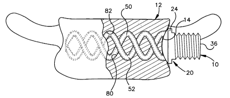
As representative of the present invention, Fig. 1
illustrates an apparatus 10 implanted in a lumbar
vertebrae 12. It should be understood that the
apparatus 10 could be implanted into any vertebral
body, including the sacrum. The lumbar vertebrae 12
has a concave side surface 14.
The apparatus 10 comprises an anchor 20 made from
a biocompatible material, such as titanium or stainless
steel. It is contemplated that the biocompatible
material used for the anchor 20 could be polymeric or
composite (i.e., carbon fiber or other biologic
composite) in nature. It is further contemplated that
the biocompatible material used to make the anchor 20
could also be biodegradable.
The anchor 20 is centered about a longitudinal
axis 22 (Fig. 3). The anchor 20 includes a platform 24
having a generally cylindrical outer surface 26
extending between oppositely disposed first and second
ends 28 and 30 of the platform. The platform 24
includes a generally rectangular slot 32 that extends
axially from the first end 28 toward the second end 30
CA 02435694 2003-07-22
WO 03/009744 PCT/US02/03450
-15-
of the platform. Adjacent the first end 28, the outer
surface 26 of the platform 24 includes first and second
segments of external threads 34 and 36 that are
separated by the slot 32. The slot 32 and the
threads 34 and 36 provide structure for connecting
spinal fixation instrumentation to the platform 24 as
discussed further below. The second end 30 of the
platform 24 includes an end surface 38 having a convex
shape that is complimentary to the shape of the concave
side surface 14 of the vertebrae 12. The end surface
38 of the platform 24 may include barbs (not shown) or
other suitable structure for fixedly engaging the side
surface 14 of the vertebrae 12. Further the end
surface 38 of the platform 24 may also be porous,
pitted, or have a biocompatible surface coating to
assist with fixation of the anchor 20 to the vertebrae
12.
First and second helical spikes 50 and 52 project
tangentially from the end surface 38 of the
platform 24. The helical spikes 50 and 52 resemble a
pair of intertwined corkscrews. As shown in Figs. 5
and 6, each of the helical spikes 50 and 52 has a
tubular cross-section defined by an outer diameter OD
and an inner diameter TD. The outer diameter OD of
CA 02435694 2003-07-22
WO 03/009744 PCT/US02/03450
-16-
each of the helical spikes 50 and 52 has a first radius
R1 and the inner diameter ID of each of the helical
spikes has a second radius R2 that is less than the
first radius R1.
According to the embodiment illustrated in
Figs. 1-6, the first and second helical spikes 50
and 52 extend around the axis 22. The spikes 50 and 52
extend in a helical pattern about the axis 22 at the
same, constant overall radius R3 (Fig. 3). It is
contemplated, however, that the first and second
helical spikes 50 and 52 could extend about the axis 22
at different radiuses. Further, it is contemplated
that the radius of one or both of the first and second
helical spikes 50 and 52 could increase or decrease as
the helical spikes extend away from the platform 24.
In order for the anchor 20 to be implanted
endoscopically through a typical cannula (not shown),
the platform 24 and the helical spikes 50 and 52 should
be less than 20mm in overall diameter. It should be
understood that the anchor 20 could have an overall
diameter that is greater than 20mm for certain
applications, and that the anchor could be also
implanted in an open surgical procedure.
CA 02435694 2003-07-22
WO 03/009744 PCT/US02/03450
-17-
In the illustrated embodiment of Figs. 1-6, the
first and second helical spikes 50 and 52 have the same
axial length, and also have the same tubular
cross-sectional shape, It is contemplated, however,
that the first and second helical spikes 50 and 52
could have different axial lengths. Further, it is
contemplated that the helical spikes 50 and 52 could
have a different cross-sectional shape, such as an oval
shape. It also contemplated that the first and second
helical spikes 50 and 52 could have different outer
diameters (i.e., one spike being thicker than the other
spike). Finally, it is contemplated that the helical
spikes 50 and 52 should have the same pitch, and that
the pitch of the helical spikes would be selected based
on the specific surgical application and quality of the
bone in which the anchor 20 is to be implanted.
Each of the first and second helical spikes 50
and 52 can be divided into three portions: a connecting
portion 54, an intermediate portion 56, and a tip
portion 58. The connecting portion 54 of each of the
helical spikes 50 and 52 is located at a proximal
end 60 that adjoins the end surface 38 of the
platform 24. The connecting portion 54 may include
barbs (not shown) for resisting pull-out of the helical
CA 02435694 2003-07-22
WO 03/009744 PCT/US02/03450
-18-
spikes 50 and 52 from the vertebrae 12. According to
one method for manufacturing the anchor 20, the
connecting portion 54 of each of the helical spikes 50
and 52 is fixedly attached to the platform 24 by
inserting, in a tangential direction, the proximal
ends 60 of the helical spikes into openings (not shown)
in the end surface 38 and welding the connecting
portions 54 to the platform. The inserted proximal
ends 60 of the helical spikes 50 and 52 help to reduce
tensile bending stresses on the helical spikes under
tensile (or pull-out) loads.
Alternatively, the helical spikes 50 and 52 may be
formed integrally with the platform 24, such as by
casting the anchor 20. If the anchor 20 is cast, it is
contemplated that a fillet (not shown) may be added at
the junction of the helical spikes 50 and 52 and the
platform 24 to strengthen the junction and minimize
stress concentrations at the connecting portions 54.
The fillet at the junction of the helical spikes 50
and 52 and the platform 24 also helps to reduce bending
stresses in the connection portions 54 of the helical
spikes under tensile (or pull-out) loads.
As best seen in Fig. 4, the connecting portions 54
at the proximal ends 60 of the first and second helical
CA 02435694 2003-07-22
WO 03/009744 PCT/US02/03450
-19-
spikes 50 and 52 are spaced 180° apart about the
axis 22 to balance the anchor 20 and evenly distribute
loads on the helical spikes. The connecting portion 54
of each of the helical spikes 50 and 52 has a first
wall thickness T1 (Fig. 3) defined between the first
radius R1 and the second radius R2.
The tip portion 58 of each of the helical
spikes 50 and 52 is located at a distal end 62 of the
helical spikes. The intermediate portion 56 of each of
the helical spikes 50 and 52 extends between the tip
portion 58 and the connecting portion 54. The
intermediate portion 56 and the tip portion 58 of each
of the helical spikes 50 and 52 have an outer diameter
that is less than or equal to the outer diameter of the
connecting portions 54. If the outer diameter of the
intermediate portion 56 and the tip portion 58 is less
than the outer diameter of the connecting portion 54 of
each of the helical spikes 50 and 52, the increased
thickness of the connecting portions will help to
provide the anchor 20 with increased tensile strength
at the junction of the helical spikes and the
platform 24.
The intermediate portion 56 of each of the helical
spikes 50 and 52 has a second wall thickness T2 (Figs.
CA 02435694 2003-07-22
WO 03/009744 PCT/US02/03450
-20-
and 6) defined between the first radius R1 and the
second radius R2. The second wall thickness T2 of the
intermediate portion 56 is less than or equal to the
first wall thickness T1 of the connecting portion 54.
5 If the first wall thickness T1 is greater than the
second wall thickness T2, the additional wall thickness
in the connecting portions 54 of the helical spikes 50
and 52 will help to increase the tensile strength of
the anchor 20.
It is contemplated that the tip portions 58 of the
helical spikes 50 and 52 will have a wall thickness
(not numbered) that is greater than or equal to the
wall thickness T2 of the intermediate portions 56.
Additional wall thicknesses in the tip portions 58 will
provide additional strength that may be beneficial
during the initial stages of implantation of the anchor
20.
It is further contemplated that the wall
thiCknesses T1 and T2 of each of the helical spikes 50
and 52 may be varied, and selected, depending on the
specific application for the anchor 20. By varying the
wall thickness, the wall thickness can be selected to
match the modulus of elasticity of the bone, which can
CA 02435694 2003-07-22
WO 03/009744 PCT/US02/03450
-21-
improve fixation strength and load-sharing
characteristics of the anchor 20 and the bone.
Figs. 30-33 illustrate modified configurations for
the helical spikes 50 and 52 in accordance with the
present invention. As shown in Fig. 30, an anchor 20'
has helical spikes 50' and 52'. Figs. 30-33 illustrate
that the connecting portions 54 and/or the tip portions
58 of the helical spikes 50' and 52' could have a solid
cross-section, while the intermediate portions 56 have
a tubular cross-section. Such modified configurations
of the anchor 20' provide additional means for matching
the modulus of elasticity of the bone. The
aforementioned variations in the configuration of the
anchors 20, 20' allow the surgeon to select a
particular configuration based on the specific surgical
application and quality of the bone in which the anchor
is to be implanted.
The tip portion 58 of each of the helical
spikes 50 and 52 illustrated in Figs. 1-6 has an
elongated conical shape with a sharp pointed tip 68 for
penetrating into the vertebrae 12 as the platform 24 of
the anchor 20 is rotated in a clockwise direction.
Fig. 7 illustrates an alternative, self-tapping
configuration for the tip portions 58 which includes a
CA 02435694 2003-07-22
WO 03/009744 PCT/US02/03450
-22-
planar surface 66 for driving into the vertebrae 12, in
the same manner that a wood chisel turned upside-down
drives into wood, as the platform 24 is rotated. It is
contemplated that the tip portions 58 could also have a
pyramid shape (not shown), similar to the tip of a
nail.
Although the outer surfaces of the helical
spikes 50 and 52 are shown as being smooth in the
Figures, it is contemplated that the outer surfaces may
instead be porous, pitted, or,have a biocompatible
coating to assist with fixation of the anchor 20 to the
vertebrae 12.
It is further contemplated that the tip
portions 58 of the helical spikes 50 and 52 could be
covered with tip protectors (not shown) to prevent
accidental sticks to surgical staff and accidental
damage to tissue surrounding the vertebrae. Such tip
protectors could be made of a bio-absorbable material,
such as polylactiC acid, or non-bio-absorbable
material, such as medical grade silicon. The tip
protectors would be manually removed or pushed-off
during implantation of the anchor 20.
To implant the anchor 20, a tool (not shown) is
used to punch two holes (not shown) in the cortical
CA 02435694 2003-07-22
WO 03/009744 PCT/US02/03450
-23-
bone (not shown) of the vertebrae 12. The holes are
punched in locations that correspond to the spacing of
the tip portions 58 of the helical spikes 50 and 52 on
the anchor 20. It should be noted that one or both of
the configurations of the tip portions 58 illustrated
in Figs. 1-7 may be able to punch through the cortical
bone upon rotation of the anchor 20, thus eliminating
the need for the aforementioned tool to punch holes in
the cortical bone.
The tip portions 58 are then placed in the holes
in the vertebrae 12 and a rotatable driver (not shown)
is inserted into the slot 32 in the platform 24. The
driver is then rotated, causing the anchor 20 to rotate
as well. It is contemplated that a cylindrical sleeve
(not shown) may be placed around the intermediate
portions 56 and the connecting portions 54 of the
helical spikes 50 and 52 to prevent the helical spikes
from deforming radially outward during the initial
rotation of the anchor 20.
Rotation of the anchor 20 screws the helical
spikes 50 and 52 into the cancellous bone of the
vertebrae 12. The tangentially-oriented connection
between the connecting portions 54 of the helical
spikes 50 and 52 and the platform 24 minimizes bending
CA 02435694 2003-07-22
WO 03/009744 PCT/US02/03450
-24-
loads on the connecting portions during rotation of the
anchor 20. Further, the tangentially-oriented
connection ensures that the force vector resulting from
torque and axial force applied by the driver to
platform 24 is transmitted along the helical centerline
(not shown) of each of the helical spikes 50 and 52.
As the anchor 20 is rotated, the tip portion 58 of
the first helical spike 50 penetrates the cancellous
bone and cuts a first helical tunnel 80 (Fig. 1)
through the vertebrae 12. Simultaneously, the tip
portion 58 of the second helical spike 52 penetrates
the CanCellous bone of the vertebrae 12 and cuts a
second helical tunnel 82. The first and second helical
tunnels 80 and 82 are shaped like the helical spikes 50
and 52, respectively. Continued rotation of the
anchor 20 embeds the helical spikes 50 and 52 deeper
into the Cancellous bone of the vertebrae 12. The
anchor 20 is rotated until the convex end surface 38 of
the platform 24 seats against the concave side
surface 14 of the vertebrae 12 as shown in Fig. 1. It
should be noted that in the event that the anchor 20 to
be implanted is made from a polymeric or composite
material, it may be necessary to use a metal anchor as
a "tap" to cut the helical tunnels 80 and 82 in the
CA 02435694 2003-07-22
WO 03/009744 PCT/US02/03450
-25-
vertebrae 12 prior to implantation of the polymeric or
composite anchor.
Because the helical spikes 50 and 52 of the
anchor 20 displace much less of the cancellous bone of
the vertebrae 12 during implantation than a
conventional solid shank bone screw, much less torque
is required to implant the anchor in the vertebrae than
is required by a conventional bone screw. Further,
because the helical spikes 50 and 52 displace only a
small amount of bone, the helical spikes do not create
a core defect that could lead to bone deformation.
Fig. 2 illustrates how the anchor 20 is used for
segmental spinal fixation of lumbar vertebrae to treat
a patient with scoliosis. Lumbar vertebrae Z3-Z5,
indicated by reference numbers 90, 91, and 92,
respectively, are shown in Fig. 2. Normally, disk
material 94 separates each of the lumbar
vertebrae 90-92. However, in order to correct the
scoliosis, the surgeon removes the disk material 94
between the vertebrae 90-92. The spaces left between
the vertebrae 90-92 are subsequently filled with bone
graft material 96 (shown schematically in Fig. 2) that
fuses the vertebrae together over time. Spinal
fixation instrumentation, such as a rod or a beam 100,
CA 02435694 2003-07-22
WO 03/009744 PCT/US02/03450
-2 6-
is used to support the vertebrae 90-92 until the
vertebrae fuse together.
As shown in Fig. 2, the vertebrae 90-92 are each
implanted with the anohor 20 according to the present
invention as described above. The beam 100, which is
bent into a desired shape by the surgeon, is placed
into the slot 32 in each of the anchors 20. A nut 102
is then screwed onto the threads 34 and 36 on each of
the platforms 24 and is tightened to secure the
beam 100 to each of the anchors 20.
When implanted, the anchors 20 are subjected to
substantial forces caused by human body movement and
muscle memory. In some cases, these forces can tend to
pull the known screws used in such an application out
of the vertebrae 90-92 or can cause the screws to
toggle in the vertebrae. However, when the helical
spike 50 and 52 are embedded in the vertebrae 90-92,
the two helical spikes of the anchors 20 provide the
anchors with a high resistance to pull-out forces.
Preliminary cadaver testing indicates that the
anchor 20 is so resistant to being pulled axially from
a vertebral body that the vertebral body itself is
likely to fail before the anchor pulls out under high
tensile load. Further, the helical spikes 50 and 52,
CA 02435694 2003-07-22
WO 03/009744 PCT/US02/03450
-27-
and their tangential connection with the platform 24,
provide the anchors 20 with a high resistance to
toggling in the vertebrae 90-92.
Figs. 8-12 illustrate an apparatus 210 constructed
in accordance with a second embodiment of the present
invention. In the second embodiment of Figs. 8-12,
reference numbers that are the same as those used in
the first embodiment of Figs. 1-6 designate parts that
are the same as parts in the first embodiment.
According to the second embodiment, the
apparatus 210 comprises an anchor 220 having three
helical spikes 230, 231, and 232 projecting
tangentially from the end surface 38 of the
platform 24. The~spikes 230-232 extend around the
axis 22. As shown in Figs. 10-12, each of the helical
spikes 230-232 has a tubular cross-section defined by
an outer diameter OD and an inner diameter ID. The
outer diameter OD of each of the helical spikes 230-232
has a first radius R1 and the inner diameter ID of each
of the helical spikes has a second radius R2 that is
less than the first radius R1.
As shown in Fig. 9, the connecting portions 54 at
the proximal ends 60 of the helical spikes 230-232 are
spaced 120° apart about the axis 22, which balances the
CA 02435694 2003-07-22
WO 03/009744 PCT/US02/03450
_28-
anchor 220 and evenly distributes loads on the helical
spikes. As in the first embodiment of Figs. 1-6, in
the second embodiment of Figs. 8-12, the outer diameter
of the connecting portions 54 of the helical
spikes 230-232 is greater than or equal to the outer
diameter of the intermediate portions 56 and the tip
portions 58 of the helical spikes.
Each of the three helical spikes 230-232 extends
in a helical pattern about the axis 22 at the same,
constant radius R3 (Fig. 8). It is contemplated,
however, that one or more of the helical spikes 230-232
could extend about the axis 22 at different radiuses.
Further, it is contemplated that the radius of one or
more helical spikes 230-232 could increase or decrease
as the helical spikes extend away from the platform 24.
As shown in Fig. 8, the three helical
spikes 230-232 have the same axial length and also have
the same tubular cross-sectional shape. It is
contemplated, however, that one or more of the helical
spikes 230-232 could have different axial lengths.
Further, it is contemplated that one or more of the
helical spikes 230-232 could have a different
cross-sectional shape, such as an oval shape. It also
contemplated that the one or more of the helical
CA 02435694 2003-07-22
WO 03/009744 PCT/US02/03450
-29-
spikes 230-232 could have different outer diameters
(i.e., one spike being thicker or thinner than the
other spike(s)). Finally, it is contemplated that the
helical spikes 230-232 should have the same pitch, and
that the pitch of the helical spikes would be selected
based on the specific surgical application and quality
of the bone in which the anchor 20 is to be implanted.
As in the first embodiment of Figs. 1-6, the
intermediate portion 56 of each of the helical spikes
230-232 has a second wall thickness T2 (Figs. 10-12),
defined between the first radius R1 and the second
radius R2, that is less than or equal to the first wall
thickness T1 (Fig. 8) of the connecting portions 54.
If the first wall thickness T1 is greater than the
second wall thickness T2, the additional wall thickness
in the connecting portions 54 of the helical spikes
230-232 helps to increase the tensile strength of the
anchor 220.
It is contemplated that the tip portions 58 of the
helical spikes 230-232 will have a wall thickness (not
numbered) that is greater than or equal to the wall
thickness T2 of the intermediate portions 56.
Additional wall thicknesses in the tip portions 58 will
provide additional strength that may be beneficial
CA 02435694 2003-07-22
WO 03/009744 PCT/US02/03450
-30-
during the initial stages of implantation of the anchor
220.
The wall thicknesses T1 and T2 of each of the
helical spikes 230-232 may be varied, and selected,
depending on the specific application for the anchor
220. By varying the wall thickness, the wall thickness
can be selected to match the modulus of elasticity of
the bone, which can improve fixation strength and
load-sharing characteristics of the anchor 220 and the
bone.
It is contemplated that the modified
configurations of the helical spikes 50 and 52
illustrated in Figs. 30-33 could also be applied to the
second embodiment of Figs. 8-12. Specifically, the
connecting portions 54 and/or the tip portions 58 of
the helical spikes 230-232 could have a solid cross-
section, while the intermediate portions 56 have a
tubular cross-section. Such modified configurations of
the anchor 220 provide additional means for matching
the modulus of elasticity of the bone and allow the
surgeon to select a particular configuration based on
the specific signal application and quality of the bone
in which the anchor is to be implanted.
CA 02435694 2003-07-22
WO 03/009744 PCT/US02/03450
_31-
The tip portion 58 of each of the helical
spikes 230-232 illustrated in Fig. 8 has an elongated
conical shape for penetrating into a vertebrae as the
platform 24 of the anchor 220 is rotated in the
clockwise direction. It should be~understood that the
tip portions 58 of the helical spikes 230-232 of the
anchor 220 could alternatively be configured like the
tip portions illustrated in Fig. 7.
Although the outer surfaces of the helical
spikes 230-232 are shown as being smooth in Figs. 8-12,
it is contemplated that the outer surfaces may instead
be porous, pitted, or have a biocompatible coating to
assist with fixation of the anchor 220 to the
vertebrae.
It is further contemplated that the tip
portions 58 of the helical spikes 230-232 could be
covered with tip protectors (not shown) to prevent
accidental sticks to surgical staff and accidental
damage to tissue surrounding the vertebrae. Such tip
protectors could be made of a bio-absorbable material,
such as polylactic acid or a non-bio-absorbable
material, such as medical grade silicon. The tip
protectors would be manually removed or pushed-off
during implantation of the anchor 220.
CA 02435694 2003-07-22
WO 03/009744 PCT/US02/03450
-32-
The anchor 220 according to the second embodiment
of Figs. 8-12 is implanted in a vertebrae in the same
manner as the anchor 20 according to the first
embodiment. Further, the anchor 220 according to the
second embodiment may also be used to mount spinal
fixation instrumentation in same manner as the
anchor 20 according to the first embodiment.
Because the helical spikes 230-232 of the
anchor 220 displace less cancellous bone during
implantation than a conventional solid shank bone
screw, less torque is required to implant the anchor in
a vertebrae than is required by a conventional bone
screw. Further, because the helical spikes displace
only a small amount of bone, the helical spikes do not
create a core defect that could lead to bone
destruction. Finally, the anchor 220 according to the
second embodiment, when implanted in a vertebrae, is
highly resistant to being pulled out of the vertebrae
and to toggling in the vertebrae despite being
subjected to substantial forces caused by human body
movement and muscle memory.
Figs. 13-16 illustrate an apparatus 410
constructed in accordance with a third embodiment of
the present invention. In the third embodiment of
CA 02435694 2003-07-22
WO 03/009744 PCT/US02/03450
-33-
Figs. 13-16, reference numbers that are the same as
those used in the. first embodiment of Figs. 1-6
designate parts that are the same as parts in the first
embodiment.
According to the third embodiment, the
apparatus 410 comprises an identical pair of anchors
420 extending around a longitudinal axis 422. Each of
the anchors 420 includes a platform 424 that is
substantially wider than the platform 24 of the
anchor 20 in the first embodiment of Figs. 1-6. The
platform 424 has a cylindrical outer surface 426 that
extends between oppositely disposed first and second
end surfaces 428 and 430. An attachment tab 440
projects axially away from the first end surface 428 of
the platform 424. The attachment tab 440 includes a
pair of oppositely disposed planar surfaces 442 and a
pair of oppositely disposed arcuate surfaces 444.
The attachment tabs 440 provide structure for
connecting spinal fixation instrumentation to each of
the platforms 424 and for driving the anchors 420. The
second end surface 430 of the platform 424 of each
anchor 420 has a shape that is complimentary to the
shape of an upper or lower surface of a vertebrae. The
second end surface 430 of the platform 424 may be
CA 02435694 2003-07-22
WO 03/009744 PCT/US02/03450
-34-
porous, pitted, or have a biocompatible surface coating
to assist with fixation of the anchors 420 to the
vertebrae.
Similar to the first embodiment of Fig. 1-6, the
anchors 420 have first and second helical spikes 450
and 452 that project from the second end surface 430 of
the platform 424. The helical spikes 450 and 452
extend along the axis 422, but are significantly larger
in diameter than the helical spikes 50 and 52 in the
first embodiment of Figs. 1-6. It should be understood
that the anchors 420 could alternatively have three
helical spikes as shown in the second embodiment of
Figs. 8-12.
Although the outer surfaces of the helical spikes
450 and 452 are shown as being smooth in Figs. 13-16,
it is contemplated that the outer surfaces may instead
be porous, pitted, or have a biocompatible coating to
assist with fixation of the anchors 420 to the
vertebrae. It is further contemplated that the tip
portions of the helical spikes 450 and 452 could be
covered with tip protectors (not shown) to prevent
accidental sticks to surgical staff and accidental
damage to tissue surrounding the vertebrae. Such. tip
protectors could be made of a bio-absorbable material,
CA 02435694 2003-07-22
WO 03/009744 PCT/US02/03450
-35-
such as polylactic acid, or a non-bio-absorbable
material, such as medical grade silicon. The tip
protectors would be manually removed or pushed-off
during implantation of the anchors 420.
As shown in .Figs. 15 and 16, each of the helical
spikes 450 and 452 has a tubular cross-section defined
by an outer diameter OD and an inner diameter ID. The
outer diameter OD of each of the helical spikes 450
and 452 has a first radius R1 and the inner diameter ID
of each of the helical spikes has a second radius R2
that is less than the first radius R1.
The intermediate portion of each of the helical
spikes 450 and 452 has a second wall thickness T2
(Figs. 15 and 16) defined between the first radius R1
and the second radius R2. The second wall thickness T2
of the intermediate portion is less than or equal to a
first wall thickness T1 (Fig. 13) of the connecting
portion of each of the helical spikes 450 and 452. If
the first wall thickness T1 is greater than the second
wall thickness T2, the additional wall thickness in the
connecting portions of the helical spikes 450 and 452
will help to increase the tensile strength of the
anchors 420.
CA 02435694 2003-07-22
WO 03/009744 PCT/US02/03450
-36-
It is contemplated that the tip portions of the
helical spikes 450 and 452 will have a wall thickness
(not numbered) that is greater than or equal to the
wall thickness T2 of the intermediate portions.
Additional wall thickness in the tip portions will
provide additional strength that may be beneficial
during the initial stages of implantation of the
anchors 420.
The wall thicknesses T1 and T2 of each of the
helical spikes 450 and 452 may be varied, and selected,
depending on the specific application for the
anchors 420. By varying the wall thickness, the wall
thickness can be selected to match the mo'dulus of
elasticity of the bone, which can improve fixation
strength and load-sharing characteristics of the anchor
420 and the bone.
It is contemplated that the modified
configurations of the helical spikes 50 and 52
illustrated in Figs. 30-33 could also be applied to the
third embodiment of Figs. 13-16. Specifically, Ithe
connecting portions and/or the tip portions of the
helical spikes 450 and 452 could have a solid cross-
section, while the intermediate portions have a tubular
cross-section. Such modified configurations of the
CA 02435694 2003-07-22
WO 03/009744 PCT/US02/03450
-37-
anchors 420 provide additional means for matching the
modulus of elasticity of the bone and allow the surgeon
to select a particular configuration based on the
specific surgical application and quality of the bone
in which the anchor is to be implanted.
The apparatus 410 according to the third
embodiment of Figs. 13-16 is particularly useful for a
corpectomy application in which a damaged vertebrae is
removed. As is shown in Fig. 13, after a portion of a
damaged vertebrae 460 is removed, a first one of the
pair of anchors 420 is implanted into a vertebrae 462
directly above the removed vertebrae 460 and a second
one of the pair of anchors 420 is implanted into a
vertebrae 4~4 directly below the removed vertebrae.
The anchors 420 are implanted in the vertebrae 462
and 464 in much the same manner as the anchor 20
according to the first embodiment. A rotatable tool
(not shown) engages the planar surfaces 442 on the
attachment tab 440 and rotates each of the anchors 420
to screw the helical spikes 450 and 452 of each of the
anchors into the respective vertebrae 462 and 464. The
anchors 420 are implanted so that they extend
co-linearly along the axis 422. V~7hen implanted, the
helical spikes 450 and 452 of the anchor 420 in the
CA 02435694 2003-07-22
WO 03/009744 PCT/US02/03450
-38-
vertebrae 462 extend in an upward direction from the
platform 430 of the upper (as viewed in Figs. 13
and 14) anchor, while the helical spikes 450 and 452 of
the other anchor in the vertebrae 464 extend in a
downward direction from the platform 430 of the lower
(as viewed in Figs. 13 and 14) anchor.
A spinal fixation implant in the form of a
cylinder member 480 connects the pair of anchors 420 to
structurally support the vertebral column in the
absence of the removed vertebrae 460. The cylinder
member 480 has a cylindrical outer surface 482 and an
eccentric inner surface 484. The cylinder member 480
has a first slot 486 at a first end 488 and a second
slot 490 at a second end 492. The first and second
slots 486 and 490 receive the attachment tabs 440 on
the anchors 420 and allow the cylinder member 480 to be
inserted between the anchors. Once inserted between
the anchors 420, the cylinder member 480 is then
rotated relative to the anchors about the axis 422.
Rotation of the cylinder member 480 brings the arcuate
surfaces 444 on the attachment tabs 440 of the
anchors 420 into frictional engagement with the
eccentric inner surface 484 of the cylinder member,
thereby securing the cylinder member.
CA 02435694 2003-07-22
WO 03/009744 PCT/US02/03450
-39-
As with the previous embodiments, the anchors 420
according to the third embodiment, when implanted, are
highly resistant to being pulled out of the
vertebrae 462 and 464 and to toggling in the vertebrae
despite being subjected to substantial forces caused by
human body movement and muscle memory. Further,
because the helical spikes 450 and 452 of the
anchors 420 displace relatively little of the
cancellous bone of the vertebrae during implantation, a
relatively small amount of torque is required to
implant the anchors in the vertebrae. Further, because
the helical spikes 450 and 452 displace only a small
amount of bone, the helical spikes do not create a core
defect that could lead to bone destruction.
Figs. 17-22 illustrate an apparatus 510
constructed in accordance with a fourth embodiment of
the present invention. The fourth embodiment of the
present invention is particularly directed to an
apparatus for attaching and stabilizing adjacent
vertebral bodies while the vertebral bodies fuse
together. As representative of the fourth embodiment,
Fig. 17 illustrates the apparatus 510 implanted into an
adjacent pair of lumbar vertebrae 512 and 514 in a
vertebral column (not shown). It should be understood
CA 02435694 2003-07-22
WO 03/009744 PCT/US02/03450
-40-
that the apparatus 510 could be implanted into any
adjacent pair of vertebrae. The vertebrae 512 has a
side surface 516 and a lower surface (or end plate) 517
(Fig. 18). The vertebrae 514 has a side surface 518
and an upper surface (or end plate) 519.
The apparatus 510 comprises an interbody
stabilizer 520 made from a biocompatible material, such
as titanium or stainless steel. It is contemplated
that the biocompatible material used for the interbody
stabilizer 520 could be polymeric or Composite (i.e.,
carbon fiber or other biologic composite) in nature. It
is further contemplated that the biocompatible material
used to make the interbody stabilizer 520 could also be
biodegradable.
The interbody stabilizer 520 is centered about a
longitudinal axis 522 (Fig. 19). The interbody
stabilizer 520 includes a platform 524 having a
generallyjCylindrical outer surface 526 extending
between oppositely disposed first and second ends 528
and 530. The second end 530 of the platform 524
includes an end surface 538 that extends transverse to
the side surfaces 516 and 518 of the adjacent
vertebrae 512 and 514, respectively. The end
surface 538 of the platform 524 has a shape that is
CA 02435694 2003-07-22
WO 03/009744 PCT/US02/03450
-41-
complimentary to the side surfaces 516 and 518 of the
vertebrae 512 and 514, respectively. The end
surfaces 538 of the platform 524 may be porous, pitted,
or have a biocompatible surface coating to assist with
fixation of the interbody stabilizer to the
vertebrae 512 and 514.
The platform 524 of the interbody stabilizer 520
further includes an axial passage 540 that extends from
the first end 528 to the end surface 538. The
passage 540 has a hexagonal configuration for receiving
a rotatable driver (not shown).
First and second helical spikes 550 and 552
project from the end surface 538 of the platform 524.
The helical spikes 550 and 552 resemble a pair of
inter-twined corkscrews. As shown in Figs. 21 and 22,
each of the helical spikes 550 and 552 has a tubular
cross-section defined by an outer diameter OD and an
inner diameter ID. The outer diameter OD of each of
the helical spikes 550 and 552 has a first radius R1
and the inner diameter ID of each of the helical spikes
has a second radius R2 that is less than the first
radius R1.
According to the fourth embodiment illustrated in
Figs. 17-22, the first and second helical spikes 550
CA 02435694 2003-07-22
WO 03/009744 PCT/US02/03450
-42-
and 552 extend around the axis 522. The spikes 550
and 552 extend in a helical pattern about the axis 522
at the same, constant radius R1. It is contemplated,
however, that the first and second helical spikes 550
and 552 could extend about the axis 522 at different
radiuses. Further, it is contemplated that the radius
of one or both of the first and second helical
spikes 550 and 552 could increase or decrease as the
helical spikes extend away from the platform 524. In
order for the interbody stabilizer 520 to be implanted
endoscopically through a typical cannula (not shown),
it is preferred that the platform 524 and the helical
spikes 550 and 552 are less than 20mm in overall
diameter. It should be understood that the interbody
stabilizer 520 could have an overall diameter that is
greater than 20mm for certain applications, and that
the interbody stabilizer could also be implanted in an
open surgical procedure.
In the fourth embodiment of Figs. 17-22, the first
and second helical spikes 550 and 552 have the same
axial length, and also have the same tubular cross-
sectional shape. It is contemplated, however, that the
first and second helical spikes 550 and 552 could have
different axial lengths. Further, it is contemplated
CA 02435694 2003-07-22
WO 03/009744 PCT/US02/03450
-43-
that the helical spikes 550 and 552 could have a
different cross-sectional shape, such as an oval shape.
It also contemplated that the first and second helical
spikes 550 and 552 could have different outer diameters
(i.e., one spike being thicker than the other spike).
Finally, it is contemplated that the helical spikes 550
and 552 should have the same pitch, and that the pitch
of the helical spikes would be selected based on the
specific surgical application and quality of the bone
in which the interbody stabilizer 520 is to be
implanted.
Each of the first and second helical spikes 550
and 552 can be divided into three portions: a
connecting portion 554, an intermediate portion 556,
and a tip portion 558. The connecting portion 554 of
each of the helical spikes 550 and 552 is located at a
proximal end 560 that adjoins the end surface 538 of
the platform 524. The connecting portion 554 may
include barbs (not shown) for resisting pull-out of the
helical spikes 550 and 552 from the vertebrae 512
and 514. According to one method for manufacturing the
interbody stabilizer 520, the connecting portion 554 of
each of the helical spikes 550 and 552 is fixedly
attached to the platform 524 by inserting, in a
CA 02435694 2003-07-22
WO 03/009744 PCT/US02/03450
-44-
tangential direction, the proximal ends 560 of the
helical spikes into openings (not shown) in the end
surfaces 38 and welding the connecting portions 554 to
the platform. The inserted proximal ends 560 of the
helical spikes 550 and 552 help to reduce tensile
bending stresses on the helical spikes under a tensile
load.
Alternatively, the helical spikes 550 and 552 may
be formed integrally with the platform 524, such as by
casting the interbody stabilizer 520. If the interbody
stabilizer 520 is cast, it is contemplated that a
fillet (not shown) may be added at the junction of the
helical spikes 550 and 552 and the platform 524 to
strengthen the junction and minimize stress
concentrations at the connecting portions 554. The
fillet at the junction of the helical spikes 550 G
and 552 and the platform 524 also helps to reduce
bending stresses in the connecting portions 554 of the
helical spikes under a tensile load.
As best seen in Fig. 20, the connecting
portions 554 at the proximal ends 560 of the first and
second helical spikes 550 and 552 are spaced 180° apart
about the axis 522 to balance the interbody
stabilizer 520 and evenly distribute loads on the
CA 02435694 2003-07-22
WO 03/009744 PCT/US02/03450
-45-
helical spikes. The connecting portion 554 of each of
the helical spikes 550 and 552 has a first wall
thickness T1 (Fig. 19) defined between the first
radius R1 and the second radius R2.
The tip portion 558 of each of the helical
spikes 550 and 552 is located at a distal end 562 of
the helical spikes. The intermediate portion 556 of
each of the helical spikes 550 and 552 extends between
the tip portion 558 and the connecting portion 554.
The intermediate portion 556 and the tip portion 558 of
each of the helical spikes 550 and 552 have an outer
diameter that is less than or equal to the outer
diameter of the connecting portions 554. If the outer
diameter of the intermediate portions 556 and the tip
portions 558 is less than the outer diameter of the
connecting portions 554, the increased thickness of the
connecting portions 554 of the helical spikes 550
and 552 will help to provide the interbo~dy
stabilizer 520 with increased tensile strength at the
junction of the helical spikes and the platform 524.
The intermediate portion 556 of each of the
helical spikes 550 and 552 has a second wall
thickness T2 (Figs. 21, 22) defined between the first
radius R1 and the second radius R2. The second wall
CA 02435694 2003-07-22
WO 03/009744 PCT/US02/03450
-46-
thickness T2 of the intermediate portion 556 is less
than or equal to the first wall thickness T1 of the
connection portion 554. If the first wall thickness T1
is greater than the second wall thickness T2, the
additional wall thickness in the connecting portions
554 of the helical spikes 550 and 552 will help to
increase the tensile strength of the interbody
stabilizer 520.
It is contemplated that the tip portions 558 of
the helical spikes 550 and 552 will have a wall
thickness (not numbered) that is greater than or equal
to the wall thickness T2 of the intermediate portions
556. Additional wall thickness in the tip portions 558
will provide additional strength that may be beneficial
during the initial stages of implantation of the
interbody stabilizer 520.
It is further contemplated that the wall
thicknesses T1 and T2 of each of the helical spikes 550
and 552 may be varied and selected, depending on the
specific application for the interbody stabilizer 520.
By varying the wall thickness, the wall thickness can
be selected to match the modulus of elasticity of the
bone, which can improve fixation strength and
CA 02435694 2003-07-22
WO 03/009744 PCT/US02/03450
-47-
load-sharing characteristics of the interbody
stabilizer 520 and the bone.
It is contemplated that the modified
configurations of the helical spikes 50 and 52
illustrated in Figs. 30-33 could also be applied to the
third embodiment of Figs. 17-22. Specifically, the
connecting portions and/or the tip portions of the
helical spikes 550 and 552 could have a solid cross-
section, while the intermediate portions 556 have a
tubular cross-section. Such modified configurations of
the interbody stabilizer 520 provide additional means
for matching the modulus of elasticity of the bone and
allow the surgeon to select a particular configuration
based on the specific surgical application and quality
of the bone in which the interbody stabilizer is to be
implanted.
The tip portion 558 of each of the helical
spikes 550 and 552 is self-penetrating and provides the
helical spikes with the ability to penetrate into a
respective one of the vertebrae 512 and 514 as the
platform 524 of the interbody stabilizer 520 is rotated
in a clockwise direction. The tip portions 558
illustrated in Figs. 17-22 have an elongated conical
shape with a sharp pointed tip 568. Fig. 23
CA 02435694 2003-07-22
WO 03/009744 PCT/US02/03450
-48-
illustrates an alternative, self-tapping configuration
for the tip portions 558 which includes a planar
surface 566 for driving into the vertebrae 512 and 514,
in the same manner that a wood chisel turned
upside-down drives into wood, as the platform 524 is
rotated. It is contemplated that the tip portions 558
could also have a pyramid shape, similar to the tip of
a nail.
Although the outer surfaces of the helical
spikes 550 and 552 are shown as being smooth in
Figs. 17-22, it is contemplated that the outer surfaces
may instead be porous, pitted, or have a biocompatible
coating to assist with fixation of the interbody
stabilizer 520 to the vertebrae 512 and 514.
It is further contemplated that the tip portions
558 of the helical spikes 550 and 552 could be covered
with tip protectors (not shown) to prevent accidental
sticks to surgical staff and accidental damage to
tissue surrounding the vertebrae. Such tip protectors
could be made of a bio-absorbable material, such as
polylactic acid, or a non-bio-absorbable material, such
as medical grade silicon. The tip protectors would be
manually removed or pushed-off during implantation of
the interbody stabilizer 520.
CA 02435694 2003-07-22
WO 03/009744 PCT/US02/03450
-49-
Figs. 17 and 18 illustrate the interbody
stabilizer 520 implanted in the adjacent lumbar
vertebrae 512 and 514 to stabilize the vertebrae.
First, disk material that normally separates the
vertebrae 512 and 514 is removed by the surgeon.
Removal of the disk material leaves an interbody
space 560 (Fig. 18) between the vertebrae 512 and 514.
A tool (not shown) is then used to punch a hole (not
shown) in the cortical bone (not shown) of each of the
vertebrae 512 and 514. The hole in the vertebrae 512
may be punched in either the side surface 516 or the
lower surface 517. The hole in the vertebrae 514 may
be punched in either the side surface 518 or the upper
surface 519. The holes in the vertebrae 512 and 514
are punched in locations that correspond to the spacing
of the tip portions 558 of the helical spikes 550
and 552 of the interbody stabilizer 520. The holes in
the vertebrae 512 and 514 are intended to make the
initial rotation of the stabilizer 520 easier. It
should be noted that one or both of the configurations
of the tip portions 558 illustrated in Figs. 17-23 may
be able to punch through the cortical bone upon
rotation of the interbody stabilizer 520, thus
CA 02435694 2003-07-22
WO 03/009744 PCT/US02/03450
-50-
eliminating the need for the aforementioned tool to
punch holes in the cortical bone.
The tip portions 558 of the interbody
stabilizer 520 are placed in the holes in the
vertebrae 512 and 514 and a rotatable driver (not
shown) is inserted into the passage 540 in the
platform 524. The driver is then rotated, causing the
interbody stabilizer 520 to rotate as well. It is
contemplated that a cylindrical sleeve (not shown) may
be placed around the intermediate portions 556 and the
connecting portions 554 of the helical spikes 550
and 552 to prevent the helical spikes from deforming
radially outward during the initial rotation of the
interbody stabilizer 520.
Rotation of the interbody stabilizer 520 screws
the helical spikes 550 and 552 into the vertebrae 512
and 514, respectively. The tangentially-oriented
connection between the connection portions 554 of the
helical spikes 550 and 552 and the platform 524
minimizes bending loads on the connecting portions
during rotation of the interbody stabilizer 520.
Further, the tangentially-oriented connection ensures
that the force vector resulting from axial force torque
and applied by the driver to the platform 524 is
CA 02435694 2003-07-22
WO 03/009744 PCT/US02/03450
-51-
transmitted along the helical centerline (not shown) of
each of the helical spikes 550 and 552.
As the interbody stabilizer 520 is rotated, the
tip portion 558 of the first helical spike 550
penetrates the cancellous bone in the vertebrae 512
and cuts a ffirst helical segment 582 of a first
tunnel 580 (Fig. 17) in the vertebrae 512.
Simultaneously, the tip portion 558 of the second
helical spike 552 penetrates the cancellous bone of the
vertebrae 514 and cuts a first helical segment 602 of a
second tunnel 600 in the vertebrae 514.
At some point between 90° and 180° of rotation of
the interbody stabilizer 520, the tip portions 558 of
the helical spikes 550 and 552 penetrate back out of
the vertebrae 512 and 514, respectively and into the
interbody space 560. More specifically, the tip
portion 558 of the first helical spike 550 projects
through the lower surface 517 of the vertebrae 512 and
into the interbody space 560. Simultaneously, the tip
portion 558 of the second helical spike 552 projects
through the upper surface 519 of the vertebrae 514 and
into the interbody space 560.
As the interbody stabilizer 520 is rotated
beyond 180°, the tip portions 558 of the helical
CA 02435694 2003-07-22
WO 03/009744 PCT/US02/03450
-52-
spikes 550 and 552 move through the interbody space 560
and engage the vertebrae 514 and 512, respectively.
The tip portion 558 of the first helical spike 550
penetrates into the upper surface 519 of the
vertebrae 514, while the tip p~rtion 558 of the second
helical spike 552 projects through the lower
surface 517 of the vertebrae 512. Continued rotation
of the interbody stabilizer 520 causes the tip
portion 558 of the first helical spike 550 to cut a
second helical segment 584 of the first tunnel 580 in
the vertebrae 514. Similarly, the continued rotation
causes the tip portion 558 of the second helical
spike 552 to cut a second helical segment 604 of the
second tunnel 600 in the vertebrae 512.
After another 90° to 180° of rotation of the
interbody stabilizer 520, the tip portions 558 of the
helical spikes 550 and 552 penetrate back out of the
vertebrae 514 and 512, respectively, and into the
interbody space 560. More specifically, the tip
portion 558 of the first helical spike 550 projects
through the upper surface 519 of the vertebrae 514 and
the tip portion 558 of the second helical spike 552
projects through the lower surface 517 of the
vertebrae 512.
CA 02435694 2003-07-22
WO 03/009744 PCT/US02/03450
-53-
As the interbody stabilizer 520 is rotated
further, the tip portions 558 of the helical spikes 550
and 552 move through the interbody space 560 and
re-engage the vertebrae 512 and 514, respectively. The
tip portion 558 of the first helical spike 550
penetrates the lower surface 517 of the vertebrae 512
and cuts a third helical segment 586 of the first
tunnel 580 in the vertebrae 512. Simultaneously, the
tip portion 558 of the second helical spike 552
penetrates the lower surface 519 of the vertebrae 514
and cuts a third helical segment 606 of the second
tunnel 600 in the vertebrae 514.
After further rotation of the interbody
stabilizer 520, the tip portions 558 of the helical
spikes 550 and 552 again penetrate back out of the
vertebrae 512 and 514, respectively and into the
interbody space 560. The tip portion 558 of the first
helical spike 550 projects through the lower
surface 517 of the vertebrae 512, while the tip
portion 558 of the second helical spike 552 projects
through the upper surface 519 of the vertebrae 514.
The interbody stabilizer 520 is then rotated so that
the tip portions 558 of the helical spikes 550 and 552
moue through the interbody space 560 and re-engage the
CA 02435694 2003-07-22
WO 03/009744 PCT/US02/03450
-54-
vertebrae 514 and 512, respectively. The tip
portion 558 of the first helical spike 550 again
penetrates into the upper surface 519 of the
vertebrae 514, causing the tip portion 558 of the first
helical spike 550 to cut a fourth helical segment 588
of the first tunnel 580 in the vertebrae 514.
Similarly, the tip portion 558 of the second helical
spike 552 again penetrates through the lower
surface 517 of the vertebrae 512, causing the tip
portion 558 of the second helical spike 552 to cut a
fourth helical segment 608 of the second tunnel 600 in
the vertebrae 512.
This pattern of screwing the helical spikes 550
and 552 of the interbody stabilizer 520 into and out of
each of the vertebrae 512 and 514 in an alternating
manner continues with each revolution of the
platform 524 by the driver. The continued rotation of
the platform 524 embeds the helical spikes 550 and 552
of the interbody stabilizer 520 into the vertebrae 512
and 514 and attaches the interbody stabilizer to each
of the vertebrae. With each rotation of the interbody
stabilizer 520, the connection between the interbody
stabilizer and each of the vertebrae 512 and 514 gets
stronger. The attachment of the interbody
CA 02435694 2003-07-22
WO 03/009744 PCT/US02/03450
-55-
stabilizer 520 to each of the vertebrae 512 and 514
thus fastens, or pins, the vertebrae together, yet
spaced apart. Rotation of the platform 524 is
terminated when the end surface 538 of the platform
seats against one or both of the side surfaces 516
and 518 of the vertebrae 512 and 514, respectively. It
should be noted that in the event that the interbody
stabilizer 520 to be implanted is made from a polymeric
or composite material, it may be necessary to use a
metal interbody stabilizer as a "tap" to cut the
helical tunnels 580 and 680 in the vertebrae 512 and
514, respectively, prior to implantation of the
polymeric or composite interbody stabilizer.
Once the interbody stabilizer 520 is implanted,
bone graft material 590 (shown schematically in
Figs. 17 and 18) for permanently fusing the
vertebrae 512 and 514 is placed into the interbody
space 560. More specifically, the bone graft
material 590 is placed into a cavity 592 defined by the
helical spikes 550 and 552, the lower surface 517 of
the vertebrae 512, and the lower surface 519 of the
vertebrae 514. The bone graft material 590, which may
comprise bone chips andlor synthetic bone material, is
placed into the cavity 592 through the axial
CA 02435694 2003-07-22
WO 03/009744 PCT/US02/03450
-56-
passage 540 in the platform 524 of the interbody
stabilizer 520. A sufficient amount of the bone graft
material 590 is placed into the cavity 592 to fill not
only the cavity, but also the entire interbody
space 560.
When implanted, the interbody stabilizer 520 is
attached to both of the vertebrae 512 and 514 and
securely fastens the vertebrae together. Because each
of the helical spikes 550 and 552 penetrates into and
subsequently out of each of the vertebrae 512 and 514,
the helical spikes provide multiple fixation locations
between the interbody stabilizer 520 and the vertebrae
that pin the vertebrae together. The interbody
stabilizer 520 is therefore able to resist relative
movement of the vertebrae 512 and 514 toward or away
from each other, and does not rely on surrounding
ligaments to stabilize the vertebrae. More
specifically, the interbody stabilizer 520 resists
relative movement of the vertebrae 512 and 514, through
bending or rotation, along any one of the three planes
of motion (sagittal, coronal, or horizontal). Thus,
the interbody stabilizer 520 is able to maintain proper
intervertebral spacing and provide effective temporary
stabilization of the adjacent vertebrae 512 and 514,
CA 02435694 2003-07-22
WO 03/009744 PCT/US02/03450
-57-
despite substantial forces on the interbody stabilizer
caused by human body movement and muscle memory, while
the bone graft material 590 fuses the vertebrae
together. Advantageously, the interbody stabilizer 520
has a simple one-piece construct that does not require
a large amount of torque to implant, and does not
require substantial cutting of cortical bone (i.e., a
reaming or tapping procedure) to prepare the
vertebrae 512 and 514 to accept the interbody
stabilizer. Thus, the interbody stabilizer 520 is not
only a simplified construct, but also simplifies the
steps required for implantation into adjacent
vertebrae.
Fig. 24 illustrates an apparatus 610 constructed
in accordance with a fifth embodiment of the present
invention. In the fifth embodiment of Fig. 24,
reference numbers that are the same as those used in
the fourth embodiment of Figs. 17-22 designate parts
that are the same as parts in the fourth embodiment.
According to the fifth embodiment, the
apparatus 610 comprises an interbody stabilizer 620
having a platform 624. The platform 624 includes a
generally rectangular slot (not numbered) that extends
axially from an open end 620 of the platform toward an
CA 02435694 2003-07-22
WO 03/009744 PCT/US02/03450
-58-
opposite end 630 of the platform. Adjacent the open
end 628, the platform 624 includes first and second
segments of external threads 634 (only one of which is
shown) that are separated by the slot. The slot and
the threads 634 provide structure for connecting spinal
fixation instrumentation to the platform 624. The
first and second helical spikes 550 and 552 project
from the end surface 538 at the second end 630 of the
platform 624.
Fig. 24 illustrates how the interbody
stabilizer 620 may be used for segmental spinal
fixation. Lumbar vertebrae Z3 and Z4, indicated by
reference numbers 690 and 692, respectively, are shown
in Fig. 24. The interbody stabilizer 620 according to
the fifth embodiment of the present invention is
implanted in the interbody space between the
vertebrae 690 and 692. The interbody stabilizer 620 is
implanted into the vertebrae 690 and 692 in much the
same manner as described above regarding the first
embodiment. A rotatable driver (not shown) fits into
the slot in the interbody stabilizer 620 and is used to
rotate the interbody stabilizer.
Once the interbody stabilizer 620 is implanted,
spinal fixation instrumentation such as a beam 680
CA 02435694 2003-07-22
WO 03/009744 PCT/US02/03450
-59-
which has been bent into a desired shape by the
surgeon, is placed into the slot in the interbody
stabilizer. A nut 682 is then screwed onto the
threads 634 on the platform 624 and tightened to secure
the beam 680 to the interbody stabilizer 620. As in
the first embodiment, the interbody stabilizer 620
fastens the vertebrae 690 and 692 together and
stabilizes the vertebrae until the bone graft
material 590 placed in the cavity 592 defined inside
each of the interbody stabilizers fuses the vertebrae.
The beam 680 helps to further support the vertebrae 690
and 692 until the vertebrae fuse together.
Figs. 25-29 illustrate an apparatus 710
constructed in accordance with a sixth embodiment of
the present invention. In the sixth embodiment of
Figs. 25-29, reference numbers that are the same as
those used in the fourth embodiment of Figs. 17-22
designate parts that are the same as parts in the
fourth embodiment.
According to the sixth embodiment, the
apparatus 710 comprises an interbody stabilizer 720
having three helical spikes 730, 731, and 732
projecting tangentially from the end surface 538 of the
platform 524. The spikes 730-732 are centered about
CA 02435694 2003-07-22
WO 03/009744 PCT/US02/03450
-60-
the axis 522. As shown in Figs. 27-29, each of the
helical spikes 730-732 has a tubular cross-section
defined by an outer diameter OD and an inner diameter
ID. The outer diameter OD of each of the helical
spikes 50 and 52 has a first radius R1 and the inner
diameter ID of each of the helical spikes has a second
radius R2 that is less than the first radius R1.
As shown in Fig. 26, the connecting portions 554
at the proximal ends 560 of the helical spikes 730-732
are spaced 120° apart about the axis 522, which
balances the interbody stabilizer 720 and evenly
distributes loads on the helical spikes. As in the
fourth embodiment of Figs. 17-22, in the sixth
embodiment of Figs. 25-29, the outer diameter of the
connecting portions 554 of the helical spikes 730-732
is greater than or equal to the outer diameter of the
intermediate portions 556 and the tip portions 558 of
the helical spikes.
Each of the three helical spikes 730-732 extends
in a helical pattern about the axis 522 at the same,
constant radius R1. It is contemplated, however, that
one or more of the helical spikes 730-732 could extend
about the axis 522 at different radiuses. Further, it
is contemplated that the radius of one or more helical
CA 02435694 2003-07-22
WO 03/009744 PCT/US02/03450
-61-
spikes 730-732 could increase or decrease as the
helical spikes extend away from the platform 524.
As shown in Fig. 25, the three helical
spikes 730-732 have the same axial length. and also have
the same tubular cross-sectional shape. It is
contemplated, however, that one or more of the helical
spikes 730-732 could have different axial lengths.
Further, it is contemplated that one or more of the
helical spikes 730-732 could have a different
cross-sectional shape, such as an oval shape. It also
contemplated that the one or more of the helical
spikes 730-732 could have different outer diameters
(i.e., one spike being thicker or thinner than the
other spike(s)). Finally, it is contemplated that the
helical spikes 730-732 should have the same pitch, and
that the pitch of the helical spikes would be selected
based on the specific surgical application and quality
of the bone in which the interbody stabilizer 720 is to
be implanted.
As in the fourth embodiment of Figs. 17-22, the
intermediate portion 556 of each of the helical spikes
730-732 has a second wall thickness T2 (Figs. 27-29)
defined between the first radius R1 and the second
radius R2. The second wall thickness T2 of the
CA 02435694 2003-07-22
WO 03/009744 PCT/US02/03450
-62-
intermediate portion 556 is less than or equal to the
first wall thickness T1 (Fig. 25) of the connecting
portions 554. If the first wall thickness T1 is
greater than the second wall thickness T2, the
additional wall thickness in the connecting
portions 554 of the helical spikes 730-732 will help to
increase the tensile strength of the interbody
stabilizer 720.
It is further contemplated that the tip portions
558 of the helical spikes 730-732 will have a wall
thickness (not numbered) that is greater than or equal
to the wall thickness T2 of the intermediate portions
556. Additional wall thickness in the tip portions 558
will provide additional strength that may be beneficial
during the initial stages of implantation of the
interbody stabilizer 720.
It is further contemplated that the wall
thicknesses T1 and T2 of each of the helical spikes
730-732 may be varied, and selected, depending on the
specific application for the interbody stabilizer 720.
By varying the wall thickness, the wall thickness can
be selected to match the modulus of elasticity of the
bone, which can improve fixation strength and
CA 02435694 2003-07-22
WO 03/009744 PCT/US02/03450
-63-
load-sharing characteristics of the interbody
stabilizer 720 and the bone.
It is contemplated that the modified
configurations of the helical spikes 50 and 52
illustrated in Figs. 30-33 could also be applied to the
sixth embodiment of Figs. 25-29. Specifically, the
connecting portions and/or the tip portions of the
helical spikes 730 and 732 could have a solid cross-
section, while the intermediate portions 556 have a
tubular cross-section. Such modified configurations of
the interbody stabilizer 720 provide additional means
for matching the modulus of elasticity of the bone and
allow the surgeon to select a particular configuration
based on the specific surgical application and quality
of the bone in which the interbody stabilizer is to be
implanted.
The tip portion 558 of each of the helical
spikes 730-732 illustrated in Fig. 25 has an elongated
conical shape for penetrating into a vertebrae as the
platform 524 of the interbody stabilizer 720 is rotated
in the clockwise direction. It should be understood
that the tip portions 558 of the helical spikes 730-732
of the interbody stabilizer 720 could alternatively be
CA 02435694 2003-07-22
WO 03/009744 PCT/US02/03450
-64-
configured like the tip portions illustrated in
Fig. 23.
Although the outer surfaces of the helical
spikes 730-732 are shown as being smooth in
Figs. 25-29, it is contemplated that the outer surfaces
may instead be porous, pitted, or have a bioCOmpatible
coating to assist with fixation of the interbody
stabilizer 720 to the vertebrae. It is further
contemplated that the tip portions 558 of the helical
spikes 730-732 could be covered with tip protectors
(not shown) to prevent accidental sticks to surgical
staff and accidental damage to tissue surrounding the
vertebrae. Such tip protectors Could be made of a bio-
absorbable material, such as polylactic acid, or a non-
bio-absorbable material, such as medical grade silicon.
The tip protectors would be manually removed or pushed-
off during implantation of the interbody stabilizer
720.
The interbody stabilizer 720 aCCOrding to the
sixth embodiment of Figs. 25-29 is implanted into an
adjacent pair of vertebrae in the same manner as the
interbody stabilizer 720 according to the fourth
embodiment. Further, the interbody stabilizer 720
according to the sixth embodiment may also be used to
CA 02435694 2003-07-22
WO 03/009744 PCT/US02/03450
-65-
mount spinal fixation instrumentation as shown in the
fifth embodiment of Fig. 24. When implanted, the
interbody stabilizer 720 is attached to both of the
adjacent vertebrae and fastens the vertebrae together.
Further, the interbody stabilizer 720 maintains proper
intervertebral spacing and provides effective temporary
stabilization of the adjacent vertebrae while the bone
graft material placed in the cavity in the interbody
stabilizer fuses the vertebrae together.
Advantageously, the interbody stabilizer 720 is a
simple one-piece construct that does not require a
large amount of torque to implant and does not require
substantial cutting of cortical bone (i.e., a reaming
or tapping procedure) to prepare the adjacent vertebrae
to accept the interbody stabilizer.
Fig. 34 illustrates a cervical application of the
apparatus 510 of Fig. 19 in accordance with the present
invention. In Fig. 34, reference numbers that are the
same as those used in the third embodiment of Figs.
17-22 designate parts that are the same as parts in the
third embodiment.
As shown in Fig. 34, the interbody stabilizer 520
has the first and second helical spikes 550 and 552
with tubular cross sections. The interbody stabilizer
CA 02435694 2003-07-22
WO 03/009744 PCT/US02/03450
-66-
520 is implanted into two cervical vertebrae 312 and
314 in the same manner as described above regarding the
first embodiment. The end surface 538 of the interbody
stabilizer 520 seats against anterior surfaces 316 and
318 of the vertebrae 312 and 314, respectively. As in
the first embodiment, the interbody stabilizer 520
fastens the vertebrae 312 and 314 and stabilizes the
vertebrae until the bone graft material 590 placed in
the cavity 592 in the interbody stabilizer fuses the
vertebrae.
It should be noted that the interbody stabilizers
according to the present invention can be used not only
to stabilize a degenerative disc, but can also be used
to correct spinal deformity such as scoliosis,
kyphosis, lordosis, and spondylosisthesis.
Figs. 35 and 36 illustrate an apparatus 810
constructed in accordance with a seventh embodiment of
the present invention. In the seventh embodiment of
Figs. 35 and 36, reference numbers that are the same as
~ those used in the first embodiment of Figs. 1-6
designate parts that are the same as parts in the first
embodiment.
According to the seventh embodiment, the
apparatus 810 comprises an anchor 820 having a
CA 02435694 2003-07-22
WO 03/009744 PCT/US02/03450
-67-
platform 824. The platform 824 has a threaded outer
surface 830 adjacent a first end portion 832 and a
cylindrical outer surface 840 adjacent a second end
portion 842. The first end portion 832 of the
platform 824 further includes an axial recess 834. The
recess 834 has a hexagonal configuration for receiving
a tool (not shown) for drivingly rotating the
anchor 820. The first and second helical spikes 50
and 52 have a tubular cross-section and project from
the end surface 38 of the platform 824.
The apparatus 810 further includes a plate 850 and
a nut 860. The plate 850 has a first opening 852 for
receiving the portion of the platform 824 which has the
threaded outer surface 830. The plate 850 has a second
opening 854 for receiving a second anchor 820 (see
Fig. 36) or other fixation instrumentation (not shown).
When the anchor 820 is implanted in a vertebrae, the
nut 860 screws onto the threaded outer surface 830 of
the platform 824 to secure the plate 850 to the
anchor 820.
The anchor 820 according to the seventh embodiment
of Figs. 35 and 36 is implanted in a vertebrae in the
same manner as the anchor 20 according to the first
embodiment. Fig. 36 shows a pair of the anchors 820
CA 02435694 2003-07-22
WO 03/009744 PCT/US02/03450
-68-
implanted in two cervical vertebrae 870 and 880. The
end surface 38 of each of the anchors 820 engages a
respective anterior surface on each of the
vertebrae 870 and 880. The plate 850 connects the
anchors 820 to help support the vertebrae 870 and 880
and transfer loads between the vertebrae until bone
graft material 890 fuses the vertebrae. Like the
anchor 820 according to the seventh embodiment, the
anchor 320 according to the third embodiment, when
implanted in the vertebrae, is highly resistant to
being pulled out of the vertebrae and to toggling in
the vertebrae despite being subjected to substantial
forces caused by human body movement and muscle memory.
From the above description of the invention, those
skilled in the art will perceive improvements, changes
and modifications. Tt should be understood that the
present invention can be used for a variety of purposes
and can be implanted in other bones besides bones in
the vertebrae column. Further, the present invention
could be used to attach and stabilize other adjacent
bones, not just bones in the spine or pelvis. It is
further contemplated that the present invention could
comprise a single helical spike, or more than three
spikes. Such improvements, changes and modifications
CA 02435694 2003-07-22
WO 03/009744 PCT/US02/03450
-69-
within the skill of the art are intended to be covered
by the appended claims.