Note: Descriptions are shown in the official language in which they were submitted.
CA 02435766 2003-07-25
WO 02/060334 PCT/US02/02488
IMPROVED DENTAL MODEL BASE ASSEMBLY
This application is being filed as a PCT International Patent
application in the name of Ronald E. Huffinan, a U.S. citizen and resident,
designating all countries except the US, on 28 January 2002.
Background of the Invention
This invention relates generally to a dental model base assembly and
more particularly to such an assembly in which a dental base body having a
plurality
of apertures may be attached to a disposable dental articulator or a metal
articulator.
Damaged teeth may be repaired or replaced by crowns, bridge inlays
or other common dental prosthesis. A successful repair requires accurate
alignment
and visual uniformity of the repaired tooth with the patient's other teeth.
Typically, a
model is made of the patient's teeth and the prosthesis is fitted to the model
and
adjusted to achieve proper aligmnent and visual uniformity.
The model is typically formed by having a patient bite into a pliant
casting material which cures to create a mold cavity having a negative
impression of
the patient's teeth and gums. The mold can be of all or any portion of the
patient's
gum line. A castable material is then poured into the negative impression to
create a
stone replica or dental model of the patient's teeth and gums.
To facilitate prosthesis development, the replica of the damaged tooth
or teeth is severed from the remainder of the dental model. Severability is
achieved
by positioning the knurled end of a tapered dowel pin in the uncured stone
material
in correspondence with the damaged tooth or teeth. The dowel pin or pins must
be
carefully aligned and held in position which requires skill and time. Once the
casting
of the gum and teeth has hardened, the cured dental model is positioned
adjacent an
uncured dental model base which is held in a dental base mold. The tapered
portion
of the dowel pins protruding from the dental model are positioned in the
uncured
dental model base. To prevent bonding with the dental model base, wax may be
placed between the base and the dental model and around the tapered portion of
the
dowel pins.
Once the dental model base has cured a saw cut on each side of the
damaged tooth model is made down to the dental model base which allows removal
of the damaged tooth model and the attached dowel from the rest of the dental
model.
Once the damaged tooth model is removed, the prosthesis can be
fitted and adjusted without the spatial limitations encountered when the
damaged
tooth model is joined to the full dental model. After the prosthesis is made
and
CA 02435766 2003-07-25
WO 02/060334 PCT/US02/02488
attached to the dental model segment, the tapered dowel attached to the dental
model
segment is guided into its respective aperture in the dental model base which
guides
the dental model segment to its position in the dental model. Alignment and
visual
conformity are then assessed.
Alignment is ascertained by evaluating the registration of the repaired
tooth with the dental model of the patient's opposing teeth. This is achieved
by
connecting the upper and lower dental model with an articulator. If the
prosthesis is
out of alignment or does not visually conform to the rest of the patient's
teeth, the
dental model segment containing the damaged tooth can be removed adjusted and
returned to the dental model base. This process is repeated until proper
alignment
and visual conformity is achieved. Thus, the model of the damaged tooth may be
removed aszd inserted into the base repeatedly. This repeated removal and
reinsertion
can damage the fit of the tapered portion of the dowel pin within the cast
dental
model base which decreases the accuracy of the alignment procedure.
The Vertex~ articulator is one disposable articulator typically used to
checlc the alignment of repaired teeth. The Vertex~ articulator is glued to a
slot in
the rear portion of the cast dental model bases. Other typical articulators
are metal
and the dental model is attached semi-permanently by applying a bonding agent,
such as plaster, to the dental model base and the articulator. While metal
articulators
may be separated at the hinge, protruding portions of the articulator obstruct
access
to the dental model from certain directions. A technician may prefer using one
type
of articulator in certain circumstances and the other when circumstances are
different.
The above described process requires time for the dental model and
dental model base castings to cure. Also, skill and time are both required to
accurately place the dowel pins in the dental model. Any misaligmnent may
result in
an unusable casting. Thus, considerable time is spent achieving proper
alignment
and allowing the dental model base casting to cure.
Some dental model bases are fabricated from plastic. In one version, a
technician must drill a tapered aperture in the dental model base to
accommodate the
placement of the dowel pin in the dental model casting. Skill and time are
required
to align the dowel pin with the damaged tooth model and the plastic base and
to
accurately drill the tapered aperture which receives the tapered dowel pin.
Another
available plastic dental model base has a plurality of pre-formed apertures
for
receiving dowel pins which eliminate the above-mentioned drilling step.
However,
the apertures are not positioned to correspond with normal tooth placement.
Also, in some existing full arch plastic bases, plastic extends from the
right molars to the left molars, creating a platform for excess casting
material in the
2
CA 02435766 2003-07-25
WO 02/060334 PCT/US02/02488
lingual area. It may be desirable to remove this excess casting material as
part of the
model preparation process. The plastic platform interferes with this removal
step.
The platform also may hinder assessment of visual conformity.
In summary, the dowel pins may be accurately aligned with the
damaged tooth in a cast dental model base; however, the casting procedure
takes
time and requires skill. Plastic bases avoid the expense of casting a dental
model
base but may require additional steps, such as drilling, for accurate
placement of a
dowel within the dental model. If the plastic base has preformed apertures for
dowel
placement, the apertures often do not correspond to normal tooth placement and
skill
is required to accurately place the dowels within the dental model. Inaccurate
placement of the dowel in a cast or preformed dental model base may result in
an
unusable dental model as the dental model segment may be unseverable from the
dental model.
As mentioned above, brass dowels or pins are typically used to
detachably engage a dental model segment to the dental model base. However,
brass
dowels are undesirable in some circumstances. For example, porcelain facings
are
often created to repair damaged teeth. The green porcelain material is applied
to a
damaged tooth model and the dental model segment containing the tooth model is
heated to set the porcelain material. This heating temperature is elevated and
will
adversely affect typical metal dowels.
United States Patent No. 5,788,489 addresses many of the concerns
raised above, and is incorporated herein by reference.
Summary of the Invention
The inventions claimed herein are directed to improvements to prior
art dental model bases.
One embodiment has a dental model mounting surface. A wall
extends from the second side of the dental model mounting surface. The
interior
surface of the wall defines a cavity. A plurality of projections extend from
the
second side of the dental model mounting surface into the cavity.
The projections define a plurality of tapered apertures extending from
the first side of the dental model mounting surface.
In another embodiment, a single projection extends into the cavity
described above. The projection defines a plurality of tapered apertures
extending
from the dental model support surface described above.
CA 02435766 2003-07-25
WO 02/060334 PCT/US02/02488
Brief Description of the Drawings
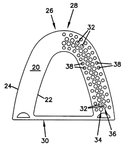
FIG. 1 is a top plan view of an embodiment of the present invention.
FIG. 2 is a bottom plan view of an embodiment of the present
invention.
FIG. 3 is a section view of an embodiment of the present invention.
FIG. 4 is a rear elevation view of an embodiment of the present
invention.
FIG. 5 is a top plan view of an embodiment of the present invention.
FIG. 6 is a rear elevation view of an embodiment of the present
invention.
FIG. 7 is a side elevation view of an embodiment of the present
invention.
FIG. 8 is a top plan view of an embodiment of the present invention.
FIG. 9 is a bottom plan view of an embodiment of the present
invention.
Description of the Preferred Embodiments
FIG. 1 depicts a full arch embodiment of one aspect of the present
invention. In this embodiment, the dental model support surface 20 has an
interior
edge 22 and an exterior edge 24. The interior edge 22 defined an unobstructed
lingual area. The dental model support surface 20 is the portion of the dental
model
base 26 adapted to support a dental model. The dental model base 26 has a
first end
28 and a second end 30.
A plurality of apertures 32 are formed in the dental model support
surface 20. In this embodiment, the apertures 32 form an interior row 34 and
an
exterior row 36. The apertures of the exterior row 36 are off set from and
adjacent
the apertures of the interior row 34. The interior row 34 and the exterior row
36 are
positioned to track the normal tooth line of a patient. A plurality of
indexing studs
38 are in rows adjacent the interior edge 22 and exterior edge 24.
As shown in FIG. 2, a receiver 40 is at the dental model base first end
28. A pair of hemispheric sockets 42 are at the dental model base second end
30. A
bar 44 is interposed between the sockets 42 at the second end 30 of the dental
model
base 26. In one embodiment, the dental model base 26 is made of a
polycarbonate
such as Lexan.
FIG. 3 is a cross-section of the dental model base 26. An exterior
wall 46 extends from the dental model support surface 20. An interior wall 48
also
extends from the dental model support surface 20. A cavity 50 is defined by
the
4
CA 02435766 2003-07-25
WO 02/060334 PCT/US02/02488
interior wall 48, the exterior wall 46 and the dental model support surface
20. A
projection 52 extends from the dental model support surface 20 into the cavity
50.
Apertures 32 extend through the projection 52 from the dental model support
surface
20. The apertures 32 have an interior wall 54. In one embodiment, the majority
of
the aperture interior wall 54 tapers at a two degree angle relative to the
centerline of
the aperture 32. In this embodiment, the 0.05 inches of the aperture remote
from the
dental model support surface 20 is not tapered.
The apertures 32 are adapted to slidingly receive pins 56. Pins 56
have a tapered end 58 and a knurled end 60. In one embodiment, pins 56 are
tapered
at a two degree angle relative to the centerline of the pin. In one
embodiment, the
pins 50 are made of stainless steel and have at least a twenty micron finish.
The
twenty micron finish promotes the sliding engagement of the pin 56 with the
aperture 32 and reduces the likelihood a pin 56 will stick to the aperture 32.
In one
embodiment, the radial dimensional tolerances for the pins 56 are held within
0.005
inches. In one embodiment, the tapered portion of the pins 60 extends beyond
the
projection 52 into the cavity 50.
FIG. 4 is a view of the dental model base second end 30. The bar 44
has a slot 62 extending along the bar 44 between the hemispheric sockets 42.
In one
embodiment, the slot 62 extends through the bar. In another embodiment, the
slot
62 only extends into the bar 44. In one embodiment, the bar 44 is formed with
the
dental model base 26. In another embodiment, the bar 44 slidingly engages the
dental model base 26 and may be glued to the base 26. In one embodiment, the
slot
62 is adapted to receive an articulator attachment tongue. In another
embodiment,
the hemispheric sockets 42 are adapted to receive a ball connected to an
articulator.
FIG. 5 is a view of one embodiment of the present invention adapted
for use with a quadrant dental model. FIG. 6 is view of the second end 30a of
the
quadrant dental model base 26a. One hemispheric socket 42a is located at the
quadrant dental model second end 30a. In one embodiment, the socket 42a is
adapted to receive an articulator ball. A slot 64 is formed across the
hemispheric
socket 42a. The slot 64 is adapted to receive an articulator attachment
tongue.
FIG. 7 is a side view of the quadrant base 26a. The slot 64 is
depicted at the second end 30a of the quadrant base 26a. The receiver 40a is
at the
first end 28a of the quadrant base.
FIG. 8 is a view of one embodiment of the present invention having a
single row of apertures 32c in the dental model support surface 20b. The
apertures
32b are placed to correspond to the normal position of a patient's teeth. As
shown
in FIG. 9, a plurality of projections 52b extend into the cavity 50b formed by
the
exterior wall 46b, the interior wall 48b and the dental model support surface
20b.
5
CA 02435766 2003-07-25
WO 02/060334 PCT/US02/02488
Tapered apertures 32b extend through the dental model support surface 20b and
the
projections 52b. The tapered apertures 32b are adapted to slidingly receive a
tapered
pin as described above.
The dental model base described above may be connected to an
articulator through an articulator plate or through the slot at the base's
second end as
described in co-pending application No. 09/770,322 entitled Encased Stone
Dental
Model Base Body.
The foregoing describes various embodiments of the claimed
invention. The claimed inventions are not limited to the embodiments described
above. Numerous alternative constructions exist that fall within the following
claims.
6
