Note: Descriptions are shown in the official language in which they were submitted.
CA 02436235 2003-06-04
WO 02/46354 PCT/USO1/43880
AUTOMATED IMAGING AND HARVESTING OF COLONIES
ON THIN FILM CULTURE DEVICES
BACKGROUND
This invention relates , to a method for imaging and harvesting cells from a
microbial colony on a thin film culture device.
Many recombinant and molecular cloning techniques rely on the ability to
culture
bacteria on an agar plate and to select particular colonies from the agar for
further study.
Each colony is typically selected manually with a sterile toothpick, which can
be quite
laborious. In addition, there can be uncertainty when a researcher attempts to
relocate the
same colony from the original agar plate.
Accordingly, automated systems have been used to identify and mark a colony
growing on a culture device. For example, automated colony picking systems,
such as a
BIO-PICK automated colony picking system sold by Biorobotics, Inc., Cambridge,
U.K.,
have been developed to increase the speed with which recombinant E. eoli
colonies can be
processed for genetic research. Typically, these systems include an imaging
component,
such as a CCD camera, and a robotic arm that positions a "pin" over each
colony and
mechanically "picks" a portion of the colony material from agar culture
plates. The
colony material from the agar plates is then transferred to culture medium or
reagents for
growth of the cells or for amplification or analysis of the genetic material
within the
transferred material.
SUMMARY
The invention features thin film culture devices with positioning structures
and
methods for harvesting cells from colonies present on such culture devices.
Images of the
culture devices are obtained and positions of colonies growing or present on
such culture
devices are identified relative to the positioning structures to allow cells
to be harvested
from colonies based on the identified positions of the colonies. The
positioning structures
are useful for realigning the culture device such that cells from colonies on
the culture
device can be harvested at any time.
In one embodiment, the invention is a culture device for the propagation or
storage
of microorganisms. The device includes a self supporting, waterproof substrate
and a
cover sheet (e.g., a transparent cover sheet), wherein a gelling agent is
contained on the
-1-
CA 02436235 2003-06-04
WO 02/46354 PCT/USO1/43880
self supporting substrate, and wherein the self supporting substrate and the
cover sheet
include positioning structures, e.g., holes, slits, slots, beveled edges,
notches, or raised
structures. The culture device may further include a barcode label on a
surface of the
culture device. The self-supporting substrate may further include a spacer
and/or a growth
medium (e.g., containing one or more nutrients). The culture device may
further include
an indicator and a corresponding inducer. The cover sheet may further include
a gelling
agent and/or a reinforcement layer, such as a foam, a film, or a non-woven
material.
In another embodiment, the invention is a culture device for the propagation
or
storage of microorganisms that includes first and second layers that are
separable from
each other. The first and second layers may include a gelling agent such as
guar gum,
xanthan gum, locus bean gum, polyvinyl alcohol, carboxymethylcellulose,
alginate,
polyvinylpyrrolidone, gellan, or low monomer content polyacrylic acid. The
first and
second layers also include positioning structures such as holes, beveled
edges, slits, slots,
notches, or raised structures. The first or second layers may include a
spacer. The first
layer may further include a growth medium. The growth medium may include a
detergent
or a salt. The first layer may also include a selectable agent. The first or
second layer may
further include a reinforcement layer.
In another embodiment, the invention is a system for harvesting cells from a
colony on a thin film culture device having positioning structures. The system
includes a
scanner, a processing unit and a picking apparatus. The scanner obtains and
provides an
image file to the processing unit. The processing unit identifies and selects,
if necessary,
cell colonies on the culture device and provides the position of the colonies
relative to the
positioning structures to the picking apparatus. The picking apparatus
harvests the cells
from the colonies based on the position. The picking apparatus may have an
orienting
unit, wherein the orienting unit has receiving structures adapted to receive
corresponding
positioning structures in the culture device. The orienting unit may further
include a
compliant pad. The picking apparatus can include a liquid handling tip.
In yet another embodiment, the invention is a picking apparatus for harvesting
cells
from a colony on a thin film culture device having positioning structures. The
picking
apparatus includes an orienting unit, wherein the orienting unit positions the
colony
relative to the positioning structures; and a picking arm, wherein the picking
arm is
programmed with the position of a selected colony relative to the positioning
structures
-2-
CA 02436235 2003-06-04
WO 02/46354 PCT/USO1/43880
and is adapted to contact cells of the selected colony based on the position.
The orienting
unit has receiving structures adapted to receive corresponding positioning
structures in the
culture device.
A method for harvesting cells from colonies on a culture device also is
another
embodiment of the invention. The method includes the steps of providing a thin
film
culture device having positioning structures; obtaining an image of the
culture device
including cell colonies on the surface of the device (e.g., by scanning the
culture device);
processing the image to provide positions of cell colonies relative to the
positioning
structures of the device; optionally selecting particular cell colonies; and
then contacting
the cell colonies with a picking apparatus based on the position and for
selection of cell
colonies to harvest the cells. The picking apparatus may be moved in at least
one or at
least two directions from the contact point to harvest the cells. Processing
the image may
include identifying a location of the positioning structures; identifying a
location of one or
more colonies, optionally selecting a specific colony; and calculating a
position of the
selected colony relative to the positioning structures. The position of the
colonies relative
to the positioning structure may include X-Y coordinates.
In another embodiment, the invention is a computer readable medium having
instructions thereon causing a programmable processor to display an image of a
thin film
culture device having positioning structures on a display device;
differentiate positioning
structures from colonies on the culture device; identify locations of the
positioning
structures; identify locations of the colonies and/or selected colonies; and
calculate
positions of the colonies relative to the positioning structures. The computer
readable
medium may be a storage medium for storing instructions or may' be a
transmission
medium for transmitting the instructions.
The invention includes a computer readable medium having an image stored
therein, wherein the image contains image data representative of colonies on a
thin film
culture device having positioning structures and a computer readable medium
having data
stored therein, wherein the data are the coordinates of colonies on a culture
device relative
to positioning structures on the culture device.
Unless otherwise defined, all technical and scientific terms used herein have
the
same meaning as commonly understood by one of ordinary skill in the art to
which this
invention belongs. Although methods and materials similar or equivalent to
those
-3-
CA 02436235 2003-06-04
WO 02/46354 PCT/USO1/43880
described herein can be used to practice the invention, suitable methods and
materials are
described below. In case of conflict, the present specification, including
definitions, will
control. In addition, the materials, methods, and examples are illustrative
only and not
intended to be limiting.
Other features and advantages of the invention will be apparent from the
following
detailed description, and from the claims.
DESCRIPTION OF DRAWINGS
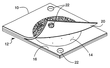
FIG 1 is a schematic of a thin film culture device having positioning
structures.
FIG 2 and FIG 3 are schematics of a thin film culture device having
positioning
structures (FIG 2) and partitioning of a microorganism colony (FIG 3).
FIG 4 is a diagram of a system for harvesting cells from a colony on a culture
device.
FIG 5 is a flow diagram of processing steps for calculating the positions of
colonies relative to each other and to positioning structures on the culture
device.
FIG 6 is a diagram of a picking apparatus.
FIG 7 is a diagram of an orienting unit.
FIG & is a diagram of a culture device being placed on an orienting unit.
FIG 9 is a color digital image of a thin film culture device containing lac
(+) and
lac (-) E. coli colonies.
DETAILED DESCRIPTION
Thin film culture devices of the invention are useful for molecular cloning
techniques, and provide many advantages over traditional culture devices, such
as petri
dishes, which typically contain semisolid nutrient agar medium, and mufti-well
devices
containing nutrient broth medium. One advantage of the thin film culture
devices of the
invention is that they are "sample-ready" and require no preparation before
use. Thin film
culture devices of the invention also are more compact than traditional petri
dishes,
making them highly suitable for imaging microbial colonies contained on these
devices
with an inexpensive, flatbed scanning device.
Incorporation of positioning structures, such as holes, slots, and notches, on
the
thin film devices of the invention allows the devices to be oriented such that
the precise
-4-
CA 02436235 2003-06-04
WO 02/46354 PCT/USO1/43880
position of the microbial colonies within the culture device can be mapped
and,
subsequently, allows cells from the mapped colonies to be picked using an
automated
picking apparatus. In addition, colonies, either similar or different from one
another, from
multiple thin film culture devices can be mapped simultaneously. The
positioning
structures provide a reference position such that at a future time, the
culture devices can be
realigned and cells from the colonies may be harvested based on the~original
map.
Culture Devices
Suitable thin film culture devices can be constructed generally as described
in U.S.
Pat. Nos. 4,565,783; 5,089,413; 5,232,838; and 5,601,998. For example, culture
device
10, which includes a body member having a self supporting, waterproof
substrate 12 may
be used (see FIG 1). Substrate 12 is preferably a relatively stiff material
made of a
waterproof or water impermeable material (i.e., does not absorb water) such as
polyester,
polypropylene, or polystyrene. Other suitable waterproof materials include
substrates such
as paper containing a waterproof polyethylene coating.
The upper surface of substrate 12 may contain a layer of culture medium 14,
which
is dried to provide a dry medium on substrate 12. Alternatively, a layer of
adhesive may
be coated on substrate 12, which serves to hold a culture medium that may be
applied as a
powder. The adhesive should be sufficiently transparent when hydrated to allow
viewing
of microbial colonies, e.g., bacterial colonies, growing on the surface of the
substrate
through the coated substrate. The adhesive should also be coated on the
substrate in a
thickness that allows the substrate to be uniformly coated with dry culture
medium without
completely embedding the medium in the adhesive.
If the culture medium is to be used in a dry form or as a dry powder, the
components, e.g., nutrients, gelling agents, and indicator may be added as a
liquid to the
substrate and then dried. The culture medium may be readily dried by heating
liquid
medium in an oven at about 104°C until essentially all of the water in
the liquid has
evaporated. If the medium is heated after the water has evaporated, however,
the medium
begins to degrade.
A spacer 16 having a circular opening in the center may be adhered to the
medium
coated surface of substrate 12. The portion of spacer 16 that covers the
periphery of
substrate 12 defines the area that is to be inoculated with a sample and
serves to prevent
-5-
CA 02436235 2003-06-04
WO 02/46354 PCT/USO1/43880
the sample from leaking from the substrate. Spacer 16 may be any non-absorbent
material
such as plastic, including foamed plastic (i.e., a foam) or a non-absorbent
non-woven
material. Alternatively, a device may not include spacer 16. In this device,
the amount of
sample is contained on the substrate by the components of the medium alone.
Cover sheet 20 may be attached to one edge of an upper surface of spacer 16.
Cover sheet 20 is preferably made of a transparent film or sheet material in
order to
facilitate visualizing of microbial colonies present on the substrate. In
addition, cover
sheet 20 is preferably impermeable to bacteria and water vapor in order to
avoid the risk of
contamination and deterioration of the components A preferred material for use
as a cover
sheet 20 is biaxially oriented polypropylene. The cover sheet is typically
coated with a
gelling agent such as a gum and, in some embodiments, a second indicator.
Cover sheet
may include a reinforcement layer, such as a non-woven material, foam (e.g., a
polystyrene foam), or film (e.g., a polycarbonate film), for additional
support.
Self supporting substrate 12 and cover sheet 20 each contain positioning
structures
15 22, which allow the culture device to be oriented. Positioning structures
22 may be holes
(as pictured in FIG 1), slits, slots, beveled edges, protrusions or other
raised structures,
notches, or any other structure that may be used to orient the culture device.
Typically,
two or more positioning structures are contained on the thin film culture
device. It should
be noted, however, that a thin film culture device may contain a single
positioning
20 structure if, during the harvesting step, the single positioning structure
may be used in
combination with the overall configuration of the thin film culture device to
be oriented.
In addition, combinations of positioning structures may be used, e.g., a hole
and a notch.
A barcode label may also be on a surface of the culture device to aid sample
tracking and
identification of the culture device.
In use, a predetermined amount of inoculum, typically about 1 to 5 ml (e.g.,
2-3 ml) of inoculum, is added to the device illustrated in FIG 1 by pulling
back cover
sheet 20 and adding the inoculum (e.g., an aqueous microbial suspension) to
the middle of
culture medium 14. Cover sheet 20 is then replaced over substrate 12 and the
inoculum is
evenly spread on the substrate. A convenient tool to do this is a weighted
circular
template, which also is used to confine the inoculum to a specific area of
substrate 12. As
the inoculum contacts and is spread on substrate 12, the culture medium on
substrate 12
hydrates to form a growth-supporting nutrient gel. The inoculated device is
then
-6-
CA 02436235 2003-06-04
WO 02/46354 PCT/USO1/43880
incubated for a predetermined time after which the number of microbial
colonies growing
on the substrate may be visualized, and, optionally, counted through the
transparent cover
sheet 20. Alternatively, a gelling agent is contained on substrate 12 in place
of culture
medium 14. In such an embodiment, culture medium is added before inoculation
or
during the inoculation step.
Another suitable culture device that contains multiple layers is shown in FIG
2. As
used herein, the term "layer" includes a solid substrate and any adhesives,
indicators,
inducers, nutrients, gelling agents, or other reagents coating the solid
substrate. The
devices may be constructed generally as described above. The device 40
includes a first
layer made from a self supporting solid substrate, such as water impermeable
substrate 42.
Bottom substrate 42 typically is a relatively stiff material made of a water
impermeable
material that does not absorb water, such as polyester, polypropylene,
polystyrene, or
glass. Polyester material is a particularly useful substrate. Other suitable
waterproof
materials include water permeable substrates such as paper containing a water
impermeable polyethylene coating such as "Schoeller Type MIL" photoprint paper
(Schoeller, Inc., Pulaski, N.Y.). In general, devices of the invention are
constructed using
substrates that are transparent or translucent to allow colonies to be viewed.
In
embodiments where viewing of the colonies is not necessary, opaque substrates
may be
used. Thickness of the substrate can range from about 0.08 mm to 0.5 mm. For
example,
polyester films typically are about 0.10 to about 0.18 mm thick, polypropylene
films are
about 0.10 to about 0.20 mm thick, and polystyrene films are about 0.38 mm
thick.
The upper surface of substrate 42 may be coated with growth medium 44, which
is
then dried to provide a dry medium on substrate 42. Alternatively, adhesive
may be
coated on substrate 42, which serves to hold a growth medium that may be
applied as a
powder. The adhesive should be sufficiently transparent when hydrated to allow
visualization of microbial colonies growing on the surface of the substrate
when viewed
through the coated substrate. The adhesive should also be coated on the
substrate at a
thickness that allows the substrate to be uniformly coated with dry growth
medium
without completely embedding the medium in the adhesive.
A spacer 46 having a circular opening may be attached to the medium coated
surface of substrate 42. Spacer 46 covers the periphery of substrate 42, and
defines an
area that is to be inoculated with a sample and also serves to prevent the
sample from
CA 02436235 2003-06-04
WO 02/46354 PCT/USO1/43880
leaking from the substrate. Spacer 46 may be any non-absorbent material such
as plastic,
including foamed plastic (i.e., a foam) or a non-absorbent non-woven material.
The
diameter of the circular opening may be altered. For example, a polystyrene
foam web
may have 5 cm to 6 cm diameter die-cut circular holes and be used with the
same volume
of sample (approximately 1 ml). It should be noted that for larger surfaces,
spacer 46 may
have multiple circular openings such that multiple plating surfaces are formed
on
substrate 42. For example, substrate 42 may be the size of a sheet of paper
(e.g.,
21.6 cm x 27.94 cm), or any other size that is convenient for scanning, and
spacer 46 may
have multiple openings in it to allow, e.g., multiple platings from the same
transformation
or platings of different dilutions of the same transformation. In an alternate
embodiment,
a device may not include a sample-containing spacer. In this device, the
amount of sample
is contained and sequestered on the substrate by the components of the medium
alone.
Top cover sheet 50 is disposed on one edge of an upper surface of spacer 46.
Cover
sheet 50 is the second layer and is preferably made of a transparent film or
sheet material
in order to facilitate visualizing, and optionally, counting of microbial
colonies present on
the substrate. In addition, cover sheet 50 is preferably impermeable to
bacteria and water
vapor in order to avoid the risk of contamination and deterioration of the
components.
Materials for cover sheet 50 may be selected to provide the amount of oxygen
transmission necessary for the type of microorganism to be grown. For example,
polyester
films have a low-oxygen permeability and are suitable for growing anaerobic
bacteria,
while polyethylene films have a high-oxygen permeability and are suitable for
growing
aerobic bacteria. A preferred material for use as cover sheet 50 is biaxially
oriented
polypropylene. The cover sheet includes gelling agents, and optionally may
include
microbial growth medium, inducers, indicators, and/or an adhesive. In
addition, the cover
sheet can include a reinforcement layer, such as a non-woven material, foam
(e.g.,
polystyrene foam), or film (e.g, a polycarbonate film), for additional
support.
It should be noted that the top-bottom orientation of the first and second
layers can
be reversed from that described above.
The first and second layers of the device may be removably or permanently
attached to each other by various methods. For example, hinges, clasps, glue,
tape,
staples, or clamps may be used to attach the first and second layers to each
other. In one
-g_
CA 02436235 2003-06-04
WO 02/46354 PCT/USO1/43880
embodiment, a pressure-sensitive adhesive is used to attach the first and
second layers to
each other.
The first and second layers of the culture device each contain positioning
structures
52, which allow the culture device to be oriented. Positioning structures 52
may be holes
(as pictured in FIG. 2), slits, slots, beveled edges, protrusions or any other
raised structure,
notches, or any other structure that may be used to orient the culture device.
Typically,
two or more positioning structures are contained on the thin film culture
device. It should
be noted, however, that a thin film culture device may contain a single
positioning
structure if, during the harvesting step, the single positioning structure may
be used in
combination with the overall configuration of the thin film culture device to
be oriented.
In addition, combinations of positioning structures may be used, e.g., a hole
and a notch.
A barcode label may also be on a surface of the culture device to aid sample
tracking and
identification of the culture device.
In general, the first and second layers of the device include a gelling agent
in an
effective amount, i.e., such that, upon separating the layers, portions of
most, and
preferably, of at least 80% of the visible microorganism colonies are retained
on both
layers of the device. In other words, at least 80% of the visible
microorganism colonies
partition to form replicates on the first and second layers after separating
the layers. See,
FIG. 3 for a diagram of the partitioning of the colony to form replicates. For
example, at
least 85%, 90%, 95%, or 99% of the colonies can partition and form replicates
on the first
and second layers. Non-limiting examples of gelling agents include guar,
xanthan, locust
bean gum, polyvinyl alcohol, carboxymethylcellulose, alginate,
polyvinylpyrrolidone,
gellan, and polyacrylic acid (low monomer content). Guar is a particularly
useful gelling
agent. Suitable concentrations for a gelling agent may be determined by using
the
methods described herein. In general, a device is produced with varying
amounts of the
gelling agent on the first and second layers. The device is inoculated with an
aqueous
sample containing microorganisms (e.g., 1 to 5 mls) and incubated for an
appropriate
length of time (e.g., 16-24 hours). The layers of the device are separated,
and the fraction
of colonies that are retained on both the first and second layers is
determined.
The first layer further may include a growth medium. In some embodiments, the
growth medium may be on both the first and second layers. Typically, a gelling
agent and
growth medium are applied together to the substrate included in the first
layer. A suitable
-9-
CA 02436235 2003-06-04
WO 02/46354 PCT/USO1/43880
growth medium typically contains gelling agent at a concentration of less than
1 % weight/volume of solution before dehydration. For example, the gelling
agent
concentration before dehydration can be 0.4% to 0.9% weight/volume or 0.6% to
0.8% weight/volume. Final amounts of gelling agent in the first layer range
from 20 mg to
100 mg/24 in2 after drying. For example, the final amount of gelling agent may
be 30 to
80 or 40 to 50 mg/24 in2 in the first layer. The amount of gelling agent in
the second layer
typically is at least five times (5X) greater or more than five times (e.g.,
7X, 8X, 9X, or
10X) than the amount in the first layer. For example, the amount of gelling
agent in the
second layer may range from 300 to 500 mg/24 in2 or 400 to 450 mg/24 in2.
Alternatively,
the growth medium may be applied before or during inoculation.
Nutrients in the growth medium may vary depending on the microorganism to be
cultured. See, the Handbook of Microbiological Media (2°d Ed., by
Atlas, L.C. Parks (ed),
1996, CRC Press, Boca Raton, FL) for a description of growth media for culture
of
bacteria, yeast, and fungi. A growth medium may include a detergent (e.g., an
ionic
detergent) at a concentration from about 0.5% to about 2% weight/volume of
solution
before dehydration. Non-limiting examples of detergents include deoxycholate,
bile salts
and sodium lauryl sulfate.
Additional components of the growth medium can include salts, such as calcium
chloride and magnesium chloride, selectable agents, indicators, and inducers.
For
example, selectable agents may be antibiotics such as such as kanamycin,
ampicillin,
carbenicillin, spectinomycin, streptomycin, vancomycin, tetracycline, or
chloramphenicol.
Other selectable agents may be deficiencies in particular amino acids.
Indicators may be
_ precipitable, chromogenic, or fluorescent and/or fluorogenic. Suitable
fluorescent or
fluorogenic indicators include, for example, 4-methylumbelliferyl phosphate
(disodium
. salt trihydrate or free acid), 4-methylumbelliferyl-beta-D-glucopyranoside,
4-methylumbelliferyl-beta-D glucuronic acid, 4-methylumbelliferyl-beta-D-
galactopyranoside, fluoroscein diacetate, or fluoroscein antibody conjugates.
A
precipitable indicator may be, for example, 2,3,5-triphenyltetrazolium
chloride.
Chromogenic indicators typically are colorless until activation by the
microorganism, e.g.,
enzymatic hydrolysis or reduction of a chemical bond. Non-limiting examples of
chromogenic indicators include 5-bromo-4-chloro-3-indoxyl-(3-D-glucuronic
acid,
L-Alanine-5-bromo-4-chloro-3-indoxyl ester (trifluoroacetate salt), 5-bromo-4-
chloro-3-
-10-
CA 02436235 2003-06-04
WO 02/46354 PCT/USO1/43880
indoxyl-1-acetate, S-bromo-4-chloro-3-indoxyl-3-acetate, 5-bromo-4-chloro-3-
indoxyl-N-
acetyl-(3-D-galactosaminide, 5-bromo-4-chloro-3-indoxyl-N-acetyl-(3-D-
glucosaminide,
5-bromo-4-chloro-3-indoxyl butyrate, 5-bromo-4-chloro-3-indoxyl caprylate, 5-
bromo-4-
chloro-3-indoxyl-[3-D-cellobioside, 5-bromo-4-chloro-3-indoxyl- a,-L-
fucopyranoside,
5-bromo-4-chloro-3-indoxyl-(3-D-fucopyranoside, 5-bromo-4-chloro-3-indoxyl-(3-
L-
fucopyranoside, 5-bromo-4-chloro-3-indoxyl-oc -D-galactopyranoside, 5-bromo-4-
chloro-
3-indoxyl-[3-D-galactopyranoside, 5-bromo-4-chloro-3-indoxyl-oc-D-
glucopyranoside,
5-bromo-4-chloro-3-indoxyl-~3-D-glucopyranoside, 5-bromo-4-chloro-3-indoxyl-(3-
D-
glucuronic acid (cyclohexylammonium salt), 5-bromo-4-chloro-3-indoxyl-(3-D-
glucuronic
acid (sodium salt), 5-bromo-4-chloro-3-indoxyl myo-inositol-1-phosphate
(ammonium
salt), 5-bromo-4-chloro-3-indoxyl-a-D-maltotriose, 5-bromo-4-chloro-3-indoxyl
myristate, 5-bromo-4-chloro-3-indoxyl-a-D-mannopyranoside, 5-bromo-4-chloro-3-
indoxyl-nonanoate, 5-bromo-4-chloro-3-indoxyl oleate, 5-bromo-4-chloro-3-
indoxyl
palmitate, 5-bromo-4-chloro-3-indoxyl phosphate (di{2-amino-2-methyl-1,3-
propanediol}salt), 5-bromo-4-chloro-3-indoxyl phosphate (dilithium salt
hydrate),
5-bromo-4-chloro-3-indoxyl phosphate (dipotassium salt), 5-bromo-4-chloro-3-
indoxyl
phosphate (disodium salt sesquihydrate), 5-bromo-4-chloro-3-indoxyl phosphate
(potassium salt), 5-bromo-4-chloro-3-indoxyl phosphate (p-toluidine salt), 5-
bromo-4-
chloro-3-indoxyl sulfate (potassium salt), 5-bromo-4-chloro-3-indoxyl sulfate
(p-toluidine
salt), 5-bromo-4-chloro-3-indoxyl thymidine-3'-phosphate (cyclohexylammonium
salt), or
5-bromo-4-chloro-3-indoxyl-~3-D-xylopyranoside. Sodium tellurite also is a
suitable
indicator.
Inducers stimulate an enzyme to cleave a corresponding indicator. Fox example,
1-O-methylglucuronic acid is an inducer that stimulates glucoronidase to
cleave 5-bromo-
4-chloro-3-indoxyl-[3-D-glucuronic acid (indicator) to produce a colored
product. Other
inducer and indicator pairs include 5-bromo-4-chloro-3-indoxyl-(3-D-glucuronic
acid,
sodium salt or 3-indoxyl-(3-D-glucuronic acid, sodium salt and isopropyl-(3-D-
thioglucuronic acid, sodium salt; 5-bromo-4-chloro-3-indoxyl-(3-D-
galactopyranoside or
indoxyl-(3-D-galactopyranoside and isopropyl-(3-D-thiogalactopyranoside; and 5-
bromo-4-
chloro-3-indoxyl-(3-D-glucopyranoside, 3-indoxyl-~i-D-glucopyranoside, or 5-
bromo-6-
chloro-3-indoxyl-(3-D-glucopyranoside and 1-O-Methyl-(3-D-glucopyranoside.
-11-
CA 02436235 2003-06-04
WO 02/46354 PCT/USO1/43880
A further embodiment of one embodiment of a thin film culture device includes
a
lac differentiation mechanism. When a thin film culture device includes two
chromogenic
indicators, the (3-galactosidase deficient colonies activate a first indicator
and the
~3-galactosidase producing colonies activate a second indicator, thereby
producing two
color differentiation. For example, some Esclaerichia coli host-vector systems
use
a (3-galactosidase reporter gene, to denote the presence or absence of foreign
DNA
inserted into a bacterial plasmid vector. When foreign DNA is not in the
vector, the cells
express ~3-galactosidase, which hydrolyzes 5-bromo-4-chloro-3-indoxyl-(3-D-
galactopyranoside (X-gal) to form an insoluble blue precipitate. When foreign
DNA is
inserted into the lacZ gene in the plasmid vector, the cells are unable to
hydrolyze X-gal.
These cells may be readily identified using another reagent, 2,3,5-
triphenyltetrazolium
chloride (TTC), which turns red in the presence of such cells. In sum, lac+
colonies
appear blue and lac colonies appear red.
Method and System for Harvesting Cells
With reference to FIG. 4, culture devices of the invention can be scanned
using
scanner 100 to obtain an image of the culture device, e.g., a TIFF image, JPEG
image,
GIFF image, or bitmap. Minimal requirements for a scanner include 500
dots/inch (dpi)
for resolving microbial colonies on culture devices of the invention.
Commercially
available flatbed scanners such as the Astra 2000 (1200 dpi, UMAX
Technologies, Inc.,
Freemont, CA) are suitable for scanning and provide adequate resolution. As
thin film
culture devices typically are transparent from the top and from below, the
devices may be
scanned from either direction. Furthermore, culture devices of the invention
may be
scanned at varying magnifications and orientations without loss of fidelity as
the
positioning structures have a known geometry.
Processing unit 120 stores the scanned image and processes the image using an
algorithm that provides location of each of the colonies relative to the
positioning
structures on the culture device. Processing unit 120 includes a central
processing unit
(CPU) that forms part of a general purpose computer, such as a PC, Macintosh,
or
workstation and a display device that includes a viewing screen for graphic
output.
Processing unit 120 is capable of storing program code, and contains an input
device for
user input, such as a keyboard or mouse. Processing unit 120 communicates with
input
-12-
CA 02436235 2003-06-04
WO 02/46354 PCT/USO1/43880
devices, a display device, and in some embodiments, a printer, via one or more
input/output controllers. Processing unit 120 also is in communication with
picking
apparatus 200 (see FIG. 6) such that a stored file in processing unit 120 is
accessible to
picking apparatus 200. In addition, processing unit 120 can be communicatively
linked to
one or more processing units by a network such that a user can remotely access
the raw or
processed image. For example, if the raw or processed image is remotely
accessible,
scanner 100 and processing unit 120 can be in one location, while picking
apparatus 200 is
at a different location.
As indicated in FIG. 5, processing of the image includes differentiating
positioning
structures and microbial colonies based on a pre-determined threshold,
identifying location
of positioning structures (by, for example, size discrimination), identifying
location of
microbial colonies (by, for example, size discrimination), optionally
selecting specific
microbial colonies and calculating position of microbial colonies relative to
the
positioning structures (e.g., providing X-Y coordinates of colonies relative
to positioning
structures).
Component Works 1MAQ Vision Software from National Instruments (Austin,
TX) may be used for the processing. This software package processes the image
by first
creating a histogram by converting color or black and white images into a gray
scale pixel
map. Positioning structures and microbial colonies are distinguished by
segmenting the
pixels into a binary map, based on set levels. Positioning structures and
microbial
colonies each are identified by grouping pixels into local objects,
calculating area of each
object, and calculating center of mass coordinates (i.e., X,Y).
Parameters can be set such that colonies of a certain size, e.g., 0.5 to 1.0
mm in
diameter, or of a certain color are chosen. Other selection options are also
available. One
available option uses color differentiation. Using a color imaging system,
rather than a
black/white imaging system, a RGB (red-green-blue) histogram of each colony
may be
used to distinguish the color intensity, such as blue vs. xed, for each colony
on the plate.
Another available option uses filter differentiation. Using one or two filters
enables a
blacklwhite imaging system to distinguish different colored colonies, such as
red from
blue colonies. For example, with a blue filter, only red colonies would be
substantially
"visible" to the black/white imaging system. With a red filter, only blue
colonies would be
"visible". Imaging a culture device with both filters (sequentially) and using
a system to
-13-
CA 02436235 2003-06-04
WO 02/46354 PCT/USO1/43880
maintain registration of the camera with the device also may account for
coincidental or
overlapping red and blue colonies, thus allowing identification of only pure
colonies.
Still another available option uses size differentiation. Appropriate use of
control
culture devices may facilitate selection of specific colonies on a culture
device containing
differentiable microorganisms when the selection is based on the size of such
colonies.
For example, when cells transformed with a plasmid containing a desired DNA
insert are
to be selected by the size of the colonies, two controls may be used. A first
control culture
device includes cells transformed with a plasmid that does not have a DNA
insert and a
second control culture device includes cells transformed with a plasmid that
has a DNA
insert. Unknown, first control and second control culture devices are all
incubated at the
same temperature (e.g., 37°C) for the same length of time and then all
three devices are
imaged. The average colony size (and standard deviation) of each control
culture device
strain is determined and a suitable statistical test (e.g., a T-test) is
applied to ascertain
whether any observed difference in colony sizes between the two control
devices are
statistically significant. Ideally, the difference in size between "small" and
"large"
colonies would be greater than the standard deviations of both groups of
colony sizes. A
threshold value, either an upper or a lower threshold, based on colony sizes
of the two
control sizes is then used to select desired colonies from the unknown culture
device. The
unique combination of chromogenic, precipitable indicators in a thin film
culture device
such as a CLONdisc plates (Clontech Laboratories,' Inc., Palo Alto CA) affords
a
technique of distinguishing colony lac phenotypes using colony size. FIG. 9
illustrates a
plate containing the indictor system used in the CLONdisc plates. Both lac+
and lac
derivatives of an E. coli strain were inoculated and grown overnight. The
figure illustrates
the significant differences in the sizes of the red colonies (lac ) and the
blue colonies
(lac+). In some cases, an indicator combination of one indicator that remains
essentially
physically associated with the bacteria after changing color (e.g., TTC) and
one indicator
that results in an accumulation of intracellular and extracellular color
formation (e.g.,
X-gal) results in a measurable differentiation of colony sizes.
Coordinates of the colonies are stored in an appropriate file for the picking
apparatus such that cells from colonies on the culture device can be
harvested. Custom
Visual Basic (VB) software can be used to coordinate processing of the image
with the
-14-
CA 02436235 2003-06-04
WO 02/46354 PCT/USO1/43880
picking apparatus. VB utilizes dynamic linked libraries (DLLs) and ActiveX
controls
from the IMAQ vision package.
The commercially available Biomek 2000 fluidic workstation from Beckman
Instruments is an example of a suitable picking apparatus. In the case of the
Biomek
2000, coordinates of the colonies are stored in a tool command language (TCL)
file. With
reference to FIG. 6, picking apparatus 200 contains a processing unit, picking
arm 210,
liquid handling tip 212, orienting unit 220, and base 250 for receiving the
orienting unit
(illustrated in FIG. 7). Orienting unit 220 contains receiving structures 230
and,
optionally, may include compliant pad 240 (see FIG. 7). Receiving structures
230 receive
corresponding positioning structures of the culture device. As illustrated in
FIG. 8, if
positioning structures 22 or 52 are holes, receiving structures 230 are
circular posts.
Similarly, if positioning structures 22 or 52 are notches or other structures,
receiving
structures 230 are complementary to those positioning structures. Compliant
pad 240 is
composed of a material that can be compressed such that the pipette tip can
push into the
thin film culture device to harvest cells from colonies without damaging the
film.
Non-limiting examples of compliant materials that may be used include rubber
or foam.
Picking apparatus 200 also is adapted such that it can contact a colony on the
culture device to harvest cells by contact and/or aspiration, then transfer
the harvested
cells to a container, such as a 96-well plate or test tube. For example,
picking arm 210 can
be configured with liquid handling tip 212, e.g., a pipette tip, plastic tube,
or glass tube, for
contacting and/or aspirating colonies. Picking arm 210 also can be configured
with a solid
rod, e.g., a plastic probe or toothpick for contacting colonies. In addition,
picking arm 210
can be configured with multiple liquid handling tips and controlled such that
a particular
tip can be selected (e.g., an active tip could be moved such that it protrudes
beyond the
other tips). Preferably, liquid handling tip 212 is disposable and is
discarded after contact
with a colony. For example, the picking apparatus can be programmed such that
it
retrieves a pipette tip from a container of pipette tips, contacts a colony to
harvest cells
from that colony, transfers the cells to a defined location on a 96-well
plate, and disposes
of the pipette tip. Alternatively, the pipette tip can be placed back in the
same container
from which it was retrieved. In other embodiments, liquid handling tip 212 is
cleaned
on-line (e.g., washed in circulating water, alcohol, then vacuum dried before
use) or
-15-
CA 02436235 2003-06-04
WO 02/46354 PCT/USO1/43880
cycled through a recycling station where liquid handling tip 212 is cleaned
without
hindering the picking of colonies.
To increase yield of cells from the colony and to offset any errors in the
calculation
of the colony coordinates, picking arm 210 can be moved in at least one
direction from the
contact point, e.g., two or more directions from the contact point. For
example, picking
arm 210 can moved in a circular or zigzag pattern from the contact point. The
uniform
surface topography of the culture device allows the picking arm to move over
the surface
of the colony, whereas for traditional culture devices (e.g., agar plates),
the surface
topography is more variable, making it less likely that the picking arm
contacts a useful
amount of colony surface.
Computer Readable Media
The invention also features a programmable processor configured to execute
instructions from a computer readable medium, such as a hard-disk, floppy-
disk,
networked storage device or the like. The computer program is arranged such
that when
the program is executed, an image of a culture device of the invention is
displayed on a
display device, positioning structures are differentiated from colonies on the
culture
device, location of the positioning structures is identified, location of the
colonies is
identified, and position of the colonies is calculated relative to the
positioning structures.
In other embodiments, a computer readable medium is featured that has an image
stored
therein, wherein the image represents the colonies on a culture device of the
invention, or
that has data stored therein, wherein the data are the coordinates of colonies
on a culture
device of the invention relative to positioning structures. In addition, the
instructions,
images, or positioning data may be transmitted within a computer readable
medium such
as a global computer network for remote processing according to the invention.
The invention will be further described in the following examples, which do
not
limit the scope of the invention described in the claims.
EXAMPLES
Example 1
Bacterial Cultures:
The strains listed in Table 1 were stored on LB agar plates (Miller
Formulation,
Becton Dickinson Microbiological Systems, Sparks, MD) containing 50 ~.glmL
ampicillin
-16-
CA 02436235 2003-06-04
WO 02/46354 PCT/USO1/43880
(sodium salt, Sigma Chemical Co., St. Louis, MO), 40 p,M isopropyl-~3-D=
galactopyranoside (Sigma Chemical Co., St. Louis, MO), and 8 mg/L 5-bromo-4-
chloro-3-
indoxyl-(3-D-galactopyranoside (Biosynth AG, Staad, Switzerland). E. coli DH5a
was
obtained from Clontech Laboratories, Inc. (Palo Alto, CA). E. coli XL1-Blue
was
obtained from Stratagene, Inc., La Jolla, CA. Plasmid pHB2 is a derivative of
pUCl9 in
which DNA has been inserted into the Multiple Cloning Site of the lac-
complementing
region. Plasmid pGFPuv was obtained from Clontech Laboratories and had a very
low
level of residual (3-galactosidase activity, thus making the colonies appear
lac' on
CLONdisc plates and on agar plates containing X-gal. Colonies from each strain
were
inoculated into 17 x 100 mm sterile snap-cap plastic tubes containing 5-mL of
LB broth
containing 50 ~,g/mL ampicillin. The tubes were capped, placed into a
37°C
environmental shaker, and agitated at 220 rpm.
Table 1
3M Strain NumberE. coli StrainPlasmid Lac Phenotype
GPM-1 DHSoc pUC 19 Positive
GPM-51 DH5 a pHB 2 Negative
GPM-350 XL1-Blue ' pUCl9 Positive
GPM-351 XL1-Blue pGFPuv Negative
Plate Inoculation
Two sterile diluents were prepared: I) 0.85% NaCl (Hardy Diagnostics, Santa
Maria, CA) containing 50 ~,g/mL ampicillin and 40 ~.M isopropyl-(3-D-
galactopyranoside
and II) LB Broth containing 50 ~,g/mL ampicillin and 40 ~,M isopropyl-(3-D-
galactopyranoside. After 7 hours of incubation, the cultures were diluted
serially (10-fold
steps) into each of the diluents listed above. A 5-mL diluting pipettor (3M
Microbiology
Products, St. Paul, MN) was used to prepare the inocula as follows: 1) 2.9 mL
of the
diluent was withdrawn into a sterile pipette tip, 2) 0.1 mL of the diluted
cell suspension
was withdrawn into the same pipette tip, and 3) the entire 3.0 mL mixture was
used to
inoculate CLONdisc plates (lot # 2002 08 PB, Clontech Laboratories, Palo Alto,
CA)
according to the manufacturer's instructions. After inoculation, the plates
were incubated
in stacks up to 8 per stack at 37°C for 24 hours.
-17-
CA 02436235 2003-06-04
WO 02/46354 PCT/USO1/43880
Plate Ima_ging~ and Anal,
The incubated plates were scanned using a ScanJet 6100C flat-bed scanner
(Hewlett-Packard, Palo Alto, CA). The scanned images were analyzed using Adobe
Photoshop software version 5.0 (Adobe Systems, Inc., San Jose, CA). Ten
colonies were
randomly chosen from the images of four different plates containing bacterial
strains
GPM-l, GMP-51, GPM-350, or GPM-351. The images were zoomed to 1600X and the
number of pixels with the darkest color intensity were counted for each
colony. Table 2
shows the number of pixels for each colony type and the average colony size
(in pixels).
Table 2
Number of pixels comprising the darkest-colored areas of random colonies
chosen from
the plates inoculated with bacterial strains GPM-l, GPM-51, GPM-350 or GPM-351
GPM-1 GPM-51 GPM-350 GPM-351
Colony 1 42 9 81 9
Colony 2 42 9 81 16
Colony 3 56 6 64 12
Colony 4 36 25 100 9
Colony 5 49 16 64 16
Colony 6 36 6 81 9
Colony 7 42 9 81 12
Colony 8 42 16 100 16
Colony 9 42 16 56 16
Colony 10 49 25 81 16
Average 43.6 +/- 13.7 +/- 78.9 +/-14.513.1 +/-
6.1 7.1 3.2
On average, the size (area) of the blue lac+ colonies was larger than the size
of the
corresponding red lac colonies of the same host E. coli strain. Furthermore,
the lac+
colonies were larger than then corresponding lac colonies whether the diluent
consisted
of saline or a nutrient solution, such as LB broth.
-18-
CA 02436235 2003-06-04
WO 02/46354 PCT/USO1/43880
Example 2
E. coli strain DHSa cells were made competent using CaCl2 then transformed
with
pUC 19 or pUC 19 derivatives containing inserts of various sizes. After
transformation and
recovery, all cells were mixed and diluted in Butterfield's buffer containing
ampicillin
(50~,g/ml) and 1 ml of the diluent was plated on a thin film culture device
capable of
differentiating recombinants and non-recombinants. The culture device was
constructed
as described in Example 1 of U.S. Patent Application No. 09/541,416, filed
April 3, 2000,
except that the culture device had two 0.32 cm positioning holes in opposite
corners and a
reinforcing foam sheet was adhered to the cover sheet. Plates were incubated
at 37°C for
14 to 18 hours then scanned.
The culture device was placed face down on a Umax 2000 flatbed scanner (Model:
Astra 1200P, 1200 dpi, Freemont, CA) and a bitmap file of the culture device
was
obtained. The bitmap file was processed such that colonies were identified by
color,
intensity level, and minimum/maximum size. Colonies were mapped into picture
units
with respect to the positioning structures. The colony map was resized and
rotated into
coordinates using the known geometric location of the positioning structures.
As the
culture device was designed to be peeled open before picking colonies, the
mirror image
was generated for the robotic workstation to produce transformed colony
coordinates.
Transformed colony coordinates were downloaded into an appropriate instruction
file for a
Biomek robot (TCL file). Beckman Biomek software was initiated from the
program
processing the image, and the Biomek software executed the revised colony
picking
algorithm based on the colony coordinates.
The culture device was positioned on the orienting unit of the workstation
that
contained receiving structures adapted to receive the corresponding
positioning structures
on the culture device. The robotic arm used a P20 pipetting tool and selected
pipette tips
from a pipette holder. A 1 mm zigzag motion was used to increase the yield of
bacteria
picked from the colony and to compensate for any mapping error. Picked
bacteria were
transferred into incubation broth at a unique location in a 96-well plate. The
pipette tip
was returned back to its original location in the pipette holder, and a new
pipette tip was
selected for the next pick.
Each well of the 96-well plate contained 1.2 ml of LB and 50~g/ml ampicillin.
Cultures were grown at 37°C for 16 hours with shaking (200 rpm). Growth
was observed
-19-
CA 02436235 2003-06-04
WO 02/46354 PCT/USO1/43880
in 85 of the wells (88.5%) and plasmid DNA was isolated from the cultures
using the
alkaline Iysis method. Plasmids were cut with EcoRl and electrophoresed
through an
0.7% agarose gel. Ethidium bromide staining of the gel indicated that
different colonies
were picked and plasmids of varying sizes were isolated.
OTHER EMBODIMENTS
It is to be understood that while the invention has been described in
conjunction
with the detailed description thereof, the foregoing description is intended
to illustrate and
not limit the scope of the invention, which is defined by the scope of the
appended claims.
Other aspects, advantages, and modifications are within the scope of the
following claims.
-20-