Note: Descriptions are shown in the official language in which they were submitted.
CA 02437029 2003-07-25
WO 01/54770 PCT/CA00/00081
Title: METHOD AND APPARATUS FOR LOCALIZED LOW
ENERGY PHOTON THERAPY (LEPT)
FIELD OF THE INVENTION
The invention relates to an apparatus for treating disorders of
biological tissues with light of selected optical parameters. The invention
also relates to methods for stimulating healing of disorders of biological
tissue with light having selected optical parameters and to methods of
stimulating healing of lesions using such light.
BACKGROUND OF THE INVENTION
Curing with light was known and used in medicine in ancient
times. Red or ultraviolet light was successfully used in the 19th century
for the treatment of pockmarks and lupus vulgaris by Danish physician,
N.R. Finsen, the father of contemporary phototherapy.
Biological phenomena induced by ultraviolet light have been
intensively investigated in photobiology and photomedicine for several
decades. Ultraviolet light as a phototherapy for some dermatological
diseases (mainly psoriasis) has been used since the early twenties.
However, ultraviolet light is an ionizing radiation, and therefore has a
damaging potential for biomolecules and has to be used in
photomedicine with certain precautions.
Biological and healing phenomena induced by optical
wavelength (visible) and infrared (invisible) light have been intensively
investigated in the last decade. Electromagnetic waves with optical
(visible light) and near infrared (invisible irradiation) wavelengths (~, _
400 - 2,000 nm) provide non-ionizing radiation and have been used in
vivo, in vitro and in clinical studies, as such radiation does not induce
mutagenic or carcinogenic effects.
Lasers, specific light sources which provide narrow-band
monochromatic, coherent, polarized light with wide range of powers and
intensities, have been widely used in medicine. Medical lasers may be
subdivided into three groups according to their power and ability to
CA 02437029 2003-07-25
WO 01/54770 PCT/CA00/00081
-2_
produce heat: hot lasers, which are used in surgery; mid power lasers
which are used in photodynamic therapy for cancer treatment or in
dermatology to treat telangiectasia, port-wine stains, etc.; and low energy
(or low intensity, cold or low level) lasers which deliver several orders of
magnitude less energy to the tissue than surgical lasers. They produce
very little heat in biological tissue or no heat at all.
Low energy lasers have been used in dermatology, traumatology
and somey other areas to enhance healing phenomena in the body
(Mester et al., Lasers Surg. Med. 5:31-39, 1985; Trelles et al., Lasers Surg.
Med. 7:36-45, 1987; Ohshiro T., Laser Therapy: Practical Applications, (Ed.
T. Ohshiro), John Wiley, Chichester, 1991). The most frequently used
terms for this area of physiotherapy are low Energy Laser Therapy (LELT),
Low reactive Level Laser Therapy (LLLT), or Laser Therapy. The first
successes of LELT were demonstrated in the treatment of chronic ulcers
and persistent wounds of different etiology (Mester et al., Lasers Surg.
Med. 5:31-39, 1985).
Anecdotal case studies have suggested that LELT is beneficial for
a number of dermatological and musculoskeletal conditions. However,
LELT has failed to provide good results in well-controlled randomized
double-blind studies designed in accordance with rigorous North-
American standards (Gogia and Marquez, O s t o m y/W o a n d
Management, 38:38-41, 1992; Lundeberg and Malm, Ann. Plant. Surg.,
27:53).
Coherence and polarization are the main features which
differentiate laser light from regular monochromatic light. Many
photoinduced phenomena in cell cultures and biotissue are reported to
be induced by noncoherent, nonpolarized monochromatic light (Karu,
Health Physics, 56:691-704, 1989 and Karu, IEEE J. of Quantum Electronics,
QE23:1703-1717,1987).
Laser beams lose coherence and polarization because of
scattering very quickly after entering tissue and thus deeper tissue layers
CA 02437029 2003-07-25
WO 01/54770 PCT/CA00/00081
-3-
"do not distinguish" laser from non-laser light.
Low energy photon therapy (LEPT), also known as low energy,
low level, low intensity laser therapy, photobiomodulation, is the area of
photomedicine where the ability of monochromatic light to alter cellular
function and enhance healing non-destructively is a basis for the
treatment of dermatological, musculosketal, soft tissue and neurological
conditions.
Low energy photons with wavelengths in the range of 400nm -
2,000 nm have energies much less than ultraviolet photons, and
therefore, low energy photons do not have damaging potential for
biomolecules as ionizing radiation photons have.
The area of LEPT research is controversial and has produced
very variable results, especially in clinical studies. Almost every
mammalian cell may be photosensitive, e.g. could respond to
monochromatic light irradiation by changes in metabolism, reproduction
rate or functional activity. Monochromatic light photons are thought to
be absorbed by some biological molecules, primary photoacceptors,
presumably enzymes, whieh change their biochemical activity. If enough
molecules are affected by photons, this may trigger (accelerate) a complex
cascade of chemical reactions to cause changes in cell metabolism. Light
photons may just be a trigger for cellular metabolism regulation. This
explains why low energies are adequate for these so called
"photobiomodulation") phenomena. However, it is difficult to induce
and observe these phenomena both in vivo and in vitro using the
same optical parameters. Specific optical parameters are required to
induce different photobiomodulation phenomena (Karu, Health Physics,
56:691-704, 1989; Karu, IEEE J. of Quantum Electronics, QE23:1703-1717,
1987). The range of optical parameters where "photobiomodulation"
phenomena are observed may be quite narrow. The specificity and
narrowness of the optical parameters required for "photobiostimulatiori'
in LEPT therapy distinguishes LEPT therapy from the photodestruction
CA 02437029 2003-07-25
WO 01/54770 PCT/CA00/00081
-4-
phenomena induced by hot and mid power lasers (e.g. in surgery and
PDT).
Devices for stimulating biological tissue using low energy light
are disclosed for example in U.S. Patent No. 4,930,504 to Diamantopoulos
et al. and U.S. Patent No. 4,686,986 to Fenyo et al., U.S Patent No
4,535,784 to Rohlicek describes an apparatus for stimulating acupuncture
points using light radiation. U.S Patent No. 4,672,969 to Dew describes a
method and apparatus for closing wounds using a laser tuned to a
wavelength of 1.33 ~.m to produce thermal heating of the tissue to
denature the protein.
To meet the changing requirements for optical parameters for
different experimental and clinical applications, there is a need for an
optical system for "photobiomodulation" having flexible parameters,
adjustable for particular applications. In particular, there is a need for an
apparatus capable of treating a range of biological disorders by reliably
providing light to the affected three dimensional biological tissue, which
light has the optical parameters necessary for inducing the appropriate
photobiomodulation for the particular disorder and tissue to be treated.
There is also a need for a method for reliably providing light having such
parameters to a biological tissue having a disorder in order to effect
healing.
SUMMARY OF THE INVENTION
The present inventors have determined that for each disorder
of biological tissue there is a set of optical parameters which constitute
the optimal protocol for treating the disorder by LEPT. The optimal
protocol depends on a range of factors such as the type of tissue affected,
the disorder, the stage of tissue healing (acute, subacute, tissue
regeneration stage) and the size and three dimensional placement of the
affected area. The optical parameters which make up .the protocol
include optical power, dose, intensity, wavelength, bandwidth, beam
diameter and divergence, frequency and pulse duration. The present
CA 02437029 2003-07-25
WO 01/54770 PCT/CA00/00081
-5-
inventors have also determined that these protocols may be developed,
stored, selected, retrieved from a microprocessor and utilized to provide
optimal LEPT treatment for a range of biological disorders efficiently and
reliably.
The present invention thus provides an apparatus for treating a
disorder of a biological tissue in a mammal by stimulating the biological
tissue with Iight having selected optical parameters. The apparatus
comprises a power source for providing power to a central
microprocessor; a central microprocessor having stored optical parameter
protocols suitable for treating a range of disorders of biological tissue and
means for selecting one or more stored optical parameter protocols for
the disorder to be treated; including at least one wireless optical probe,
having a microprocessor in communication with the central
microprocessor, to receive the selected optical parameter protocol and
having at least one probe containing an optical sources) for generating a
beams) of light having the selected optical parameter protocol and for
directing the beam of light to the biological tissue to be treated; and
communication means for transmitting the optical parameter protocol
from the central microprocessor to the probes, or remotely via telephone
and satellite links to any location around the world or outer space.
In an embodiment, the beam of light having the selected optical
parameter protocol is substantially monochromatic and has a
wavelength of from 400 to 2,000 nm and preferably has a wavelength in
the range of from 500 to 2,000 nm, more preferably from 600 to 1,100 nm.
In particular, embodiments, preferred ranges include from 360 to 440 nrn,
from 630 to 700 nm, from 740-760 nm, or from 800-1,100 nm. The optical
source may be, for example a laser, laser diode, superluminous or light
emitting diode. In an embodiment, the optical source is in pulsed mode
with an operating frequency in a range of from 0 to 200 Hz and 1,000 -
10,000 Hz for short pulses. In a further embodiment, the optical
parameters are optical power, dose and intensity, frequency, modulation
CA 02437029 2003-07-25
WO 01/54770 PCT/CA00/00081
-6-
frequency and phase of stimulation.
The wireless communication means may be acoustic, magnetic
or optical.
In a still further embodiment, the apparatus further comprises
means for monitoring the condition of the mammal and providing
feedback to the central microprocessor to adjust the selected optical
parameter protocol based on the condition of the mammal. The means
for monitoring the condition of the mammal may be for example EEG
(electroencefalography), E MG ( electromyography), E CG
20 (electrocardiography), CL (chemoluminescence) or a respirator, or a
combination thereof.
In a further embodiment the apparatus comprised and utilized
means to modulate treatment optical parameters by endogenous (such as
respiratory, ECG, EEG, etc.) frequencies and to provide on-line feedback
for selection of stimulation phase in respect to any endogenous rhythm
phases.
Another aspect of the invention relates to a method for
stimulating healing of a disorder of a biological tissue in a mammal by
stimulating the biological tissue with light having selected optical
parameters provided by a central microprocessor having stored optical
parameter protocols suitable for treating a range of disorders of biological
tissue; selecting one or more stored optical parameter protocols for the
disorder to be treated; generating a beam of light having the selected
optical parameter protocol and directing the beam of light to the
biological tissue to be treated. "
In an embodiment, the invention provides a method of
stimulating healing of a lesion in a mammal, comprising: irradiating the
lesion with a substantially monochromatic beam of light having
predetermined optical parameters, wherein the predetermined optical
parameters include a dose of from 0.2 to 10 J/cm~, an intensity of from 0.2
to 5,OOOmW/cm2 and a wavelength of from 400 to 2,000 nm.
CA 02437029 2003-07-25
WO 01/54770 PCT/CA00/00081
-7-
In a particular embodiment of the method, the lesion is a
chronic ulcer or wound and the selected optical parameters include a
dose of from 0.2 to 1.0 J/cm2, an intensity of from 0.2 to lOmW/cm~ and a
wavelength of from 600 to 700 nm. In another embodiment of the
method, the lesion is an acute ulcer or wound and the selected optical
parameters include a dose of from 2.0 to 5.0 J/cm2, an intensity of from
10.0 to 30 mW/cm2 and a wavelength of from 600 to 700 nm. In yet
another embodiment, the lesion is an infected wound and the selected
optical parameters include a dose of from 3.0 to 7.0 J/cm2, an intensity of
from 50.0 to 80 mW/crn2 and a wavelength of from 600 to 700 nm.
Other objects, features and advantages of the present invention
will become apparent from the following detailed description. It should
be understood, however, that the detailed description and the specific
examples while indicating preferred embodiments of the invention are
given by way of illustration only, since various changes and
modifications within the spirit and scope of the invention will become
apparent to those skilled in the art from this detailed description.
DESCRIPTION OF THE DRAWINGS
The invention will be better understood with reference to the
drawings in which:
Figure 1 is a schematic flow chart illustrating the development
of LEPT optical parameter protocols;
Figure 2 is a graph showing the percentage of the dose on the
skin surface (D(z)/(1-R)Do)x100 % received by cells at skin depth Z and at
wavelengths ~,=630 nm (wavelength 1) and ~,=1,060 nm (wavelength 2),
(for explanation of parameters Do, D and R see the following detailed
description);
Figure 3 is a graph showing the results of Monte-Carlo
simulation of photon propagation in the skin;
Figure 4 is a schematic view showing major optical pathways in
CA 02437029 2003-07-25
WO 01/54770 PCT/CA00/00081
human skin;
Figure 5 shows a simplified two-dimensional diagram of desired
parameters for low energy photon therapy for a particular condition and
an example of the range produced by a typical given laser;
Figure 6 is a plan view showing how Figure 6-1, Figure 6-2,
Figure 6-3 and Figure 6-4 are assembled to form a complete circuit
diagram of a base unit according to the invention;
Figure 6-1 is the first part of the circuit diagram referred to in
Figure 6;
Figure 6-2 is the second part of the circuit diagram referred to in
Figure 6;
Figure 6-3 is the third part of the circuit diagram referred to in
Figure 6;
Figure 6-4 is the fourth part of the circuit diagram referred to in
Figure 6;
Figure 7 is a plan view showing how Figure 7-1, Figure 7-2 and
Figure 7-3 are assembled to form a complete circuit diagram of a probe
unit according to the invention;
Figure 7-1 is the first part of the circuit diagram referred to in
Figure 7;
Figure 7-2 is the second part of the circuit diagram referred to in
Figure 7;
Figure 7-3 is the third part of the circuit diagram referred to in
Figure 7;
Figure ~ is a diagrammatic' view of a remote probe unit
according to the invention;
Figure 9 is a perspective view of a probe unit according to the
invention;
Figure 10 is a perspective view of the probe unit of Fig. 9 from
the opposite side;
Figure 11 is a diagrammatic view showing various patterns of
CA 02437029 2003-07-25
WO 01/54770 PCT/CA00/00081
-9-
diodes for use with the invention;
Figure 12 is a schematic view showing alternative patterns of
diodes according to the invention;
Figure 13 is a perspective view of a flexible probe unit according
to the invention; and
Figure 14 is a perspective view of a ring probe according to the
invention.
DETAILED DESCRIPTION OF PREFERRED EMBODIMENTS
As hereinbefore mentioned, the present invention provides an
apparatus for treating a disorder of a biological tissue in a mammal by
stimulating the biological tissue with light having selected optical
parameters. The apparatus comprises a power source for providing
power to a central microprocessor; a central microprocessor having
stored optical parameter protocols suitable for treating a range of
disorders of biological tissue and means for selecting one or more stored
optical parameter protocols for the disorder to be treated; at least one
wireless probe, having a microprocessor in communication with the
central microprocessor, to receive the selected optical parameter protocol
and having at least one probe containing an optical source for generating
a beam of light having the selected optical parameter protocol and for
directing the beam of light to the biological tissue to be treated; and
wireless communication means for transmitting the optical parameter
protocol from the central microprocessor to the wireless heads.
A wide range of disorders of biological tissue or their symptoms
may be treated by the apparatus of the invention, including acute and
chronic musculoskeletal conditions, such as arthritis, degenerative disc
and joint diseases, bone spurs, back and joint pain, tendinitis, muscle
pain and stiffness, myofascial pain; post surgical complications, such as
swelling, inflammation, scarring and stiffness; acute trauma and chronic
post-traumatic conditions in the soft tissues and bones, including sprains,
CA 02437029 2003-07-25
WO 01/54770 PCT/CA00/00081
-10-
strains, wounds, whiplash; repetitive strain injuries such as carpal
tunnel syndrome, tennis and golfer's elbow; neurological and
neuromuscular conditions; dermatological conditions such as burns,
acne, herpes simplex, and ulcers, including infected or non-infecfed
chronic ulcers of different etiology such as venous ulcers, diabetic ulcers,
decubitus ulcers, pressure sores, burns and post-traumatic ulcers.
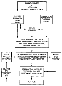
There are many optical parameters, including the type of the
light source, optical power, intensity, dose, frequency and pulse duration,
wavelength and bandwidth, beam diameter and divergence, three-
dimensional light distribution etc. which may be selected to provide an
optimized protocol to treat the disorder. The individual . optical
parameters may be selected based on the disorder to be treated, as
described below. The development of appropriate treatment protocols
was carried out as indicated by the flow chart of Fig. 1.
Optical Power
Optical power may be provided in continuous wave mode or
pulse mode. In continuous wave mode for a single optical source, optical
power P is a total energy of emitted light per second and measured in
Watts (W) or Milliwatts (mW). Th.e total power Pt in a cluster probe is
Pt=nxP (1)
P is the power of a single optical source, and n is the number of optical
sources per probe.
The power of a single optical source or cluster probe has to
correspond to the type of tissue disorder to be treated. For example, for
the treatment of so-called acupuncture points or spinal nerve roots, less
power in a single probe is required compared to the treatment of trigger
points. The total power in a cluster should be physiologically justified
and can vary from application to application. For example, neck and face
areas are more sensitive, in general, to LEPT compared to the rest of the
CA 02437029 2003-07-25
WO 01/54770 PCT/CA00/00081
-11-
body and, therefore, their treatment requires less power in the cluster
probe. There is some maximum total power in a cluster probe for every
particular wavelength up to which patients can respond to LEPT with
comfort. Exceeding certain limits in the total power of a cluster probe
could lead to overdose, excessive stress for the patient and sometimes to
exacerbation of the patient's condition.
Optical sources in a pulsed mode are described by peak power
Pp, average power Pav, pulse frequency F(Hz) and pulse duration i(s),
Average power Pav is less than peak power Pp and can be controlled by
changing the frequency in accordance with the following formula
Pay=PpxFx i (2)
Power by itself is not a decisive factor for LEPT, the power density
(intensity mW/cm~) is more important for "photobiomodulation"
phenomena. Physiologic tissue response first of all depends on light
intensity and dose. Intensity (mW/cm2), and dose (J/cm2) are optical
parameters which skin or biotissue can "feel".
Table 1 below shows suitable power ranges for different tissue
pathologies.
Table 1
Tissue pathology,Wavelength Single optical Cluster
area range, probe.
(points) to be ,(n m~ probe. Range Range of
treated of powers powers
(mW) (mW)
Spinal nerve 800-1,100 1-70 ---
roots
Tender trigger a) 630-700 5-50 ---
points
b) 800-1,000 5-150
CA 02437029 2003-07-25
WO 01/54770 PCT/CA00/00081
-12-
Tissue pathology,Wavelength Single optical Cluster probe.
area range,
(points) to be ~n m~ probe. Range Range of
treated of powers powers
(mW) (rnW)
ITlcer, wound a) 630-700 5-30 30-150
b) 800-1,100 10-50 30-200
Acute post-traumatica) 630-700 --- 30-150
inflammation b) 800-1,100 --- 50-200
in soft
tissue
(all body, except
face)
Chronic inflammation800-1,100 --- 20-100
(flare-up stage)
in soft tissue
(all body except
face)
Chronic inflammation800-1,100 --- 50-200
(no flare-up)
in soft tissue
(all body except
face)
All types of a) 630-700 --- 20-50
inflammation b) 800-960 --- 30-100
in face
soft tissues
Intensif~r (power densii~r)
Intensity is the rate of light energy delivery to 1 cm~ of skin or
biotissue. Intensity is measured in milliwatts per cm2 (mW/cm2). Real
intensity on the skin surface depends on light reflection and scattering
from the skin and underlying tissue layers. The light intensity on the
skin surface can be calculated with the following formula
I = (I - R) x 4 x P/~d2 (3)
where P (or Pav for pulsed mode) is the optical power, d(cm) is the beam
diameter and R is the reflection coefficient. Coefficient R can vary from
CA 02437029 2003-07-25
WO 01/54770 PCT/CA00/00081
-13-
0.4 up to 0.75 for different wavelengths and depends also on the skin type
and condition. For applications using non-contact techniques a portion
of the optical power (and dose) equal to R x P is lost because of the
reflection. Back scattering has to be taken into account for LEPT
dosimetry as well. For contact technique applications, less power is lost
due to the repeating light reflection back to the skin surface from optical
source parts. Therefore, for the same optical source LEPT dosimetry
would be different depending on the type of technique used (contact or
noncontact). Particular "photobiomodulation" phenomenon can best be
activated within narrow ranges of parameters (e.g. see Tables 2, 5, which
appear later in this description). For example, collagen type 1 production
is thought to be affected by LEL in an inverse manner to fibroblast
proliferation: when cell proliferation is increased, collagen type 1
production is decreased and vice versa (van Breugel and Bar, 1992, Laser
Surg. Med. 12:528-537). In cell culture experiments thin cell layers are
usually uniformly exposed to light therefore intensity does not change
significantly within the sample. For biotissue stimulation, the whole
picture is different because light intensity (and dose) decreases with depth
z. In the skin and subcutaneous tissue layers light intensity can be
approximately described by the following formula (Beer's law):
I(z) = Io (I - R)exp( - ccz)
(I- R) x 4 x P x exp(- ocz) (4)
I(z) _
where I(z) (w/cm2 or mw/cmc) - is the fluence rate (intensity or power
density) at the depth z (mm); Io = P/S - incident intensity; P - beam power;
S = ~d2/4 is a beam area for a cylindrical parallel beam of diameter d (cm);
and a (mm 1) is the attenuation coefficient which depends on light
absorption and scattering. This formula may be used to calculate
CA 02437029 2003-07-25
WO 01/54770 PCT/CA00/00081
-14-
intensity and dose for every particular tissue layer.
Suitable intensities for biostimulation are in the range of from
0.1 to 5,000 mW/cm2~ For stimulating healing of chronic ulcers or
wounds intensity may preferably be in the range of from 0.2 to 10
mW/cm2, for ulcers or wounds in acute inflammatory stage a preferred
range is from 10.0 to 30 mW/cm~ and for infected wounds a preferred
range is from 50 to 80 mW/cm~. Table 2 below shows suitable ranges of
intensities for different tissue pathologies.
Table 2
Ranges of Intensities for Different Tissue Pathologies
ProtocolTissue pathology, area Wavelength Intensity
(point) to be range, range
# txeated nm mW/cm~
1 1 2 3
Ulcers or wounds, 630-700 0.2-10
stimulation of repair
processes
2 Ulcers, wounds, acute 630-700 10-30
inflammatory condition
3 Infected wounds 630-700 50-80
4 Area of ulcers or wounds800-1,100 300-600
with
impaired microcirculation,
or to
treat such areas and
also the area
surrounding the ulcer,
wound
5 Post-surgical scar, acute630-700 10-30
inflammatory condition
6 Post-surgical scar, sub-acute800-1,100 10-40
inflammatory condition 60-100
300-600
1,000-5,000
7 ~ Herpes simplex and acne 630-700 ~ 20-60
CA 02437029 2003-07-25
WO 01/54770 PCT/CA00/00081
-15-
ProtocolTissue pathology, area Wavelength Intensity
(point) to be range, range
# treated nm mW/cm2
8 Acute post-traumatic a) 630-700 10-40
inflammation
in soft tissue b) 800-1,100 30-100
9 Post-traumatic condition630-700 20-50
in soft
tissue accompanied by
hematoma,
bruise
Post-traumatic condition,800-1,100 a) 10-40
sub-acute
stage b) 60-100
c) 300-600
d) 1,000-5,000
11 Post-traumatic condition,800-1,100 a) 60-100
regeneration of tissue, b) 300-600
normalization of function c) 1,000-5,000
12 Chronic inflammation a) 630-700 1-5
in soft tissue
(flare-up stage), treatmentb) 880-1,100 1-10
of the
affected area
13 Chronic inflammation 630-700 1-10
in soft tissue
(flare-up stage), treatment800-1,100 10-30
at
selected points' or areas*~' 100-300
on the
body
14 Chronic inflammation a) 630-700 5-30
in soft tissue
(no flare-up) b) 800-1,100 10-40
60-100
300-600
1,000-5,000
Degenerative joint diseases630-700 1-10
(arthritis, rheumatoid 800-1,100 10-30
arthritis,
degenerative disk disease, 60-100
etc.),
treatment of the affected 300-600
area
1,000-5,000
CA 02437029 2003-07-25
WO 01/54770 PCT/CA00/00081
-16-
ProtocolTissue pathology, area Wavelength Intensity
(point) to be range, range
# treated nm my~l/~2
16 Degenerative joint diseases630-700 1-10
(arthritis, rheumatoid 800-1,100 100-300
arthritis,
degenerative disk disease,
etc.),
treatment of selected
points* or
areas)** on the body
17 Muscle spasm relief a) 630-700 1-10
b) 800-1,000 1-40
60-100
300-600
1,000-10,000
18 Localized pain relief 800-1,100 60-100
300-600
1,000-10,000
19 Tender, trigger point 800-1,100 300-600
therapy
1,000-10,000
20 5o called acupuncture a) 630-700 1-10
point
therapy 60-100
300-600
b) 800-1,100 3-15
60-100
300-600
21 Carpal tunnel syndrome 800-1,100 1-10
60-100
300-600
1,000-10,000
22 Neuritis, neuralgia, 800-1,100 1-10
trigeminal
neuralgia 20-40
100-400
800-3,000
CA 02437029 2003-07-25
WO 01/54770 PCT/CA00/00081
-17-
ProtocolTissue pathology, area Wavelength Intensity
(point) to be range, range
# treated nm mW/cm2
23 Post-traumatic, post-surgical630-700 10-40
complications, arthritis800-1,100 300-600
accompanied by swelling, 1,000-5,000
edema,
pain
'~ Selected points on the body may include tender and trigger points,
related acupuncture points, spinal nerve roots, points along related
nerves' pathways.
~'* Selected areas) on the body may include related dermatomes, spine
areas, nerves' pathways.
Beam Diameter and Diver ence
Beam diameter and divergence are important features of single
optical sources. Beam size affects light intensity values on the skin
surface and within the tissue in accordance with formulae (3, 4). Beam
divergence affects light distribution and dosimetry for different tissue
layers. For non-contact techniques light spot size and irradiated area S
on the skin surface depend on the distance to the irradiated surface h as
follows:
S = 4 x (d+2h x TAN a)2 (5)
where d is the beam diameter near the probe tip, 2a is the diverging
angle, 2h x TAN a is the additional beam diameter due to beam
divergence.
Different optical sources (lasers, laser diodes, light emitting
diodes, etc.) have different beam divergences. Lasers usually have small
CA 02437029 2003-07-25
WO 01/54770 PCT/CA00/00081
-18-
beam divergency, laser diodes and LED's have bigger divergences. For
different applications particular beam divergences are more convenient.
For example, for the treatment of wounds and ulcers, almost parallel
beams are less desirable because of the large areas to be treated, and
optical sources with some particular divergence are more convenient.
The beam diameter and divergence should be selected based on
the three dimensional size and shape of the tissue area affected.
Preferably, the beam diameter and divergence should be selected such
that the area receiving LEPT is just slightly larger in size than the area
affected. The appropriate radius of the beam may be calculated by the
following formula
(R+ 1)2/R2
where R (cm) equals the radius of the area affected by the disorder. In the
case of lesions, such as ulcers or other open skin wounds, it is particularly
important that too large an area not be illuminated as, where the
illuminated area is much larger than the lesion, the skin ulcer (wound)
healing rate is not optimized. As the ulcer is treated and healed the area
requiring treatment and the beam diameter will have to be reduced.
Dose
The dose D is the light energy provided to the unit of surface
(lcm~) during a single irradiation and measured in J/cm~ or mJ/cm2.
The light dose received by the skin surface is
D=I x t (6)
where I is the intensity on the skin surface, and t is the exposure time (s).
The dose received by subcutaneous tissue layer at the depth z for a
parallel beam can be calculated by the following formula:
D=I(z) x t (7)
CA 02437029 2003-07-25
WO 01/54770 PCT/CA00/00081
-19-
where I(z) is given by formula (4).
As mentioned above, the dose alone does not ensure particular
photoeffect or healing.phenomenon. Only proper selection of the whole
set of optical parameters including dose will provide the desirable
therapeutic effect. The selection of optical parameters depends on the
medical condition, location of the affected areas, person's age, etc.
Particular examples will be provided below.
The percentage of dose (D(z)/(1-R)Do)x100% on the skin surface
received by cells at a skin depth Z at wavelengths of 630 nm and 1,060 nm
is shown in Table 3 and is illustrated in Fig. 2. All curves D(z) for
different wavelengths in the interval (630-1,060) nm depend on
corresponding reflectance R~ and attenuation a~ coefficients (see Table 4)
and are located between curves 1 and 2 on Fig. 2.
1. D(J/cm~) is the actual dose (fluence) received by cells at depth
z.
2. R=RS + RT - diffuse reflectance
RS - regular reflectance from the skin surface
RT - is the remittance from within the tissue
3. D~ = Io x t - conventionally calculated dose
t = exposure time
4. Io = P/S - incident intensity
(P - beam power; S - beam area)
The actual doses received by different cell types in the skin
exposed to LEPT are illustrated in Table 3. The maximum dose (D ~ (0.5-
1.3) Do is received by keratinocytes and Langerhans cells from the
epidermis. Cells from dermis (fibroblasts, mast cells, blood and nerve
cells are exposed to significantly less dose than the incident one D ~ (0.05-
CA 02437029 2003-07-25
WO 01/54770 PCT/CA00/00081
-20-
0.3) Do. The least dose D ~ 0.3 Dolt is received by blood cells moving
through capillaries (velocity ~ 1 mm/sec).
Table 3
Actual dose received by different cells in
the skin exposed to LEP radiation
Cell type Formulae for actualTypical range
dose calculation of doses
Keratinocytes (0.5-1.8) Do (1-R) (0.5-1.3) Do
Langerhans cells
Fibroblasts (0.05-0.5) Do (1-R)(0.05-0.3) Do
mast cells
nerve cells
not moving blood
cells
moving blood cells (0.05-0.5) Do (1-R)x(2/t)0.3 D~/t
~
where Do (J/cm2) is a conventionally calculated dose
Table 4
Diffuse reflectance R~,, attenuation coefficients oc~
and penetration depths d~ for some wavelenst~---hs
~, (nm) Ra, I- Ra, a~ (mrri da, (~.)
1 )
630 0.6 0.4 1.6 0.6
300-600
b) 800-1,100 3-15
60-100
300-600
CA 02437029 2003-07-25
WO 01/54770 PCT/CA00/00081
-21-
820 0.5 0.5 1.0 1
900 0.45 0.55 0.8 1.25
1,060 ~ 0.5 ~ 0.5 ~ 0.6 1.7
d(mm) - penetration depth
a~d~ = I I (d) = 0.37Io
Suitable doses for photobiomodulation are in the range of from
0.1 to 20 J/cm2, preferably from 0.2 to 5 J/cm~. For stimulating healing of
chronic ulcers or wounds doses may preferably be in the range of from
0.05 to 0.2 J/cm~, for ulcers or wounds in acute inflammatory stage a
preferred range is from 2 to 5 J/cm2 and for infected wounds a preferred
range is from 3.0 to 7.0 J/cm2. See Table 5 below for ranges and doses (in
Joules/cm~) for different tissue pathologies.
Table 5
Ranges of doses for different tissue pathologi, es
ProtocolTissue pathology, area Wavelength Dose range,
(point) to be range,
# treated nm j/~n~.
1 1 2 3
Chronic ulcers or wounds,630-700 0.05 -
0.2
stimulation of healing
2 Ulcers, wounds, acute 630-700 2-5
inflammatory condition
3 Infected wounds 630-700 ~ 3-9
4 Area of ulcers or wounds800-1,100 0.1-9
with
impaired microcirculation,
or to
treat such area and also
the area
surrounding the ulcer,
wound
SUBSTITUTE SHEET (RULE 26)
CA 02437029 2003-07-25
WO 01/54770 PCT/CA00/00081
-22-
ProtocolTissue pathology, area Wavelength Dose range,
(point) to be range,
# treated nm j/~2
Post-surgical scar, acute630-700 2-5
inflammatory condition
6 Post-surgical scar, sub-acute800-1,100 3-7
inflammatory condition 4-25
Herpes simplex 630-700 4-9
and acne
8 Acute post-traumatic a) 630-700 3-9
inflammation
in soft tissue b) 800-1,100 3-10
9 Post-traumatic condition630-700 5-14
in soft
tissue accompanied by
hematoma,
bruise
Post-traumatic condition,800-1,100 3-7
subacute
stage 4-25
11 Post-traumatic condition,800-1,100 3-5
regeneration of tissue, 4-25
normalization of function
12 Chronic inflammation a) 630-700 0.1-0.5
in soft tissue
(flare-up stage), b) 880-1,100 0.1-0.5
treatment of the affected
area
13 Chronic inflammation 630-700 0.1-0.6
in soft tissue
(flare-up stage), treatment800-1,100 1-5
of
selected points' or area(s)'~~'
on the
body
14 Chronic inflammation a) 630-700 2-7
in soft tissue
(no flare-up) b) 80Q-1,100 2-9
3-25
25-100
SUBSTITUTE SHEET (RULE 26)
CA 02437029 2003-07-25
WO 01/54770 PCT/CA00/00081
-23-
ProtocolTissue pathology, area Wavelength Dose range,
(point) to be range,
# treated nm j/~2
15 Degenerative joint diseases630-700 0.1-0.5
(arthritis, rheumatoid 800-1,100 2-9
arthritis,
degenerative disk disease, 3-25
etc.),
treatment of the affected 25-100
area
16 Degenerative joint diseases630-700 0.1-0.5
(arthritis, rheumatoid 800-1,100 1-5
arthritis,
degenerative disk disease,
etc.),
treatment of selected
points"' or
areas)** on the body
17 Muscle spasm relief a) 630-700 0.1-0.3
b) 800-1,100 0.1-0.5
3-5
4-25
25-100
18 Localized pain relief 800-1,100 8-150
19 Tender, trigger point 800-1,100 4-150
therapy
20 So-called acupuncture a) 630-700 0.02-0.2
point
therapy 0.1-1.0
2-4
b) 800-1,100 0.06-0.4
0.1-2.0
2-4
21 Carpal tunnel syndrome 800-1,100 0.05-0.3
0.2-4.0
5-10
25-150
SUBSTITUTE SHEET (RULE 26)
CA 02437029 2003-07-25
WO 01/54770 PCT/CA00/00081
-24-
ProtocolTissue pathology, area Wavelength Dose range,
# (point) to be range, J~~2
treated nm
22 Neuritis, neuralgia, 800-1,100 0.1-0.3
trigeminal
neuralgia 1-3
5-25
25-80
23 Post-traumatic, post-surgical630-700 5-14
complications, arthritis,800-1,100 25-100
accompanied by swelling,
edema,
pain
~' Selected points on the body may include tender and trigger points,
related acupuncture points, spinal nerve roots, points along related
nerves' pathways.
~'~' Selected areas) on the body may include related dermatomes, spine
areas, nerves' pathways.
Frequency and Pulse Duration
Low range frequencies of 0-200Hz may sensitize release of key
neurotransmitters and/or neurohormones (e.g. endorphins, cortisol,
serotonin). These frequencies correspond to some basic electromagnetic
oscillation frequencies in the peripheral and central nervous system
(brain). Qnce released these neurotransmitters and/or neurohormones
can modulate inflammation, pain or other body responses. Analogous
phenomena can be expected with "photobiomodulation" within the
same range of low frequencies. Certainly, the interaction between living
cell and pulsed electromagnetic wave depends on wavelength as well as
pulse duration. Pulse repetition rates within the range 1,000-10,OOOHz
with different pulse durations (milli-, micro- or nanoseconds) can be
SUBSTITUTE SHEET (RULE 26)
CA 02437029 2003-07-25
WO 01/54770 PCT/CA00/00081
-25-
used to change average power. Specific pulse repetition rates to induce
particular healing mechanism are reflected in Table 6 below.
Table 6
Ranges of frequencies for stimulation of particular healing mechanisms
Healing mechanisms Basic frequency Endogenous modulation
(Hz) or
stimulated continuous wave frequency (Hz)
mode
Endorphin release 1-5 ---
Capillar microcirulation9-11 1-1.2 (average frequency
of
improvement heart beating)
50-200 0.2-0.3 (average
frequency
of breathing cycle)
"' Localized muscle 50-120 1-5
spasm
and pain relief or 0.2-0.3
Continuous wave
mode
Lymph flow enhancementcontinuous wave 1-1.2
mode
0.2-0.3
Stimulation of tissuecontinuous wave 1.2
repair mode
or 100 Hz 0.2-0.3
Three Dimensional Light Distribution
Depending on the target tissue for LEPT (e.g. skin, muscle,
ligament) a proper three-dimensional light distribution should be
provided to get the desirable physiologic and therapeutic response. For
single optical sources important parameters affecting light distribution
are beam size, divergence, light wavelength as well as biotissue optical
properties (reflection, absorption, scattering, refraction). Total reflectance
is equal to the sum of the regular reflectance from the skin surface and
the remittance from within the tissue (see Fig. 4).
CA 02437029 2003-07-25
WO 01/54770 PCT/CA00/00081
-26-
For cluster probes, additional contributive parameters are the
distance between diodes and the cluster probe's three-dimensional shape.
All these parameters should be physiologically justified to provide
optimal biotissue response and requirable three-dimensional light
distribution. For example, the distance between diodes can affect
vasoactive blood vessel response and average energy density delivered to
the treated area. For proper vasoactive response a definite distance
between diodes has to be provided depending on particular parameters of
a singular diode (power, beam, diameter, divergence).
The three-dimensional light distribution in tissues such as the
skin and underlying tissue layers may be calculated based on diffusion
approximation and/or the Monte Carlo approach (L. Wang and S.
Jacques, Hybrid model of Monte Carlo Simulation and diffusion theory
for light reflectance by turbid media, J. Opt. Soc. Am. A/Vol. 10, No. 8,
1993, pp 1746-1752; A. Welch et al., Practical Models for Light Distribution
in Laser-Irradiated Tissue, Lasers in Surg. Med. 6: 488-493, 1987). Results
of Monte-Carlo stimulation of photon propagation in the skin with a flat
beam, R=1 cm are shown in Figure 3. Examples of diffuse reflectance R~,,
attenuation coefficients a~, and penetration depths d~, for some
wavelengths are shown in Table 4. A schematic representation of the
major optical pathways in human skin is shown in Figure 4.
Waveleng h
Wavelength ~, (nm) is the basic electromagnetic wave feature
which is directly linked to the energy of an individual light quantum
(photon). The more wavelength the less photon energy. Wavelength is
also linked to the monochromatic light color. Visible monochromatic
light changes its color with wavelength, increasing from violet and blue
(shorter wavelengths) to orange and red (longer wavelengths). Cell
culture experiments have indicated that there is a selectivity in
photoinduced phenomena related to wavelengths. Experiments on
CA 02437029 2003-07-25
WO 01/54770 PCT/CA00/00081
-27-
different cell cultures (microbe and mammalian) have revealed the
ranges of wavelengths (360-440nm, 630-680 nm. 740-760 nm) where
photoinduced phenomena are observed (Karu, Health Physics, 56:691-
704, 1989; Karu, IEEE J. of Quantum Electronics, QE23:1703-1717, 1987).
Photoeffect can be induced by monochromatic light, only in cases, where
a cell contains photoacceptors, substances which are able to absorb
monochromatic light of this particular wavelength. No photoinduced
cell phenomena can be observed if there are no wavelength specific
photoacceptors in a cell.
The following factors have to be taken into account when
considering LEPT dosimetry for monochromatic light of a particular
wavelength ~,. The dose required for "photobiomodulation" strongly
depends on the wavelength. In general, the longer the wavelength the
more dose is required to induce photoeffect. For example, in
experiments on cell cultures, doses required for DNA synthesis
stimulation are 10-100 times less with blue light (~, = 404 nm) than with
red (~, = 680 nm) or near infrared (~, = 760 nm) light.
Wavelengths in the range of from 400 to 10,000 nm may be used
for LEPT, preferably from 500 to 2,000, more preferably from 600 to 1,100,
most preferably from 600 to 700 nm and 800-1,100. There appears to be
some optimal wavelength range to induce every particular photoeffect or
healing phenomenon. For example, light having a wavelength of from
600 to 700, preferably from 630 - 680 nm, may be used for wound and
ulcer healing. For chronic soft tissue pathology monochromatic light in
near infrared wavelength range (800-1,100) is more suitable.
Biotissue optical parameters (reflection, scattering, refraction,
absorption and depth penetration) depend on wavelength. Therefore,
light wavelength affects three-dimensional light distribution in biotissue.
For example in a specific wavelength range, the longer wavelength the
more light penetration depth. The darker skin the more light
CA 02437029 2003-07-25
WO 01/54770 PCT/CA00/00081
-28-
absorption, therefore the dose for a black skin has to be less then for a
white skin.
Monochromaticity (Bandwidth)
Light source is described by its spectrum, which shows the range
of wavelengths of the emitted light. Strictly monochromatic light source
is a source of radiation with exactly the same wavelengths. This is never
achieved in practice even with a laser. Every light source can be
described by its spectrum bandwidth ~~,(nm). The smaller the bandwidth
the more monochromaticity of the light source. The following
considerations are important in regards to light source
monochromaticity.
Biological objects became adapted to wide-band solar radiation
through evolution. Therefore, pronounced photoinduced phenomena
in living cells can be observed only under irradiation by a light source
with narrow enough bandwidth. The exact restrictions on light
bandwidth may differ for various biological objects.
Simultaneous irradiation by wide bandwidth and
monochromatic light can lead to decrease or even disappearance of
"photobiomodulation" effect. Therefore, it is recommended to provide
some LEPT treatments in a darkened room.
Difference in wavelengths emitted by optical source is leading to
dispersion in light reflection, scattering, refraction and absorption which
can affect three-dimensional light distribution and LEPT dosimetry.
Bandwidth of the optical source can affect optimal intensity and
dose values required to induce a particular healing phenomenon. The
full bandwidth of monochromatic light to activate healing phenomena
should not exceed 30-40 nm.
Selection of Optical Parameter Protocols for LEPT
Optical parameter protocols, may be established by combining
the above-noted parameters. Once established the protocols may be
entered and stored in the central microprocessor. A user of the apparatus
CA 02437029 2003-07-25
WO 01/54770 PCT/CA00/00081
can then select the appropriate protocol for the disorder to be treated.
The clinical practitioner must examine the patient, establish diagnosis
and. the following particulars of tissue pathology:
(a) for museuloskeletal conditions:
(i) the stage of inflammatory process (acute, subacute
inflammation, chronic inflammation with or without flare-
up of preexisting pathological condition)
(ii) localization of soft tissue affected areas, muscle spasm,
tender and trigger points
(b) for skin conditions:
the stage of inflammatory process (acute or chronic
inflammation, presence or absence of bacteria contamination)
LEPT optical parameters are chosen from Tables 1-3, 5-7, based on
diagnosis and soft tissue condition.
Some suitable protocol ranges are shown in Table 7 below.
Table 7
WavelengthPower Beam DiameterIntensity Dose
or
Covered Area
Size
Red (1-40) (0.1-15) cm (1-220) mW/cmZ(0.05-20)
mW J/cmz
Infrared (10-200) (0.1-15) cm (1-1220) (0.5-150)
mW mW/cm2 J/cmz
Intensity and dose are important parameters in providing the
proper optical parameter protocols to induce photobiomodulation
phenomena. The total volume and amount of cells exposed to LEPT
depends on the light incident intensity and beam size.
Powers of optical sources used for LEPT differ by one order of
magnitude and the rest of the parameters (treated area size, intensity and
dose) differ by more than two orders of magnitude from each other.
CA 02437029 2003-07-25
WO 01/54770 PCT/CA00/00081
-30-
There are other optical parameters which can affect
"photobiomodulation" phenomena. To be in a position to repeat
particular in vitro or in vivo studies an investigator has carefully to
reproduce the same experimental conditions. Failure to control properly
all significant optical parameters may lead to nonreproducibility of
results both in experiments and clinical trials.
Optical parameter protocols for various disorders were
determined by the present inventors and examples of protocols are
provided below (see Tables 8, 9 and 10). In Table 8, in the column
entitled "Probe", R means red and IR means infrared, and the number
refers to the number of optical sources used. Thus, for example, R-7
refers to a probe having 7 light emitting or superluminous diodes, each
red. IR-LD means infrared laser or laser diode. Also in Table 8, the
"frequency" refers to the light source being continuously on ("CW" -
continuous wave mode) or pulsed, in which case the number given is
the pulse repetition rate per second.
In Table 9, the "Protocol #" refers to the protocol number in
Table 8. When the treatment consists of more than one protocol, they
are administered sequentially, in the order shown, one immediately after
the other.
CA 02437029 2003-07-25
WO 01/54770 PCT/CA00/00081
- 31 -
~
o
. o ~ o ~
,~
~ O V t/~ V V
V ~ O , _.
~ V N
+Q-~ O ~ ~U N ~ ~N ~N
N
z
' V V
'
" cn cd p cd
.
v
~
O O N V O .~-~ O ~ O ~J
'J ~ x ~ a U U
x~ '~ U
~
o o ~ ~ o o o
~
a
c ~ s.., N
n N i
~
p Q i
m ~ N o
~ o
.d W ~ n
:G
p
O
O
~ m vo
~ da Q N di ~ di O~
n
c
,
4!
-~ ,-, U
~ ~ 0 0 0 0
n Vi ~ ~ 0
o
a., U U r' U U
w
0
0 0
o
o ~ ~ ~ ~.
''
'" ~ ' o '
'
v o o ~ ~
o o
o ~s m ~ m
0
0
dWD N ~ dWD ~ di vD ~ dy0 ~ W O
N N N N
N N ~ ~ '-~ ~ ~
c~ N
' '~ N-~ N- ~ N-y
'~ N-~ N-~ ~ N-
N-~ N-~ N-~ ~ N-
' '
,~
~
~'
a ~ ~, ~
0 0 0 ~ :~ o :~ o
0 .N ~
:~ ~ ~ o
~ ~ ~
G~ ~ m , m V V
-~
V _V ~ .~ _V c~ ~ ,Q-~ ~~.,
c~ N N ',.4, ,
~'
O O
cCt ~ ~ ~ .,~ ~--i ~ ~
r, H
~'' ~ V O O
d t
' O .,,, c a
V ~ V Q"' ~ ~"'
'
p .,- ~ c~ .~ Q .u N ~
p -~ ''~ . .~
~ 'i'' O
~ ~'
'
.t , F..~ Q N N
., -~
~ ~ '~
'"C3
~ :C~ $"'
O
O '~'' ~ ~ ~ ~ V 'n V
U 3 '~' O ~ ~
U ~
~., o .,~ V U .~ ~ . ~ .~ ;.,~
:~
0
0
~ N m
0
SUBSTITUTE SHEET (RULE 26)
CA 02437029 2003-07-25
WO 01/54770 PCT/CA00/00081
- 32 -
~ ~ 'n U
~ 'n
o U o -"
., ~ .
p.,
v
ca ca ~ o ~ o
~
~
~ p
~ ~ ~t ~a
~ , ~ ~
~ ~
a ~
V
Q c~ ~ wt O ctf
.,~ ., cCS O
'~' ",'
-~; .4;
~ ~
O . . ~ n ~ . S~ r''
y ~ '~ ~ ~ ~ ~ '~ ~ ~
a~
~ ~~
.
+V. ~ .~VN +~.VN
d~ ~ ~+, ~ w
~
01 G~
is ca
~r V r-a O ~.-iO ~ ~, V F..,,p U~ N p cn
~ ~ O ,-~ ~
~
o H ~ ~., o E-~ ,-~ ~+..~x x ~s .~
~ ~ .~
0 0 0 0
o ~. r. m c
~ ~n n n
~ ~ ~
a~ '
r'
x w ~r ~ er
~
W
a~
0
y n
a
o ~ o
~
.~
o
N
O O O O
c-1 ~-1
O rN', O rN..'i ~ N t-a N
~
U U U
U
w
0
.-. 0 0 0
0
~ 0 0 0
0
'dW O N
~ 'dW O N ~ ~ ~ N LW di ~ N n d1 ~ N
~
~ H H
V +' ~ V V +' V V
V
v-~
.
~
p~~ ~N
- cn bl~
~, '
cn blD '+' +
Q .,~
'
'
Q
O .S''-i ~ .Sri "~ ~
~ .S"~., .~''., V
V
V .(''-i ~ ~ ~ V ~ V
~ ~ V
~
a
"
O Q O
~ c
n
a
c~ ~ ~ ~ c~ c~C ~ 4J
'
p c~ ca V~ ,.., +. ~
~ cn ,.., :+~
o s~ ~.., , .
~ a~ ~ ~ ~ v
~~
G ~ ~ ~' '
U
~ :~ a ~ U
~ :~ i ~~
. .
. ~,,
.
0
o ~ ~ ~ a\
0
G
SUBSTITUTE SHEET (RULE 26)
CA 02437029 2003-07-25
WO 01/54770 PCT/CA00/00081
- 33 -
x ~ ~ ~ v
.,.r V i..,.ice ",_, .+, +~ ',~ +~ ',~
V ~ +~ O +~ O
O w
O ~ p _
N c~
,.Q ,S~
N ~ '~ cl~ V V ~ V
~,
~
~ fa ,
V N +~ r'' ~ O O V V N U
V
V V '~ V "'" ~-.i ~ V V
V QJ
~
O ~ V O w O ~ O ~ p O ~
~ o ~ p ~~ ~~
~~
x U U x x
00
p N O O O
O ~
N
p ~, p m O O
~
O N
~
c
nN
O
''O
~
~, 0l p o
di O
. '~,~'
O
O O O
O
O N O N ~ O cV
O N
p ~ ~ ~ U , ~
U U U
w
0 0 0 0 0
0
~ 0 0 0 o
~.
~
~N ~ N ~ di ~ I~ da ~ ~ dW0 N
N N
~ ~ CV , ~ ~ N ,~ ~ r, , rl ~ N
H H r, N ~
~ ~
~ N ~ ~ ~ ~ ~ ~ ~ ~ H H
_ V +~ _~ _
a ~ V
~ v
O Q., :G O i~ :~ O p., :~
- -
~ QJ .'~ ~ '~ .'", v~ ~ N
,~ V ~ '~ U
~ N ''L~ ~ O "~ ~', V 'L3 ~ N
~ ~ ~
~ . ~ . i.-W .-i
~ . ~ ~ W ~
~ ~
~ ~ ~ ~
~ ~ ~
V '~'~ V V
,
.
~ N ~ N
~
V ~ N ~ V
(~ cCi (a
N
by t.~ '. ~ t~ ~ ~ tw. ~ +~ v~ vo
+, +~ ~
~ V ~ V ~ V _ _
N N
~ . ~
~
~
c~ ~ ~ ~ c~ ~
~ ~ ~
'
''p
'a'' w u~ ~, w cn t~
~, ~n
R-I U .~ -~ ~ .~ (a ~ .~ ca
(~ V V V
U U
. .
O
CIO c-~ ~N-I v('(5-1 vdi-1
O
SUBSTITUTE SHEET (RULE 26)
CA 02437029 2003-07-25
WO 01/54770 PCT/CA00/00081
- 34 -
air .~ ~ ~ ~ ~ ~ o ~ a~
~ ~ ~
, v ~ . ,.
V +~ ~ ~ ' ,-
V .I~ Q +~ ~ ~ T~-i
~ N ~ m V V V
~ ~'
V , ~ N i.-~
w ~ ~ ca ~ t"~ N
V c~ p o
4; ~ ~ ~ O V ~ ~
,
O N
, ~ ''~; ~ o O
O ~ ~,
~ . ~ ~ c~
~ '~ .O V
N 0
V
N
V"d V' ~V V-N V cdN
~ ~ ~
'~'
~
ca N v~ ~ ~ V c~
T..~
V
O O O O O
~ ~ ~
~
~ p ,. ~ cct
U ~ a ., x ~ o ~ U
U ~ ~ a
x ~
., . Q..
0 0
O O O N O
O N O O
N c'r1 m N
W ~ ~
V
'~ ~ ~ o
N
N ~ p O O O
'v
~ O
,
.
O O
O O p
r1 r1 p
V O N ~, N ~, N ~ N
ri U O ,-i O ,~ ~ ,--i
V U
w
~ 0 0 0 0 0
r;
~ 0 0 0 0 0
0 0 0 0 0
~ ~H ~ N (~ ~ di ~ ~ di ~ ~ ~H ~D
N N N
~ ~ N a i ~ ~ ~ ~ ~ N i ~ ~
N N
-I I-i F-1 I-I I-I 1-I h-i 1-i I-i
h-I h-I N I-I
r-I ~1 r-~
Q ~ ~ ~
V ~i
~ ~i
O i"' f '
V
~ ~ ~ ,_, V ,_, V r,,, V
O cCi c~ c~
,~ ~ ~ ~ O ~ O ~ O
'
c~ ~ , c~
U c~ ~ n
n ~
~ U U ~
0
o ~ 'r,
o
SUBSTITUTE SHEET (RULE 26)
CA 02437029 2003-07-25
WO 01/54770 PCT/CA00/00081
- 35 -
y ~ a~a~ ''Li~ a~a~ v ~ a~ '~
~ '"~ "~
p ~ ~ +~
, V
V
~
O V ~ V
t-~ ''Ci i-i
V O .~''.,N c~ N V.~,,O c~ ~ ~ F,OiV
~ ~ ~
4; ~ ~ c~~ ~ ~ .''~ca,y.~.,~ O V c~ ~ ~ v V
O O ~ O O ~
r, ~
, a~
'
c~ (~ V .~ V ,-~
f~~ ~ ""~V (CiV1 ~ .'..~~ .''j~ ~ ~ ca ~
~ ,~
a. i-i , tl~ tl~
.,
'~O ~ O ~ ~ ~ V ~ '~ ~ '~
i ~ O O O
-~
U ~ ~ ~ U ~ ~ ~ ~ U a i U ~
xzi U p: a
. ,
O O O O
O O O ~ O
vo , '~'' , '
~
p., m i.ra ~ N ~ d
~ I-c7 O try ~n
N N N
V _
''Ci -~- O ~ ~ ~-1
~
~ N -~ -~ ~ ~'
I c c
N ~ O O O O
e-1 e--i r1 ,~
,
.~ O
N
t-
,-~ O O O
W
O O O
N 0 N
N
N r1 U O 0 O
r~ ~ ri r~
U U U
v
w
0 0 0 0 0 0
0 0 0 0 0 0
a ~
c o o o o o o
<H ~ di v0
N ~
~ ~ ~
N
H
H H H H H
-1 ~~ ~ (~ CCZ (C3 C~ C~
O ~ N (~
-i
-
'
.v-i r .,.-I
r1 I e
.r1
(~ ~ ,~-1 ~ 3-I ~ ~i
O
V N O N N N
.N +~
.~ O O ~ ~ ~ cCi
,~ ~ ~ ~
O c~~ c~ ~ '+ ,+ . :~
.
~ ' V ~ V ' V '
~ ~
N ~
U ,5, U J, z:~ - . z
~ ~ z z ~
~ ~
.~ .~ .~
O
N N
O N N N N
O
SUBSTITUTE SHEET (RULE 26)
CA 02437029 2003-07-25
WO 01/54770 PCT/CA00/00081
- 36 -
0
...,
V
O ~ O ~ O
'L~
m ~ n
o~ + o~ ~ ~ a\ +
m ~s
* . .'.,
ci ~
V
O
V
O ~ + ~ O 0 +
.~.i .'r .O _y
~1 >~
n
.~ ~ ~ + r.(/~i ~ ~ ~ ~ +
H ~ i N .~.r ~ ~
N
O
td
m + ~ cn di ~ c~ +
N ~ ..W O
~
N _
O ,.-1V O
N N ~ N 'd~p N
O
V ~ ''; O
.~.~ c-~-E y -t d~ ~ ~ c~ +
N O O w
~., :~ ~'1
V
V ~ O ~ ~k ~ ~ ~ ~k
~ O .~ O
H O O ~ O
N * tn
SUBSTITUTE SHEET (RULE 26)
CA 02437029 2003-07-25
WO 01/54770 PCT/CA00/00081
- 37 -
a,
0 0 0 + o ,-+.., o +
O~ N ~ r--i
a\ ~ d\
N
00 p o0 + ~ 00 +
:..: 00 ,--I c-~
N
V O~ N LW + ''a
_G~ LW O LW c-+i ~ CW
N
,.~i ~ cN- y 0 + ~ O
D p D + ~ +
c-i
_
~
O cN- y .O c-y O
~ tn O ~ tn ~ ' r"i ~ +
,r
Ov ~ di ~ O
,,.,G ~ p p~
O
i3.'~+ ~, ~+ ,..i m O N
m o o m ,-a ~ c~ o+o
x
W
o p
N ~ '~ N O '~'~ N p ~ N +
O ~ '~ ~ O ~ LW
'"'i
.t', N
~ ,,-.ml-
0 .V.' O
~k ~ ~ ~ ~ ~ ~k
(, ~ O V ~ O ~'~ ~ O V ~ O
O ~ O ~ O ~ O
O O O O
i
SUBSTITUTE SHEET (RULE 26)
CA 02437029 2003-07-25
WO 01/54770 PCT/CA00/00081
- 38 -
O N
m
N
~r
O~ + '"
M
N
O
N
00 + O
%.: N
o o x
H e-~ N
i H H
O O O
~,
~r N ~i
i ~ M II i~
m u~
N ~G ; ~ cZ ; d
' V
a I I ,~ ,.t~
~1 ~ ~ ~ .~ ~ ~ O
~
a + ~ ~'' 3 ~ w ~ o
~ . O ~ ~ ~ ~
'~'~
~
N . ~ ~ o ~.-, r
,
0
'
3 ~ ~ s~
s~ a
~, ~ o
r +
N O ~n "s~
by bp ~
.""
~ O
N ~ ~ O x d
M N ~
OJ ~ ~ ~ O w O
O
N ~ ~ O O
bA :N 'Q
~'
O
a ,,r . d
o
~,
~
r, to
N + ~ ' G cVJ~ ~
~ d~ V
'bp N _ ~ _ O
~"r ~ i~-i ~
Cj G~
d
O d
~
r~ G
7
~ ~
N Q., ~ ~,
N y
N i ~ ~ w V O .~. y,
, t0 i-~ 4J
N ~ ~
'i
~k ~
d ~ ~ ~ H
~ ~
xr ~r N "~
O w i.O.W-1 .~ i A
'd~-I ~ GJ
t
W
o ~ ~, ~ ~ ~, ~ a
~
~H ~ ~HV~
SUBSTITUTE SHEET (RULE 26)
CA 02437029 2003-07-25
WO 01/54770 PCT/CA00/00081
- 39 -
a~
x
V
U O O
(~
N
i
O
c-1 O
* c-I
M
try
O O
i
O
~--I O
* r1
m
0
~3 0 0
E-
o ~"
0
m
0 0
0 0
0 0
m
0
0
o
0 0
X
~, O O
O
O
V U
N
~ .r N
.,
V
.bf
~
~ i.
a
W
SUBSTITUTE SHEET (RULE 26)
CA 02437029 2003-07-25
WO 01/54770 PCT/CA00/00081
-40-
Central Microprocessor
The selected optical parameter protocols may be stored in a
central microprocessor, which may be powered by any standard electrical
power source.
Figure 5 shows an illustrative relationship between dosage and
intensity in three cases. Reference numeral 1 refers to the relationship
between intensity and dosage which may be required for ulcer, wound
healing, or smooth scar formation. Block ~ 2 shows the required
relationship for ulcer, wound healing in acute and sub acute conditions.
Block 3 shows a different relationship which may be required for infected
wound healing. Block 4 shows the relationship between intensity and
dosage provided by a typical laser, which as will be seen may completely
miss the areas needed.
Base Unit and Probe
The selected optical parameter protocols may be stored in a
central microprocessor of a base unit, which may be powered by any
standard electrical power supply. The base unit may include a keypad for
the user input interface and a display for the user output interface. The
system contains a microprocessor and 8 kilobytes of non-volatile
memory which will normally hold all the optical parameter protocols
needed.
A typical base unit 8 is shown in Fig. 6 as having a keyboard 10
connected to a microcontroller 12 (a single chip) having an EPROM RAM
memory 14. Circuit 16 provides the required clock signal. Information
entered from the keyboard is decoded and processed by microcontroller
12 and is instantly displayed and updated on a liquid crystal display
module 18. The display has a backlight feature which is controlled by the
microcontroller via circuit 20. The user can select the on/off state of the
liquid crystal display module backlight from the keypad. Circuit 22 is a
digital potentiometer which provides contrast adjust by the user keypad
via the microcontroller.
CA 02437029 2003-07-25
WO 01/54770 PCT/CA00/00081
-41-
The memory 14 is a 10 kilobyte serially interfaced EEPROM
memory used to contain the protocol data for up to 1,000 protocols.
Battery level is monitored, and displayed on display module 18
by the microcontroller using R1, R2, C1 as a voltage mirror. The battery
charge rate is fixed by R5. Diode D2 eliminates a voltage drop across R5
during, normal operation. Diode D1 protects the entire circuit against
incorrect polarity on the charger.
Visible or invisible light coming from the probes can be tested
by phototransistor Q2. Tf the incoming light exceeds a preset threshold,
set by V1, a display message on the display module 18 indicates that a
present light signal exists on the probe unit.
Audio beeper 24 is used to provide acoustical feedback for event
confirmation such as keystroke or low battery level. Capacitors C2, C3,
and C5 to C8 provide noise decoupling and voltage stabilization for the
circuit.
The protocols stored in the base unit shown in Fig. 6 may be
transferred to wireless probe units (to be described) using output plug 26.
Wireless Probe Units
The wireless probe units receive the optical parameter protocols
from the base unit and are used to apply LEPT to the patient. A typical
wireless probe unit is shown at 40 in Fig. 7 and includes a
microcontroller and timing circuit 42 clocked by a 4 MHz ceramic
resonator 44. Microcontroller U2 is a single chip with EPROM program
memory and suitable RAM. Capacitor C6 provides noise decoupling for
the microcontroller.
Battery voltage monitoring and charging is performed once per
second by the microcontroller with the help of R5, R6 and C7 as a battery
voltage mirror and A1, A2 as power relays to provide
connections/disconnections of the batteries to the input charge power.
The battery voltage mirror is read by the microcontrollers' built-in analog
to digital converter and is translated into battery charge before
CA 02437029 2003-07-25
WO 01/54770 PCT/CA00/00081
-42-
determining the on/off state of the relays A1, A2.
R4, C4 provide proper power on reset for the microcontroller
boot start.
Audio beeper 48 provides an audio indication upon different
events such as timer start, timer stop, low battery and the like.
Display 50 is a four digit alpha-numeric LED display which will
show system parameters such as timer, power, frequency, unit serial
number and the like.
Switch S1 is a momentary single pole, single throw switch used
to start or stop the timer or initiate a complete system parameter display.
A software digital to analog pulse width modulated pulse
generated on pin 2 of the microcontroller 42 is converted into an analog
voltage by R7, C8. This voltage is then buffered by operational amplifier
54. The voltage is then conditioned by adjusting voltage V1 which
operates in conjunction with R8 to establish a voltage divider circuit.
The resultant voltage is fed to operational amplifier 56 which is part of
the transconductance (voltage to current converter) amplifier generally
indicated at 58.
Pulses sent to pin 2 of Q1-A by the microcontroller will control
the frequency of the signal by switching the output power between on
and off modes.
Circuit 60 is a 5 volt voltage regulator which provides power for
the total circuit.
Jack 62 acts as a charger connection for the unit (to charge its
battery) and also acts as the programming connection, to transfer
protocols to the memory of microcontroller chip 42 from pin 26 of the
base unit. When a charger is plugged in, the voltage at pin 1 of R3 goes
high and informs the microcontroller 42 that the charger is plugged in. If
the base unit programmer pin 26 is plugged in instead of the charger, a
low voltage on pin 1 of R3 indicates the presence of the base unit
programmer connection to jack 62. Diode D1 protects the circuit in the
CA 02437029 2003-07-25
WO 01/54770 PCT/CA00/00081
-43-
event of an incorrect polarity connection to jack 62.
In use, one central base unit may be used for a clinic, holding all
of the required protocols. Individual users may have wireless probe
units 40, which they will plug into the base unit 8 (Fig. 6). They will then
operate the keyboard on base unit 8 to transfer the required protocols to
wireless probe 40 (see Fig. 7), after which they will take the wireless probe
unit for treatment. As shown, the wireless probe 40 (Fig. 7) includes
single or multiple laser or light emitting diodes 68 which are activated in
the required sequence and combinations by the protocols stored in the
microcontroller 42 memory.
When a user of a probe unit wishes to change the protocols in
the probe unit, he/she simply plugs the wireless unit into the base unit
as described and operates the keypad 10 of the base unit 8 to modify or
change the protocols which have been stored in the probe unit. Then,
when the probe unit is used (by operating switch S1), the lights sourees 68
are suitably illuminated under control of the microcontroller and timer
42 to illuminate the area to be treated in accordance with the protocol or
protocols stored in the wireless probe 40.
If desired; and as shown in Fig. 8, the probe unit may be a home
unit 70 which includes a modem (not shown) so that it can be
programmed remotely by a telephone link 72 (which can include a
satellite link), from the base unit 74 which itself operates via an interface
unit or modem 76. Thus, customers with the home unit, in their far
away locations, can call the location having the base unit and after
consulting with a therapist, they can have their home unit programmed
for a selected period of operation time and power settings. Alternatively
the protocols may be stored in an office computer 76 for transmission to
the home unit. The therapist will then enter the desired protocol
settings into the computer to be sent to the home unit 70, including the
length of time (e.g. one month) during which the home unit 70 is
permitted to be operative (all controlled by the microcontroller 42 in the
CA 02437029 2003-07-25
WO 01/54770 PCT/CA00/00081
-44-
home unit). When the therapist enters a send command to the
computer 76, protocol information is transferred to the home unit 70 via
the telephone link 72. The remaining time of permitted use, previous
protocol numbers and serial number information is sent from the home
unit and displayed on the computer, utilizing the home unit
microcontroller 42. The therapist can view this information and confirm
the home unit's proper time and protocol settings. The computer 76 may
be programmed to permit the therapist to keep track of all of the home
units and enter his/her comments into the computer as the therapy
progresses.
After data is sent by the therapist to the home unit 70 by
telephone, the computer 76 may confirm the new data transferred by
reading it back to the therapist after the transmission has been completed.
Two views of a typical home unit 70 are shown in Figs. 9 and 10.
The home unit 70 is able to store four protocols (all programmable) and
its face 80 has four LEDs 82 marked I, II, III and IV, to show which
protocol has been selected. A single start/stop button or switch S2 will
select the desired protocol, by keeping the switch S2 pressed for more
than five seconds to step through the protocols and releasing it on the
desired protocol number. The LEDs 82 marked I to IV will light in
sequence as switch S2 steps the unit through the various protocols. The
switch S2 will also start or stop the treatment by pressing it for less than
five seconds and then releasing it. These operations are controlled by the
microcontroller 42.
Each or all of the four protocols can be modified as mentioned
using a computer and a telephone line, and each protocol can be disabled
in the same manner.
The parameters in each protocol include:
Shut down timer: select from one of the following - 15 minutes
to 30 hours in 15 minute intervals.
Power: Power can be selected from 1 to l2mW in 3 steps (4mW,
CA 02437029 2003-07-25
WO 01/54770 PCT/CA00/00081
-45-
BmW, l2mW) for IR LEDs and 0 to 6 mW in 3 steps (2mW, 4mW, 6mW)
for Red LEDs.
Frequency: can be selected From Zero (CW) to 10 Hz in 1Hz steps
and from 10 to 100 in 10 Hz steps.
Modulation Frequency: Can be turned on or off (Modulation
freq. e.g. =1.2 Hz).
Point timer: is a count down timer and can be selected from a
range of Zero (Manual) to 600 seconds in 5 second intervals.
Probe type selection: Since each unit has a single IR and a
multiple (14) red or multiple (14) TR, the selection is included in the
protocol.
User Protocol Select Enable/Disable: Home unit can be set to
enable or disable the user from selecting the protocols.
User Protocol Selection: Since each unit has 4 programmable
(by phone) protocols, the user can select the desired protocol by pressing
the start/stop button and scroll through the protocol (when button is
released on desired protocol, indicated by one of the 4 LEDs, that protocol
will be selected). If any one of the 4 protocols is disabled, it will not
light
up in the rotation. A maximum of 3 protocols only can be turned off
(one protocol must always be enabled).
The other face 84 of the home unit 70 contains 14 light sources
(laser diodes) 68 arranged in three rows of five, four and five, so that
appropriate patterns of illumination can be provided as required by the
protocol selected. A single light source 68a is also provided, located in a
stalk 86 extending from the unit so that illumination can be provided
using the single source 68a if required by the protocol selected.
Single optical sources are suitable for tender points, trigger
points, selected points in the affected area, points on the skin overlying
the treatment target (e.g. tendon, spur, calcification deposit), spinal nerve
roots, points on the skin overlying selected nerve pathways and other
localized (e.g. acupuncture) points.
CA 02437029 2003-07-25
WO 01/54770 PCT/CA00/00081
-46-
Multiple optical source (cluster) probes may be used to stimulate
affected area (e.g. joint, muscle, ulcer), selected dermatomes, skin
overlying selected nerve pathways and reflexogenic zones (e.g.
stimulation of the skin overlying carotid sinus with proper optical
parameters can reduce elevated blood pressure).
Cluster probes are helpful to reduce treatment time and provide
simultaneous three-dimensional treatment. Some LEPT applications are
impossible without using Bluster probes, because treatment time would
otherwise be enormously long (for example, in the case of large ulcers).
In addition, even more importantly, cluster probe application can lead in
some cases to different physiological body responses compared to point by
point stimulation. For example, cluster probe application provides
much more pronounced anti-inflammatory and antiedematous effect for
acute post-traumatic conditions compared to point by point stimulation.
Cluster probe design has to be geared to provide the required three-
dimensional light distribution within biotissue to produce the desired
physiologic response and therapeutic effect.
Many different types of probes may be used for generating beams
of light. If desired the optical source can be a white light source which
provides a beam that can be collimated, focussed or defocussed by passing
it through a lens and light of particular wavelength may be selected using
filters. The beam may be directed to the tissue to be treated through
variously shaped bodies with holes to permit passage of the light and to
control the pattern of light delivered to the skin. The probe may be
shaped to fit the body part to be treated. Examples of suitable probes are
shown in Figs. 11A to 11F, which show a suitable probe having a five by
seven matrix of optical diodes 68 illuminated in various patterns as
required by the protocol selected.
Figs. 12A to 12F show an array 90 of optical diodes 68 arranged in
a circular pattern. Again, different patterns of LEDs 68 are shown as
illuminated, depending on the protocol selected.
CA 02437029 2003-07-25
WO 01/54770 PCT/CA00/00081
-47-
If desired, the probe may be constructed of flexible plastic or
other suitable material as shown at 100 in Fig. 13, so that it 'can be
wrapped around the surface contours of the body. The probe 100 may be
secured in position by adhesive or VELCRO (trade mark) tape as required.
Cabling 102 and a plug 104 may be used to plug the flexible part 100 of the
probe into the remainder of the probe unit which will contain the
circuitry described for selecting and illuminating light sources 68 in the
required pattern for the required lengths of time.
Alternatively, as shown in Fig. 14, a probe unit 106 may be
formed in the shape of a ring, having light sources 68 arranged on its
inner surface, to apply treatment to a finger. Different size rings may be
made for different finger sizes, or an adjustment such as a spring (not
shown) may be provided so that the ring will fit different size fingers.
Appll~g the beam of light to the biological tissue
There are different LEPT techniques depending on the
application. In the contact technique, a probe (single or multiple source)
is applied directly to the skin surface. In the contact with pressure
technique, a probe is applied to the skin surface with pressure. This
technique allows deeper light penetration to the tissue due to the
following phenomena: light scattering is significantly less in compressed
tissue and light absorption by blood is less because blood is partially
squeezed out of the compressed biotissue.
The non-contact technique can be used with both single or
multiple source probes. The non-contact technique is used to increase
the treated area size. For example, for big size ulcers, the non-contact
technique allows to stimulate more ulcer and surrounding tissue surface
improving therefore, healing effect; to decrease light intensity on the
skin surface and increase simultaneously the treated area size, for
example, to induce analgesic effects for tooth extraction specific
acupuncture points could be stimulated by He-Ne laser at a distance of
about 0.5 m (depending on the beam divergency) for minutes. The
CA 02437029 2003-07-25
WO 01/54770 PCT/CA00/00081
-48-
contact stimulation by laser with high intensity and short time exposure
is not that effective for this purpose.
The scanning technique is used for the treatment of large areas
with single or cluster probes. The probe is moved by the therapist or
special scanning device along the skin surface with definite speed and the
affected area is irradiated by a defocussed laser beam. This technique is
used when the laser beam has very small divergence and the area to be
treated is large. Defocussing lenses can be used to increase beam size and
reduce light intensity. This technique could be used, for example, for the
treatment of ulcers and skin dermatosis.
Method for stimulating healing of a disorder
As hereinbefore noted, the present invention provides a
method for stimulating healing of a disorder of a biological tissue in a
mammal by stimulating the tissue with light having the selected optical
parameters as discussed above. Although not wishing to be bound by any
particular mode of action, a brief discussion follows of some possible
mechanisms of the healing action of LEPT having the selected optical
parameters.
At the biomolecular level, LEPT induces changes in the levels of
enzymatic activity, including activation of the enzymes of the respiratory
chain and of Na, K ATPases. LEPT also induces synthesis of DNA, RNA,
ATP and proteins, such as collagen, and alters the levels of cAMP in the
cells. At the cellular level, LEPT activates cell metabolism and
respiration and secretory activity, such as mast cell degranulation. Cell
motility may also be enhanced in motile cells, such as keratinocytes and
spermatozoa. Transmembrane transport alterations and cell permeability
changes may also result in alterations in intracellular and extracellular
ion concentrations. LEPT may also modulate the release of Bell cytokines,
such as [32 transforming growth factor and platelet derived growth factor.
At the tissue level, LEPT may cause vasodilation,
vasoconstriction, anastomosis opening, angiogenesis, blood vessel
CA 02437029 2003-07-25
WO 01/54770 PCT/CA00/00081
-49-
permeability changes, wound granulation, epithelization activation, skin
collagen content and tensile strength increase, increase in mast cell count
and degranulation, increase nerve action potential, stimulation of
regeneration of damaged nerve tissue and acceleration of bone fracture
consolidation.
At the systemic level, LEPT treatment may improve
microcirculation in the targeted area, increase lymph flow and lymphatic
drainage and result in faster resolution of post traumatic hematoma and
edema. LEPT may also exert specific and non-specific effects on the
immune system by affecting phagocytosis activation, modulation of
reactive oxygen species release by neutrophilic granulocytes, neutrophil
chemotaxis enhancement, T-lymphocyte blast transformation, T-Rosette
formation activation, killer cell activation and alteration in blood levels
of complement and immunoglobulins, including IgA, IgG and IgM.
LEPT may also influence the hemopoietic system by increasing the
counts of red and white blood cells, including lymphocytes and
polymorphonuclear leukocytes and of hemoglobin. LEPT may also
exert the following effects: increase in blood protein, alteration in
prostaglandin level, decrease in blood viscosity and activation of the
blood antioxidant system by influencing levels of catalase, superoxide-
dismutase and ceruloplasmin. LEPT may also decrease the erythrocyte
sedimentation rate in patients with rheumatoid arthritis. LEPT may
further have the following therapeutic effects: antiinflammatory,
antiedematous, immunomodulative, microcirculation improvement in
precisely targeted areas, soft tissue regeneration acceleration, bone
fracture consolidation acceleration and muscle spasm relaxation.
The optical parameter protocols of the treatment may be derived
based on the biological tissue or cells size to be treated. The appropriate
pulse duration based on he size of the biological structure to be targeted
is shown in Table 10.
The method and apparatus of maximal temperature and
CA 02437029 2003-07-25
WO 01/54770 PCT/CA00/00081
-50-
pressure gradients are generated within biological structures of various
sizes by pulsed electromagnetic waves of properly selected wavelength
and pulse duration.
To produce an energy gradient within the target biological
structure while its electromagnetic wave is propagating through the
structure, the following conditions are required:
1. The target biological structure (e.g. vessels, nerve, cell,
membrane, mitochondrion, etc.) has to have
(a) increased electromagnetic wave absorbtion coefficient for
definite wavelength (lengths) comparing to the
surrounding tissue,
(b) or decreased specific heat comparing to the surrounding
tissue,
(c) or combination of the conditions a) and b).
Notice: condition a) may be created in target biological structure
artificially by introduction of photosensitizer which is selectively bound
to this biological structure and has high light absorbtion coefficient for
the definite wavelength Szo.
2. The maximal energy gradient in the target biological
structure of size ~ and with the maximum light absorbtion coefficient
(comparing with surrounding tissue for wavelength 52,0, will be provided
by pulsed electromagnetic wave irradiation with narrow band around
wavelength S2o and pulse duration To equal to To = ~/D where
D(cm~/sec) is the thermal diffusion constant of surrounding tissue.
The following non-limiting examples are illustrative of the
present invention:
EXAMPLES
EXAMPLE 1
Chronic Leg Ulcers
CA 02437029 2003-07-25
WO 01/54770 PCT/CA00/00081
-51-
Fifty patients with infected or non-infected chronic leg ulcers,
which had failed to respond to conventional therapy for more than 10
weeks, were treated by LEPT. The chronic leg ulcers were of different
etiology and included venous ulcers, diabetic ulcers, decubitus ulcers,
burns and post-traumatic ulcers. A combination of protocols 1 and 3
(Table 8) was used for non-infected wounds and a combination of
protocols 1 and 2 (Table 8) was used for infected wounds. Following
LEPT treatment, 40 of the ulcers (80 %) were completely healed, 5 ulcers
(10 %) were reduced in size and 5 ulcers (10 °l°) were
unchanged.
Twenty four patients with 32 infected or non-infected ulcers of
various etiology (venous ulcers, diabetic ulcers, decubitus ulcers, post-
traumatic ulcers, etc.), which had failed to respond to conventional
therapy, were treated by LEPT. Conventional therapy was the same for
all three groups of patients and included cleansing with saline,
application of wet-to-dry dressing followed by a kling. Total of 15 patients
with 18 ulcers were treated with real LEPT, 5 patients with 10 ulcers
received placebo LEPT and 4 patients with 4 ulcers received conventional
therapy only. Placebo LEPT was provided with the LEPT device looking
identical to a real LEPT, however it was producing no output optical
parameters. Neither the patient nor the personnel involved in the study
were aware of the treatment area the patient was in. This methodology
completely satisfied double-blind study requirement. After 10 weeks of
treatment the total ulcer size decrease in the real LEPT group was 79.4%,
in the placebo group total ulcer size decrease was 31.9% from baseline,
and in the control group (conventional therapy only) 45.8% total ulcer
size decrease from baseline.
A comparison of the results obtained in the present study with
those previously obtained by others using conventional therapy or low
energy laser therapy are shown in Table 11, which illustrates the superior
results obtained with the LEPT treatment of the present invention.
64 year-old patient, female, presented with the leg ulcer of post-
CA 02437029 2003-07-25
WO 01/54770 PCT/CA00/00081
-52-
traumatic origin and venous insufficiency. The first onset of the ulcer
was 45 years ago after a car accident. The patient had three attempts at
skin grafts which had failed. The patient did not allow another surgery.
Allergy to antibiotics and to some dressings, severe pain and
inflammation developed.
The ulcer's size was 3.9 cm2. The patient received LEPT
treatments with the following protocol: IR-7-probe, 880 nm, 12 mW, 10
Hz, modulation frequency 1.2 Hz, 25 sec contact on the skin surrounding
ulcer and R-22 probe, 660 nm, 6 mw, CW, for 180 s at the distance 7 cm
over the ulcer area. After three courses of LEPT (60 treatments total) the
ulcer healed completely.
EXAMPLE 2
Carpal Tunnel Syndrome
Twenty one patients with carpal tunnel syndrome who had
been receiving conventional therapy were treated with LEPT three times
a week for five weeks. A combination of protocols 8, 19 and 20 (Table 8)
was used. Conventional therapy included wrist immobilization at night,
specific chiropractic manipulations and vitamins C, E and B6. No
corsticosteroid injections during the course of LEPT were applied. Fifteen
patients (71.4 %) were free of symptoms and had returned to work after
treatment. These patients remained free of symptoms after ~. 6_-18
months of follow up examinations. Two patients (9.5 %) had reduced
symptoms and 4 patients (19.1%) did not respond.
A comparison of the results obtained in the present study with
those previously obtained by others using conventional therapy or low
energy laser therapy are shown in Table 12, which illustrates the superior
results obtained with the LEPT treatment of the present invention.
EXAMPLE 3
Acute Whiplash Injiury
CA 02437029 2003-07-25
WO 01/54770 PCT/CA00/00081
-53-
Fifty four patients with acute whiplash injury were randomly
assigned to three groups. Group 1 (17 patients) received manipulation
therapy (MT), Group 2 (18 patients) received MT plus exercise and Group
3 (19 patients) received MT, exercise and LEPT (protocols 6 and 7, Table 8)
were administered three times a week for eight weeks. In both protocols
100 Hz frequency and 1.2 Hz modulation frequency were used.
Analysis of variance (ANOVA) was used to test if the patients
were properly randomized prior to the study. ANOVA test did not
reveal any statistically significant difference between 3 groups in the
extensor neck muscle strength (EMS) and uninterrupted sleep (US) at
night prior to the study.
The Newman-Keuls' multiple-range test was used to obtain
more complete and accurate analysis of data obtained after the course of
therapy.
The Newman-Keuls' test revealed statistically significant
improvement (SS) in both, EMS and US parameters measured in Group
3 vs. Group 1 and SS improvement in US in Group 3 vs. Group 2. The
Newman-Keuls' test did not reveal any SS improvement in US in Group
1 during the course. In Group 2 SS improvement was first observed only
after 8 weeks of therapy. In Group 3 SS improvement in EMS was
observed much earlier: after 4 weeks of therapy. (See Table 13.)
EXAMPLE 4
Acute and chronic musculoskeletal conditions
Patients having a range of acute and chronic musculoskeletal
conditions were treated by LEPT according to the protocol of the
invention as described herein. Most of the patients treated had failed to
respond earlier to conventional treatments such as pharmacotherapy and
physiotherapy using heat, transcutaneous nerve stimulation,
interferential and ultrasound. One hundred ninety nine patients
received a course of LEPT. LEPT treatment provided significant
CA 02437029 2003-07-25
WO 01/54770 PCT/CA00/00081
-54-
improvement of 60-85% of musculosketal conditions.
Eighty five patients with osteoarthritis were treated with LEPT
(protocols 8-10, Table 8), of these patients, 59 showed significant
improvement and 5 showed some improvement.
Fifty patients with low back pain were treated with LEPT
(protocols 8, 11, 13, 24, Table 8), of these patients, 39 were free of
symptoms or showed significant improvement and 5 showed some
improvement.
Seventy three patients with degenerative disc disease were
treated with LEPT (protocols 8, 11, 13, 24, Table 8), of these patients, 44
showed significant improvement.
Thirty two patients with tension headache and neck pain were
treated with LEPT (protocols 13, 14, 15, 24, Table 8), of these patients, 24
showed significant improvement and 2 showed some improvement.
Twenty patients with spurs were treated with LEPT (protocol 15,
Table 8), of these patients, 17 were free of symptoms.
The results from the above-noted LEPT treatments are
summarized in Table 14.
Examples of case histories are provided below.
Female, age 39-Acute left shoulder capsulitis: abduction
increased 15° after the first treatment with 60% decrease in pain.
Abduction increased to 90° following second treatment. Following
three
LEPT treatments (protocols 8, 14, 24, Table 8) abduction almost normal,
pain minimal and no further treatment required.
Male, age 37- Acute right medial collateral ligament strain: after
one LEPT treatment (protocols 4, 5, Table 8) was able to resume playing
hockey and following two additional LEPT treatments patient was
completely pain free and totally able to resume all normal activity.
Male, age 55 three year old rotator cuff tear, which was
aggravated by swimming and golf: Had surgery contemplated and
CA 02437029 2003-07-25
WO 01/54770 PCT/CA00/00081
-55-
utilized cortisone injections, anti-inflammatory and analgesic
medications. Pain free and full range of motion following five LEPT
treatments (protocols 8, 9, 11, 24, Table 8).
Male, age 74 developed decreased flexion of right elbow. He
stopped playing tennis, could not bring a cup of tea to his lips, or shave
his neck. X-rays showed a spur on the proximal tip of the radius. After 24
LEPT treatments (protocols 11, 24, Table 8) and manipulation, X-rays
showed a spur of 1/3 the original size. Patient could now raise a eup of
tea for drinking, able to shave and returned to playing tennis. Follow-up
18 months examination revealed that the patient was still free of
symptoms.
Female, age 31 years with rheumatoid arthritis for 10 years, had
generalized stiffness, with aches and pains. She came into the clinic with
a walking cane. Forward flexion of her body showed 'finger to floor' was
24", and it took her 15 minutes to climb the subway stairs. After 15
consecutive LEPT treatments (protocols 8, 9, 10, 11, Table 8) and
manipulative therapy, she no longer uses the cane, 'finger to floor' is 6"
and she is up the subway stairs in 5 minutes.
Female, age 41 with chronic cervical degenerative disc and joint
disease along with pain for 15 years. After 15 consecutive LEPT
treatments (protocols 6, 7, 11, 24, Table 8), manipulative therapy and
specific neck exercises, she no longer experiences pain, has increased
motion, sleeps better and smokes 50% fewer cigarettes per day.
Modulation frequency 1.2 Hz in CW mode was used in protocols 6 and 7.
Male, age 42 with chronic low back pain and radiation into the
left hamstring and popliteal muscles. MIR studies indicated a mild disc
bulge at L4-L5. After 8 consecutive LEPT treatments (protocols 13-15, 24,
Table 8), manipulative therapy and specific exercises, patient has
returned to jogging without pain, radiation or stiffness, and has increased
range of movement.
Female, age 71 year complained of severe knee pain with
CA 02437029 2003-07-25
WO 01/54770 PCT/CA00/00081
-56-
frequent episodes of swelling, causing her to be practically immobile. She
was able to trace back her knee problem as having begun at age 51 and
could recall only a few periods of remission, lasting no longer than three
weeks, after having received steroid injection. Functional inquiry
indicated episodes of frequent abdominal pain (very sensitive to weather
changes) and back pain which, on their own, she felt she would have
been able to tolerate. However, in conjunction with the knee pain, she
was not able to tolerate these symptoms. Her past history indicated that
she had good health until age 50, it was later found that she has
polylithiasis, for which she had not been operated on. She also has a
tendency towards constipation and epigastric pain. She suffers from
arteriosclerotic heart disease, with stable angina pectoris, and is on multi-
pharmacy treatment. In addition, she is constantly taking different types
of non-steroid anti-inflammatories. Musculoskeletal examination
revealed the presence of moderate varus alignment of the knees, of at
least 7-10 degrees. There was moderate joint effusion in the left knee,
with significant patellofemoral and medial joint compartment
crepitation, on the left more so than on the right. X-ray showed
considerable osteoarthritis changes involving both knees.
The patient underwent 12 LEPT treatments (protocols 8, 9, 10, 11,
Table 8). During and after treatment, the patient was able to report a
decrease in the frequency of her non-steroid anti-inflammatory intake,
and said that she had remained free of severe knee pain for the past 1 1 /2
months.
Male, 70 year old complained of right shoulder pain, which had
become particularly bothersome over the past few weeks, and which had
begun depriving him of adequate sleep. The patient stated that the pain
had developed gradually over the last three months, and that it had not
been responding to conventional physiotherapeutic treatment
modalities, using heat and TNS. Interferential treatment had also failed
to improve his condition. The patient was unable to tolerate anti-
CA 02437029 2003-07-25
WO 01/54770 PCT/CA00/00081
-57-
inflammatory drugs because of gastrointestinal upset. He stated that,
using potent analgesics such as Tylenol #3, he was only able to sleep for
short periods. The patient mentioned that in the past he had experienced
a few similar-type episodes affecting the right shoulder, but that these
had always been easily controlled by means of conventional
physiotherapy treatment. Examination of ranges of right shoulder
movement showed significant decrease. In particular, abduction was
approximately 70 degrees, and external rotation, with elbow flexed, was
no more than 5 degrees. He had pain and tenderness over the rotator cuff
area, with clicks and crepitation on external rotation. X-rays of the
cervical spine and left shoulder were not contributory and, in particular,
there was no calcification present. The patient was treated as suffering
from right rotator cuff tendinitis.
He obtained 10 LEPT treatments (protocols 8, 9, 10, 11, 24, Table
8), and was able to report a significant improvement in his level of pain,
as well as an improvement in his sleep pattern, which began developing
gradually following the third LEPT treatment.
Female, 47 year old Esthetician, came to the doctor's office
complaining of right heel pain, which was particularly bothersome in the
mornings when the heel had to support the full pressure of her body
weight. The patient stated that this problem had developed gradually
over the preceding six to seven months. The patient was diagnosed to be
suffering from plantar tendinitis and was treated by means of
conventional modalities, such as ultrasound, heat, and steroid injections
to the heel, alI of which failed to achieve appropriate results. Her past
history revealed that she suffers from peptic ulcer disease and is,
therefore, unable to tolerate any non-steroid anti-inflammatory
medication. Examination showed no neurological deficit present. There
was localized pain on the middle of the heel.
The patient reported a significant improvement on her fourth
LEPT treatment session (protocols 11, 24, Table 8) and after 10 treatments
CA 02437029 2003-07-25
WO 01/54770 PCT/CA00/00081
-58-
her pain had disappeared completely.
Male, 43 year old with chronic neck stiffness, decreased range of
movement and intermittent radiation into the right biceps. After 15
LEPT treatments (protocols 13-15, 24, Table 8), manipulative therapy and
specific neck exercises he has no longer neck stiffness, increased range of
movement and no radiation into the right biceps. He also sleeps better.
Female, 43 year old with chronic low back pain and burning
sensation in her feet-"feels like I am walking on hot coals". After 10
treatments of LEPT (protocols 13-15, 24, Table 8) and manipulative
therapy, she is experiencing a 50% decrease in both her low back pain and
the burning sensation in her feet. She reports also that she is walking
nearly normally.
Male, 25 year old, diagnosed with bursitis in the sub deltoid
bursa for the past year, resulting in decreased range of motion,
inflammation and pain. Treatment strategy was implemented and after a
full course of LEPT treatments (protocols 4, 11, 24, Table 8), patient
retained complete range of motion, experienced no further
inflammation or pain.
EXAMPLE 5
Post-Sur~;;ical Complications
More than 100 patients having a range of post-surgical
complications, such as swelling, scars, were treated by LEPT according to
the protocol of the invention as described herein. LEPT treatment
resulted in faster resolution of post-surgical complications, smooth and
tender scar formation and improvement of old scar tenderness, elasticity
and softness.
Examples of case histories are provided below.
Male, age 47 - two previous laminectomies and surgeon hesitant
CA 02437029 2003-07-25
WO 01/54770 PCT/CA00/00081
-59-
regarding a third: Patient was reduced to a Ievel of an invalid with
constant left thigh pain and severe walking limitations. Following ten
LEPT treatments (protocols 8-11, 24, Table 8), patient was able to stand
normally and walked relatively normally with diminished pain by 60%.
Female, age 24 suffered chronic headaches and dizziness. As a
child she had TMJ surgery to control her headaches. After 8 consecutive
treatments of LEPT (protocols 22-24, Table 8), manipulative therapy and
specific upper cervical exercises, she no longer experiences headaches and
dizziness. She sleeps better and has more energy in the day.
Male, surgical face lift which did not take well. There was a
tremendous amount of swelling and redness around the neck area of the
scar. He has completed 6 LEPT treatments (protocols 4-5, Table 8), and the
swelling-has significantly subsided, the redness has disappeared and the
scar healing has taken effect, and is hardly noticeable.
EXAMPLE 6
Acute trauma and chronic post-traumatic conditions in the soft tissues
and bones
One hundred twenty patients having a range of acute trauma
and chronic post-traumatic conditions in the soft tissues and bones were
treated by LEPT according to the protocol of the invention as described
herein. Conditions treated include post-traumatic conditions in muscles,
ligaments, joints, bonest etc. such as whiplash, sprains, strains and sport
injuries in the foot, ankle, leg , knee, neck, shoulder and elbow. LEPT
treatments were administered in accordance with protocols 4, 5 (Table 8)
at acute stage followed by protocols 8, 11, 24 (Table 8) for subacute stage of
post-traumatic conditions.
Treatment with LEPT resulted in fast resolution of swelling,
hematoma, inflammation and pain and accelerated regeneration of
injured soft tissue, bone fracture consolidation, soft scar formation,
muscle function and general recovery. Examples of case histories are
CA 02437029 2003-07-25
WO 01/54770 PCT/CA00/00081
-60-
provided below.
Female, multiple injuries, car ran over her. Back sprain, knee
swelling. After treating her 2 x week with LEPT for a period of 4 weeks,
she is 85% recovered. Before starting LEPT MD wanted to operate on
knee to remove calcium build-up causing immobility. Now she can walk
and has significant movement in knee, the surgery has been put off.
Male, 33 year old with severe lacerations on hand. Extension
palmar branch of ulnar nerves damage, patient unable to flex or grip
with hand. Before completion of full course of LEPT treatments patient
had significant improvement in grip and flex abilities, no pain or
inflammation was apparent. Patient discontinued treatment.
Crushed hand injury from job accident. Fracture of the
metacarpal bone. Extreme swelling, edema and pain, suggested treatment
was to surgical incise hand to alleviate extreme edema and
inflammation. LEPT treatment was offered and after first treatment 85%
of swelling subsided, pain was almost completely diminished. Patient
returned to work the next day. After 3 LEPT treatments condition was
under control.
Non-Union of the Tibia- After 21-22 LEPT treatments, re X-ray
was performed. No change in bone fusion noted. However, soft tissue
was completely healed and pain was substantially reduced.
Gun Shot Wound- A wound inflicted in the web space of hand
between thumb and index finger. Conventional therapy failed to
improve severe swelling, deep, open wound of 1/4 inch, tremendous
pain and slight infection. After first LEPT treatment swelling had
subsided, inflamed area was greatly reduced and cicatrising had
commenced. By 4 LEPT treatment the total wound was closed and
normal healing was underway.
Female, age 46- Low back pain injury 2 1/2 years ago: in constant
pain and specialists contemplating surgery. Patient had difficulty
walking, sitting and sleeping. After 8 LEPT treatments pain free and able
CA 02437029 2003-07-25
WO 01/54770 PCT/CA00/00081
-61-
to engage in normal activity with minimal pain.
Male, 44 years had a history of 3 whiplash injuries 13 years ago.
Presently has left cervical pain with radiation into left shoulder and
headaches. X-rays showed moderate degeneration of the C5-6 disc with a
straight cervical spine. After 15 LEPT treatments and specific exercises,
patient has no headaches, no cervical pain or radiation and increased
ability to sleep. X-rays showed a return to the normal lordotic cervical
curve.
Female, 56 year old who operates a Ballet School, in addition to
teaching ballet. She complained of severe back pain, which she had
developed suddenly after bending forcefully while still trying to retrieve
something from the floor. Over the next few days, her back pain
deteriorated further, to the point where she required assistance in order
to even dress herself. Analgesic and myorelaxants failed to help her. The
patient was desperate to return to work for fear of losing her business.
She was advised to immediate LEPT therapy. On examination ten days
after the onset of symptoms, she still appeared to be in acute distress, as
she required help in order to get up out of the chair' and onto the
examining table. On examination her range of back movement were
significantly reduced, as she was hardly able to reach to the knees on
flexion. She had pain and tenderness over the lower paravertebral
muscle group. The patient's response to LEPT therapy was immediate,
with a gradual improvement in pain level and ranges of back movement
after 12 treatments. A two week observation period following treatment,
indicated that the patient was free of symptoms.
Male, 41 year old was referred to Toronto Chiropractor Clinic.
He had two motor vehicle accidents- "hyperextension whiplash
injuries", the last being 12 years ago. At consultation, patient was
experiencing neck pain on the left side with low grade radiation into the
left biceps. This condition was of permanent nature. Under strenuous
activities such as gardening or carrying heavy grocery bags on the side,
CA 02437029 2003-07-25
WO 01/54770 PCT/CA00/00081
-62-
the radiation would progress to his left hand and index finger. Physical
examination revealed normal reflexes and grip strength. The flexor-
extensor neck strength ratio was inappropriate, whereby the flexor
strength had increased compared to the extensor. X-rays showed that the
cervical spine had lost its normal lordotic curve. There was also
degenerative changes at C5C6.The course consisted of 15 LEPT treatments
to the posterior and left lateral cervical spine, 3x week. A few sessions
were missed, so that the total course duration was six weeks. The special
exercises were done 5x week at nights before bed. After the course of LEPT
and exercises the patient was free of symptoms. Re X-rays showed that
the patient's cervical lordosis became close to normal. Follow-up 18
months examination revealed that the patient continued to do exercise
and was free of symptoms.
EXAMPLE 7
Repetitive strain inj ui ries
Patients having a range of repetitive strain injuries were treated
by LEPT according to the protocol of the invention as described herein.
Male, 43 years old with history of 'carpal tunnel' symptoms for
one year. After 4 treatments with the LEPT (protocols 17-20, Table 8) and
specific manipulation of the wrist, he no longer suffers with these
symptoms.
Male, 66 year old was presented with bilateral wrist pain. He had
previously consulted an orthopedic surgeon and was booked for bilateral
surgical release of the median nerve for carpal tunnel syndrome. The
patient works in a dental lab and is required to perform repeated
movements of the wrist and forearm while pinching and grasping small
instruments. He first noticed pain in the wrists and thumb while riding
his motorcycle. This progressed to pain and numbness while sleeping
which awakened him. Relief was achieved by shaking the hands. Finally,
he was unable to perform his job adequately as his grip was too weak.
CA 02437029 2003-07-25
WO 01/54770 PCT/CA00/00081
-63-
The patient was undergoing treatment for low back pain when he
informed me of his wrist pain and forthcoming surgery. We discussed
LEPT therapy for his carpal tunnel syndrome and treatments began
immediately.
The diagnosis was based on patient history, the nerve
conduction and EMG studies done by the orthopedic surgeon. In
addition, the Phalens test and Tinels sign were positive and reproduced
pain along the median nerve. There was bilateral atrophy of thenar
eminence. Therapy included LEPT 3x week, vit. C, vit. E, vit. B6
supplementation, also the patient was instructed to wear wrist braces to
immobilize the wrists at night. Specific chiropractic manipulation was
carried out on dyskinetic joint of the wrists, elbow and neck as required.
Soft tissue therapy included trigger point therapy along the forearm wrist
flexor muscles and myofascial releaser as required. Complete resolution
of the symptoms was achieved after 35 LEPT treatments (protocols 17-20,
Table 8). The surgery was cancelled and there has been no reoccurrence to
date, 15 months following the end of treatment.
EXAMPLE 8
Neurological and neuromuscular conditions
Patients having a range of neurological and neuromuscular
conditions were treated by LEPT according to the protocol of the
invention as described herein.
Female, 51 year old who works as a medical secretary,
complained of the spontaneous onset of numbness and a tingling
sensation involving the left hand. Shortly after, these symptoms settled
into the inner border of the left forearm, and the fourth and fifth digits of
the left hand. Her functional inquiry was unremarkable. Prior to this
development she has been in a good health. Objectively, the patient was
free of any neurological deficit, and in particular, was found to have no
organic pathology present in the distribution of the medial or ulnar
CA 02437029 2003-07-25
WO 01/54770 PCT/CA00/00081
-64-
nerves. The patient was seen by a orthopedic surgeon and a neurologist,
as well as by a rehabilitation medicine specialist, all of whom agreed that
there were no objective findings, compatible with nerve degeneration,
present. This was substantiated by X-ray examination as well as by EMG
studies. All the specialists agreed that the patient was suffering from left
ulnar neuropathy. Initially, the patient was treated by means of TNS,
analgesics, and vitamins, for a period of four weeks, displaying very little
improvement in her symptoms. Ten LEPT treatments (protocols 21-24,
Table 8) have proven to be successful, as following the LEPT therapy the
patient has been asymptomatic for the last two months.
EXAMPLE 9
Dermatological conditions
Patients having a range of dermatological conditions were
treated by LEPT according to the protocol of the invention as described
herein.
The patient with chronic ulcers were treated in accordance with
protocols 1-3, Table 8, depending on the ulcer condition (infected, acute
inflammatory condition or non-infected ulceration).
47 year old patient, diabetic-had bilateral toe amputation
followed by skin grafting. Skin graft healed slowly and 3 ulcers developed
on both feet which did not respond to any conventional therapy. After
the first course of 19 LEPT sessions administered for 2 out of 3 ulcers (3x
week) 1 ulcer healed and 1 improved. After the second course of 29 LEPT
treatments all ulcers healed.
65 year old female presented with 2 ulcers: L tibial ulcer which
persisted for 3 years and right distal tibial ulcer which did not heal for 1
year. There were 3 skin grafts attempts over the years and all failed. The
patient complained of burning sensation around all ulcers that kept her
up at night. After 6 LEPT treatments (3x week) she was able to expose
ulcers to air without pain. After 8-10 treatments her night pain was
CA 02437029 2003-07-25
WO 01/54770 PCT/CA00/00081
-65-
markedly reduced. Her right tibial ulcer healed after 30 sessions of LEPT
and left tibial ulcer healed after 48 sessions of LEPT. Six months follow-
up the patient is free of ulceration.
73 year old patient, male presented with 35 cm2 ulcer on his big
toe and adjacent foot area. The ulcer did not respond to conventional
therapy for more than 3 months and was heavily bacteria-contaminated
and had a lot of necrotic tissue. LEPT treatments were provided 3x week.
After 20 LEPT treatments ulcer decreased in size by 50°l°
and the patient
had 2 weeks interval in LEPT. After this interval in LEPT 2 new
breakdown areas developed on the same foot. LEPT treatments were
resumed on all 3 ulcers, 3x week. After total 42 LEPT treatments all ulcers
completely healed. At the 14 months follow-up the patient is still free of
ulceration.
60 year old female was admitted to Hospital burn-unit with
burns 20 cm2 to the right foot, scalded by hot tea. Burn was infected and
did not respond to antibiotic treatment and daily cleaning for 3 weeks.
Plastic surgeon intended to do skin grafts, but decided to try LEPT first.
The patient was treated daily (5x week). After 14 sessions burns
completely healed.
96 year old female with pressure ulcer 3 cm diameter on heel
which did not heal for 1 year. Plastic surgeon tried to close the defect but
skin graft broke down. Patient was being treated with dressings but they
did not help. Patient complained of pain and was taking 3-4 Tylenol #3
daily. Patient received 10 consecutive LEPT Treatments. After 10
treatments pain medications reduced to 1-2 Tylenol #1 tablets per day.
After 25 treatments (5 weeks) ulcer healed.
Female, 77 year old presented with mixed arteriovenous
etiology ulcer on the right foot more than 200 cm2. The ulcer onset
happened in 1976 and had been open since then with occasional closing.
This ulcer persisted growing in size and became bacteria-contaminated
despite different dressings and antibiotics used. The underlying causes of
CA 02437029 2003-07-25
WO 01/54770 PCT/CA00/00081
-66-
this ulcer was venous insufficiency, ischemia and osteomyelitis. Three
previous skin grafts failed. Recently, the patient developed allergy to
some dressings. Besides ulcer history, patient had in 1986 hysterectomy
followed by radiation therapy after being diagnosed with adenocarcinoma
uteri. The course of LEPT started at the end of Sept.'93. The LEPT therapy
was provided 2-3 x week. The ulcer remained bacteria-free for a few
months (~55 treatments) and size decreased by 50% and a bridge of new
skin formed on the anterior part of the right foot separated one huge into
2 smaller ulcers. Taking into account the patient's age, nutritional status,
failed skin grafts, bacteria contamination of the wound , prior our
treatment commencement the decrease in size by 50% was a major
achievement.
Female, 82 years old had 2 venous stasis ulcers for about 2
months prior to receiving LEPT. During this period, nurses were visiting
her for about 3x week for dressing changes with the wound showing little
or no healing. Nursing visits were reduced after starting LEPT, although
continued to monitor other health problems. The wounds (the largest
approximately 1.5 cm in diameter) after initiating LEPT were healed
following 27 sessions over 9 weeks. The staff also noted improved color
in the affected limb after only a few treatments. The lady also had
cellulitis, dementia and anemia. Although this case was difficult with
other aspects of home-care, the compliance with LEPT treatments was
total.
57 year old diabetic patient had a 2 cm in diameter ulcer on the
metatarsal head of his left foot. He had a history of ulcers over a period of
10 years with his diabetes. In late November he was admitted to DECH
with cellulitis and received surgical debridement of this current ulcer.
EMII nurses began following him in mid December for IV therapy and
dressing changes, with the wound showing only limited or no healing by
mid March. He also had peripheral neuropathy, hypertension and
nephropathy. After 30 LEPT treatments, this wound had closed. Nurses
CA 02437029 2003-07-25
WO 01/54770 PCT/CA00/00081
-67-
originally were visiting 3x week diminishing to only 1x week as IV
therapy discontinued and patient was managing own dressing changes at
the time LEPT started. These visits were discontinued shortly after as the
physiotherapist carried out the care required.
Female, 58 years old with Multiple Sclerosis had two wounds (2
cm and 1 cm in diameter) in her coccygeal region for over two years. The
wounds showed little success in healing. Nurses had been visiting 2-3x
week since mid Nov.'93 but this had decreased to lx week in Feb.'93. The
lady was paraplegic with decreased sensation in her lower limbs. The
wounds were due to friction resulting from inefficient transfers. 28 LEPT
treatments were given over a 9 1/2 week period, resulting in closure of
both wounds.
A 48 year old female had been suffering from herpes simplex
attacks on her lips, for 30 years. These attacks were especially frequent in
a cold time of year and under stressful conditions. Painful herpes
simplex lesions did not respond to any conventional therapy and usually
it took from 9 to 15 days for a lesion to get healed. Low Energy Photon
Therapy with a dose 8J/cm2 and a wavelength of 660 nm was used to
treat the lesion locally. The patient experienced immediate pain relief
and lesion became dry within 1-2 days after the first treatment. One to
three LEPT treatments were enough to reduce lesion healing time to 3 to
6 days. The next lesion never appeared again at the spot previously
treated by LEPT. The patient successfully used a home LEPT unit to treat
herpes simplex lesion early on the lesion onset and to prevent lesion
development as well during one year. For three years follow-up after the
last LEPT course the patient did not have any herpes simplex attacks.
Tables 11, 12, 13 and 14 which follow show comparative analysis
of LEPT versus conventional therapy for skin ulcers, carpal tunnel
syndrome, and acute whiplash injury respectively, as well as a summary
of LEPT results which have been achieved to date. In addition, the
information given in the appended claims is hereby incorporated into
CA 02437029 2003-07-25
WO 01/54770 PCT/CA00/00081
-68-
the disclosure.
CA 02437029 2003-07-25
WO 01/54770 PCT/CA00/00081
- 69 -
~ y ~ ~
c~ N.
~
~ .
~ .N c~, ~ .
;~ ~
3 ~
'~
v
Q , ,--
.,
w ~ o
~' ~
H ~y G ~ ~
3 a,
a~
s G o can
~, + a~
~ o ~
v ~w 3 ~'~ V'
: ' ~
p
.-1 t~,'r~. Q. ~ N
~
D ~~., ~ ..~ Q, ~O G
'~ v '
w
o '~ ~ o ~ ~ '~
y ~ ~CS o
x ~~'i o
m
p,p :~ ~o'c~
u G
zI ~ ~ i~ ~ N
a .~ .~
.~
0
z
y.~
~ ~
N ,~ ~ H O ,, C r'~
~' ~
O ' v ~ ~ a v a .fl
~N ' v ~' z~~ ~
v ~ ~ ~ ~u' x
d ~ a ~n ~ a ~n .~
.
o o H a aor~
~
,~ a~ ~
z ~ ~ z va z ~ ~ ~ 3
~
3 a o ~ a a~ ~
o ~ on
, . o
~ ~ a
~' ~
, l
)
Z ~ 3 o
n ~, z
c ~
~
a o 0
W ~ ~" O
a o ~-~ ~ o ~ ~ ~ ~
o 3
'~
1 ~ ~ ~ o ~
E
a ~ ~: ~ ~ ~
~,
~ ~~~ a H ~ oy v
a ~ .~ x
a o ~, ~ W m ~; '~ ~o
C7
"W.1 i G vo v ~'~~ ~
~ ~~ v
~
'
n o ~ ~ ~ ' y ~ ~ o
' ~
s~,~' m d
'
a ~ w ! .~ 0
0 , o 0 .,r ~, C7 ~,
0 0~ ~, .~
v ~~~ o~o~
D,
.~ ~~n R.~ ~~ ~ ~"~ v 3 a
~ ~ 3 W ~ ~s~
a~ E~ s o a ~ , ' a ~ 3 d
'~ ~ ~'
oo ~
o ~
00 ~ ~ m o ~ ~ ~ ~ y
a. N .~ ' o 3 ' 3 4, o
n .~' ~ -~
s
o wi .a., . ~ ~
~ ,
T ~
G~n~
a
~ . ~,
a o ~ ~ a ., ~
~ o .o .-
~ ~a v
. ~ ~ ~
3
o H c~ ~' o '~ m ~ _ W
a~ ~ . p"~ W
~, ~ o a a ~ ~ d
~ a ~
~ ~ ~ ~ ~
z ~ ~ ~
~
z .
.
~
V '.'~r~a ~d -N
a
N
W v O ~ H O~
is -Ni
b~D'~ ~
'~
G ~ .~ i.~
'~" ~ H v N
~ '6~n
V ~ v ~ O~
O ~~ t~C .f., O
G
~
a ~ G ~ ~
J ~Ha~~
3
a'
~ H~ ci,
~~ a
~
, ,
0
w
G
Ga~ biD w
a
G
~.,a aa~ ~
SUBSTITUTE SHEET (RULE 26)
CA 02437029 2003-07-25
WO 01/54770 PCT/CA00/00081
- 70 -
r o '
G w G bD G
D,d 3 ~ ~n ~ o ~1
G. ca .
~7 a o o t~
"'' .O 'Cf :r:.mn p, ~d >;
R + y -_,
...~
~n ACS'~ C O
~ !~ O y M OD
,, ..N, n ~
~ r r .--n
0 ''3 tr m
~' ~
C~
O y . ~
J .w ~ G .G
H +.
v ~ ~Cf
a ea ~ o ca ~ ;C o ~ :-.
d G
~ ~, ~p ~ ~ o .~ ~ cri c'~a~"
~ G cGC
o m w ~ ~ ~ ~ t av G
,~ a~ ~
v G w m O ...~ -~ ~ G w
c % ~
o ~ c ,~ + ,
o v Sia-'~ o P as ai O
U o *' G .-. ~ '~ '~
~ V
en d G .~-~.~ ~,." :., , ~ o ,_,
l'~ s m
~
~ w ~ z~ G ~ ~ ~ i ~ ai
v G
"
~n . ~
0
G
m ~o ~ ~ v ~s ~ ~ ~ '~ 'o
N V ~ ~ N "~
N
~ ''.' ~
p 64 ~
.'"' :w
aW.~ G cI7
" t~
" X
a p t-i N
t'
C C, O O C/~ ~ N
te .. G z
~ ~"~.
~
v .~ ~ ~a o
-
m
w ~., ~
.
. ~,
~
'~ v ~ .G o o ~ ~ ' ~
N E
P ~ ''
~o ~ E-~o ;~ .~ ~ ~.7 w " ~ H ~ ''~
o ~ m E., G. Q.
o e
-~
~
,.a ,x ~ ~, '' U
~ o x ~
' o
o ~ ~ \E o a ~~ ~
~ ~?.r a o
" ~ ~
P, N . 4i ~~ :,r N w
.G ~ S N ~ a V a
d ' a
~ 3 ,
~ G o ~ a o ~ ~ ~
c~ a a~
w U o ir, ~ '~s G
.~ ~ o G '
.~ ~ ~z v o
o '~ . ~ ~' E
" . G
~ N -~
'
d >, ~ :G v "-'
y, ~ G .., o~ ~ ~ 'd~ : d
o " ~ v : ;G ~ o c.
O G ~' ~'
v G
n ~' O ~~ ~ A , .~;
~ o ' ~ ~ H
a
~
v
~o . ~
m
,
c "" '~~ ~ ~ v
-a W x m ~ a? o ~ .~'n
~ ~ ,--. ;~
L c
G o ~a u,
3 . ~ ~ z
~
o
v o x .
.
~
lGli ~1-1 ~ VJ .N N
~ ~O
~
~' W ~ d ~ 0
N ' .t'
'~ C~
"CS
' ~ ~
~ ~
C ' ~ G ~ o w
'n .~" v '
4i ~
d v ,~ ~ ~ N V N
r C V V
~
. ' ~ V
~ Ti
,~,
~
v
v O ~ a~ U
~
y ~ a, ~ G
C
o a o 3 3 ~; yn
4.
~ ~
G d w ~ o 0 .~n
.., y--.H
O o W
o0
0
~
U o ~~ O .~,~'~~wcj'pi,o
~, ,W '
,v
~~
~ ~
d
H~N~ ~
~ N
z
~ w ,~; ~ n.,
C7
~
G ~ f'-'~W '~~N.:
~~~
o ~n ,~ r=; ~~cvi
Ox~
v ~
~ ' ~ o
.~.3 ~~
~
' v ~ ~
G v ;~ '~ ,~
~ ~ a,
N ~'C'"~rpN~b~~,~
'~U
3 o o c. ~ ~~,
~ ~waa~ o
o ~ u~~~
~
a. v
G
U do
U a
w
,. a ~ ri W ri
a ai
~'~.,
SUBSTITUTE SHEET (RULE 26)
CA 02437029 2003-07-25
WO 01/54770 PCT/CA00/00081
- 71 -
d
0
0
a, T,
w
x
0
d d
~
W o cn o
~ W
.~
w
~'n ~
m
o ~o
H
v
W
~
d a
'
~O
o .,
~r V
N
d m d
.
w~
w ~ a. ~
w p
c + ., .-
~ ~,
~ ~ .a'
' ~~ o
~ '
s v
v o 0
.." N M t-~ ~ ,~
N
3
.,
~w ~
' ~ a~ v o
W '+p
G
~
n
w
W
.
X O
,~, ~
m -E-
X
tz, ~ x
m
.~ t~
a ,
. 3
~
v
~~~
v ~
~
:p
v
5
1-~
ICI
N i-i !n
u 3
~,~~.,a,~
~~~,~
..
SUBSTITUTE SHEET (RULE 26)
CA 02437029 2003-07-25
WO 01/54770 PCT/CA00/00081
-~z-
0 0
o ~ m
W
~ ~ ~ '~ ::
o a , o
N
a
o \ o 0
0
'"''d: ~ ~ m
N N
ed
H
0 0 ~ o
,_,oo tn N ~ c--r
' N
N N
m
cd
O
O O
D, v~ ~
'
+~ O V Cj _ V1 ~ .,
Cl~ ~
~ ye +
~ ~~
V r VS O ~ fd
7
~0
~ ~ ~ O '
cd cd "'~ ctt ~ V
'+'' O
'+ .S'..~
~ ~
O +,
+~ O :"'~O
"'
U O ~'' on
~
. .
SUBSTITUTE SHEET (RULE 26)
CA 02437029 2003-07-25
WO 01/54770 PCT/CA00/00081
-73-
While preferred embodiments of the invention have been
described, it will be appreciated that various changes may be made within
the scope and spirit of the invention.