Note: Descriptions are shown in the official language in which they were submitted.
CA 02437484 2003-08-05
WO 02/063012 PCT/US02/03346
RAGWEED ALLERGENS
FIELD OF THE INVENTION
The present invention relates to allergenic proteins from pollen of ragweed
and
fragments, derivatives and homologues thereof, and to allergenic proteins
immunologically
related thereto. More particularly, the present invention relates to a major
allergenic 30 kDa
disulfide protein isolated from complete ragweed pollen, an 8-10 kDa complete
ragweed
pollen extract disulfide protein, a 30kDa defatted ragweed pollen extract
disulfide protein and
fragments, derivatives and homologues thereof.
BACKGROUND OF THE INVENTION
Genetically predisposed individuals, who make up at least 10% of the
population,
become hypersensitized (allergic) to antigens from a variety of environmental
sources to
which they are exposed. Those antigens that can induce immediate and/or
delayed types of
hypersensitivity are known as allergens. Anaphylaxis or atopy, which includes
the symptoms
of hay fever, asthma, and hives, is one form of immediate allergy. It can be
caused by a
variety of atopic allergens, such as products of grasses, trees, weeds, animal
dander, mites,
insects, food, drugs and chemicals. Many individuals are allergic to ragweed
pollen. In fact ,
ragweed is the major cause of pollen related allergies in much of the United
States.
However, some of these ragweed sufferers do not test positively for allergic
reactions
in conventional tests suggesting that there may be as yet unidentified ragweed
allergens.
There is thus an urgent need to identify additional ragweed allergens.
SUMMARY OF THE INVENTION
In accordance with the present invention, it has been discovered that a number
of
ragweed proteins are only marginally extracted by prior art ragweed protein
purification
protocols. These proteins are, however, readily extracted by reversing the
order of the
extraction solutions--i.e., by applying the aqueous buffer first and then
extracting this fraction
with ether to remove interfering lipids. With this procedure, we have detected
several novel
ragweed pollen proteins. These proteins include a 30kDa complete pollen
extract disulfide
glyco-protein also referred to as Ambt 7 herein, an 8-lOkDa complete pollen
extract disulfide
protein and a 30kDa defatted pollen extract disulfide protein. Ambt 7 appears
to be a major
CA 02437484 2003-08-05
WO 02/063012 PCT/US02/03346
allergen: it has a high specificity for IgE from ragweed-sensitive patients
and elicits a positive
skin test in dogs sensitized to ragweed. This invention is directed to the
isolation,
purification and use of this glycoprotein, refererred herein as the "30 kDa
ragweed pollen
protein allergen" and "Ambt 7." This invention is further directed to
isolation, purification
and use of an 8-lOkDa complete ragweed pollen extract disulfide protein and a
30kDa
defatted ragweed pollen extract disulfide protein
The present invention provides at least one purified 30 kDa ragweed complete
pollen
extract disulfide protein, at least one 8-lOkDa complete ragweed pollen
extract disulfide
protein and at least one 30kDa defatted ragweed pollen extract disulfide
protein or at least
one antigenic fragment thereof, or derivative or homologue. A further aspect
of the present
invention provides an isolated antigenic fragment of an allergen from ragweed
pollen, from a
30 kDa ragweed complete pollen extract disulfide protein allergen, from an 8-
lOkDa
complete ragweed pollen extract disulfide protein or from a 30kDa defatted
ragweed pollen
extract disulfide protein.
The present invention is further directed to isolated peptides having the
following
peptide sequences:
1. L/I L/I SGISNTVYANPK (SEQ ID NO: 1)
2. PTSFN L/I ATK (SEQ ID NO: 2)
3. L/I YGLVQFNR (SEQ ID NO: 3)
4. FY L/I FSTK (SEQ ID NO: 4)
5. FYATEV L/I D L/I D (SEQ ID NO: 5)
6. LLDNLHQQTPPDGFGR (SEQ ID NO: 6)
7. MYATEVLDLDGSK (SEQ ID NO: 7)
8. YSDGNFFGAGLDHQ (SEQ ID NO: 8)
9. LLNNMR (SEQ ID NO: 9)
10. VEASAELR (SEQ >D NO: 10)
11. LLSGLSDTV (SEQ ID NO:11)
The present invention is directed to a method of purifying a 30 kDa ragweed
complete
pollen extract disulfide protein allergen. The present invention is further
directed to a method
of purifying an 8-l OkDa complete ragweed pollen extract disulfide protein and
a method of
purifying a 30kDa defatted ragweed pollen extract disulfide protein.
2
CA 02437484 2003-08-05
WO 02/063012 PCT/US02/03346
In one embodiment, the present invention is directed to the purification
scheme
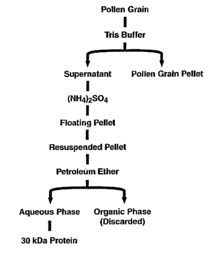
depicted in Figure 2B.
The present invention further provides purified nucleic acid sequences coding
for the
30 kDa ragweed complete pollen extract disulfide protein allergen, the 8-lOkDa
complete
ragweed pollen extract disulfide protein and the 30kDa defatted ragweed pollen
extract
disulfide protein or at least one antigenic fragment thereof, or derivative or
homologue
thereof, or the functional equivalent of the nucleic acid sequences. In
particular, the present
invention further provides purified nucleic acid sequences coding for peptides
depicted in
SEQ ID NO:1-11. The present invention also provides expression vectors
comprising a
nucleic acid sequence coding for at least one 30 kDa ragweed complete pollen
extract
disulfide protein, one 8-l OkDa complete ragweed pollen extract disulfide
protein and one
30kDa defatted ragweed pollen extract disulfide protein or at least one
antigenic fragment
thereof, or derivative or homologue thereof, or the functional equivalent of
the nucleic acid
sequence. The present invention further provides host cells transformed to
express a protein
or peptide encoded by the nucleic acid sequences of the invention.
Still another aspect of the invention provides a modified ragweed pollen
protein
allergen which, when administered to a ragweed pollen-sensitive individual,
reduces the
allergic response of the individual to ragweed pollen. Preferably the ragweed
pollen allergen
is a modified 30 kDa ragweed complete pollen extract disulfide protein
allergen, a modified
8-lOkDa complete ragweed pollen extract disulfide protein or a modified 30kDa
defatted
ragweed pollen extract disulfide protein or derivative or homologue thereof.
The present
invention also provides at least one modified fragment of ragweed pollen
protein allergen
which, when administered to a ragweed pollen-sensitive individual, reduces the
allergic
response of the individual to ragweed pollen. Preferably the ragweed pollen
protein allergen
is a 30 kDa ragweed complete pollen extract disulfide protein, an 8-l OkDa
complete ragweed
pollen extract disulfide protein or a 30kDa defatted ragweed pollen extract
protein or
antigenic fragment thereof, immunologically related to the 30 kDa ragweed
complete pollen
extract disulfide protein allergen, the 8-l OkDa complete ragweed pollen
extract disulfide
protein or the 30kDa defatted ragweed pollen extract protein or fragment or
derivative thereof
is also provided by the present invention. The ragweed pollen protein allergen
is generally in
the form of a pharmaceutical composition.
3
CA 02437484 2003-08-05
WO 02/063012 PCT/US02/03346
In yet another aspect of the present invention, there is provided non-native
(i.e.,
recombinant or chemically synthesized) 30 kDa ragweed pollen protein family
members or
their derivatives or homologues, or a non-native allergenic protein
immunologically cross-
reactive to antibodies to one or more 30 kDa ragweed complete pollen extract
disulfide
proteins, one or more 8-l OkDa complete ragweed pollen extract disulfide
proteins or one or
more 30kDa defatted ragweed pollen extract protein family members or their
derivatives or
homologues. The present invention also provides purified native 30 kDa ragweed
complete
pollen disulfide protein allergens, purified native 8-lOkDa complete ragweed
pollen extract
disulfide protein allergens or purified native 30kDa defatted ragweed pollen
extract disulfide
protein allergens or at least one fragment or derivative or homologue thereof.
Non-native 30 kDa ragweed complete pollen extract disulfide protein, non-
native 8-
l OkDa complete ragweed pollen extract disulfide protein or non-native 30kDa
defatted
ragweed pollen extract disulfide protein and fragments or portions derived
therefrom
(peptides) can be used in methods of diagnosing, treating and preventing
allergic reactions to
ragweed pollen. Purified native 30 kDa ragweed complete pollen extract
protein, purified
native 8-l OkDa complete ragweed pollen extract disulfide protein or purified
native 30kDa
defatted ragweed pollen extract protein fragments thereof, and homologues or
derivatives
thereof are also useful in methods of diagnosing, treating and preventing
allergic reactions to
ragweed pollen.
Still yet another aspect of the present invention relates to antibodies to non-
native 30
kDa ragweed complete pollen extract disulfide protein, non-native 8-l OkDa
complete
ragweed pollen extract disulfide protein or non-native 30kDa defatted ragweed
pollen extract
disulfide protein or derivatives or homologues thereof as well as antibodies
raised against
purified native 30 kDa ragweed complete pollen extract disulfide protein,
purified native 8-
l OkDa ragweed complete pollen extract disulfide protein or purified native
30kDa defatted
ragweed pollen extract protein or derivatives or homologues thereof.
The present invention is thus directed to an isolated protein having an amino
acid
sequence wherein the amino acid sequence is selected from SEQ >D NO:1, SEQ ID
N0:2,
SEQ >D N0:3, SEQ >D N0:4, SEQ ID NO:S, SEQ >D N0:6, SEQ >D N0:7, SEQ )D N0:8,
SEQ >D N0:9, SEQ >D NO:10, and SEQ )D NO:I 1.
4
CA 02437484 2003-08-05
WO 02/063012 PCT/US02/03346
The present invention is further directed to a pharmaceutical composition
including an
isolated protein having an amino acid sequence wherein the amino acid sequence
is selected
from SEQ ID NO:l, SEQ ID N0:2, SEQ >D N0:3, SEQ ID N0:4, SEQ ID NO:S, SEQ ID
N0:6, SEQ ID N0:7, SEQ ff~ N0:8, SEQ ID N0:9, SEQ ID NO:10, and SEQ ID NO:11.
The pharmaceutical composition may be utilized in a method of treating pollen
allergy in a
mammal by administering a therapeutically effective amount of the protein to a
mammal.
The pharmaceutical composition may be also be utilized in a method of treating
sensitivity to
pollen in a mammal sensitive to pollen by administering to the mammal a
therapeutically
effective amount of the protein to a mammal. The mammal may be a human.
The present invention is further directed to a diagnostic composition for
detecting
pollen allergy wherein the diagnostic composition includes an isolated protein
having an
amino acid sequence wherein the amino acid sequence is selected from SEQ ID
NO:1, SEQ
ID N0:2, SEQ ID N0:3, SEQ ID N0:4, SEQ ID NO:S, SEQ ID N0:6, SEQ ID N0:7, SEQ
ID N0:8, SEQ ID N0:9, SEQ ID NO:10, and SEQ ID NO:11.
In the invention, the pollen may be from any source. In one embodiment, the
pollen
is selected from walnut, ryegrass and ragweed pollen.
The present invention is further directed to an isolated nucleic acid having a
nucleotide sequence encoding an amino acid sequence wherein the amino acid
sequence is
selected from SEQ 117 NO:1, SEQ ID N0:2, SEQ ID N0:3, SEQ ID N0:4, SEQ ID
NO:S,
SEQ ID N0:6, SEQ ID N0:7, SEQ ID N0:8, SEQ ID N0:9, SEQ ID NO:10, and SEQ ID
NO:11. The nucleic acid of the invention may be in an expression vector. The
expression
vector may be in a host cell.
The present invention is further directed to an isolated pollen allergen
substantially
free of any other pollen proteins characterized by the following
physiochemical and
biological properties: a) being contained in pollen extracts, b) a
glycoprotein, c) a sulfhydryl
group containing protein, d) a molecular weight about 30,000 as determined by
SDS-
polyacrylamide gel electrophoresis and e) possessing allergen activity. The
allergen may be
from any source. In a preferred embodiment, the allergen may be from walnut,
ryegrass or
ragweed pollen.
The present invention is further directed to a pharmaceutical composition
having a
pollen allergen substantially free of any other pollen proteins characterized
by the following
CA 02437484 2003-08-05
WO 02/063012 PCT/US02/03346
physiochemical and biological properties: a) being contained in pollen
extracts, b) a
glycoprotein, c) a sulfhydryl group containing protein, d) a molecular weight
about 30,000 as
determined by SDS-polyacrylamide gel electrophoresis and e) possessing
allergen activity.
The allergen may be from any source. In a preferred embodiment, the allergen
may be
selected from walnut, ryegrass and ragweed pollen.
The present invention is further directed to a diagnostic composition for
detecting
allergic diseases which includes as the active ingredient a diagnostically
effective amount of
a pollen allergen substantially free of any other pollen proteins
characterized by the following
physiochemical and biological properties: a) being contained in pollen
extracts, b) a
glycoprotein, c) a sulfliydryl group containing protein, d) a molecular weight
about 30,000 as
determined by SDS-polyacrylamide gel electrophoresis and e) possessing
allergen activity.
The allergen may be from any source. In a preferred embodiment, the allergen
may be
selected from walnut, ryegrass and ragweed pollen.
The present invention is further directed to a method of treating pollen
allergy in a
mammal by administering a pharmaceutically effective amount of an pollen
allergen
substantially free of any other pollen proteins characterized by the following
physiochemical
and biological properties: a) being contained in pollen extracts, b) a
glycoprotein, c) a
sulfhydryl group containing protein, d) a molecular weight about 30,000 as
determined by
SDS-polyacrylamide gel electrophoresis and e) possessing allergen activity to
a mammal,
preferrably a human.
The present invention is further directed to a therapeutic composition having
an
isolated antigenic fragment of a ryegrass pollen allergen Ambt 7 wherein the
antigenic
fragment includes one or more amino acid sequences selected from SEQ >D NO:1,
SEQ m
N0:2, SEQ >D N0:3, SEQ >D N0:4, SEQ >D NO:S, SEQ )D N0:6, SEQ )Z7 N0:7, SEQ >D
N0:8, SEQ )D N0:9, SEQ >D NO:10, and SEQ ID NO:11 wherein the antigenic
fragment
has at least one epitope of the pollen allergen. The therapeutic composition
generally
includes a pharmaceutically effective carrier.
The epitope may be a T cell epitope or a B cell epitope.
The therapeutic composition may be administered to a mammal such as a human to
treat sensitivity to ryegrass pollen.
6
CA 02437484 2003-08-05
WO 02/063012 PCT/US02/03346
The present invention is father directed to a therapeutic composition having
an Ambt7
pollen allergen which is a polymorphic variant of a ryegrass Ambt7 pollen
allergen wherein
the polymorphic variant has an amino acid sequence selected from the group
consisting of
SEQ ID NO:1, SEQ ID N0:2, SEQ >D N0:3, SEQ ID N0:4, SEQ ID NO:S, SEQ ID N0:6,
SEQ ID N0:7, SEQ ID N0:8, SEQ 1D N0:9, SEQ ID NO:10, and SEQ ID NO:11. The
therapeutic composition may include a pharmaceutically acceptable Garner.
The therapeutic composition may be administered to a mammal in a method of
treating sensitivity to ryegrass pollen
The present invention is father directed to a kit for detecting Ambt7 pollen
allergen
wherein the kit includes one or more isolated proteins having an amino acid
sequence
selected from SEQ ID NO:1, SEQ ID N0:2, SEQ ID N0:3, SEQ ID N0:4, SEQ ID NO:S,
SEQ ID N0:6, SEQ ID N0:7, SEQ ID N0:8, SEQ ID N0:9, SEQ ID NO:10, and SEQ ID
NO:11. The kit of may further include protein detection components such as
antibodies.
The kit may also further include directions for use of the kit.
The present invention is further directed to a method of purifying a pollen
allergen,
by: a) suspending the pollen in a liquid to form a pollen solution; b)
centrifuging the pollen
solution to produce a pollen protein supernatant; c) precipitating the protein
in the pollen
protein supernatant to form a protein precipitate; d) resuspending the protein
precipitate in a
protein precipitate buffer to form a resuspended protein mixture; e)
extracting the
resuspended protein mixture in organic solvent to form an aqueous phase and an
organic
phase; and f) purifying the pollen allergen from the aqueous phase.
In one embodiment of the method of purifying a pollen allergen, the protein in
the
pollen solution is precipitated with (1VH4)2 504,
In another embodiment of the method of purifying a pollen allergen the organic
solvent is petroleum ether.
In another embodiment of the method of purifying a pollen allergen, the pollen
allergen is purified from the aqueous phase by chromatography or
electrophoresis procedures.
In the method, the chromatography procedure may be gel filtration or affinity
chromatography.
7
CA 02437484 2003-08-05
WO 02/063012 PCT/US02/03346
The present invention is further directed to an isolated antibody that binds
specifically
to a protein comprising an amino acid sequence wherein the amino acid sequence
is selected
from the group consisting of SEQ >D NO:1, SEQ >D N0:2, SEQ ID N0:3, SEQ ID
N0:4,
SEQ 117 NO:S, SEQ >D N0:6, SEQ )D N0:7, SEQ m N0:8, SEQ ID N0:9, SEQ >D NO:10,
and SEQ >D NO:11.
The present invention is further directed to an isolated antibody that binds
specifically
to a pollen allergen substantially free of any other pollen proteins wherein
the pollen allergen
is characterized by the following physiochemical and biological properties: a)
being
contained in pollen extracts, b) a glycoprotein, c) a sulfliydryl group
containing protein, d) a
molecular weight about 30,000 as determined by SDS-polyacrylamide gel
electrophoresis
and e) possessing allergen activity.
The antibodies of the invention may be a polyclonal or monclonal antibodies
Further features of the present invention will be better understood from the
following
detailed description of the preferred embodiments of the invention in
conjunction with the
appended figures.
BRIEF DESCRIPTION OF THE FIGURES
The invention will be better understood by reference to the Figures in which:
Figure 1 shows the structure of the wall of ragweed pollen including: (1) the
intine;
(2) the exine; (3) the nexine; (4) the sexine; (S) the lipid layer; (6) the
micropore; (7) the
spine, (8) the protein; (9) cavity and (10) the protoplast.
Figure 2a shows the prior art procedure for producing clinical pollen
preparations.
Figure 2b outlines a procedure for extracting the 30 kDa protein and other
proteins
from ragweed pollen.
Figure 3 shows total protein, sulfllydryl protein and allergen profiles of
aqueous
extracts from complete and defatted ragweed pollen. Extracts were fractionated
by Sephadex
GSOF chromatography and separated by SDS-PAGE (10-20%). Figure 3A. shows gels
stained for total protein. Gels were stained with Coomassie blue; each lane
contained 3 to 30
pg protein. Figure 3B shows sulfllydryl determination with monobromobimane.
Sulfliydryl
8
CA 02437484 2003-08-05
WO 02/063012 PCT/US02/03346
groups of proteins were labeled with monobromobimane and analyzed by UV light.
Figure
3C shows allergen determination by IgE immunoblotting. Proteins were
transferred to
nitrocellulose and probed with IgE of sera combined from 10 ragweed patients.
The symbols
depicted in Figure 3c are: (1) 30 kDa protein, (*) 8-10 kDa protein (complete
pollen) and
(~)second 30 kDa protein (defatted pollen).
Figure 4 shows some properties of 30 kDa protein. SDS-PAGE (10-20%). Figure 4A
demonstrates that 30 kDa protein is glycosylated. The gel in Figure 4A was
stained for
glycoprotein. Lane 1 contains soybean trypsin inhibitor (negative control, 5
fig); lane 2
contains 30 kDa protein (10 fig); and lane 3 contains horseradish peroxidase
(positive control,
pg). Figure 4B demonstrates that the 30 kDa protein contains at least one
disulfide bond.
The gel was examined under UV light following reaction with monobromobimane
(mBBr).
Ten pg protein was used.
Figure 5 shows the response of sera from grass-sensitive patients to pollen
preparations by SDSIPAGE (10-20%)/IgE immunoblotting. Figure SA shows the
response
of sera from 35 patients to pure 30 kDa protein. Patients showing binding to
the 30kDa
protein are designated with a "+". Those patients also showing a positive
ImmunoCAP test
are identified with an circle surrounding the "+". Each lane on the gel
contained 1.6 pg
protein. Figure SB shows the response of sera from selected grass positive
patients to
commercial ragweed extract (left panel), complete pollen extract (middle
panel) and purified
30 kDa protein (right panel). The commercial and complete extracts and the 30
kDa protein
contained 25, 25 and 1.6 p,g protein, respectively. In the control (C )
treatment, sera and
secondary antibody were omitted; the secondary antibody was omitted in lanes
designated
"Ab2."
Figure 6 shows the immunoblot inhibition of the 30 kDa protein with walnut and
ryegrass pollen extracts and the demonstration of cross-reactivity. Figure 6A
is the control
with no inhibitor protein added (C), ovalbumin as inhibitor protein (O,
negative control),
walnut (Juglans nigra) complete pollen extract (W) and ryegrass (Lolium
perenne) complete
pollen extract (R) added to sera prior to immunoblotting. In Figure 6B the
results with sera
from an additional 17 patients positive to the 30 kDa protein are shown. In
the control lanes
(C) no allergen is added. In the lanes identified with an (R) ryegrass
complete pollen extract
is added as an inhibitor protein.
9
CA 02437484 2003-08-05
WO 02/063012 PCT/US02/03346
Figure 7 shows the percentage of human IgE binding to known ragweed allergens
vs.
30 kDa protein. An ELISA determination was carried out with sera from 10
ragweed-
sensitive patients. 1 p,g per ml protein was tested for each allergen. The
values represent
percent of total IgE bound by each allergen tested.
DETAILED DESCRIPTION OF THE INVENTION
Giant ragweed allergen extract was purchased from Bayer, Inc. (Spokane, WA).
Complete and defatted giant ragweed (Ambrosia trifida) pollen grains were
purchased from
Greer Laboratories (Lenoir, N.C.). These sources of pollen are not intended to
limit the
scope of the invention since they only represent one convenient supply of the
pollen. The
present invention can be practiced using ragweed pollen from any location or
source.
In the following discussion, the 30 kDa ragweed complete pollen extract
disulfide
protein allergen is described and is identified as "the 30 kDa ragweed protein
allergen" and as
"Ambt 7." This discussion is not limited to the 30 kDa ragweed protein
allergen but applies
equally to other ragweed protein allergens including the 8-l OkDa ragweed
complete pollen
extract disulfide protein, the 30kDa ragweed defatted pollen extract disulfide
protein and
derivatives or homologues thereof.
Purification of Pollen Proteins
The present invention is directed to a method of purifying pollen proteins.
The
method finds particular use with ragweed pollen but is not limited to ragweed
pollen. The
method is outlined in Figure 2b. Pollen is suspended in buffer such as 50 mM
Tris-HCI, pH
7.4. One or more protease inhibitors such as phenylmethylsulfonyl fluoride and
EDTA may
be added to the buffer to reduce protein degradation. The suspended pollen is
stirred gently
at room temperature for a time sufficient to release pollen proteins,
typically 30 minutes. The
suspension is then centrifuged to precipitate the insoluble pollen material.
The supernatant is
then filtered. The proteins in the supernatant are precipitated with ammonium
sulfate, for
example at a 95% saturation. The floating pellet is then recovered by
centrifugation and
resuspended in buffer containing salt. In one embodiment, the buffer is 20 mM
Tris-HCl (pH
7.5) and the salt is 200mM NaCI. Lipids are then removed by extracting the
resuspended
protein pellet in an organic solvent such as petroleum ether. The mixture is
then centrifuged,
for example for 10 min at 48,OOOg at 4°C, and the organic phase is
discarded. The resulting
clarified aqueous solution is filtered, for example, through a 0.2 uM filter.
After filtration,
CA 02437484 2003-08-05
WO 02/063012 PCT/US02/03346
the filtrate is separated on a gel filtration column to separate the various
pollen proteins.
After separation, the pollen proteins such as Ambt 7 can be further purified
by procedures
well known in the art such as SDS-PAGE, chromatography, etc.
The invention encompasses isolated or substantially purified nucleic acid or
protein
compositions. In the context of the present invention, an "isolated" or
"purified" DNA
molecule or an "isolated" or "purified" polypeptide is a DNA molecule or
polypeptide that,
by the hand of man, exists apart from its native environment and is therefore
not a product of
nature. An isolated DNA molecule or polypeptide may exist in a purified form
or may exist
in a non-native environment such as, for example, a transgenic host cell. For
example, an
"isolated" or "purified" nucleic acid molecule or protein, or biologically
active portion
thereof, is substantially free of other cellular material, or culture medium
when produced by
recombinant techniques, or substantially free of chemical precursors or other
chemicals when
chemically synthesized. Preferably, an "isolated" nucleic acid is free of
sequences
(preferably protein encoding sequences) that naturally flank the nucleic acid
(i.e., sequences
located at the 5' and 3' ends of the nucleic acid) in the genomic DNA of the
organism from
which the nucleic acid is derived. For example, in various embodiments, the
isolated nucleic
acid molecule can contain less than about Skb, 4 kb, 3 kb, 2 kb, 1 kb, 0.5 kb,
or 0.1 kb of
nucleotide sequences that naturally flank the nucleic acid molecule in genomic
DNA of the
cell from which the nucleic acid is derived. A protein that is substantially
free of cellular
material includes preparations of protein or polypeptide having less than
about 30%, 20%,
10%, 5%, (by dry weight) of contaminating protein. When the protein of the
invention, or
biologically active portion thereof, is recombinantly produced, preferably
culture medium
represents less than about 30%, 20%, 10%, or S% (by dry weight) of chemical
precursors or
non-protein of interest chemicals.
Genes) Encoding Ragweed Protein Allergens
"Gene," is used, in respect of the present invention, in its broadest sense
and refers to
any contiguous sequence of nucleotides, the transcription of which leads to a
mRNA
molecule, which mIRNA molecule is capable of being translated into a protein.
The gene
encoding a 30 kDa ragweed protein allergen family member means the nucleotide
sequence
encoding the protein or derivatives or homologues of the protein which may
contain single or
multiple amino acid substitutions, deletions or additions. A 30 kDa ragweed
protein allergen
11
CA 02437484 2003-08-05
WO 02/063012 PCT/US02/03346
gene also refers to cDNAs complementary to the mRNAs corresponding to the full
or partial
length of a 30 kDa protein.
It is expected that there are sequence polymorphisms in the nucleic acid
sequence
coding for each 30 kDa ragweed protein allergen family member, and it will be
appreciated
by one skilled in the art that one or more nucleotides in the nucleic acid
sequence coding for a
30 kDa ragweed protein allergen family member may vary among individual
ragweed plants
due to natural allelic variation. Any and all such nucleotide variations and
resulting amino
acid polymorphisms are within the scope of the invention. It may also be
appreciated by one
skilled in the art that the 30 kDa ragweed protein allergen is a family of
highly related genes
whose proteins are present in ragweed pollen. Nucleotide sequences and
corresponding
deduced amino acid sequences of any and all such related family members
including 30 kDa
are within the scope of the invention.
Accordingly, it is within the scope of the present invention to encompass all
proteins
belonging to the 30 kDa ragweed protein allergen family, at least one fragment
(peptide) of a
30 kDa ragweed protein allergen family member, and amino acid derivatives
thereof, and to
encompass nucleotide sequences, including DNA, cDNA and mRNA and homologue or
degenerate forms thereof, encoding 30 kDa ragweed protein allergen family
members or
fragments thereof, or derivatives thereof. It is also within the scope of the
invention to
encompass purified native 30 kDa ragweed protein allergen, at least one
fragment (peptide)
thereof, and derivatives or homologues thereof. It is further in accordance
with the present
invention to include molecules such as polypeptides fused to a 30 kDa protein,
or at least one
fragment thereof, or derivatives thereof or to nucleotide sequences contiguous
to such
fragment and/or derivative-encoding nucleotide sequences.
For example, for some aspects of the present invention, it is desirable to
produce a
fusion protein comprising a 30 kDa ragweed protein allergen family member or
at least one
fragment thereof or their derivatives and an amino acid sequence from another
peptide or
protein, examples of the latter being enzymes such as beta-galactosidase,
phosphatase, urease
and fusion proteins incorporating purification moieties such as His-tags and
the like. Most
fusion proteins are formed by the expression of a recombinant gene in which
two coding
sequences have been joined together such that their reading frames are in
phase.
Alternatively, proteins or peptides can be linked in vitro by chemical means.
All such fusion
protein or hybrid genetic derivatives of a 30 kDa ragweed protein allergen or
its encoding
12
CA 02437484 2003-08-05
WO 02/063012 PCT/US02/03346
nucleotide sequences are encompassed by the present invention. Furthermore, by
homologues and derivatives of a 30 kDa ragweed protein allergen is meant to
include
synthetic derivatives thereof. The nucleotide sequences encoding the 30 kDa
ragweed
protein allergen can be used to chemically synthesize the entire protein or
generate any
number of fragments (peptides) by chemical synthesis by well known methods
(e.g., solid
phase synthesis). All such chemically synthesized peptides are encompassed by
the present
invention. Accordingly, the present invention extends to isolated 30 kDa
ragweed protein
allergen family members, fragments thereof and their derivatives, homologues
and
immunological relatives made by recombinant means or by chemical synthesis.
The terms "isolated" and "purified" are used interchangeably herein and refer
to
peptides, protein, protein fragments, and nucleic acid sequences substantially
free of cellular
material or culture medium when produced by recombinant DNA techniques, or
chemical
precursors or other chemicals when synthesized chemically. The term "native
purified" as
used herein refers to proteins or fragments thereof purified from Ambrosia
trifida pollen or
other plant part. Furthermore, the present invention extends to proteins or
fragments
(peptides) corresponding in whole or part.
Fragments of nucleic acid within the scope of the invention include those
coding for
parts of the 30 kDa ragweed protein allergen that elicit an immune response in
mammals,
preferably humans, such as the stimulation of minimal amounts of IgE; binding
of IgE; '
eliciting the production of IgG and IgM antibodies; or the eliciting of a T
cell response such
as proliferation and/or lymphokine secretion and/or the induction of T cell
anergy. The
foregoing fragments of the 30 kDa ragweed protein allergen are referred to
herein as
antigenic fragments. Fragments within the scope of the invention also include
those capable
of hybridizing with nucleic acid from other plant species for use in screening
protocols to
detect allergens that are cross-reactive with the 30 kDa ragweed protein
allergen. As used
herein, a fragment of the nucleic acid sequence coding for the 30 kDa ragweed
protein
allergen refers to a nucleotide sequence having fewer bases than the
nucleotide sequence
coding for the entire amino acid sequence of the 30 kDa ragweed protein
allergen and/or a
mature 30 kDa ragweed protein allergen family member. Generally, the nucleic
acid
sequence coding for the fragment or fragments of a the 30 kDa ragweed protein
allergen
family member will be selected from the bases coding for the mature 30 kDa
ragweed protein
allergen family member, however, in some instances it may be desirable to
select all or a part
13
CA 02437484 2003-08-05
WO 02/063012 PCT/US02/03346
of a fragment or fragments from the leader sequence portion of a nucleic acid
sequence of the
invention. A nucleic acid sequence of the invention may also contain linker
sequences,
restriction endonuclease sites and other sequences useful for cloning,
expression or
purification of the 30 kDa ragweed protein allergen or fragments thereof.
Antigenic Fragments of Ragweed Protein Allergens
Antigenic fragments of an allergen from ragweed pollen, preferably the 30 kDa
ragweed protein allergen, may be obtained, for example, by screening peptides
produced by
recombinant methods from the corresponding fragment of the nucleic acid
sequence of the
invention coding for such peptides, synthesized chemically using techniques
known in the art,
or by degrading of the purified allergen. The peptide fragments of the protein
allergen may
be obtained by any method known in the art such as chemical cleavage of the
allergen,
arbitrary division of the allergen into fragments of a desired length with no
overlap of the
peptides, or preferably division of the allergen into overlapping fragments of
a desired length.
The fragments are tested to determine their antigenicity and allergenicity.
Fragments of recombinantly or synthetically produced 30 kDa ragweed protein
allergen or of purified native 30 kDa ragweed protein allergen which are
capable of eliciting
a T cell response such as stimulation (i.e., proliferation or lymphokine
secretion) and/or are
capable of inducing T cell anergy are particularly desirable. Fragments of
recombinantly or
synthetically produced 30 kDa ragweed protein allergen or purified native 30
kDa ragweed
protein allergen which do not bind immunoglobulin E (IgE) and/or which have
minimal IgE
stimulating activity are also desirable. If the fragment or fragments of a
recombinantly or
synthetically produced 30 kDa ragweed protein allergen family member or
purified native 30
kDa ragweed protein allergen bind IgE, it is preferable that such binding does
not lead to
histamine release, e.g., such binding does not cause cross-linking of IgE on
mast cells or
basophils. Minimal IgE stimulating activity refers to IgE stimulating activity
that is less than
the amount of IgE production stimulated by whole recombinantly or
synthetically produced
30 kDa ragweed protein allergen or whole purified native 30 kDa ragweed
protein allergen.
Preferred fragments also include antigenic fragments which, when administered
to a
ragweed pollen-sensitive individual or an individual allergic to an allergen
cross-reactive
with ragweed pollen allergen, are capable of modifying the allergic response
to ragweed
pollen allergen of the individual, and antigenic fragments which, when
administered to a
14
CA 02437484 2003-08-05
WO 02/063012 PCT/US02/03346
ragweed pollen-sensitive individual, are capable of modifying B-cell response,
T-cell
response or both B-cell and T-cell response of the individual to a ragweed
pollen allergen.
As used herein modification of the allergic response of an individual
sensitive to ragweed
pollen allergen can be defined as non-responsiveness or diminution in symptoms
to the
allergen, as determined by standard clinical procedures (see e.g. Varney et
al, British
Medical Journal, (1990), 302:265-269), including diminution in grass pollen
induced
asthmatic symptoms (Suphioglu et al. (1992) Lancet 339: 569-572).
Antigenic fragments of the present invention which have T cell stimulating
activity,
and thus comprise at least one T cell epitope are particularly desirable. T
cell epitopes are
believed to be involved in initiation and perpetuation of the immune response
to a protein
allergen which is responsible for the clinical symptoms of allergy. These T
cell epitopes are
thought to trigger early events at the level of the T helper cell by binding
to an appropriate
HLA molecule on the surface of an antigen presenting cell and stimulating the
relevant T cell
subpopulation. These events lead to T cell proliferation, lymphokine
secretion, local
inflammatory reactions, recruitment of additional immune cells to the site,
and activation of
the B cell cascade leading to production of antibodies. One isotype of these
antibodies, IgE,
is fundamentally important to the development of allergic symptoms and its
production is
influenced early in the cascade of events, at the level of the T helper cell,
by the nature of the
lymphokines secreted. A T cell epitope is the basic element or smallest unit
of recognition by
a T cell receptor, where the epitope comprises amino acids essential to
receptor recognition.
Amino acid sequences which mimic those of the T cell epitopes and which modify
the
allergic response to protein allergens are within the scope of this invention.
Exposure of patients to purified protein allergens of the present invention or
to the
antigenic fragments of the present invention which comprise at least one T
cell epitope and
are derived from protein allergens may tolerize or anergize appropriate T cell
subpopulations
such that they become unresponsive to the protein allergen and do not
participate in
stimulating an immune response upon such exposure. In addition, administration
of the
protein allergen of the invention or an antigenic fragment of the present
invention which
comprises at least one T cell epitope may modify the lymphokine secretion
profile as
compared with exposure to the naturally-occurring protein allergen or portion
thereof (e.g.
result in a decrease of IL-4 and/or an increase in IL-2). Furthermore,
exposure to such
antigenic fragment or protein allergen may influence T cell subpopulations
which normally
CA 02437484 2003-08-05
WO 02/063012 PCT/US02/03346
participate in the response to the allergen such that these T cells are drawn
away from the
sites) of normal exposure to the allergen (e.g., nasal mucosa, skin, and lung)
towards the
sites) of therapeutic administration of the fragment or protein allergen. This
redistribution of
T cell subpopulations may ameliorate or reduce the ability of an individual's
immune system
to stimulate the usual immune response at the site of normal exposure to the
allergen,
resulting in a diminution in allergic symptoms.
Screening for IgE binding to the protein or fragments thereof may be performed
by
scratch tests or intradermal skin tests on laboratory animals or human
volunteers, or in in
vitro systems such as RAST (radioallergosorbent test), RAST inhibition, ELISA
assay or
radioimmunoassay (RIA).
Expression Vectors and Host Cells
The present invention provides expression vectors and host cells transformed
to
express the nucleic acid sequences encoding the ragweed protein allergen of
the invention.
Expression vectors of the invention comprise a nucleic acid sequence coding
for at least one
30 kDa ragweed pollen allergen, or at least one antigenic fragment thereof, or
derivative or
homologue thereof, or the functional equivalent of such nucleic acid sequence.
Nucleic acid
sequences coding for 30 kDa ragweed protein allergen family members including
30 kDa
ragweed protein allergen, or at least one fragment thereof may be expressed in
prokaryotic or
eukaryotic host cells. Suitable host cells include bacterial cells such as E.
coli, insect cells,
yeast, or mammalian cells such as Chinese hamster ovary cells (CHO). Suitable
expression
vectors, promoters, enhancers, and other expression control elements may be
found in
Sambrook et al. Molecular Cloning: A Laboratory Manual, second edition, Cold
Spring
Harbor Laboratory Press, Cold Spring Harbor, New York, 1989. Suitable vectors
for
expression in yeast include YepSecl (Baldari et al. (1987) Embo J. 6: 229-
234); pMF
(Kurjan and Herskowitz (1982) Cell 30: 933-943); and JRY88 (Schultz et al.
(1987) Gene
54: 113-123).
For expression in E. coli, suitable expression vectors include pTRC (Amann et
al.
(1988) Gene 69: 301-315); pET-l 1d (Novagen, Madison, Wis.); pGEX (Amrad
Corp.,
Melbourne, Australia); pMAL (N.E. Biolabs, Beverly, Mass.); pRITS (Pharmacia,
Piscataway, N.J.); and PSEM (Knapp et al. (1990) BioTechniques 8: 280-281).
The use of
pTRC and pET-l 1d will lead to the expression of unfused protein. The use of
pGEX, pMAL,
16
CA 02437484 2003-08-05
WO 02/063012 PCT/US02/03346
pRITS and pSEM will lead to the expression of allergen fused to glutathione S-
transferase
(pGEX), maltose E binding protein (pMAL), protein A (pRITS), or truncated 13-
galactosidase
(PSEM). When a 30 kDa ragweed protein allergen family member, fragment, or
fragments
thereof is expressed as a fusion protein, it is particularly advantageous to
introduce an
enzymatic cleavage site at the fusion junction between the carrier protein and
the 30 kDa
protein family member or fragment thereof. A 30 kDa ragweed protein allergen
family
member or fragment thereof may then be recovered from the fusion protein
through
enzymatic cleavage at the enzymatic site and biochemical purification using
conventional
techniques for purification of proteins and peptides. Suitable enzymatic
cleavage sites
include those for blood clotting Factor Xa or thrombin for which the
appropriate enzymes and
protocols for cleavage are commercially available from for example Sigma
Chemical
Company, St. Louis, Mo. and N.E. Biolabs, Beverly, Mass.
Host cells can be transformed to express the nucleic acid sequences encoding
the 30
kDa ragweed protein allergen of the invention using conventional techniques
such as calcium
phosphate or calcium chloride co-precipitation, DEAF-dextran-mediated
transfection, or
electroporation. Suitable methods for transforming the host cells may be found
in Sambrook
et al. supra, and other laboratory textbooks. The nucleic acid sequences of
the invention may
also be synthesized using standard techniques.
Production of Recombinant 30 kDa Ragweed Protein
Accordingly, another aspect of the present invention provides a method of
producing
recombinant 30 kDa ragweed protein allergen, or at least one fragment thereof,
or their
derivatives or homologues, or their immunological relatives (as hereinbefore
defined)
comprising culturing an organism containing a replicable recombinant DNA
molecule, the
molecule comprising a promoter capable of expression in the organism, a gene
encoding a 30
kDa ragweed protein allergen family member, at least one fragment thereof, or
homologue or
derivative thereof, or immunological relatives thereof, located downstream of
and transcribed
from the promoter, a selectable marker and a DNA vehicle containing a
prokaryotic or
eukaryotic origin of replication, under conditions and for a time sufficient
for the
recombinant DNA molecule to be stably maintained and direct the synthesis of
the 30 kDa
ragweed protein allergen, at least one fragment thereof, or derivatives,
homologues or
immunological relatives thereof and then optionally isolating same.
17
CA 02437484 2003-08-05
WO 02/063012 PCT/US02/03346
30 kDa ragweed protein allergen and fragments (peptides) thereof can be
purified
from cell culture medium, host cells, or both using techniques known in the
art for purifying
peptides and proteins, including ion-exchange chromatography, hydrophobic
chromatography, gel filtration chromatography, ultrafiltration,
electrophoresis and
immunopurification with antibodies specific for 30 kDa ragweed protein
allergen or
fragments thereof. The terms isolated and purified are used interchangeably
herein and refer
to peptides, protein, protein fragments, and nucleic acid sequences
substantially free of
cellular material or culture medium when produced by recombinant DNA
techniques, or
chemical precursors or other chemicals when synthesized chemically.
Another aspect of the invention provides protein preparations comprising
isolated 30
kDa ragweed protein allergen or at least one fragment of 30 kDa ragweed
protein allergen. In
preferred embodiments of this aspect of the invention, the 30 kDa ragweed
protein allergen or
at least one fragment of the 30 kDa ragweed protein allergen is produced in a
host cell
transformed with a nucleic acid sequence coding for the protein or fragment.
Modifying an Individual's Allergic Response
It is possible to design peptides derived from the 30 kDa ragweed protein
allergen
which, when administered to a ragweed pollen sensitive individual in
sufficient quantities,
will modify the individual's allergic response to ragweed pollen. This can be
done, for
example, by examining the structure of 30 kDa ragweed protein allergen,
producing peptides
(via an expression system, synthetically or otherwise) to be examined for
their ability to
influence B-cell and/or T-cell responses in ragweed pollen sensitive
individuals and selecting
appropriate epitopes recognized by the cells. In referring to an epitope, the
epitope will be
the basic element or smallest unit of recognition by a receptor, particularly
immunoglobulins,
histocompatibility antigens and T cell receptors where the amino acids
essential to the
receptor recognition may be contiguous and/or non-contiguous in the amino acid
sequence.
Amino acid sequences which mimic those of the epitopes and which are capable
of down
regulating allergic response to ragweed pollen allergen can also be used.
It is now also possible to design an agent or a drug capable of blocking or
inhibiting
the ability of ragweed pollen allergen to induce an allergic reaction in
ragweed pollen
sensitive individuals. Such agents could be designed, for example, in such a
manner that they
would bind to relevant anti-30 kDa ragweed protein allergen-IgE's, thus
preventing IgE-
18
CA 02437484 2003-08-05
WO 02/063012 PCT/US02/03346
allergen binding and subsequent mast cell or basophil degranulation.
Alternatively, such
agents could bind to cellular components of the immune system, resulting in
suppression or
desensitization of the allergic response to Ambrosia trifida pollen allergens.
A non-restrictive
example of this is the use of appropriate B- and T-cell epitope peptides, or
modifications
thereof, based on the cDNA/protein structures of the present invention to
suppress the allergic
response to ragweed pollen. This can be carried out by defining the structures
of B- and T-
cell epitope peptides which affect B- and T-cell function in in vitro studies
with blood
components from ragweed pollen sensitive individuals.
Diagnosing Pollinosis
Protein, peptides or antibodies of the present invention can also be used for
detecting
and diagnosing ragweed pollinosis. For example, this could be done by
combining blood or
blood products obtained from an individual to be assessed for sensitivity to
ragweed pollen
with an isolated antigenic peptide or peptides of recombinantly or
synthetically produced 30
kDa ragweed protein allergen or native purified 30 kDa ragweed protein
allergen, under
conditions appropriate for binding of components (e.g., antibodies, T-cells, B-
cells) in the
blood with the peptides) or protein and determining the extent to which such
binding occurs.
The extent to which binding occurs can be determined, for example, by
assessing T cell
function, T cell proliferation, B cell function, or binding of the protein, or
fragment thereof,
or derivative or homologue thereof to antibodies present in the blood or a
combination
thereof.
Additionally, sensitivity of a mammal to ragweed pollen may be determined by
administering to a mammal a sufficient quantity of the 30 kDa ragweed pollen
allergen, or at
least one antigenic fragment thereof, or derivative or homologue thereof to
provoke an
allergic response in the mammal and determining the occurrence of an allergic
response in
the mammal to the ragweed pollen allergen. The ragweed pollen allergen(s),
fragments) or
derivative or homologue thereof used in this aspect of the present invention
can be produced
recombinantly or synthetically. Purified native 30 kDa ragweed protein
allergen or fragments
thereof may be substituted for a recombinantly or synthetically produced 30
kDa ragweed
protein allergen or fragments thereof and used in the above method to
determine sensitivity of
the mammal to ragweed.
19
CA 02437484 2003-08-05
WO 02/063012 PCT/US02/03346
The invention further includes isolated allergenic proteins or fragments
thereof that
are immunologically related to 30 kDa ragweed protein allergen, including
fragments, or
derivatives or homologues thereof, such as by antibody cross-reactivity
wherein the isolated
allergenic proteins or fragments thereof are capable of binding to antibodies
specific for the
protein and peptides of the invention, or by T cell cross-reactivity wherein
the isolated
allergenic proteins or fragments thereof are capable of stimulating T cells
specific for the
protein and peptides of this invention.
Work by others has shown that high doses of allergens generally produce the
best
results (i.e., best symptom relief). However, many people are unable to
tolerate large doses
of allergens because of allergic reactions to the allergens. Modification of
naturally-
occurnng allergens can be designed in such a manner that modified peptides or
modified
allergens which have the same or enhanced therapeutic properties as the
corresponding
naturally-occurnng allergen but have reduced side effects (especially
anaphylactic reactions)
can be produced. These can be, for example, a protein or peptide of the
present invention
(e.g., one having all or a portion of the amino acid sequence of 30 kDa
ragweed protein
allergen, including purified native 30 kDa ragweed protein allergen), or a
modified protein or
peptide, or the protein or peptide analogue. It is possible to modify the
structure of a protein
or peptide of the invention for such purposes as increasing solubility,
enhancing therapeutic
or preventive efficacy or stability (e.g., shelf life ex vivo and resistance
to proteolytic
degradation in vivo. A modified protein or peptide can be produced in which
the amino acid
sequence has been altered, such as by amino acid substitution, deletion or
addition, to modify
immunogenicity and/or reduce allergenicity or to which a component has been
added for the
same purpose. A modified protein can further be produced by the use of thiol
redox proteins
to reduce protein intramolecular disulfide bonds as described in U.S: Patent
No.6,114,504.
Treating Allergic Responses
Thus, the present invention provides modified ragweed pollen protein allergens
which, when administered to a ragweed pollen-sensitive individual, reduce the
allergic
response of the individual to ragweed pollen. Preferred modified ragweed
pollen protein
allergens include modified 30 kDa ragweed pollen allergen or derivative or
homologue
thereof. The present invention also provides at least one modified fragment of
ragweed
pollen protein allergen which, when administered to a ragweed pollen-sensitive
individual,
reduces the allergic response of the individual to ragweed pollen. Preferably
such modified
CA 02437484 2003-08-05
WO 02/063012 PCT/US02/03346
fragments are at least one modified fragment of the 30 kDa ragweed pollen
allergen or
derivative or homologue thereof.
Another example of a modification of protein or peptides is substitution of
cysteine
residues preferably with alanine, serine, threonine, leucine or glutamic acid
to minimize
dimerization via disulfide linkages. Another example of modification of the
peptides of the
invention is by chemical modification of amino acid-side chains or cyclization
of the peptide.
In order to enhance stability and/or reactivity, the protein or peptides of
the invention
can also be modified to incorporate one or more polymorphisms in the amino
acid sequence
of the protein allergen resulting from natural allelic variation.
Additionally, D-amino acids,
non-natural amino acids or non-amino acid analogues can be substituted or
added to produce
a modified protein or peptide within the scope of this invention.
The purification of native 30 kDa ragweed pollen allergen is described in the
examples herein.
Cloning of cDNAs
The DNA used in any embodiment of this invention can be cDNA obtained as
described herein, or alternatively, can be any oligodeoxynucleotide sequence
having all or a
portion of a sequence represented herein, or their functional equivalents.
Such
oligodeoxynucleotide sequences can be produced chemically or mechanically,
using known
techniques.
The following terms are used to describe the sequence relationships between
two or
more nucleic acids or polynucleotides: (a) "reference sequence",
(b)"comparison window",
(c) "sequence identity", (d) "percentage of sequence identity", and (e)
"substantial identity".
(a) As used herein, "reference sequence" is a defined sequence used as a basis
for
sequence comparison. A reference sequence may be a subset or the entirety of a
specified
sequence; for example, as a segment of a full length cDNA or gene sequence, or
the complete
cDNA or gene sequence.
(b) As used herein, "comparison window" makes reference to a contiguous and
specified segment of a polynucleotide sequence, wherein the polynucleotide
sequence in the
comparison window may comprise additions or deletions (i.e., gaps) compared to
the
21
CA 02437484 2003-08-05
WO 02/063012 PCT/US02/03346
reference sequence (which does not comprise additions or deletions) for
optimal alignment of
the two sequences. Generally, the comparison window is at least 20 contiguous
nucleotides
in length, and optionally can be 30, 40, 50, 100, or longer. Those of skill in
the art
understand that to avoid a high similarity to a reference sequence due to
inclusion of gaps in
the polynucleotide sequence a gap penalty is typically introduced and is
subtracted from the
number of matches.
Methods of alignment of sequences for comparison are well known in the art.
Thus,
the determination of percent identity between any two sequences can be
accomplished using
a mathematical algorithm. Preferred, non-limiting examples of such
mathematical algorithms
are the algorithm of Myers and Miller, 1988; the local homology algorithm of
Smith et al.
1981; the homology alignment algorithm of Needleman and Wunsch 1970; the
search-for-
similarity-method of Pearson and Lipman 1988; the algorithm of Karlin and
Altschul, 1990,
modified as in Karlin and Altschul, 1993.
Computer implementations of these mathematical algorithms can be utilized for
comparison of sequences to determine sequence identity. Such implementations
include, but
are not limited to: CLUSTAL in the PC/Gene program (available from
Intelligenetics,
Mountain View, California); the ALIGN program (Version 2.0) and GAP, BESTFIT,
BLAST, FASTA, and TFASTA in the Wisconsin Genetics Software Package, Version 8
(available from Genetics Computer Group (GCG), 575 Science Drive, Madison,
Wisconsin,
USA). Alignments using these programs can be performed using the default
parameters. The
CLUSTAL program is well described by Higgins et al. 1988; Higgins et al. 1989;
Corpet et
al. 1988; Huang et al. 1992; and Pearson et al. 1994. The ALIGN program is
based on the
algorithm of Myers and Miller, supra. The BLAST programs of Altschul et al.,
1990, are
based on the algorithm of Karlin and Altschul supra.
Software for performing BLAST analyses is publicly available through the
National
Center for Biotechnology Information at the web site ncbi.nlm.nih.gov. This
algorithm
involves first identifying high scoring sequence pairs (HSPs) by identifying
short words of
length W in the query sequence, which either match or satisfy some positive-
valued threshold
score T when aligned with a word of the same length in a database sequence. T
is referred to
as the neighborhood word score threshold (Altschul et al., 1990). These
initial neighborhood
word hits act as seeds for initiating searches to find longer HSPs containing
them. The word
hits are then extended in both directions along each sequence for as far as
the cumulative
22
CA 02437484 2003-08-05
WO 02/063012 PCT/US02/03346
alignment score can be increased. Cumulative scores are calculated using, for
nucleotide
sequences, the parameters M (reward score for a pair of matching residues;
always > 0) and
N (penalty score for mismatching residues; always < 0). For amino acid
sequences, a scoring
matrix is used to calculate the cumulative score. Extension of the word hits
in each direction
are halted when the cumulative alignment score falls off by the quantity X
from its maximum
achieved value, the cumulative score goes to zero or below due to the
accumulation of one or
more negative-scoring residue alignments, or the end of either sequence is
reached.
In addition to calculating percent sequence identity, the BLAST algorithm also
performs a statistical analysis of the similarity between two sequences (see,
e.g., Karlin &
Altschul (1993). One measure of similarity provided by the BLAST algorithm is
the smallest
sum probability (P(I~), which provides an indication of the probability by
which a match
between two nucleotide or amino acid sequences would occur by chance. For
example, a test
nucleic acid sequence is considered similar to a reference sequence if the
smallest sum
probability in a comparison of the test nucleic acid sequence to the reference
nucleic acid
sequence is less than about 0.1, more preferably less than about 0.01, and
most preferably
less than about 0.001.
To obtain gapped alignments for comparison purposes, Gapped BLAST (in BLAST
2.0) can be utilized as described in Altschul et al. 1997. Alternatively, PSI-
BLAST (in
BLAST 2.0) can be used to perform an iterated search that detects distant
relationships
between molecules. See Altschul et al., supra. When utilizing BLAST, Gapped
BLAST,
PSI-BLAST, the default parameters of the respective programs (e.g. BLASTN for
nucleotide
sequences, BLASTX for proteins) can be used. The BLASTN program (for
nucleotide
sequences) uses as defaults a wordlength (W) of 1 l, an expectation (E) of 10,
a cutoff of 100,
M=5, N=-4, and a comparison of both strands. For amino acid sequences, the
BLASTP
program uses as defaults a wordlength (W) of 3, an expectation (E) of 10, and
the
BLOSUM62 scoring matrix (see Henikoff & Henikoff, 1989). See the web site
located at
ncbi.nlm.nih.gov. Alignment may also be performed manually by inspection.
For purposes of the present invention, comparison of nucleotide sequences for
determination of percent sequence identity to the promoter sequences disclosed
herein is
preferably made using the BlastN program (version 1.4.7 or later) with its
default parameters
or any equivalent program. By "equivalent program" is intended any sequence
comparison
program that, for any two sequences in question, generates an alignment having
identical
23
CA 02437484 2003-08-05
WO 02/063012 PCT/US02/03346
nucleotide or amino acid residue matches and an identical percent sequence
identity when
compared to the corresponding alignment generated by the preferred program.
(c) As used herein, "sequence identity" or "identity" in the context of two
nucleic acid
or polypeptide sequences makes reference to the residues in the two sequences
that are the
same when aligned for maximum correspondence over a specified comparison
window.
When percentage of sequence identity is used in reference to proteins it is
recognized that
residue positions which are not identical often differ by conservative amino
acid
substitutions, where amino acid residues are substituted for other amino acid
residues with
similar chemical properties (e.g., charge or hydrophobicity) and therefore do
not change the
functional properties of the molecule. When sequences differ in conservative
substitutions,
the percent sequence identity may be adjusted upwards to correct for the
conservative nature
of the substitution. Sequences that differ by such conservative substitutions
are said to have
"sequence similarity" or "similarity." Means for making this adjustment are
well known to
those of skill in the art. Typically this involves scoring a conservative
substitution as a partial
rather than a full mismatch, thereby increasing the percentage sequence
identity. Thus, for
example, where an identical amino acid is given a score of 1 and a non-
conservative
substitution is given a score of zero, a conservative substitution is given a
score between zero
and 1. The scoring of conservative substitutions is calculated, e.g., as
implemented in the
program PC/GENE (Intelligenetics, Mountain View, California).
(d) As used herein, "percentage of sequence identity" means the value
determined by
comparing two optimally aligned sequences over a comparison window, wherein
the portion
of the polynucleotide sequence in the comparison window may comprise additions
or
deletions (i.e., gaps) as compared to the reference sequence (which does not
comprise
additions or deletions) for optimal alignment of the two sequences. The
percentage is
calculated by determining the number of positions at which the identical
nucleic acid base or
amino acid residue occurs in both sequences to yield the number of matched
positions,
dividing the number of matched positions by the total number of positions in
the window of
comparison, and multiplying the result by 100 to yield the percentage of
sequence identity.
(e)(i) The term "substantial identity" of polynucleotide sequences means that
a
polynucleotide comprises a sequence that has at least 70%, 71%, 72%, 73%, 74%,
75%, 76%,
77%, 78%, or 79%, preferably at least 80%, 81%, 82%, 83%, 84%, 85%, 86%, 87%,
88%, or
89%, more preferably at least 90%, 91%, 92%, 93%, or 94%, and most preferably
at least
24
CA 02437484 2003-08-05
WO 02/063012 PCT/US02/03346
95%, 96%, 97%, 98%, or 99% sequence identity, compared to a reference sequence
using
one of the alignment programs described using standard parameters. One of
skill in the art
will recognize that these values can be appropriately adjusted to determine
corresponding
identity of proteins encoded by two nucleotide sequences by taking into
account codon
degeneracy, amino acid similarity, reading frame positioning, and the like.
Substantial
identity of amino acid sequences for these purposes normally means sequence
identity of at
least 70%, more preferably at least 80%, 90%, and most preferably at least
95%.
Another indication that nucleotide sequences are substantially identical is if
two
molecules hybridize to each other under stringent conditions (see below).
Generally,
stringent conditions are selected to be about 5°C lower than the
thermal melting point (Tm)
for the specific sequence at a defined ionic strength and pH. However,
stringent conditions
encompass temperatures in the range of about 1 °C to about 20°C,
depending upon the desired
degree of stringency as otherwise qualified herein. Nucleic acids that do not
hybridize to
each other under stringent conditions are still substantially identical if the
polypeptides they
encode are substantially identical. This may occur, e.g., when a copy of a
nucleic acid is
created using the maximum codon degeneracy permitted by the genetic code. One
indication
that two nucleic acid sequences are substantially identical is when the
polypeptide encoded
by the first nucleic acid is immunologically cross reactive with the
polypeptide encoded by
the second nucleic acid.
(e)(ii) The term "substantial identity" in the context of a peptide indicates
that a
peptide comprises a sequence with at least 70%, 71%, 72%, 73%, 74%, 75%, 76%,
77%,
78%, or 79%, preferably 80%, 81%, 82%, 83%, 84%, 85%, 86%, 87%, 88%, or 89%,
more
preferably at least 90%, 91%, 92%, 93%, or 94%, or even more preferably, 95%,
96%, 97%,
98% or 99%, sequence identity to the reference sequence over a specified
comparison
window. Preferably, optimal alignment is conducted using the homology
alignment
algorithm of Needleman and Wunsch (1970). An indication that two peptide
sequences are
substantially identical is that one peptide is immunologically reactive with
antibodies raised
against the second peptide. Thus, a peptide is substantially identical to a
second peptide, for
example, where the two peptides differ only by a conservative substitution.
For sequence comparison, typically one sequence acts as a reference sequence
to
which test sequences are compared. When using a sequence comparison algorithm,
test and
reference sequences are input into a computer, subsequence coordinates are
designated if
CA 02437484 2003-08-05
WO 02/063012 PCT/US02/03346
necessary, and sequence algorithm program parameters are designated. The
sequence
comparison algorithm then calculates the percent sequence identity for the
test sequences)
relative to the reference sequence, based on the designated program
parameters.
As noted above, another indication that two nucleic acid sequences are
substantially
identical is that the two molecules hybridize to each other under stringent
conditions. The
phrase "hybridizing specifically to" refers to the binding, duplexing, or
hybridizing of a
molecule only to a particular nucleotide sequence under stringent conditions
when that
sequence is present in a complex mixture (e.g., total cellular) DNA or RNA.
"Bind(s)
substantially" refers to complementary hybridization between a probe nucleic
acid and a
target nucleic acid and embraces minor mismatches that can be accommodated by
reducing
the stringency of the hybridization media to achieve the desired detection of
the target nucleic
acid sequence.
"Stringent hybridization conditions" and "stringent hybridization wash
conditions" in
the context of'nucleic acid hybridization experiments such as Southern and
Northern
hybridization are sequence dependent, and are different under different
environmental
parameters. The Tm is the temperature (under defined ionic strength and pH) at
which 50%
of the target sequence hybridizes to a perfectly matched probe. Specificity is
typically the
function of post-hybridization washes, the critical factors being the ionic
strength and
temperature of the final wash solution. For DNA-DNA hybrids, the Tm can be
approximated
from the equation of Meinkoth and Wahl, 1984; Tm 81.5°C + 16.6 (log M)
+0.41 (%GC) -
0.61 (% form) - 500/L; where M is the molarity of monovalent cations, %GC is
the
percentage of guanosine and cytosine nucleotides in the DNA, % form is the
percentage of
formamide in the hybridization solution, and L is the length of the hybrid in
base pairs. Tm is
reduced by about 1 °C for each 1 % of mismatching; thus, Tm,
hybridization, and/or wash
conditions can be adjusted to hybridize to sequences of the desired identity.
For example, if
sequences with >90% identity are sought, the T", can be decreased 10°C.
Generally, stringent
conditions are selected to be about 5°C lower than the thermal melting
point I for the specific
sequence and its complement at a defined ionic strength and pH. However,
severely stringent
conditions can utilize a hybridization and/or wash at 1, 2, 3, or 4°C
lower than the thermal
melting point I; moderately stringent conditions can utilize a hybridization
and/or wash at 6,
7, 8, 9, or 10°C lower than the thermal melting point I; low stringency
conditions can utilize a
hybridization and/or wash at 11, 12, 13, 14, 1 S, or 20°C lower than
the thermal melting point
26
CA 02437484 2003-08-05
WO 02/063012 PCT/US02/03346
I. Using the equation, hybridization and wash compositions, and desired T,
those of ordinary
skill will understand that variations in the stringency of hybridization
and/or wash solutions
are inherently described. If the desired degree of mismatching results in a T
of less than 45°C
(aqueous solution) or 32°C (formamide solution), it is preferred to
increase the SSC
concentration so that a higher temperature can be used. An extensive guide to
the
hybridization of nucleic acids is found in Tijssen, 1993. Generally, highly
stringent
hybridization and wash conditions are selected to be about 5°C lower
than the thermal
melting point Tm for the specific sequence at a defined ionic strength and pH.
An example of highly stringent wash conditions is 0.15 M NaCI at 72°C
for about 15
minutes. An example of stringent wash conditions is a 0.2X SSC wash at
65°C for 15
minutes (see, Sambrook, infra, for a description of SSC buffer). Often, a high
stringency
wash is preceded by a low stringency wash to remove background probe signal.
An example
medium stringency wash for a duplex of, e.g., more than 100 nucleotides, is 1X
SSC at 45°C
for 15 minutes. An example low stringency wash for a duplex of, e.g., more
than 100
nucleotides, is 4-6X SSC at 40°C for 15 minutes. For short probes
(e.g., about 10 to 50
nucleotides), stringent conditions typically involve salt concentrations of
less than about 1.5
M, more preferably about 0.01 to 1.0 M, Na ion concentration (or other salts)
at pH 7.0 to
8.3, and the temperature is typically at least about 30°C and at least
about 60°C for long robes
(e.g., >50 nucleotides). Stringent conditions may also be achieved with the
addition of
destabilizing agents such as formamide. In general, a signal to noise ratio of
2X (or higher)
than that observed for an unrelated probe in the particular hybridization
assay indicates
detection of a specific hybridization. Nucleic acids that do not hybridize to
each other under
stringent conditions are still substantially identical if the proteins that
they encode are
substantially identical. This occurs, e.g., when a copy of a nucleic acid is
created using the
maximum codon degeneracy permitted by the genetic code.
Very stringent conditions are selected to be equal to the Tm for a particular
probe. An
example of stringent conditions for hybridization of complementary nucleic
acids which have
more than 100 complementary residues on a filter in a Southern or Northern
blot is 50%
formamide, e.g., hybridization in 50% formamide, 1 M NaCI, 1% SDS at
37°C, and a wash in
0. 1X SSC at 60 to 65°C. Exemplary low stringency conditions include
hybridization with a
buffer solution of 30 to 35% formamide, 1 M NaCI, 1% SDS (sodium dodecyl
sulphate) at
37°C, and a wash in 1X to 2X SSC (20X SSC = 3.0 M NaCI/0.3 M trisodium
citrate) at 50 to
27
CA 02437484 2003-08-05
WO 02/063012 PCT/US02/03346
SS°C. Exemplary moderate stringency conditions include hybridization in
40 to 45%
formamide, 1.0 M NaCI, 1% SDS at 37°C, and a wash in O.SX to 1X SSC at
55 to 60°C.
The following are examples of sets of hybridization/wash conditions that may
be used
to clone orthologous nucleotide sequences that are substantially identical to
reference
nucleotide sequences of the present invention: a reference nucleotide sequence
preferably
hybridizes to the reference nucleotide sequence in 7% sodium dodecyl sulfate
(SDS), 0.5 M
NaP04, 1 mM EDTA at 50°C with washing in 2X SSC, 0.1% SDS at
50°C, more desirably in
7% sodium dodecyl sulfate (SDS), 0.5 M NaP04, 1 mM EDTA at 50°C with
washing in 1X
SSC, 0.1% SDS at 50°C, more desirably still in 7% sodium dodecyl
sulfate (SDS), 0.5 M
NaP04, 1 mM EDTA at 50°C with washing in O.SX SSC, 0.1% SDS at
50°C, preferably in
7% sodium dodecyl sulfate (SDS), 0.5 M NaP04, 1 mM EDTA at 50°C with
washing in O.1X
SSC, 0.1% SDS at 50°C, more preferably in 7% sodium dodecyl sulfate
(SDS), 0.5 M
NaP04, 1 mM EDTA at 50°C with washing in O.1X SSC, 0.1% SDS at
65°C.
A 30 kDa ragweed protein allergen cDNA, or a portion thereof can be used to
identify
similar sequences in any variety or type of plant and thus, to identify or
"pull out" sequences
which have sufficient homology to hybridize to 30 kDa cDNA or mRNA or portion
thereof,
for example, DNA from allergens of other plants under conditions of low
stringency. Those
sequences which have sufficient homology (generally greater than 40%) can be
selected for
further assessment using the method described herein. Alternatively, high
stringency
conditions can be used.as discussed above. In this manner, DNA of the present
invention can
be used to identify, in other types of plants, preferably related families,
genera, or species,
sequences encoding polypeptides having amino acid sequences similar to that of
30 kDa
ragweed protein allergen and thus to identify allergens in other species.
Thus, the present
invention includes not only 30 kDa ragweed protein allergen, but also other
allergens
encoded by DNA which hybridizes, preferably under high stringency conditions,
to DNA of
the present invention.
The cloning of the cDNAs encoding the 30 kDa ragweed pollen allergen can be
based
on the recognition of the protein expressed by Escherichia coli transformed
with lambda-gt
11 phage, using both specific monoclonal antibodies and specific serum IgE
from grass
pollen-sensitive patients.
28
CA 02437484 2003-08-05
WO 02/063012 PCT/US02/03346
The allergenic nature of the subject proteins are characterized in part, by
their binding
of the reaginic IgE antibodies which are present at high levels in sera of
allergic patients. The
IgE binding to the epitopes on allergic proteins can be tested in a
chromogenic assay in which
allergens immobilized on a solid support can be visualized by sequential
incubation in (1)
allergic patients serum; (2) enzyme-labeled anti-IgE antibodies.
A variety of expression vectors can be constructed for the production of the
30 kDa
ragweed protein allergen, at least one fragment thereof or their derivatives.
Thus, a further
aspect of the present invention provides recombinant vectors comprising DNA
sequences
encoding the 30 kDa ragweed protein allergen, or derivatives or homologues
thereof. More
particularly, the present invention relates to recombinant DNA molecules
comprising a
eukaryotic or prokaryotic origin of replication, a detectable marker, DNA
sequences
encoding the 30 kDa ragweed protein allergen family members or derivatives or
homologues
thereof, or allergenic proteins cross-reactive with antibodies to the 30 kDa
ragweed protein
allergen or derivatives or homologues thereof, and, optionally, promoter
sequences capable of
directing transcription of 30 kDa ragweed protein allergen family members.
The 30 kDa ragweed protein allergen promoter is isolatable from ragweed
genomic
DNA by any number of procedures including use of promoter probes vectors,
"chromosome
walking" and S 1 nuclease mapping and sequencing as DNA upstream of the
transcription
initiation site.
Accordingly, the present invention provides a recombinant DNA molecule
comprising
a ragweed pollen promoter sequence, and in particular a promoter for a gene
encoding a 30
kDa ragweed protein allergen family member, or homologues or degenerate forms
thereof
located on the molecule and further having one or more restriction
endonuclease sites
downstream of the promoter such that a nucleotide sequence inserted into one
or more of
these sites is transcribable in the correct reading frame and is thereby a
developmentally
regulated, pollen-specific expression vector. As used herein, the "correct
reading frame" has
the same meaning as "in phase." The aforementioned DNA molecule will
preferably also
have a selectable marker thereon, such as an antibiotic or other drug
resistance gene, such as
for example gene encoding resistance to ampicillin, carbenicillin,
tetracycline, streptomycin
and the like. The recombinant molecule will further comprise a means for
stable inheritance
in a prokaryotic and/or eukaryotic cell. This can be accomplished by the
recombinant
29
CA 02437484 2003-08-05
WO 02/063012 PCT/US02/03346
molecule carrying a eukaryotic and/or a prokaryotic origin of replication as
hereinbefore
described in relation to expression vectors.
Alternatively, the recombinant molecule will carry a means for integration
into a host
cell genome thereby permitting replication of the recombinant molecule in
synchrony with
the replication of said host cell genome. Examples of preferred prokaryotic
hosts include
cells E. coli, Bacillus and Pseudomonas amongst others. Preferred eukaryotic
hosts include
cells from yeast and fungi, insects, mammals and plants.
Antibodies to Ragweed Protein Allergens
The present invention extends to monoclonal and polyclonal antibodies to the
30 kDa
ragweed protein allergen or at least one fragment of recombinantly or
synthetically produced
30 kDa ragweed protein allergen or purified native 30 kDa ragweed protein
allergen,
produced according to methods well known to those of ordinary skill in the
art.
Monoclonal Antibodies
The monoclonal antibodies may be used to screen the cDNA library for 30 kDa
ragweed protein allergen clones to cross-reactivity with allergenic proteins
from pollen of
various related species. In the following discussion, reference to the 30 kDa
ragweed protein
allergen includes its derivatives, homologues and immunological relatives and
chemical
synthetic derivatives thereof. The following discussion also includes
antibodies specific for
purified 30 kDa ragweed protein allergen and fragments, derivative and
homologues thereof.
Such antibodies are contemplated to be useful in developing detection assays
(immunoassays) for 30 kDa ragweed protein allergens especially during the
monitoring of a
therapeutic or diagnostic regimen and in the purification of recombinantly or
synthetically
produced 30 kDa ragweed protein allergen family members or purified native 30
kDa
ragweed protein allergen. The antibodies may be monoclonal or polyclonal.
Additionally, it
is within the scope of this invention to include any second antibodies
(monoclonal or
polyclonal) directed to the first antibodies discussed above. The present
invention further
contemplates use of these first or second antibodies in detection assays and,
for example, in
monitoring the effect of a diagnostic or an administered pharmaceutical
preparation.
Furthermore, it is within the scope of the present invention to include
antibodies to any
molecules complexed with a 30 kDa ragweed protein allergen. Accordingly, an
antibody to a
30 kDa ragweed protein allergen encompasses antibodies to such 30 kDa ragweed
protein
CA 02437484 2003-08-05
WO 02/063012 PCT/US02/03346
allergen, or antigenic parts thereof, and to any associated molecules (e.g.,
lipid regions,
carrier molecules, fused proteins, and the like).
The 30 kDa ragweed protein allergen family members, or fragments thereof,
considered herein are purified then utilized in antibody production. Both
polyclonal and
monoclonal antibodies are obtainable by immunization with recombinant,
synthetic or native
30 kDa ragweed protein allergen family members, and either type is utilizable
for
immunoassays. The methods of obtaining both types of sera are well known in
the art.
Polyclonal sera are less preferred but are relatively easily prepared by
injection of a suitable
laboratory animal with an effective amount of a purified 30 kDa ragweed
protein allergen
family member, or antigenic parts thereof, collecting serum from the animal,
and isolating
specific sera by any of the known immunoabsorbent techniques. Although
antibodies
produced by this method are utilizable in virtually any type of immunoassay,
they are
generally less favored because of the potential heterogeneity of the product.
The use of monoclonal antibodies in an immunoassay is particularly preferred
because of the ability to produce them in large quantities and the homogeneity
of the product.
The preparation of hybridoma cell lines for monoclonal antibody production
derived by
fusing an immortal cell line and lymphocytes sensitized against the
immunogenic preparation
can be done by techniques which are well known to those who are skilled in the
art. (See, for
example, Kohler and Milstein (1975) Nature 256: 495-499, and Kohler and
Milstein (1986)
Eur. J. Immunol. 6:511-519.
Unlike preparation of polyclonal sera, the choice of animal is dependent on
the
availability of appropriate immortal lines capable of fusing with lymphocytes.
Mouse and rat
have been the animals of choice in hybridoma technology and are preferably
used. Humans
can also be utilized as sources for sensitized lymphocytes if appropriate
immortalized human
(or nonhuman) cell lines are available. For the purpose of the present
invention, the animal
of choice may be injected with from about 0.1 mg to about 20 mg of purified
recombinant or
native 30 kDa ragweed protein allergen, or parts thereof. Usually the
injecting material is
emulsified in Freund's complete adjuvant. Boosting injections may also be
required. The
detection of antibody production can be carried out by testing the antisera
with appropriately
labeled antigen. Lymphocytes can be obtained by removing the spleen or lymph
nodes of
sensitized animals in a sterile fashion and carrying out fusion.
Alternatively, lymphocytes
31
CA 02437484 2003-08-05
WO 02/063012 PCT/US02/03346
can be stimulated or immunized in vitro, as described, for example, in Reading
(1982) J.
Immunol. Methods 53:261-291.
A number of cell lines suitable for fusion have been developed, and the choice
of any
particular line for hybridization protocols is directed by any one of a number
of criteria such
as speed, uniformity of growth characteristics, deficiency of its metabolism
for a component
of the growth medium, and potential for good fusion frequency.
Intraspecies hybrids, particularly between like strains, work better than
interspecies
fusions. Several cell lines are available, including mutants selected for the
loss of ability to
secrete myeloma immunoglobulin.
Cell fusion can be induced either by virus, such as Epstein-Barr or Sendai
virus, or
polyethylene glycol. Polyethylene glycol (PEG) is the most efficacious agent
for the fusion
of mammalian somatic cells. PEG itself may be toxic for cells, and various
concentrations
should be tested for effects on viability before attempting fusion. The
molecular weight
range of PEG may be varied from 1000 to 6000. It gives best results when
diluted to from
about 20% to about 70% (w/v) in saline or serum-free medium. Exposure to PEG
at 37°. C.
for about 30 seconds is preferred in the present case, utilizing murine cells.
Extremes of
temperature (i.e., about 45° C.) are avoided, and preincubation of each
component of the
fusion system at 37° C. prior to fusion can be useful. The
ratio.between lymphocytes and
malignant cells is optimized to avoid cell fusion among spleen cells and a
range of from
about 1:1 to about 1:10 is commonly used.
The successfully fused cells can be separated from the myeloma line by any
technique
known by the art. The most common and preferred method is to chose a malignant
line
which is hypoxanthine guanine phosphoribosyl transferase (HGPRT) deficient,
which will
not grow in an aminopterin-containing medium used to allow only growth of
hybrids, and
aminopterin-containing medium used to allow only growth of hybrids and which
is generally
composed of hypoxanthine aminopterin, and thymidine, commonly known as the HAT
medium. The fusion mixture can be grown in the HAT-containing culture medium
immediately after the fusion or 24 hours later. The feeding schedules usually
entail
maintenance in HAT medium for two weeks and then feeding with either regular
culture
medium or hypoxanthine, thymidine-containing medium.
32
CA 02437484 2003-08-05
WO 02/063012 PCT/US02/03346
The growing colonies are then tested for the presence of antibodies that
recognize the
antigenic preparation. Detection of hybridoma antibodies can be performed
using an assay
where the antigen is bound to a solid support and allowed to react to
hybridoma supernatants
containing putative antibodies. The presence of antibodies may be detected by
"sandwich"
techniques using a variety of indicators. Most of the common methods are
sufficiently
sensitive for use in the range of antibody concentrations secreted during
hybrid growth.
Cloning of hybrids can be carried out after 21-23 days of cell growth in
selected
medium. Cloning can be preformed by cell limiting dilution in fluid phase or
by directly
selecting single cells growing in semi-solid agarose. For limiting dilution,
cell suspensions
are diluted serially to yield a statistical probability of having only one
cell per well. For the
agarose technique, hybrids are seeded in a semisolid upper layer, over a lower
layer
containing feeder cells. The colonies from the upper layer may be picked up
and eventually
transferred to wells.
Antibody-secreting hybrids can be grown in various tissue culture flasks,
yielding
supernatants with variable concentrations of antibodies. In order to obtain
higher
concentrations, hybrids may be transferred into animals to obtain inflammatory
ascites.
Antibody-containing ascites can be harvested 8-12 days after intraperitoneal
injection. The
ascites contain a higher concentration of antibodies but include both
monoclonals and
immunoglobulins from the inflammatory ascites. Antibody purification may then
be
achieved by, for example, affinity chromatography.
Detection of the 30 lcDa Ragweed Protein Allergen
The presence of 30 kDa ragweed protein allergen contemplated herein, or
antibodies
specific for same, in a patient's serum, plant or mammalian tissue or tissue
extract, can be
detected utilizing antibodies prepared as above, either monoclonal or
polyclonal, in virtually
any type of immunoassay. A wide range of immunoassay techniques are available
as can be
seen by reference to U.S. Pat. No. 4,015,043, 4,424,279 and 4,018,653. This,
of course,
includes both single-site and two-site, or "sandwich", assays of the non-
competitive types, as
well as in the traditional competitive binding assays. Sandwich assays are
among the most
useful and commonly used assays and are favored for use in the present
invention. A number
of variations of the sandwich assay technique exist, and all are intended to
be encompassed
by the present invention. Briefly, in a typical forward assay, an unlabeled
antibody is
33
CA 02437484 2003-08-05
WO 02/063012 PCT/US02/03346
immobilized in a solid substrate and the sample to be tested brought into
contact with the
bound molecule. After a suitable period of incubation, for a period of time
sufficient to allow
formation of an antibody-antigen secondary complex, a second antibody, labeled
with a
reporter molecule capable of producing a detectable signal is then added and
incubated,
allowing time sufficient for the formation of a tertiary complex of antibody-
antigen-labeled
antibody (~, antibody-30 kDa ragweed protein-antibody). Any unreacted material
is
washed away, and the presence of the antigen is determined by observation of a
signal
produced by the reporter molecule. The results may either be qualitative, by
simple
observation of the visible signal, or may be quantitated by comparing with a
control sample
containing known amounts of hapten. Variations on the forward assay include a
simultaneous assay, in which both sample and labeled antibody are added
simultaneously to
the bound antibody, or a reverse assay in which the labeled antibody and
sample to be tested
are first combined, incubated and then added simultaneously to the bound
antibody. These
techniques are well known to those skilled in the art, including any minor
variations as will
be readily apparent.
In the typical forward sandwich assay, a first antibody having specificity for
the 3~
kDa ragweed protein allergen, or antigenic parts thereof, contemplated in this
invention, is
either covalently or passively bound to a solid surface. The solid surface is
typically glass or
a polymer, the most commonly used polymers being cellulose, polyacrylamide,
nylon,
polystyrene, polyvinyl chloride or polypropylene. The solid supports may be in
the form of
tubes, beads, discs of microplates, or any other surface suitable for
conducting an
immunoassay. The binding processes are well-known in the art and generally
consist of
cross-linking covalently binding or physically adsorbing, the polymer-antibody
complex is
washed in preparation for the test sample. An aliquot of the sample to be
tested is then added
to the solid phase complex and incubated at 25° C. for a period of time
sufficient to allow
binding of any subunit present in the antibody. The incubation period will
vary but will
generally be in the range of about 2-40 minutes. Following the incubation
period, the
antibody subunit solid phase is washed and dried and incubated with a second
antibody
specific for a portion of the hapten. The second antibody is linked to a
reporter molecule
which is used to indicate the binding of the second antibody to the hapten.
By "reporter molecule," as used in the present specification, is meant a
molecule
which, by its chemical nature, provides an analytically identifiable signal
which allows the
34
CA 02437484 2003-08-05
WO 02/063012 PCT/US02/03346
detection of antigen-bound antibody. Detection may be either qualitative or
quantitative.
The most commonly used reporter molecules in this type of assay are either
enzymes,
fluorophores or radionuclide containing molecules (i.e., radioisotopes). In
the case of an
enzyme immunoassay, an enzyme is conjugated to the second antibody, generally
by means
of glutaraldehyde or periodate. As will be readily recognized, however, a wide
variety of
different conjugation techniques exist, which are readily available to the
skilled artisan.
Commonly used enzymes include horseradish peroxidase, glucose oxidase, beta-
galactosidase and alkaline phosphatase, amongst others. The substrates to be
used with the
specific enzymes are generally chosen for the production, upon hydrolysis by
the
corresponding enzyme, of a detectable color change. For example, R-nitrophenyl
phosphate
is suitable for use with alkaline phosphatase conjugates; for peroxidase
conjugates, 1,2-
phenylenediamine, 5-aminosalicylic acid, or toluidine are commonly used. It is
also possible
to employ fluorogenic substrates, which yield a fluorescent product rather
than the
chromogenic substrates noted above. In all cases, the enzyme-labelled antibody
is added to
the first antibody hapten complex, allowed to bind, and then the excess
reagent is washed
away. A solution containing the appropriate substrate is then added to the
tertiary complex of
antibody-antigen-antibody. The substrate will react with the enzyme linked to
the second
antibody, giving a qualitative visual signal, which may be further
quantitated, usually
spectrophotometrically, to give an indication of the amount of hapten which
was present in
the sample. "Reporter molecule" also extends to use of cell agglutination or
inhibition of
agglutination such as red blood cells or latex beads, and the like.
Alternately, fluorescent compounds, such as fluorescein and rhodamine, may be
chemically coupled to antibodies without altering their binding capacity. When
activated by
illumination with light of a particular wavelength, the fluorochrome-labelled
antibody
adsorbs the light energy, inducing a state of excitability in the molecule,
followed by
emission of the light at a characteristic color visually detectable with a
light microscope. As
in the EIA, the fluorescent labelled antibody is allowed to bind to the first
antibody-hapten
complex. After washing off the unbound reagent, the remaining tertiary complex
is then
exposed to the light of the appropriate wavelength, the fluorescein observed
indicates the
presence of the hapten of interest. Immunofluorescence and EIA techniques are
both very
well established in the art and are particularly preferred for the present
method. However,
other reporter molecules, such as radioisotope, chemilluminescent or
bioluminescent
molecules, may also be employed. It will be readily apparent to the skilled
technician how to
CA 02437484 2003-08-05
WO 02/063012 PCT/US02/03346
vary the procedure to suit the required purpose. It will also be apparent that
the foregoing can
be used to detect directly or indirectly (i.e., via antibodies) the 30 kDa
ragweed protein
allergen of this invention.
The term "protein chip" refers to chips for assaying proteins. Examples of
protein
chips include The Ciphergen ProteinChip~ System available from Cipherphen
which
provides scientists with a versatile, integrated platform for biological
research. Biologically
important molecules from a variety of sources may be captured and analyzed on
ProteinChip
Arrays, using ProteinChip Readers and ProteinChip Software for rapid data
analysis. The 30
kDa ragweed protein allergen of this invention may be analyzed using protein
chips.
Another aspect of the present invention provides a method of detecting the 30
kDa
ragweed protein allergen or a derivative or homologue thereof or an allergenic
protein
immunologically reactive with the 30 kDa ragweed protein allergen or
derivatives or
homologues present in serum, tissue extract, plant extract or other biological
fluid comprising
the steps of containing the serum, extract or fluid to be tested with an
antibody to the 30 kDa
ragweed protein allergen for a time and under conditions sufficient for an
allergenic protein-
antibody complex to form and subjecting the complex to a detecting means.
Kits
The present invention is also directed to a kit for the rapid and convenient
assay for
antibodies to the 30 kDa ragweed protein allergen or derivatives, homologues
or
immunological relatives thereof in mammalian body fluids (e.g., serum, tissue
extracts, tissue
fluids), in vitro cell culture supernatants, and cell lysates. The kit is
compartmentalized to
receive a first container adapted to an antigenic component thereof, and a
second container
adapted to contain an antibody to the 30 kDa ragweed protein allergen, the
antibody being
labelled with a reporter molecule capable of giving a detectable signal as
hereinbefore
described. If the reporter molecule is an enzyme, then a third container
adapted to contain a
substrate for the enzyme is provided. In one use of the subject kit, a sample
to be tested is
contacted with the contents of the first container for a time and under
conditions for an
antibody, if present in the sample, to bind to the 30 kDa ragweed protein
allergen in the first
container. If the 30 kDa ragweed protein allergen of the first container has
bound to
antibodies in the test fluid, the antibodies of the second container will bind
to the secondary
36
CA 02437484 2003-08-05
WO 02/063012 PCT/US02/03346
complex to form a tertiary complex and, since these antibodies are labeled
with a reporter
molecule, when subjected to a detecting means, the tertiary complex is
detected.
Therefore, one aspect of the present invention is a kit for the detection of
antibodies to
a protein having allergenic properties, the protein from pollen of ragweed,
the kit being
compartmentalized to receive a first container adapted to contain recombinant
30 kDa
ragweed protein allergen or its antigenic derivative or homologue or a
purified native the 30
kDa ragweed protein allergen or its antigenic derivative or homologue, and a
second
container adapted to contain an antibody to the 30 kDa ragweed protein
allergen or derivative
or homologue thereof, the antibody labelled with a reporter molecule capable
of giving a
detectable signal. The "reporter molecule" may also involve agglutination of
red blood cells
(RBC) on latex beads. In this kit the reporter molecule is a radioisotope, an
enzyme, a
fluorescent molecule, a chemoilluminescent molecule, bioluminescent molecule
or RBC.
The kit alternatively comprises a container adapted to contain recombinant 30
kDa ragweed
protein allergen or is antigenic derivative or homologue labeled with a
reporter molecule
capable of giving a detectable signal.
Immunotherapy
Because of the presence of allergens in the environment, hayfever and seasonal
asthma continue to have significant morbidity and socio-economic impact on
Western
communities, despite advances made in their pharmacology and immunology. While
the
available spectrum of drugs, including anti-histamines and steroids have
resulted in
improvement in the treatment of allergic disease, they have unfortunate side-
effects
associated with long-term usage. Because of these problems, renewed interest
has been
shown in the immunotherapy of allergic disease. Immunotherapy involves the
injection of
potent allergen extracts to desensitize patents against allergic reactions
(Bousquet, & Michel
(1989) Allergy Clin. Immunol. News l: 7-10). Unfortunately, the pollen
preparations used
as allergens are polyvalent and of poor quality. Consequently, concentrations
used are
frequently high in order to induce IgG responses, but may be lethal through
triggering of
systemic reactions, including anaphylaxis. The cloned gene product or
synthetic peptides
based on the sequence of allergens provides a safer medium for therapy since
it can be quality
controlled, characterized and standardized.
37
CA 02437484 2003-08-05
WO 02/063012 PCT/US02/03346
The precise mechanism for symptomatic relief remains hypothetical. However,
administration of a preparation comprising recombinant, synthetic or purified
native 30 kDa
ragweed protein allergen or at least one antigenic fragment thereof, of the
instant invention to
a ragweed sensitive individual will modify the allergic response of a ragweed
sensitive
individual to ragweed pollen allergens, e.~. by modifying the B-cell response
to 30 lcDa
ragweed protein allergen, the T-cell response to 30 kDa ragweed protein
allergen, or both the
B cell and T cell response to 30 lcDa ragweed protein allergen.
Accordingly, the present invention provides a method for desensitizing a human
allergic to ragweed pollens which comprises administering a desensitizing-
effective amount
of 30 lcDa ragweed protein allergen or at least one fragment or a derivative,
homologue, or
immunological relative thereof, for a time and under conditions sufficient to
effect
desensitization of the human to the grass pollen.
The present invention also provides a method of treating sensitivity to
ragweed pollen
in a mammal sensitive to such pollen, comprising administering to the mammal a
therapeutically effective amount of a therapeutic composition of the
invention. The present
invention further provides a method of treating sensitivity to ragweed pollen
allergen or an
allergen immunologically cross-reactive with ragweed pollen allergen
comprising
administering to a mammal a therapeutically effective amount of the protein
preparation of
the invention.
Through the use of the peptides and protein of the present invention,
preparations of
consistent, well-defined composition and biological activity can be made and
administered
for therapeutic purposes (e.g., to modify the allergic response of a ragweed
pollen sensitive
individual to pollen of such plants. Administration of such peptides or
protein may, for
example, modify B-cell response to 30 lcDa ragweed protein allergen, T-cell
response to 30
lcDa ragweed protein allergen, or both responses. Purified peptides can also
be used to study
the mechanism of immunotherapy of ragweed protein allergy and to design
modified
derivatives or analogues useful in immunotherapy.
Pharmaceutical Compositions
The present invention, therefore, provides a pharmaceutical composition
comprising a
desensitizing or therapeutically effective amount of 30 lcDa ragweed protein
allergens or
derivatives, homologues or immunological relatives thereof and one or more
38
CA 02437484 2003-08-05
WO 02/063012 PCT/US02/03346
pharmaceutically acceptable carriers and/or diluents. The active ingredients
of a
pharmaceutical composition comprising 30 kDa ragweed protein allergens is
contemplated to
exhibit excellent therapeutic activity, for example, in the desensitization of
humans allergic to
ragweed pollen when administered in amount which depends on the particular
case. For
example, from about 0.5 ~g to about 20 mg per kilogram of body weight per day
may be
administered. Dosage regime may be adjusted to provide the optimum therapeutic
response.
For example, several divided doses may be administered daily or the dose may
be
proportionally reduced as indicated by the exigencies of the therapeutic
situation. The active
compound may be administered in a convenient manner such as by the oral,
intravenous
(where water soluble), intramuscular, subcutaneous, intranasal, intradermal or
suppository
routes or implanting (e.g., using slow release molecules). Depending on the
route of
administration, the active ingredients which comprise the pharmaceutical
composition of the
invention may be required to be coated in a material to protect the
ingredients from the action
of enzymes, acids and other natural conditions which may inactivate said
ingredients. For
example, the 30 kDa ragweed protein allergens may be administered in an
adjuvant, co-
administered with enzyme inhibitors or in liposomes. Adjuvant is used in its
broadest sense
and includes any immune stimulating compound, such as interferon. Adjuvants
contemplated
herein include resorcinols, non-ionic surfactants such as polyoxyethylene
oleyl ether and n-
hexadecyl polyethylene ether. Enzyme inhibitors include pancreatic trypsin.
Liposomes
include water-in-oil-in-water CGF emulsions as well as conventional liposomes.
For
purposes of inducing T cell anergy, the pharmaceutical composition if
preferably
administered in non-immunogenic form (e.g. it does not contain adjuvant).
The active compounds may also be administered parenterally or
intraperitoneally.
Dispersions can also be prepared in glycerol, liquid polyethylene glycols, and
mixtures
thereof and in oils. Under ordinary conditions of storage and use, these
preparations contain
a preservative to prevent the growth of microorganisms.
The pharmaceutical forms suitable for injectable use include sterile aqueous
solutions
(where water soluble) or dispersions and sterile powders of the extemporaneous
dispersion.
In all cases the form must be sterile and must be fluid to the extent that
easy syringability
exists. It must be stable under the conditions of manufacture and storage and
must be
preserved against the contaminating action of microorganisms such as bacteria
and fungi.
The Garner can be a solvent or dispersion medium containing, for example,
water, ethanol,
39
CA 02437484 2003-08-05
WO 02/063012 PCT/US02/03346
polyol (for example, glycerol, propylene glycol, and liquid polyethylene
glycol, and the like),
suitable mixtures thereof, and vegetable oils. The proper fluidity can be
maintained, for
example, by the use of a coating such as lecithin, by the maintenance of the
required particle
size in the case of dispersion and by the use of superfactants. The
preventions of the action
of microorganisms can be brought about by various antibacterial and antifungal
agents, for
example, parabens, chlorobutanol, phenol, sorbic acid, thimerosal, and the
like. In many
cases, it will be preferable to include isotonic agents, for example, sugars
or sodium chloride.
Prolonged absorption of the injectable compositions can be brought about by
the use in the
compositions of agents delaying absorption, for example, aluminum monostearate
and
gelatin.
Sterile injectable solutions are prepared by incorporating the active
compounds in the
required amount in the appropriate solvent with various of the other
ingredients enumerated
above, as required, followed by filtered sterilization. Generally, dispersions
are prepared by
incorporating the various sterilized active ingredients into a sterile vehicle
which contains the
basic dispersion medium and the required other ingredients from those
enumerated above. In
the case of sterile powders for the preparation of sterile injectable
solutions, the preferred
methods of preparation are vacuum drying and the freeze-drying technique which
yield a
powder of the active ingredient plus any additional desired ingredient from
previously sterile-
filtered solution thereof.
When at least one 30 kDa ragweed protein allergen family member, or at least
one
fragment thereof is suitably protected as described above, the active compound
may be orally
administered, for example, with an inert diluent or with an assimilable edible
carrier, or it
may be enclosed in hard or soft shell gelatin capsule, or it may be compressed
into tablets, or
it may be incorporated directly with food of the diet. For oral therapeutic
administration, the
active compound may be incorporated with excipients and used in the form of
ingestible
tablets, buccal tablets, troches, capsules, elixirs, suspensions, syrups,
wafers, and the like.
Such compositions and preparations should contain at least 1% by weight of
active
compound. The percentage of the compositions and preparations may, of course,
be carned
and may conveniently be between about 5 to 80% of the weight of the unit. The
amount of
active compound in such therapeutically useful compositions is such that a
suitable dosage
will be obtained. Preferred compositions or preparations according to the
present invention
CA 02437484 2003-08-05
WO 02/063012 PCT/US02/03346
are prepared so that an oral dosage unit form contains between about 10 and
2000 mg of
active compound.
The tablets, troches, pills, capsules and the like may also contain the
following: A
binder such as gum tragacanth, acacia, corn starch or gelatin; excipients such
as dicalcium
phosphate; a disintegrating agent such as corn starch, potato starch, alginic
acid and the like;
a lubricant such as magnesium stearate; and a sweetening agent such as
sucrose, lactose or
saccharin may be added or a flavoring agent such as peppermint, oil of
wintergreen, or cherry
flavoring. When the dosage unit form is a capsule, it may contain, in addition
to materials of
the above type, a liquid carrier. Various other materials may be present as
coatings or to
otherwise modify the physical form of the dosage unit. For instance, tablets,
pills, or
capsules may be coated with shellac, sugar or both. A syrup or elixir may
contain the active
compound, sucrose as a sweetening agent, methyl and propylparabens as
preservatives, a dye
and flavoring such as cherry or orange flavor of course, any material used in
preparing any
dosage unit form should be pharmaceutically pure and substantially non-toxic
in the amounts
employed. In addition, the active compound may be incorporated into sustained-
release
preparations and formulations.
As used herein "pharmaceutically acceptable carrier and/or diluent" includes
any and
all solvents, dispersion media, coatings, antibacterial and antifungal agents,
isotonic and
absorption delaying agents and the like. The use of such media and agents for
pharmaceutical active substances is well known in the art. Except insofar as
any
conventional media or agent is incompatible with the active ingredient, use
thereof in the
therapeutic compositions is contemplated. Supplementary active ingredients can
also be
incorporated into the compositions.
It is especially advantageous to formulate parenteral compositions in dosage
unit form
for ease of administration and uniformity of dosage. Dosage unit form as used
herein refers
to physically discrete units suited as unitary dosages for the mammalian
subjects to be
treated; each unit containing a predetermined quantity of active material
calculated to
produce the desired therapeutic effect in association with the required
pharmaceutical Garner.
The specification for the novel dosage unit forms of the invention are
dictated by and directly
dependent on (1) the unique characteristics of the active material and the
particular
therapeutic effect to be achieved, and (b) the limitations inherent in the art
of compounding
41
CA 02437484 2003-08-05
WO 02/063012 PCT/US02/03346
such an active material for the treatment of disease in living subjects having
a diseased
condition in which bodily health is impaired as herein disclosed in detail.
The principal active ingredient is compounded for convenient and effective
administration in effective amounts with a suitable pharmaceutically
acceptable carrier in
dosage unit form as hereinbefore disclosed. A unit dosage form can, for
example, contain the
principal active compound in amounts ranging from about 10 pg to about 2000
mg.
Expressed in proportions, the active compound is generally present in from
about 10 pg to
about 2000 mg/ml of Garner. In the case of compositions containing
supplementary active
ingredients, the dosages are determined by reference to the usual dose and
manner of
administration of the ingredients.
The present invention is further illustrated by the following non-limiting
Example.
EXAMPLE
A. Materials and Methods
Pollen grains.. Complete and defatted Giant ragweed (Ambrosia trifida) pollen
grains
were purchased from Greer laboratories (Lenoir, NC). A control pollen extract
from giant
ragweed was purchased from Bayer, Inc (Spokane WA) and also used for skin
tests in dogs; a
pollen extract mixture of giant, short and Western ragweed was purchased from
Bayer, Inc.
for clinical percutaneous skin tests in humans.
Protein quantification and amino acid sequencing. Protein was quantified with
the
Bradford assay using gamma globulin as standard. Bradford, M.A. ( 1976) A
rapid and
sensitive method for the quantification of microgram quantities of protein
utilizing the
principle of protein-dye binding. Analytical Biochemistry 72: 248-254. Amino
acid
sequences were determined with tryptic peptides by mass spectroscopy by the
Protein
Structure Laboratory, University of California, Davis.
Protein labeling with monobromobimane (mBBr). Protein solutions were reduced
with 1 mM dithiothreitol at 100°C, 5 min. The sample was cooled to room
temperature and
labeled with 0.2 mM mBBr by incubating 20 min at room temperature. The
reaction was
stopped by adding 10 mM beta-mercaptoethanol and the proteins were
precipitated by adding
trichloroacetic acid to 12%. After washing with 100% acetone, the pellet was
subjected to
SDS-PAGE and the extent of protein labeling was visualized by spectroscopy at
365 nm as
42
CA 02437484 2003-08-05
WO 02/063012 PCT/US02/03346
described by along et al. along, J.H., Kobrehel, K. and Buchanan, B.B. (1995)
Thioredoxin
and seed proteins. Methods in Enzymology 252: 228-240.
Glycoprotein staining. After separation by SDS-PAGE, proteins were stained for
glycosylation with the in gel GelCode Glycoprotein Staining kit from Pierce
(Rockford, IL).
Gel electrophoresis. Samples were reduced by 1 mM dithiothreitol at
100°C, 5 min.
and after cooling to room temperature were separated in 10-20% SDS-PAGE.
Laemmli,
U.K. (1970) Cleaveage of structural proteins during the assembly of the head
of
bacteriophage T4. Nature 227: 680-5. After the run, gels were fixed, stained
with
Coomassie brilliant blue G-250 and destained in 10% acid acetic. We observed
that low
molecular weight proteins such as Amb t 5 required reduction by dithiothreitol
to be stained
effectively with Coomassie blue and that the use of methanol for destaining
removes them
from the gel.
Immunoblots. Proteins were transferred from 10-20% SDS-PAGE to a
nitrocellulose
membrane under semi-dry conditions with a 20% methanol solution (25 mM Tris
base, 192
mM glycine, 0.1 % SDS) for 1 h at 4°C. Nitrocellulose membranes were
briefly stained with
Ponceau Red to verify the extent of transfer and then blocked by incubating
twice with a 3%
cow's milk solution (20 mM Tris-HCI, pH 7.5, 150 mM NaCI and 0.2 % Triton X-
100) for
30 min at room temperature. Membranes were then incubated in 1 to 10 dilution
of sera in
the same solution overnight at 4°C. Finally, membranes were incubated
at room temperature
for 1 h in a 1000-X dilution of secondary anti-human IgE conjugated to
horseradish
peroxidase (Sigma) and reactive protein identified with 3,3',5,5'-
tetramethylbenzidine
(TMB) substrate kit for peroxidase from Vector laboratories (Burlingame, CA).
ELISA. Microplates were coated with 100 ~ l of each purified allergen (see
below) at
1 ~g per ml in 10 mM PBS, pH 7.5, by leaving overnight at 4°C. The
plates were washed 3
times with 10 mM PBS, pH 7.2, containing 0.05% Triton X-100 (Buffer A), coated
a second
time with 1 % milk in Buffer A for 2 hr at 37°C and washed as before.
Serial dilutions (10 -
50-X) of each of 10 different human sera were added in Buffer A and incubation
was
continued for an additional 2 hr at 37°C. Plates were washed as before
and incubated with a
1000-X dilution of secondary anti-human IgE conjugated to peroxidase (Sigma)
for 2 hr at
37°C. TMB substrate for measuring the conjugated peroxidase was added
according to the
manufacturer's instructions and the reaction was monitored for linearity at
650 nm for 1 h
43
CA 02437484 2003-08-05
WO 02/063012 PCT/US02/03346
using a microplate reader. The reaction was stopped with 50 X10.1 N HZS04 and
absorbance
was measured at 450 nm. The experiment was repeated 3 times and the mean was
calculated
for each allergen with the 10 human sera tested.
Skin tests. Procedures to measure the type I hypersensitivity reaction by skin
tests
with sensitized dogs have been described elsewhere. Ermel, R.W., Kock, M.,
Griffey, S.M.,
Reinhart, G.A. and Frick, O.L. (1997) The atopic dog: a model for food
allergy. Laboratory
Animal Science 47: 40-9. In brief, 0.5% Evans blue dye (0.2 ml/kg) was
injected
intravenously 5 minutes prior to skin testing. Aliquots of 0.1 ml of the test
protein solution in
half log dilutions were injected intradermally on ventral abdominal skin. Skin
tests were read
by the same experienced blinded observer scoring the two perpendicular
diameters of each
blue spot. Appropriate negative controls (diluted in PBS) were included for
each animal
tested.
Human sera. Seven sera from patients with fall pollinosis and a positive prick
skin
test to a mixture of giant, short and Western ragweed pollen extracts (wheat 3
mm greater
than the negative control) and 1 S sera from patients with a positive
Pharmacia ImmunoCAP
specific IgE assay to giant ragweed (IU/kl > 0.35) were used. Many of these
subjects were
known to have resided in the Midwest, East or Southeast areas of the United
States prior to
California residence, but full geographic history was not available on all
subjects. An
additional 20 sera from patients known to be sensitive to perennial ryegrass
(Lolium perenne)
(by prick skin testing, positive ImmunoCAP and late spring allergic rhinitis)
but negative on
ImmunoCAP assay to giant ragweed were also included.
Calculation of Relative allergenicity. Equal amounts of protein, either
purified or in
pollen extracts, were injected and assigned a relative value indicating the
minimal quantity
producing a wheat: 330 ng protein = 1, 100 ng = 2, 33 ng = 3, 10 ng = 4, 3.3
ng = 5, 1 ng = 6
and 0.33 ng = 7. We then summed the values for each purified protein or
extract for the two
groups of dogs tested [4 old (7-year-old) and 5 young (2-year-old) dogs.]
Protein extraction and allergen purification.. An adaptation of the method of
Marsh
et al. 1981 was followed. Marsh, D.G., Belin, L., Bruce, A., Lichtenstein,
L.M. and
Hussain, R. (1981) Rapidly released allergens from short ragweed pollen. I.
Kinetics of
release of known allergens in relation to biologic activity. Journal ofAllergy
and Clinical
Immunology 67 (3): 206-16. Hussain, R., Norman, P.S. and Marsh, D.G. (1981)
Rapidly
44
CA 02437484 2003-08-05
WO 02/063012 PCT/US02/03346
released allergens from short ragweed pollen. II. Identification and partial
purification.
Journal of Allergy and Clinical Immunology 67 (3): 217-22. To work out the
procedure, an
extract was prepared from 10 g of pollen (complete or defatted) and subjected
to different
exploration treatments. For the purification of proteins, 100 g of complete
pollen was used.
In brief, the pollen was suspended at 1 g to 10 ml of cold buffer [50 mM Tris-
HCl pH 7.4
containing 1 pM phenylmethylsulfonyl fluoride (PMSF) and 1 mM EDTA-Na] and
stirred
gently for 30 min at room temperature. The mixture was centrifuged for 10 min,
25,900 x g,
4°C. The pellet, containing the pollen grains, was set aside and the
supernatant fraction was
recentrifuged and filtered through Whatman quantitative filters. Ammonium
sulfate was
added to 95% saturation yielding a floating pellet that was recovered by
centrifugation (10
min, 25,900 x g, 4°C) and resuspended to a minimal volume with 20 mM
Tris-HCl pH 7.5,
containing 200 mM NaCI. With both complete and defatted pollen, the high
quantity of
lipids was removed by extraction with an equal volume of petroleum ether. The
mixture was
centrifuged (10 min, 48,400 x g, 4°C) and the organic fraction was
discarded. The petroleum
ether step was repeated at least 4 times. The resulting clarified aqueous
solution was filtered
through a 0.2 ~M filter and, in the case of 100 g pollen, separated on a
Sephadex G-SOF gel
filtration column (2.1 x 90 cm) equilibrated and eluted with the same buffer
used to dissolve
the sample. The fractions were analyzed by 10-20% SDS-PAGE, combined according
to
protein size and dialyzed against 10 mM K-phosphate buffer pH 7.0 overnight at
4°C. The
remainder of the procedure is described for 100 g pollen as starting material.
30 kDa protein. The combined Sephadex G-SOF fractions of the proteins from
complete pollen were dialyzed against 10 mM K-phosphate pH 7.0 and applied
first to a 6 ml
Resource S column and then to a 6 ml Resource Q column, both equilibrated with
20 mM K-
phosphate pH 7Ø The 30 kDa protein (known herein as the 30 kDa ragweed
protein allergen
and the 30 kDa ragweed complete pollen extract disulfide protein allergen) was
not retained
in either case and was recovered in the column pass-through fractions. A
considerable
amount of contaminants was retained on the columns and thus removed from the
30 kDa
protein. The fractions containing the 30 kDa protein were subjected to
precipitation with
ammonium sulfate, 95% saturation, and centrifuged for 10 min, 48,400 x g,
4°C. The
supernatant fraction was discarded and the pellet was resuspended in 2-3 ml
volume of 50
mM K-phosphate, pH 7.0, containing 2.0 M ammonium sulfate. The fraction was
applied to
a 1 ml Resource Isopropyl column equilibrated with the same buffer. The 30 kDa
protein
was eluted at 1.7 M using a 60 ml gradient ranging from 2 to 0 M ammonium
sulfate. The
CA 02437484 2003-08-05
WO 02/063012 PCT/US02/03346
fractions containing the 30 kDa protein were localized by SDS-PAGE (using mBBr
labeling
and Coomassie blue staining), combined and dialyzed against 5 mM K-phosphate,
pH 7Ø
Sodium-acetate pH 4.75 was then added to 30 mM and the sample was applied to a
6 ml
Resource S equilibrated with the same buffer. The 30 kDa protein was eluted at
100-200 mM
NaCI in a 120 ml gradient ranging from 0 to 300 mM NaCI. The fractions were
neutralized
by adding 50 mM K-phosphate pH 7.0, dialyzed against 10 mM of the same buffer,
concentrated by ultrafiltration with a YM-10 Amicon membrane and stored at-
70°C. Protein
was quantified using the Bradford assay.
30 kDa protein (alternate procedure). Following addition of 20 mM Tris-HCl pH
7.5, 1 mM MgCl2, 1 mM CaCl2, 0.5 M NaCI, the combined Sephadex G-SOF fractions
were
applied to a 18 ml Concanavalin A affinity column (Sigma Chemical Co., St.
Louis, MO)
equilibrated with the same buffer. The 30 kDa protein was retained and eluted
with a
solution of 20 mM Tris-HCl pH 7.5, 0.5 M NaCI and 0.5 M methyl-alpha-D-
glucopyranoside. The fractions containing the 30 kDa protein were combined and
dialyzed
against 5 mM K-phosphate buffer pH 7.0 using a membrane with a 25,000 M.W.
cutoff pore.
Finally, the protein was applied to a 6 ml Resource S column equilibrated with
20 mM Na-
acetate, pH 6.0, and was recovered in the pass-through fractions.
Amb t S. Ammonium sulfate was added to 2.6 M to the low molecular weight
Sephadex G-SOF fractions from complete pollen containing Amb t 5. The
resulting solution
was fractionated on a 1 ml HiTrap Phenyl Sepharose column equilibrated with
200 mM
phosphate buffer, pH 7.0, and eluted with a 50 ml of ammonium sulfate gradient
ranging
from 2.5 to 0 M in this same buffer. Pure Amb t 5 was recovered in a single
peak at
approximately 0.8 M ammonium sulfate, dialyzed against 5 mM K-phosphate
buffer, pH 7.0,
and stored at -70°C for further experiments. Protein was quantified
using a molar coefficient
of extinction at 278 nm of 5800. Gill, S.C. and von Hippel, P.H. (1989)
Calculations of
protein extinction coefficients from amino acid sequence data. Analytical
Biochemistry 182:
319-26.
Amb t 3 and cytochrome c. The maximal yield of Amb t 3 was obtained from
complete pollen and cytochrome c from defatted pollen. The Sephadex G-SOF
fractions
containing proteins of about 10-20 kDa from the respective pollen preparations
were
combined and applied to a 6 ml Resource S column equilibrated with 20 mM K-
phosphate
buffer, pH 7Ø Amb t 3 and cytochrome c were separated with a 120 ml gradient
from 0 to
46
CA 02437484 2003-08-05
WO 02/063012 PCT/US02/03346
500 mM NaCI in 20 mM K-phosphate buffer pH 7Ø Amb t 3 was eluted at 100-120
mM
and cytochrome c at 150-170 mM NaCI. The presence of Amb t 3 was confirmed by
adding
a crystal of potassium ferncyanide to the fractions to oxidize the copper of
Amb t 3, thereby
turning the solution blue. Fractions containing Amb t 3 were combined and made
2 M with
ammonium sulfate. The final purification of Amb t 3 and cytochrome c was
achieved by
separation through a 1 ml HiTrap Phenyl Sepharose column equilibrated with 200
mM K-
phosphate buffer, pH 7Ø The column was eluted with a 60 ml ammonium sulfate
gradient
ranging from (1) 1.75 to 0 M for Amb t 3, which was eluted at 1.4 M, and (2)
2.0 to 0 M for
cytochrome c, eluted at 1.2 M. The purified proteins were then dialyzed
against 10 mM K-
phosphate buffer, pH 7.0 and stored in aliquots at -70°C. Protein
content was quantified
using the Bradford assay and, in the case of Amb t 3, using a molar
coefficient of extinction
at 278 nm of 26600. Gill, S.C. and von Hippel, P.H. (1989) Calculations of
protein
extinction coefficients from amino acid sequence data. Analytical Biochemistry
182: 319-26.
Amb t 1 and 2. The largest quantity of Amb t 1-2 was obtained from defatted
pollen.
The Sephadex G-SOF fractions containing proteins of 35 kDa and greater were
dialyzed
against 20 mM Tris-HCI, pH 7.9, containing 14 mM beta-mercaptoethanol. The
following
steps were modified from the procedure of King's group. King, T.P. (1972)
Separation of
proteins by ammonium sulfate gradient solubilization. Biochemistry 11: 367-
371. Ishizaka,
K., Kishimoto, T. Delespesse, G. and King, T.P. (1974) Immunogenic properties
of
modified antigen E. I. Presence of specific determinants of T cells in
denaturated antigen
and polypeptide chains. Journal of Immunology 113: 70-7. King, T.P. Philip,
S.N. and Tao,
N. (1874) Chemical modifications of the major allergen of ragweed pollen,
antigen E.
Immunochemistry 11:83-92. Ishizaka, K. Okudaira, H. and King T.P. (1975)
Immunogenic
properties of modified antigen E. II. Ability of urea-denatured antigen and
alpha-
polypeptide chain to prime cells specific for antigen E. Journal of Immunology
114:110-S.
King, T.P., Kouchmian, L., Ishizaka, K., Lichtenstein, L. and Norman, P.S.
(1975)
Immunochemical studies of dextran coupled ragweed pollen allergen, antigen E.
Archives of
Biochemistry and Biophysics 169: 464-473. The protein solution was applied to
a 6 ml
Resource Q column equilibrated with 20 mM Tris-HCI, pH 7.9, and eluted with a
gradient of
240 ml ranging from 0 to 500 mM NaCI. As described earlier by King's group,
the proteins
were eluted at about 50 mM NaCI. Ammonium sulfate was added to 2.5 M and the
solution
was applied to a 1 ml Resource Isopropyl column equilibrated with 100 mM K-
phosphate,
pH 7.0, containing 2.5 M ammonium sulfate. Amb t 1 and Amb t 2 were eluted at
about 1.4
47
CA 02437484 2003-08-05
WO 02/063012 PCT/US02/03346
M ammonium sulfate in a 100 ml gradient ranging from 2.5 M to 0 M. The
positive fractions
were identified as above, combined, concentrated by ultrafiltration through a
YM-30 Amicon
membrane, dialyzed against 10 mM K-phosphate pH 7.0 and stored in aliquots at -
70°C. The
positive fractions were shown to have the correct molecular mass by SDS-PAGE;
allergenicity was confirmed by skin tests with ragweed-sensitive dogs. Protein
was
quantified with the Bradford assay.
Immunoblot inhibitions to investigate possible cross-reactivity of other
pollens with
the 30 kDa protein were performed for 4 patient sera from patients who lived
in ragweed
endemic areas prior to relocation to California. Complete pollens of perennial
ryegrass
(Lolium perenne) and black walnut (Juglans nigra) were purchased from
Hollister-Stier
(Spokane, WA). In brief, S g of pollen were extracted in PBS (1:20 w:v)
overnight at 4°C
and pelleted by centrifugation as described above. After that, the supernatant
was defatted
with ether and the organic phase was discarded. Preincubation of sera with 250
pg/ml
perennial ryegrass (Lolium perenne) pollen extract, black walnut (Juglans
nigra) pollen
extract, or ovalbumin (Sigma) as a negative control was done overnight at
4°C. The sera
were then incubated with nitrocellulose strips as above, washed, and l2sl-
labeled anti-IgE
(Hycor Biomedical, Inc., Garden Grove, CA) was used as the secondary antibody
for
immunoblotting as described by Teuber et al., 1999.
B. Results and Discussion
In a comparative allergen study, Marsh and his colleagues Marsh, et al.
(1981),
Hussain, et al. (1981) found no significant differences between complete and
defatted
ragweed pollen. By contrast, our preliminary investigation on the
hypersensitivity response
of atopics dogs suggested a difference in the allergen profile with complete
and defatted
pollen Ermel, et al. (1997) and G. del Val, et al., J. Allergy Clin. Immunol.
103, 690
(1999). The results seemed of interest because of possible relevance to the
allergenic
response to pollen. As demonstrated by Bridger and Protcor, Ann. Otol.
Ithinol. Laryngol.
80, 445 (1971), albumin beads of the pollen grain size remain in the nose and
larynx for
about a half hour prior to being swallowed. A large quantity of pollen protein
is, therefore,
released during that time. Howlett, et al. J. Cellsci, 13,603 (1973). We have,
therefore,
focused on the proteins released within the first 20 minutes of extraction,
thinking that new
allergens could be present in this fraction (the "first released proteins").
This fraction is
known to contain several allergens, Amb t 5, Amb t 3 and cytochrome c) Marsh,
et al.
48
CA 02437484 2003-08-05
WO 02/063012 PCT/US02/03346
(1981), Hussain, et al. (1981). The major allergen (Amb t 1), however,
requires several
hours for maximal release King ( 1972), Ishizaka, et al. ( 1974), King et al.
( 1974) and
Ishizaka, et al. (1974). Our initial results suggested that defatted pollen
differs from its
complete counterpart by a deficiency in the first released protein allergens
(data not shown).
Identification of the 30 kDa protein as an allergen. This finding prompted us
to
carry out an analysis of the allergens present in the first released proteins
of complete and
defatted ragweed pollen Bradford (1976). Owing to the large amount of lipid
recovered in
aqueous extracts of complete pollen grain, we devised a procedure to obtain
large quantities
of "first released proteins". In the current comparative study, the aqueous
solutions obtained
after the petroleum ether extraction and filtration steps were applied to a
Sephadex G-SOF gel
filtration column and the fractions probed with sera from ragweed-sensitive
patients. The
fractions from the two types of pollen preparations were then examined with
respect to (a)
total protein using Coomassie blue stain (Figure 3A); (b) proteins containing
sulfhydryl
groups using a fluorescent probe, monobromobimane (mBBr), applied after
reduction with
dithiothreitol (9, 24-27) (Figure 3B); and (c) allergens using pooled sera
from 10 patients
with specific IgE directed against giant ragweed (Figure 3C). S.S. Teuberm,
K.C. Jarvis,
A.M. Dandekar, W.R. Peterson, A.A. Ansari, J. Allergy Clin. Immunol. 104, 1311
( 1999).
Significant differences were noted between the complete and defatted pollens.
Standing out was a 30 kDa protein (identified with a wedge) that contained a
sulfhydryl
component and was delayed in elution from the gel filtration column such that
it was
recovered with the low molecular weight proteins, e.g., Amb t 5 (Figures 3A
and 3B). The
30 kDa protein was recognized by IgE in the pool of human sera used (Figure
3C). By
exposing the gel filtration fractions to individual sera, we found that the 30
kDa protein was
recognized by sera of all ten patients tested, whereas the other allergens
were not (data not
shown). This finding suggested that the 30 kDa protein was a major allergen
(see Figures
SA and 5B below).
In addition to the 30 kDa protein, several proteins not previously described
were
found to bind human IgE. These include (i) an 8-10 kDa disulfide protein (G-
SOF fraction
#36) in complete pollen extract, just below Amb t 3 (the 8-10 kDa protein is
identified with a
star in Figure 3C), and (ii) a second 30 kDa protein in the defatted extract
that was not
delayed by the gel filtration (G-SOF fraction #16, identified with a diamond
in Figure 3C).
49
CA 02437484 2003-08-05
WO 02/063012 PCT/US02/03346
Finally, as Marsh and collaborators reported, we found, that unlike Amb t 3
and Amb t 5, the
level of the major allergen Amb t 1-2 was low in the protein fraction from
complete pollen.
A significantly higher quantity of Amb t 1-2, was, however, found in the
corresponding
fraction from defatted pollen.
Properties of the 30 kDa protein. Because of its apparent allergenic
properties, we
purified the 30 kDa protein from complete pollen to homogeneity as detailed
above. In
characterizing the protein, we found it to have properties of many known
allergens R.D.J.
Huby, R.J. Dearman, I. Kimber, Toxicol. Sci. S5, 235 (2000). S.B. Lehrer, W.E.
Homer, G. Reese, Crit. Rev. Food Sci. Nutr.. 553-64 (1996). D.D. Metcalfe et
al., Crit.
Rev. Food Sci. Nutr. 36, S165 (1996). J.D. Astwood, J.N. Leach, R.L. Fuchs,
Nat.
Biotechnol. 14, 1269 (1996); i.e., it (i) was a glycoprotein (Figure 4A), (ii)
had at least one
disulfide bond (Figure 4B) and (iii) had a pI about 8.0 (determined by
isoelectrofocusing
electrophoresis, data not shown). The fording of a glycan moiety in the
protein led us to
another interesting property. After the gel filtration separation, the 30 kDa
protein was
strongly retained (>90%) on a glycoprotein affinity column, concanavalin A.
This feature
simplifies the purification of the protein to a few steps. Further experiments
indicated that
the 30 kDa protein was also retained at a lower level by lectin affinity
columns (data not
shown). The affinity data suggest that the glycan moiety is composed mainly of
alpha-D-
mannose and alpha-D-glucose.
Importance of the 30 kDa protein as a human allergen. The next query was to
assess the allergenic importance of the 30 kDa protein with ragweed-sensitive
patients. An
allergen is qualified as being major if recognized immunologically by at least
50% of a
minimum of 15 sensitive patients S.B. Lehrer, et al. (1966). In our case, we
initially
screened sera from 7 individuals who had a history of ragweed pollinosis
(allergic rhinitis),
positive prick skin tests to a mixture of giant, short, and Western ragweed
pollen extracts and
were positive in an approved in vitro (Pharmacia ImmunoCAP FEIA) test for IgE
to giant
ragweed (kU/1 > 0.35). All seven sera showed IgE binding to the 30 kDa
protein. To pursue
our study, we blindly analyzed 35 additional sera from patients identified as
being allergic to
grass and possibly ragweed. Among them, 31 patient sera showed binding to the
30 kDa
protein (identified with a "+" in Figure SA). Fifteen of these patient sera
had a positive
ImmunoCAP to ragweed (kU/1 > 0.35) and also showed IgE binding to the 30 kDa
protein
(identified with a circle and a "+" in Figure SA). Hence, 22 patient sera
positive to ragweed
CA 02437484 2003-08-05
WO 02/063012 PCT/US02/03346
by skin test and/or ImmunoCAP (7 from first group, 15 from grass group) showed
IgE
binding to the 30 kDa protein, qualifying it as a major allergen (Figure 5A).
Of the
remaining 20 grass allergic control sera, 16 identified with a "+" in Figure
3A showed IgE
binding to the 30 kDa protein, some very faintly, but were negative on
ImmunoCAP to
ragweed. To investigate this point further, immunoblotting was performed
against the first
released protein fraction from complete pollen containing the 30 kDa protein
and against a
commercial counterpart. Twenty-two sera were strongly positive to several
proteins in the
complete pollen extract, whereas only 18 were positive to a commercial ragweed
preparation
(positive on ImmunoCAP testing). Immunoblots showing sera from 6 patients
selected from
the group positive to complete pollen extract are shown in Figure SB.
Interestingly, 4 of 22
patient sera (patient nos. 7, 9, 18 and 26) negative with the ImmunoCAP assay
for giant
ragweed showed IgE binding to the complete pollen extract and the 30 kDa
allergen, but not
to the commercial counterpart (no. 7 is shown in Figure SB). Two patient sera
(nos. 4 and
7) that were barely positive (0.37 kU/1) or negative on ImmunoCAP were
negative when
tested with the commercial extract, but were positive with the first released
proteins of
complete pollen as well as with the 30 kDa allergen (Figure SB).
Briefly, of the 42 patient sera tested we found: 22 positive by the Pharmacia
ImmunoCAP assay or skin tests, 29 positive to first released proteins of
complete ragweed
pollen extract, and 39 positive to the purified 30 kDa protein.
To summarize, the IgE immunoblots using sera from patients with a positive
ImmunoCAP or positive skin tests to ragweed indicate that the 30 kDa protein
is a major
allergen. Furthermore, our study suggests that use of commercial defatted
extracts may give
false negatives. That is, we identified patients whose sera were positive to
complete pollen
extract, but were negative by the ImmunoCAP assay. Thus, there were at least 4
strong
reactors to complete pollen extract (patient sera nos. 7, 9, 18 and 26) who
failed to react with
the ImmunoCap screen (Figure SB for no. 7, other data not shown). Based on
this finding,
the ImmunoCap assay may miss about 18% of ragweed-sensitive patients (4 of the
22
patients whose sera IgE bound proteins in the complete pollen extract).
However, this point
would need to be correlated clinically by challenge testing in those negative
by ImmunoCAP
(defatted pollen is used in its preparation) but positive to complete pollen.
In addition, we
identified 16 out of the 20 grass allergic sera that were reactive to the 30
kDa protein (sera
from patients nos. 1, 6, 7, 8, 9, 12, 17, 18, 22, 23, 24, 25, 26, 30, 31 and
34) and were not
51
CA 02437484 2003-08-05
WO 02/063012 PCT/US02/03346
detected with the other tests. The clinical history in regard to ragweed
sensitivity was not
known for these patients.
The finding that 80%(16 of 20) of grass allergic patient sera (negative to
ragweed by
ImmunoCAP) showed binding to the 30 kDa protein raised the possibility that
this protein
cross-reacts with a counterpart protein in other allergen sources. To test
this possibility,
immunoblot inhibition was performed using two sera that strongly (nos. 2, 20)
and two that
weakly bound the 30 kDa protein (nos. 33, 35) from patients in the group of 35
grass allergic
patients whose clinical and geographic history supported a diagnosis of
ragweed pollinosis.
Figure 6A (left panel) shows that while the control protein ovalbumin (O) was
inactive and
resembled the control treatment without added inhibitor (C). By contrast,
treatment of sera
with extracts from complete pollens of black walnut (W) and perennial ryegrass
(R) partially
or totally inhibited IgE binding to the 30 kDa protein. An additional 17
patient sera were
screened for cross-reactivity between the 30 kDa protein and ryegrass pollen
extract. In each
case, ryegrass extract either partially or totally absorbed IgE directed to
the 30 kDa protein
(Figure 6B right panel). These results suggest that the 30 kDa protein from
ragweed pollen
cross reacts with a counterpart in other pollens (walnut and rye grass).
A comparative assessment of allergenicity using an ELISA protocol (38) with
sera
from 10 ragweed-sensitive patients (same as for Figure 3C), confirmed the
immunoblot
results in identifying the 30 kDa protein as an allergen (Figure 7). Moreover,
the data
indicated that the 30 kDa protein bound a higher percentage of human IgE from
these patients
than any of the known allergens tested (39,40) including Amb t 1, the allergen
proposed to be
the strongest in ragweed (t test, p = 0.02). The results provide further
evidence that the 30
kDa protein is a major allergen in ragweed pollen.
A remaining question was whether the allergenicity of the 30 kDa protein could
be
detected in vivo. To this end, we tested the purified protein with an atopic
dog colony Ermel,
et al. (1997) sensitized to giant ragweed pollen and observed a hypersensitive
response (41).
We obtained positive results with 16 of the 19 animals tested (Table 1). The
old dogs were
more sensitive to the 30 kDa protein (by 10-fold) than their young
counterparts. In addition,
whereas the old dogs were uniformly sensitive to the new allergen, 20% of the
young dogs
were not. These results indicate that the low level of 30 kDa protein present
in commercial
preparations is sufficient to sensitize dogs if injected repeatedly over an
extended period.
52
CA 02437484 2003-08-05
WO 02/063012 PCT/US02/03346
This observation also supports the conclusion that the defatted commercial
extract is deficient
in major allergens.
Table 1: Average of minimum amount of 30 kDa protein from complete ragweed
pollen producing a wheal.
Old Dogs, n =5* 1 ng
Young Dogs, n=14** 10 ng
* 5 dogs positive
** 11 dogs positive
The next question was to determine whether the 30 kDa protein had been
described.
We therefore obtained partial amino acid sequences by mass spectrometry. The
results
indicated that aside from marginal similarity to envelop glycoproteins, the 30
kDa protein had
not been previously described from pollen or other sources.
Amino acid sequence of tryptic peptidase
1. L/I L/I SGISNTVYANPK (SEQ ID NO: 1)
2. PTSFN L/I ATK (SEQ ID NO: 2)
3. L/I YGLVQFNR (SEQ ID NO: 3)
4. FY L/I FSTK (SEQ ID NO: 4)
5. FYATEV L/I D L/I D*(SEQ ID NO: 5)
6. LLDNLHQQTPPDGFGR (SEQ 117 NO: 6)
7. MYATEVLDLDGSK (SEQ ID NO: 7)
8. YSDGNFFGAGLDHQ (SEQ ID NO: 8)
9. LLNNMR (SEQ ID NO: 9)
10. VEASAELR (SEQ ID NO: 10)
11. LLSGLSDTV (SEQ ID NO:11 )
* Homology with envelope glycoproteins
We have developed a simple procedure to extract allergens associated with the
extracellular lipid layer of pollen. The procedure has yielded a major new
allergen, a 30 kDa
protein, that is released from complete ragweed pollen within minutes and is
discarded in the
53
CA 02437484 2003-08-05
WO 02/063012 PCT/US02/03346
commercial defatting process. The protein is glycosylated, has a molecular
mass of 30 kDa,
is water soluble and has at least one disulfide bond. Sequence data are
forthcoming. The
finding of the 30 kDa protein, as well as other uncharacterized allergens
among proteins
released in minutes from ragweed pollen grains, suggests that these allergens
are in close
association with the appearance of the first allergic symptoms to pollen. We
propose to name
the new allergen Amb t 7 (submitted to the WHO/ICJIS Allergen Nomenclature Sub-
committee).
The data at hand indicate that up to 18% of patients sensitive to ragweed
pollen by
IgE immunoblotting are not diagnosed owing to deficiency of the Amb t 7
protein and
possibly other allergens in current clinical preparations used for skin
testing and in vitro
specific IgE assays. Allergists encounter occasional patients who show typical
seasonal
variation in allergic rhinitis symptoms, but who are negative on prick skin
testing to the usual
allergen panels. Recognition that aqueous extracts from complete pollen
contain an allergen
such as the 30 kDa protein may be useful in producing improved formulas for
diagnosing
these patients and for immunotherapy. Skin test studies may be utilized to
compare patients
having known ragweed allergy with immunotolerant counterparts in order to
correlate
reactivity of Amb t 7 with the classical ragweed allergy syndrome.
54
CA 02437484 2003-08-05
WO 02/063012 PCT/US02/03346
SEQUENCE LISTING
<110> Buchanan, Bob B.
del Val, Gregorio
Frick, Oscar L.
Regents of the University of California
<120> RAGWEED ALLERGENS
<130> 416272000240
<150> US 60/266,686
<151> 2001-02-05
<160> 11
<170> FastSEQ for Windows Version 4.0
<210> 1
<211> 14
<212> PRT
<213> Ragweed
<220>
<221> VARIANT
<222> 1,2
<223> Xaa = Leucine or Isoleucine
<400> 1
Xaa Xaa Ser Gly Ile Ser Asn Thr Val Tyr Ala Asn Pro Lys
1 5 10
<210> 2
<211> 9
<212> PRT
<213> Ragweed
<220>
<221> VARIANT
<222> 6
<223> Xaa= Leucine or Isoleucine
<400> 2
Pro Thr Ser Phe Asn Xaa Ala Thr Lys
1 5
<210> 3
<211> 9
<212> PRT
<213> Ragweed
<220>
<221> VARIANT
<222> 1
<223> Xaa= Leucine or Isoleucine
1
CA 02437484 2003-08-05
WO 02/063012 PCT/US02/03346
<400> 3
Xaa Tyr Gly Leu Val Gln Phe Asn Arg
1 5
<210> 4
<211> 7
<212> PRT
<213> Ragweed
<220>
<221> VARIANT
<222> 3
<223> Xaa= Leucine or Isoleucine
<400> 4
Phe Tyr Xaa Phe Ser Thr Lys
1 5
<210> 5
<211> 10
<212> PRT
<213> Ragweed
<220>
<221> VARIANT
<222> 7,9
<223> Xaa= Leucine or Isoleucine
<400> 5
Phe Tyr Ala Thr Glu Val Xaa Asp Xaa Asp
1 5 10
<210> 6
<211> 16
<212> PRT
<213> Ragweed
<400> 6
Leu Leu Asp Asn Leu His Gln Gln Thr Pro Pro Asp Gly Phe Gly Arg
1 5 10 15
<210> 7
<211> 13
<212> PRT
<213> Ragweed
<400> 7
Met Tyr Ala Thr Glu Val Leu Asp Leu Asp Gly Ser Lys
1 5 10
<210> 8
<211> 14
<212> PRT
<213> Ragweed
<400> 8
Tyr Ser Asp Gly Asn Phe Phe Gly Ala Gly Leu Asp His Gln
1 5 10
2
CA 02437484 2003-08-05
WO 02/063012 PCT/US02/03346
<210> 9
<211> 6
<212> PRT
<213> Ragweed
<400> 9
Leu Leu Asn Asn Met Arg
1 5
<210> 10
<211> 8
<212> PRT
<213> Ragweed
<400> 10
Val Glu Ala Ser Ala Glu Leu Arg
1 5
<210> 11
<211> 9
<212> PRT
<213> Ragweed
<400> 11
Leu Leu Ser Gly Leu Ser Asp Thr Val
1 5
3