Note: Descriptions are shown in the official language in which they were submitted.
CA 02437644 2003-08-06
WO 02/062273 PCT/US02/02585
SPINAL BONE IMPLANT
s BACKGROUND OF THE INVENTION
The present invention relates to implantable spinal devices and methods for
their use. More particularly, the present invention relates to interbody
devices
formed of bone that may be utilized in spinal fusions.
A variety of interbody implants are available for spinal fusion procedures.
l0 These implants have been manufactured of various materials including steel,
titanium, composites, allograft, xenograft or other biocompatible materials,
and
have the necessary strength to prevent the disc space from collapsing before
fusion
has occurred. Other techniques for spinal fusion include the placement of bone
graft material in the disc space along with a plate or rod construct that
spans the
15 affected disc space. One disadvantage to the above devices is that once
fusion has
occurred, the implants and hardware used to maintain the stability of the
segment is
unnecessary and remains in the body as a foreign object.
Other types of implants have been developed from bio-compatible metals
which incorporate threads on the outer surface of the implant that retain the
implant
20 in the disc space after it is threaded therein. Still other implants have
been
developed that are made from bone. Examples of such spacers made from bone
having use in spinal procedures are disclosed in U.S. Patent No. 5,989,289.
The
spacers in the '289 patent are provided with vertebral engaging surfaces on
the
upper and lower faces of the implant to resist migration of the implant in the
disc
25 space and/or expulsion of the implant from the disc space. While spacers
made of
bone offer much improved incorporation in fusion procedures, the inherent
brittle
nature of bone resulting from a high mineral content, particularly load-
bearing
cortical bone, severely limits its potential for use in applications that
require the
implant to resist loading other than bearing or compression type loading. For
30 example, cortical bone typically consists of approximately 70% mineral
content
CA 02437644 2003-08-06
WO 02/062273 PCT/US02/02585
2
and 30% non-mineral matter. Of this non-mineral matter, approximately 95% is
type I collagen, with the balance being cellular matter and non-collagenous
proteins.
Bone grafts have commonly been used in a fixed shape, pulverized, or as
pliable demineralized bone. One form of a pliable bone graft is a
demineralized
bone material typically in the form of a sponge or putty having very little
structural
integrity. While a demineralized bone segment may retain properties suitable
to
support bone ingrowth, the structural properties of the bone are altered by
removal
of its mineral content. Thus, such bone sponges and putties may not typically
be
to used in load-bearing applications.
Therefore, there remains a need for bone implants having the requisite load
carrying capabilities for applications that require both bearing or
compression load
carrying capabilities along with capabilities for resisting loading other than
bearing
or compression type loading.
CA 02437644 2003-08-06
WO 02/062273 PCT/US02/02585
SZTMMARY OF THE INVENTION
The present invention is directed to a bone implant having a rigid portion
for insertion between adjacent bony structures and a flexible portion for
securement to the adjacent bony structures.
According to one aspect of the invention, there is provided an implant that
has a body portion positionable in the disc space between adjacent upper and
lower
vertebrae. The implant further includes an upper member and a lower member
extending from the body portion along the upper vertebral body and the lower
vertebral body, respectively. The body portion, the upper member, and the
lower
member are each made from bone material.
According to another aspect of the invention, there is provided an implant
that includes a bone body with a first bearing surface and a second bearing
surface.
An upper bone member extends from the body in a first direction and a lower
bone
member extends from the body in a second direction opposite the first
direction.
The upper and lower bone members are at least partially demineralized and
flexible.
According to a further aspect of the invention, there is provided a spinal
fusion implant that is adapted for insertion into the space between adjacent
first and
second vertebral bodies. The implant includes a bone body having a first
bearing
2o surface for contacting an endplate of the first vertebral body and a second
bearing
surface for contacting the endplate of the second vertebral body. At least one
flexible portion extends from the bone body so that it can be secured to one
of the
first or second vertebral bodies outside the disc space.
According to yet another aspect of the invention, there is provided a method
of preparing a bone implant. The method includes providing a rigid bone
segment
having a body portion with an upper bearing surface and opposite lower bearing
surface. The rigid bone segment further includes an upper flange member and an
opposite lower flange member that each extend from the body portion. The upper
and lower flange members are at least partially demineralized so as to be
flexible.
CA 02437644 2003-08-06
WO 02/062273 PCT/US02/02585
4
According to another aspect of the invention, there is provided a method of
inserting an interbody fusion implant made of bone. The method includes:
providing an implant formed of bone and having a body portion with an upper
bearing surface and opposite lower bearing surface, the rigid bone segment
including a flexible upper flange member and an opposite flexible lower flange
member each extending from the body portion; accessing the disc space between
adjacent vertebrae; inserting the body portion of the implant into the disc
space;
securing the flexible upper flange member to the upper vertebra; and securing
the
flexible Iower flange member to the lower vertebra.
According to a further aspect of the invention, a method of preparing a bone
implant, is provided. The method includes obtaining a rigid bone segment and
forming from the rigid bone segment an implant having a body portion with an
upper bearing surface and opposite lower bearing surface, the rigid bone
segment
further including an upper flange member and an opposite lower flange member
each extending from the body portion.
These and other aspects, advantages, features, embodiments, and objects of
the present invention will be apparent to those skilled in the art based on
the
following descriptions of the illustrated embodiments of the present
invention.
CA 02437644 2003-08-06
WO 02/062273 PCT/US02/02585
BRIEF DESCRIPTION OF THE DRAWINGS
FIG. 1 is perspective view of an implant according to the present invention.
FIG. 2 is a side elevational view of the implant of FIG. 1 inserted in the
disc
space between adjacent vertebrae.
FIG. 3 is a side elevational view of another embodiment implant according
to the present invention.
FIG. 4 is a perspective view of yet another embodiment implant according
to the present invention.
CA 02437644 2003-08-06
WO 02/062273 PCT/US02/02585
DESCRIPTION OF THE ILLUSTRATED EMBODIMENTS
For the purposes of promoting an understanding of the principles of the
invention, reference will now be made to the embodiments illustrated in the
drawings and specific language will be used to describe the same. It will
nevertheless be understood that no limitation of the scope of the invention is
thereby intended. Any such alterations and further modifications in the
illustrated
devices, and such further applications of the principles of the invention as
illustrated therein are contemplated as would normally occur to one skilled in
the
art to which the invention relates.
1o Referring now to FIG. 1, there is shown an implant according to one
embodiment of the present invention. Although implants according to the
present
invention may have many uses, the embodiment shown in FIG. 1 is particularly
adapted for promoting interbody fusion in the spine. Specifically, FIG. 1
illustrates
a bone implant 10 having a first substantially rigid body portion 12 that
extends
between a leading end 30 and a trailing end 32. Implant 10 further includes at
trailing end 32 a first or upper flange member 14 that extends upwardly from
body
portion 12 and a second or lower flange member 16 that extends downwardly from
body portion 12. Preferably, body portion 12 and flange members 14, 16 are
made
from a single piece of bone material, and the flange members are integral with
body portion 12. However, other embodiments contemplate that the flanges are
made from a separate piece of material, such as bone or cartilage, and secured
to
body portion 12 via fasteners or other known bonding technique.
Flange members 14 and 16 have been at least partially demineralized to
create flexible flange members extending from rigid body portion 12. The
demineralized portion of implant 10 can extend through rigid body portion 12
between upper flange member 14 and lower flange member 16 as illustrated.
Alternatively the demineralized portion can extend partially into rigid body
portion
12, or terminate at the junction between flange members 14, 16 and rigid body
portion 12. Preferably, at least flange members 14 and 16 have been completely
demineralized to provide maximum flexibility. The flexibility created by
CA 02437644 2003-08-06
WO 02/062273 PCT/US02/02585
demineralization permits flange members 14 and 16 to be movable with respect
to
rigid body portion 12 and with respect to each other, and thus function
similarly to
a ligament extending between and secured to the adjacent bony structure and to
body portion 12.
Body portion 12 of implant 10 has a cavity 18 which is preferably derived
from the intermedullary canal of the bone from which implant 10 is obtained by
a
cross-cut across the diaphysis of a fibula, femur or like long bone. Cavity 18
provides an area to receive material that promotes bony incorporation and
fusion.
Prior to positioning body portion 12 into the disc space, bone growth
promoting
l0 material 28 may be positioned in cavity 18 to encourage bone growth into
and
through body portion 12. Bone growth material can be any type of material
known
in the art. As shown further in FIG. 2, upper flange member 14 includes a
first
fastener bore 20 for receiving a first fastener 24 and lower flange member 16
has a
second fastener bore 22 for receiving a second fastener 26. The fasteners of
the
15 present invention can be in the form of a threaded screw and made from
metal,
bone, polymer, bio-absorbable material, or other material known in the art.
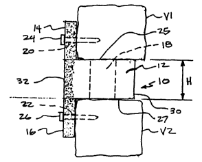
As shown in FIG. 2, one specific application of the present invention
implant 10 contemplates use for fusion of the vertebrae of the cervical spine.
In
this embodiment implant 10 is obtained from the fibula. Body portion 12 can
have
2o any shape, including a specific shape for use in the cervical region, such
as those
shapes identified in U.S. Patent No. 5,989,289 which is incorporated herein by
reference in its entirety. The vertebrae Vl and V2 are accessed from an
anterior
approach using known surgical techniques. The disc material is removed and the
disc space height is restored, if necessary, using known surgical techniques.
25 Implant 10 is inserted into the prepared disc space. Rigid body portion 12
is
adapted to provide structural support between the respective lower endplate of
upper vertebra V1 and the upper endplate of vertebra V2. In the illustrated
embodiment, rigid body portion 12 has a height H sufficient to provide support
for
and maintain the desired spacing between adjacent vertebra V1 and V2. Fusion
30 between vertebrae V 1 and V2 is obtained with bone growth through cavity
18,
CA 02437644 2003-08-06
WO 02/062273 PCT/US02/02585
which is filled with bone growth material 2~. Fusion between the vertebrae can
be
further promoted by reducing the endplates to bleeding bone prior to insertion
of
implant 10.
Implant 10 has upper bearing surface 25 that contacts and supports upper
vertebral body V 1 and lower bearing surface 27 that contacts and supports
implant
on lower vertebral body V2. Body portion 12 has height H between upper
bearing surface 25 and lower bearing surface 27 that is substantially equal to
the
height of disc space formed between vertebra Vl and vertebra V2. It will
understood by those skilled in the art that in the preferred embodiment
illustrated
10 herein, the height H is substantially constant. Furthermore, while a
uniform height
implant is shown in FIG. 2, it will be understood that the implants of the
present
invention may have a tapered height such that the implant could be utilized
for
establishing or maintaining the proper curvature in the spine. Rigid body
portion
12 has sufficient rigidity and structural integrity to substantially maintain
height H
and to withstand normal forces applied to the spinal column. Flange members 14
and 16 need not have such structural requirements, although, preferably, each
assists in the implant stability by maintaining rigid body portion 12 in the
disc
space between the two vertebrae.
Fasteners 24 and 26 are placed through the corresponding fastener bores 20
and 22 in the upper and lower flange members 14 and 16, respectively, to
stabilize
implant 10 in the disc space. Since flange members 14 and 16 are flexible,
they
can be manipulated and positioned adjacent the vertebral bodies outside the
disc
space without the creation of large shear and bending stresses in implant 10
at the
junction between flange members 14, 16 and body portion 12.
While it is contemplated in one specific embodiment that implant 10 have
application for fusion of a cervical region of the spine, application at other
regions
of the spine and at other joints where it is desirable to have a bone implant
with a
rigid body portion with a pair of flexible members extending therefrom are
also
contemplated. Bone implant 10 provides the desirable features of being formed
of
3o a highly successful bone fusion material, i.e. natural bone, with the
advantages of
CA 02437644 2003-08-06
WO 02/062273 PCT/US02/02585
having flexible members made from bone to secure the rigid bone body portion
of
the implant at the implantation location.
In another surgical technique, a tensile force can be applied to upper flange
member 14 prior to insertion of fastener 24. When fastener 24 is secured to
vertebra Vl, the tensile force is released. Fastener 26 can be similarly
inserted
through bore 22 of a tensioned lower flange member 16. The pre-tensioned upper
flange member 14 and pre-tensioned lower flange member 16 thus apply a
compressive load on body portion 12 in the disc space, further promoting
fusion
and incorporation of implant 10 and inhibiting expulsion of implant 10 from
the
disc space.
Referring now to FIG. 3, a further embodiment implant is shown and
designated as 50. Implant 50 is substantially identical to implant 10. Implant
50
includes rigid body portion 52 with flexible upper flange member 54 and
flexible
lower flange member 56 extending therefrom. A first fastener bore 60 is formed
through upper flange member 54 and a second fastener bore 62 is formed through
Iower flange member 56. Body portion 52 includes a cavity 58 in which bone
growth material 64 is placed.
Body portion 52 further includes a number of upper bone engagement
ridges 68 formed on and extending upwardly from upper bearing surface 66 with
an identical set of lower ridges 72 formed on and extending downwardly from
lower bearing surface 70. It will be understood that while ridges have been
shown
in the illustrated embodiment, it is contemplated that there are a variety of
structures, which could provide a surface for effective engagement with the
vertebral bodies to limit expulsion from the disc space. Examples of some such
2s further structures are discussed in U.S. Patent No. 5,989,289. Further, the
endplates or bearing surfaces of the adjacent bony structure can be roughened
or
otherwise shaped to retain the body portion 52 in its inserted position.
Referring now to FIG. 4, there is shown another embodiment implant 80 for
use in vertebral fusion procedures that has particular application in a
posterior
approach to the disc space, although implant 80 may be used in other
approaches,
CA 02437644 2003-08-06
WO 02/062273 PCT/US02/02585
including anterior and lateral approaches. Implant 80 has a rigid body portion
82
with an upper flange member 84 and a lower flange member 86 each extending
from rigid body portion 82 at its trailing end. Implant 80 does not have a
cavity
and can therefore have a width that is less than the width of implants 10 and
50.
5 Access to the disc space between adjacent vertebra is achieved as known in
the art.
Examples of such techniques and posterior bone implants are discussed in PCT
Publication No. WO 00/24327, which is incorporated herein by reference in its
entirety. Once access is achieved, the disc space is distracted if necessary.
Implant 80 is moved into the disc space with body portion 82 positioned
between
10 the adjacent vertebrae and upper flange member 84 and lower flange member
86
positioned adjacent the vertebral bodies outside the disc space. Once body
portion
82 is secured in the disc space D, fasteners can be used to secure the flange
members to the respective adjacent vertebral body. It will be understood that
a
second implant can be placed in the disc space adjacent the first inserted
implant to
provide further stability.
Although not illustrated, the implants of the present invention can have a
slot or threaded bore for engaging a driving tool adapted to position and push
the
implant into the disc space.
The bone for the implants of present invention is preferably selected from
one of the femur, tibia, fibula radius, or ulna or other bone segment having
the
requisite cortical bone strength. It is further contemplated that implant 10
can be
autograft, allograft, or xenograft bone with the bone being treated as known
in the
art for subsequent implantation into the recipient. Specifically, the bone
implant
may be selected from donor bone having sufficient resistance to compression
between the upper and lower surfaces to find application in the intended
environment.
Creation of the demineralized portion of the bone will now be described.
The processing involves the use of donor bone with processing in a clean room
environment within a bone processing facility. Such donor bone may include
allograft from human sources or xenograft from animal sources. Further, it is
CA 02437644 2003-08-06
WO 02/062273 PCT/US02/02585
11
contemplated that as technology advances in the area of bone processing, the
donor
bone may be generated in the manufacturing process, either by bone growth or
by a
processing of constituent components of bone to create artificial materials
having
properties very similar to bone. More specifically, while any available
allogenic or
xenogenic bone stock rnay be utilized for the procedure, cortical bone is
conventionally preferred for spinal fusion for its structural properties,
although
cortical cancellous or cancellous bone may be used depending upon the
particular
requirements for the implant.
In further processing, the connective tissues are removed and the bone is
to cleaned, rinsed, and defatted using a solvent such as ethanol or hydrogen
peroxide.
The bone is then machined or otherwise shaped using conventional techniques to
create its final shape. The upper and lower flange members and, if require,
the
body portion are demineralized to create the required flexible capability.
Penetration of the demineralization fluid into the bone adjacent the desired
area of
flexibility may be controlled by hydrostatic pressure thereby limiting the
area of
demineralization. The amount of mineral removed from the bone may be adjusted
to create the desired amount of flexibility. This demineralization
conventionally
uses an organic acid such as hydrochloric, nitric, or citric acid. Preferably,
the
demineralization solution comprises 0.1 to 1.0 N HCI, most preferably 0.3 N
HCI.
If a xenograft is used, known techniques on the utilization of organic
solvents to
inactivate bone proteins and reduce antigenecity may be applied at this point.
Additionally, the use of glutaraldehyde may take place in order to further
cross-line
the collagen structure following removal of the mineral portion. Once the
implant
has been machined and partially demineralized, it may be stored prior to
insertion.
Although the above-described processing is disclosed herein as a preferred
embodiment, it is contemplated that other suitable processes may be used.
While the invention has been illustrated and described in detail in the
drawings and foregoing description, the same is to be considered as
illustrative and
not restrictive in character, it being understood that only the preferred
embodiments
CA 02437644 2003-08-06
WO 02/062273 PCT/US02/02585
12
have been shown and described and that all changes and modifications that come
within the spirit of the invention are desired to be protected.