Note: Descriptions are shown in the official language in which they were submitted.
CA 02438806 2003-08-19
WO 02/070064 PCT/EP02/01792
INTERNAL INDIFFERENT ELECTRODE DEVICE FOR USE WITH LESION
CREATION APPARATUS AND METHOD OF FORMING
LESIONS USING THE SAME
BACKGROUND OF THE INVENTIONS
1. Field of the Inventions
The present inventions relate generally to electrophysiological devices
and, more particularly, to the indifferent electrodes that are used in
conjunction
with electrophysiological devices.
2. Description of the Related Art
There are many instances where diagnostic and therapeutic elements
must be inserted into the body. One instance involves the treatment of cardiac
conditions such as atrial fibrillation and atrial flutter which lead to an
unpleasant, irregular heart beat, called arrhythmia.
Normal sinus rhythm of the heart begins with the sinoatrial node (or
"SA node") generating an electrical impulse. The impulse usually propagates
uniformly across the right and left atria and the atrial septum to the
atrioventricular node (or "AV node"). This propagation causes the atria to
contract in an organized way to transport blood from the atria to the
ventricles,
and to provide timed stimulation of the ventricles. The AV node regulates the
propagation delay to the atrioventricular bundle (or "HIS" bundle). This
coordination of the electrical activity of the heart causes atrial systole
during
ventricular diastole. This, in turn, improves the mechanical function of the
heart. Atrial fibrillation occurs when anatomical obstacles in the heart
disrupt
the normally uniform propagation of electrical impulses in the atria. These
anatomical obstacles (called "conduction blocks") can cause the electrical
impulse to degenerate into several circular wavelets that circulate about the
obstacles. These wavelets, called "reentry circuits," disrupt the normally
uniform activation of the left and right atria.
Because of a loss of atrioventricular synchrony, the people who suffer
from atrial fibrillation and flutter also suffer the consequences of impaired
hemodynamics and loss of cardiac efficiency. They are also at greater risk of
stroke and other thromboembolic complications because of loss of effective
contraction and atrial stasis.
1
CA 02438806 2003-08-19
WO 02/070064 PCT/EP02/01792
Although pharmacological treatment is available for atrial fibrillation and
flutter, the treatment is far from perfect. For example, certain
antiarrhythmic
drugs, like quinidine, amiodarone, and procainamide, can reduce both the
incidence and the duration of atrial fibrillation episodes. Yet, these drugs
often
fail to maintain sinus rhythm in the patient. Cardioactive drugs, like
digitalis,
Beta blockers, and calcium channel blockers, can also be given to control the
ventricular response. However, many people are intolerant to such drugs.
Anticoagulant therapy also combats thromboembolic complications, but does
not eliminate them. Unfortunately, pharmacological remedies often do not
remedy the subjective symptoms associated with an irregular heartbeat. They
also do not restore cardiac hemodynamics to normal and remove the risk of
thromboembolism.
Many believe that the only way to really treat all three detrimental
results of atrial fibrillation and flutter is to actively interrupt all of the
potential
pathways for atrial reentry circuits.
One surgical method of treating atrial fibrillation by interrupting
pathways for reentry circuits is the so-called "maze procedure" which relies
on
a prescribed pattern of incisions to anatomically create a convoluted path, or
maze, for electrical propagation within the left and right atria. The
incisions
direct the electrical impulse from the SA node along a specified route through
all regions of both atria, causing uniform contraction required for normal
atrial
transport function. The incisions finally direct the impulse to the AV node to
activate the ventricles, restoring normal atrioventricular synchrony. The
incisions are also carefully placed to interrupt the conduction routes of the
most common reentry circuits. The maze procedure has been found very
effective in curing atrial fibrillation. However, the maze procedure is
technically difficult to do. It also requires open heart surgery and is very
expensive. Thus, despite its considerable clinical success, only a few maze
procedures are done each year.
Maze-like procedures have also been developed utilizing catheters
which can form lesions on the endocardium to effectively create a maze for
electrical conduction in a predetermined path. Exemplary catheters are
disclosed in commonly assigned U.S. Patent No. 5,582,609. Typically, the
2
CA 02438806 2003-08-19
WO 02/070064 PCT/EP02/01792
lesions are formed by ablating tissue with one or more electrodes carried by
the catheter. Electromagnetic radio frequency ("RF") energy applied by the
electrodes heats, and eventually kills (i.e. "ablates"), the tissue to form a
lesion. During the ablation of soft tissue (i.e. tissue other than blood, bone
and
connective tissue), tissue coagulation occurs and it is the coagulation that
kills
the tissue. Thus, references to the ablation of soft tissue are necessarily
references to soft tissue coagulation. "Tissue coagulation" is the process of
cross-linking proteins in tissue to cause the tissue to jell. In soft tissue,
it is the
fluid within the tissue cell membranes that jells to kill the cells, thereby
killing
the tissue.
Catheters used to create lesions (the lesions being 3 to 15 cm in
length) typically include a relatively long and relatively flexible body
portion
that has a plurality electrodes supported or near its distal end. The portion
of
the catheter body portion that is inserted into the patient is typically from
23 to
55 inches in length (58.4 to 139.7 cm) and there may be another 8 to 15
inches (20.3 to 38.1 cm), including a handle, outside the patient. The
proximal
end of the catheter body is connected to the handle which includes steering
controls. The length and flexibility of the catheter body allow the catheter
to be
inserted into a main vein or artery (typically the femoral artery), directed
into
the interior of the heart, and then manipulated such that the electrode
contacts the tissue that is to be ablated. Fluoroscopic imaging is used to
provide the physician with a visual indication of the location of the
catheter.
Although catheter-based soft tissue coagulation has proven to be a
significant advance in the medical arts generally and in the treatment of
cardiac conditions in particular, it is not appropriate in every situation.
Physicians may, for example, desire to perform a maze procedure as a
supplemental procedure during an open heart surgical procedure such as a
mitral valve replacement. Physicians may also desire to form lesions on the
epicardial surface. Surgical probes which include a relatively short shaft
that
supports a plurality of electrodes have been introduced in recent years to
facilitate the formation of lesions in these situations. Exemplary surgical
probes are disclosed in commonly assigned U.S. Patent No. 6,142,994, which
3
CA 02438806 2010-08-09
77742-44
is entitled "Surgical Method And Apparatus For Introducing Diagnostic And
Therapeutic Elements Within The Body."
Soft tissue coagulation that is performed using electrodes to transmit
energy to tissue, whether catheter-based or surgical probe-based, may be
performed in both bi-polar and uni-polar modes. Both modes require one or
more indifferent return electrodes. In the uni-polar mode, energy emitted by
the electrodes supported on the catheter or surgical probe is returned through
one or more indifferent patch electrodes that are externally attached to the
skin of the patient. Bi-polar devices, on the other hand, typically include a
number of bi-polar electrode pairs. Both electrodes in each pair are supported
by the catheter or surgical probe and energy emitted by one electrode in a
particular pair is returned by way of the other electrode in that pair.
The uni-polar mode has proven to be superior to the bi-polar mode
because the uni-polar mode allows for individual electrode control, while the
bi-polar mode only allows electrode pairs to be controlled. Nevertheless, the
inventor herein has determined that conventional uni-polar soft tissue
coagulation techniques can be problematic because some patients have
delicate skin and/or skin infections that preclude the attachment of an
indifferent patch electrode to their skin. Poor indifferent electrode/skin
contact
can also be a problem, as can local burning. The inventor herein has also
determined that it would be desirable to improve the likelihood that soft
tissue
coagulation procedures will result in transmural lesions, which is not always-
the case when conventional techniques are employed.
SUMMARY OF THE INVENTIONS
Accordingly, the general object of some embodiments of the present
inventions is to provide methods and apparatus that avoid, for practical
purposes, the aforementioned problems. In particular, one object of some
embodiments of the present inventions is to provide methods and apparatus
that can be used to create lesions in a more efficient manner than
conventional apparatus. Another object of some embodiments of the
present inventions is to provide methods and apparatus that facilitates
uni-polar soft tissue coagulation without the problems associated with
placing external patch electrodes on the patient's skin. Still another
object of some embodiments .of the present inventions
4
CA 02438806 2010-08-09
77742-44
is to provide methods and apparatus that are more likely to produce
transmural lesions than conventional methods and apparatus.
An internal indifferent electrode device in accordance
with an aspect a present invention includes a
flexible shaft, an energy transmission device adapted to be inserted into the
body supported on the shaft, and a connector adapted to mate with the power
return connector of a power supply apparatus. There are a number of
advantages associated with such a device. For example, the present internal,
indifferent electrode device may be placed within the patient and, therefore,
allows physicians to perform uni-polar lesion formation procedures in such a
manner that the issues associated with delicate skin and/or skin infections
are
eliminated.
A method in accordance with another aspect the
present invention includes the steps of positioning an
internal indifferent electrode device within the body on one side of a tissue
structure wall, positioning an electrophysiological device within the body on
the other side of the tissue structure wall, and transmitting energy from the
electrophysiological device to the internal indifferent electrode device.
There are a number of advantages associated with such a method. For
example, in one exemplary implementation, the internal indifferent electrode
device will be placed in the blood pool within the let, atrium and the
electrophysiological device will be placed on the epicardial surface. Such an-
arrangement improves the lesion formation process and increases the
likelihood of the formation of transmurat lesions, as compared to epicardial
processes where an external patch electrode is placed on the patient's skin,
because the resistivity of blood is lower than that of other body tissue. The
lowest resistivity path from the electrophysiological device to the
indifferent
electrode is, therefore, across the atrial wall and through the blood pool in
the
atrium. The present method also eliminates the indifferent electrode/skin
contact problems associated with conventional methods. The, flowing blood
within the atrium will also cool the indifferent electrode, thereby reducing
the
likelihood of local tissue burning that is sometimes associated with external
patch electrodes.
5
CA 02438806 2010-08-09
77742-44
According to a further aspect of the invention, there is an internal
indifferent electrode device for use with a power supply apparatus including a
power output connector and a power return connector, the internal indifferent
electrode device comprising: a flexible shaft defining a distal end, a distal
portion,
a proximal end and a proximal portion; a plurality of spaced energy
transmission
devices adapted to be inserted into a patient's body supported on the distal
portion of the flexible shaft; and an indifferent electrode connector,
including at
least one pin-connect, operably connected to the plurality of spaced energy
transmission devices such that at least two of the spaced energy transmission
devices are connected to the same pin-connect, and adapted to mate with the
power return connector.
According to a still further aspect of the invention, there is a system,
comprising: a power supply apparatus including a power output connector
defining a first configuration and a power return connector defining a second
configuration that is different than the first configuration; and internal
indifferent
electrode device including a flexible shaft defining a distal end, a distal
portion, a
proximal end and a proximal portion a plurality of spaced energy transmission
devices adapted to be inserted into a patient's body supported on the distal
portion of the flexible shaft, and an indifferent electrode connector operably
connected to the plurality of spaced energy transmission devices, defining a
configuration that substantially corresponds to a second configuration, and
adapted to mate with the power return connector.
5a
CA 02438806 2003-08-19
WO 02/070064 PCT/EP02/01792
The above described and many other features and attendant advantages
of the present inventions will become apparent as the inventions become better
understood by reference to the following detailed description when considered
in
conjunction with the accompanying drawings.
BRIEF DESCRIPTION OF THE DRAWINGS
Detailed description of preferred embodiments of the inventions will be
made with reference to the accompanying drawings.
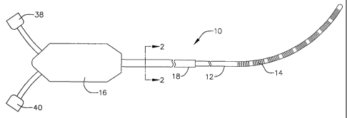
FIGURE 1 is a plan view showing an internal indifferent electrode
'- s
device in accordance with a preferred embodiment of a present invention.
FIGURE 2 section view taken along line 2-2 in FIGURE 1.
FIGURE 3 is a perspective view of one of the connectors in the internal
indifferent electrode device illustrated in FIGURE 1.
FIGURE 4 is a front elevation view of an electrosurgical unit in
accordance with a preferred embodiment of a present invention.
FIGURE 5 is a* plan view of an electrophysiological procedure kit
including a surgical probe and an internal indifferent electrode device in
accordance with a preferred embodiment of a present invention.
FIGURE 6 is a plan view of the surgical probe illustrated in FIGURE 5:
FIGURE 7 is a partial section view of the distal portion of the surgical
probe illustrated in FIGURES 5 and 6.
FIGURE 8 is a section view taken along line 8-8 in FIGURE 6.
FIGURE 9 is a section view taken along line 9-9 in FIGURE 7.
FIGURE 10 is a section view of an alternative probe distal section.
FIGURE 11 is a perspective view of a surgical probe connection device
in accordance with a preferred embodiment of a present invention.
FIGURE 12 is a section view of a human heart during a lesion
formation procedure employing the surgical probe and internal indifferent
electrode kit illustrated in FIGURE 5.
DETAILED DESCRIPTION OF THE PREFERRED EMBODIMENTS
The following is a detailed description of the best presently known modes
of carrying out the inventions. This description is not to be taken in a
limiting
6
CA 02438806 2003-08-19
WO 02/070064 PCT/EP02/01792
sense, but is made merely for the purpose of illustrating the general
principles of
the inventions.
The detailed description of the preferred embodiments is organized as
follows:
I. Internal Indifferent Electrode Device
II. Electrophysiological Procedure Kit
III. Electrodes, Temperature Sensing And Power Control
IV. Methods
The section titles and overall organization of the present detailed
description are
for the purpose of convenience only and are not intended to limit the present
inventions.
This specification discloses a number of structures, mainly in the
context of cardiac ablation, because the structures are well suited for use
with
myocardial tissue. Nevertheless, it should be appreciated that the structures
are applicable for use in therapies involving other types of soft tissue. For
example, various aspects of the present inventions have applications in
procedures concerning other regions of the body such as the prostate, liver,
brain, gall bladder, uterus and other solid organs.
1. Internal Indifferent Electrode Device
As shown by way of example in FIGURES 1-3, an internal indifferent
electrode device 10 in accordance with a preferred embodiment of a present
invention includes a shaft 12 that supports a plurality of electrodes 14. The
electrodes 14 form part of a return path for tissue coagulation energy that is
transmitted by another device in the manner discussed in greater detail below
in Section IV below. Additional information concerning the type, size,
structure
and spacing of the electrodes 14, as well as other electrodes that may be
employed in internal indifferent electrode devices, is provided in Section III
below.
The shaft 12 should be between about 18 inches and about 24 inches
(45.7 cm to 60.9 cm) in length, with an outer diameter between about 2* mm
and about 4 mm. The exemplary embodiment, which is intended for use in
cardiovascular applications, is about 18 inches (45.7 cm) in length with an
outer diameter of about 3 mm. The shaft 12 should also be very flexible.
7
CA 02438806 2003-08-19
WO 02/070064 PCT/EP02/01792
Flexible biocompatible thermoplastic tubing such as unbraided Pebax
material, polyethylene, or polyurethane tubing may be used to form the shaft
12. The proximal end of the shaft 12 is connected to a base 16 by a cable 18.
The base 16 is preferably formed from molded plastic. The cable 18, which is
preferably formed from polyurethane tubing because this material is flexible
and durable, will typically be about 10 feet long (3.05 m). An end cap (not
shown) is secured within the distal end of the shaft 12.
The exemplary internal indifferent electrode device 10 is adapted to be
used in conjunction with an automatic personality module (APM), such as the
Model 882 sold by EP Technologies Inc. of San Jose, California, or an
electrosurgical unit (ESU) such as the Model 4810 which is also sold by EP
Technologies, Inc. and is generally represented by reference numeral 20 in
FIGURE 4. The exemplary ESU 20, which is used to supply and control power
to a surgical probe or other electrophysiological device, includes a plurality
of
displays 22, as well as buttons 24, 26 and 28 that are respectively used to
control which of the electrodes on the electrophysiological device receive
power, the level of power supplied to the electrodes, and the temperature at
the electrodes.
Power is supplied to the surgical probe or other electrophysiological
device by way of a power output connector 30. Lesion creation procedures
sometimes require that up to 2 amperes be returned to the ESU 20 and, to
that end, two indifferent patch electrodes that can handle up to 1 ampere.
apiece are attached to the patient's skin and individually connected to the
APM or ESU in conventional procedures. The indifferent patch electrodes are
connected to a pair of power return connectors 32 and 34 on the ESU 20.
The exemplary internal indifferent electrode device 10 illustrated in
FIGURES 1-3 is provided with eight spaced electrodes 14 that together act
like a large single indifferent return electrode, thereby obviating the need
for
the conventional external patch electrodes described above. Each of the
electrodes is connected to a respective wire 36 that runs through the shaft 12
and the cable 18 into the base 16. There, the wires are separated. Four of the
wires 36 are connected to a connector 38 and the other four wires are
connected to a connector 40. The power return connectors 32 and 34 in the
8
CA 02438806 2003-08-19
WO 02/070064 PCT/EP02/01792
exemplary ESU 20 illustrated in FIGURE 4 each have a rectangular profile
and recessed male pins 36, while the power output connector 30 has a
circular profile. In order to mate with the rectangular power return
connectors
32 and 34, the connectors 38 and 40 on the exemplary internal indifferent
electrode device include a mating portion 42 with a rectangular profile and
longitudinally extending female pin-connects 44. The profile need not be
perfectly rectangular so long as the profile substantially corresponds to that
of
the power return connectors 32 and 34. For example, the middle of the top
and bottom surfaces of mating portion 42 may include longitudinally extending
grooves for mechanical keying with the corresponding connector.
Internal indifferent electrode devices in accordance with the present
invention are not required to be configured in the manner described above.
Instead, their configuration will depend upon the overall systems with which
they are used and the requirements thereof. If, for example, an APM or ESU
only included a single power return connector, then all of the wires 20 from
the electrodes 14 would be connected to a single connector on the internal
indifferent electrode device. Additionally, the shape and style of the power
return connectors 32 and 34 and the corresponding mating portions 42 on the
connectors 38 and 40 need not be rectangular. However, in preferred
embodiments, both should have the same general shape and this shape
should be different than the shape of the power output connector 30, which
need not be circular, to prevent users from attempting to plug an indifferent
electrode device into a power output connector and/or an electrophysiological
device into a power return connector. Alternatively, the power output power
return connectors could have the same general shape and noticeably different
sizes to prevent confusion. Color coding may also be used.
A two-part base member including a re-usable proximal portion that
supports the connectors 38 and 40, a disposable distal portion that supports
the cable 18 and shaft 12, and a pair of mating PC cards that connect the two
portions may also be used.
H. Electrophysiological Procedure Kit
As illustrated for example in FIGURE 5, the internal indifferent
electrode device 10 may form one portion of an electrophysiological
9
CA 02438806 2003-08-19
WO 02/070064 PCT/EP02/01792
procedure kit 46 that also includes.a surgical probe 48 or some other device
that is capable of transmitting energy through tissue to the internal
indifferent
electrode. Two examples of suitable surgical probes are the Cobra surgical
probe and the ThermaLineTM surgical probe, both manufactured by EP
Technologies, Inc. in San Jose, California. Additional examples of surgical
probes that may form a portion of the electrophysiological procedure kit 46
are
provided in U.S. Patent No. 6,142,994. The other tools and devices required
for a particular procedure may be provided within the kit itself or simply
provided separately.
The internal indifferent electrode device 10 and surgical probe 48 are
housed in a sterile package 50 that has a flat rigid bottom portion 52 and a
top
transparent top cover 54 that provides recesses for the internal indifferent
electrode device, surgical probe and any other included tools, thereby
providing a ready to use surgical kit. The bottom portion 52 may be formed
from Tyvek spun bonded plastic fibers, or other suitable materials, which
allow the contents of the package to be sterilized after the tools are sealed
within the package.
Turning to FIGURES 6-10, the exemplary surgical probe 48 includes a
relatively short shaft 50, a handle 52 and a distal section 54. The shaft 50
preferably consists of a hypotube 56, which is either rigid or relatively
stiff, and
an outer polymer tubing 58 over the hypotube. The shaft 50 in the illustrated
embodiment may be from 4 inches to 18 inches (10.2 to 45.7 cm) in length,
and is preferably 6 to 8 inches (15.2 to 20.3 cm), while the distal section 54
may be from 1 inch to 10 inches (2.5 cm to 25.4 cm) in length, and is
preferably 2 to 3 inches (5.1 to 7.6 cm). The handle 52 preferably consists of
two molded handle halves and is provided with strain relief element 60. A
plurality of electrodes 62 or other energy transmission devices are provided
on the distal section 54. There are seven electrodes 62 in the illustrated
embodiment. Additional details concerning the electrodes 62 are provided in
Section III below. A tissue cooling apparatus 64 is positioned over the
electrodes 62 in the exemplary embodiment to cool tissue during lesion
formation procedures.
CA 02438806 2003-08-19
WO 02/070064 PCT/EP02/01792
The distal section 54 is preferably either entirely malleable, entirely
somewhat flexible, or includes a malleable proximal portion and a somewhat
flexible distal portion. A flexible version of the distal section 54
preferably
includes a flexible spring member 66 that is secured to the hypotube 56 and
enclosed in a flexible body 68 formed from Pebax material, polyurethane, or
other suitable materials. [Figure 9.1 The distal end of the spring member 66
is
secured to a tip member 70. An insulating sleeve 72 is placed over the spring
member 66. The spring member 66 may be replaced by a malleable mandrel
74 that is secured to the hypotube 56 and tip member 70, as illustrated for
example in FIGURE 10. An insulating sleeve 76 is placed over the malleable
mandrel 74. Another alternative arrangement is to have a distal section 54
that has a malleable proximal portion and a flexible distal portion composed
of
a short malleable mandrel and a short spring member that are secured to one
another with a crimp tube. The short malleable mandrel would also be
secured to the hypotube 56, while the short spring member would be secured
to the tip member 70.
As used herein the phrase "relatively stiff" means that the shaft (or distal
section or other structural element) is either rigid, malleable, or somewhat
flexible. A rigid shaft cannot be bent. A malleable shaft is a shaft that can
be
readily bent by the physician to a desired shape, without springing back when
released, so that it will remain in that shape during the surgical procedure.
Thus,
the stiffness of a malleable shaft must be low enough to allow the shaft to be-
bent, but high enough to resist bending when the forces associated with a
surgical procedure are applied to the shaft. A somewhat flexible shaft will
bend
and spring back when released. However, the force required to bend the shaft
must be substantial. Rigid and somewhat flexible elements are preferably
formed from stainless steel, while malleable elements may be formed from
annealed stainless steel or beryllium copper. With respect to the spring
member, Nitinol as well as 17-7 and carpenter's steel are preferred.
Additional
information concerning the formation of, and materials for, the relatively
short
shaft 38 and the distal section 54 is provided in U.S. Patent No. 6,142,994.
The exemplary tissue cooling apparatus 64 illustrated in FIGURES 6
and 7 employs conductive fluid to cool tissue during coagulation procedures.
11
CA 02438806 2003-08-19
WO 02/070064 PCT/EP02/01792
More specifically, heat from the tissue being coagulated is transferred to
ionic
fluid to cool the tissue while energy is transferred from the electrodes or
other
energy transmission device(s) to the tissue through the fluid by way of ionic
transport. The conductive fluid is pumped through the tissue cooling
apparatus 64, and preferably continuously, to cool tissue and facilitate the
formation of lesions that are wider and deeper than those that could be
realized with an otherwise identical device which lacks the cooling apparatus.
The exemplary tissue cooling apparatus 64 includes a microporous
outer casing 78 mounted on the probe distal section 54 over the electrodes
62. The proximal and distal ends of the outer casing 78 are secured with
anchoring devices 80 and 82 that are preferably formed from heat shrink
tubing. A fluid transmission space 84 between the inner surface of the outer
casing 78 and the outer surface of the distal section 54 and electrodes 62
extends uninterrupted from a fluid supply line 86 to a fluid drainage tube 88.
[Note arrows F.] The ends of the supply line 86 and drainage tube 88 that
terminate within the outer casing 78 are secured with anchoring devices 80
and 82. The fluid supply line 86 is also secured to the exterior of shaft 50
with
an anchoring device 90.
The microporous outer casing 78 should be no larger than 3 times the
diameter of the electrodes 62 and will preferably be 1.2 to 2 times the
electrode diameter. This translates to a fluid transmission space 84 that is
typically about 0.005 to 0.020 inch (0.12 mm to 0.51 mm), measured inner
surface to outer surface, but can be as large as 0.1 inch (2.5 mm). Of course,
other sizes may be used if they are required by a particular application.
The ionic fluid, which is supplied under pressure from a fluid source
(not shown) to fluid supply line 86, heats up as it passes through the
transmission space 84. The drainage tube 88 directs heated ionic fluid into a
receptacle outside the patient. Removal of the heated ionic fluid is important
because it will be hot enough (typically about 60 C when it reaches the distal
end of the probe) to burn the patient if allowed to drip into the thorax.
The electrically conductive ionic fluid preferably possesses a low
resistivity to decrease ohmic loses, and thus ohmic heating effects, within
the
microporous outer casing 78. The composition of the electrically conductive
12
CA 02438806 2003-08-19
WO 02/070064 PCT/EP02/01792
fluid can vary. In the illustrated embodiment, the fluid is a hypertonic
saline
solution, having a sodium chloride concentration at or near saturation, which
is about 5% to about 25% weight by volume. Hypertonic saline solution has a
relatively low resistivity of only about 5 ohm-cm, as compared to blood
resistivity of about 150 ohm-cm and myocardial tissue resistivity of about 500
ohm-cm. Alternatively, the ionic fluid can be a hypertonic potassium chloride
solution.
With respect to temperature and flow rate, a suitable inlet temperature
for epicardial applications (the temperature will, of course, rise as heat is
transferred to the fluid) is about 0 to 25 C with a constant flow rate of
about 2
to 20 ml/min. The flow rate required for endocardial applications where blood
is present would be about three-fold higher (i.e. 6 to 60 ml/min.). Should
applications so require, a flow rate of up to 100 ml/min. may be employed. In
a closed system where the fluid is stored in a flexible bag, such as the
Viaflex bag manufactured by Baxter Corporation, and heated fluid is
returned to the bag, it has been found that a volume of fluid between about
200 and 500 ml within the bag will remain at room temperature (about 22 C)
when the flow rate is between about 2 ml/min. and 20 ml/min. Alternatively, in
an open system, the flexible bag should include enough fluid to complete the
procedure. 160 ml would, for example, be required for a 20 minute procedure
where the flow rate was 8 ml/min.
The fluid pressure within the microporous outer casing 78 should be.
about 30 mm Hg in order to provide a structure that will resiliently conform
to
the tissue surface in response to a relatively small force normal to the
tissue.
Pressures above about 100 mm Hg will cause the outer casing 78 to become
too stiff to properly conform to the tissue surface. For that reason, the flow
resistance to and from the outer casing 78 should be relatively low.
The pores in the microporous outer casing 78 allow the transport of
ions contained in the fluid through the casing and into contact with tissue.
Thus, when the electrodes 62 transmit RF energy into the ionic fluid, the
ionic
fluid establishes an electrically conductive path through the outer casing 78
to
the tissue being coagulated. Regenerated cellulose membrane materials,
typically used for blood oxygenation, dialysis, or ultrafiltration, are a
suitable
13
CA 02438806 2003-08-19
WO 02/070064 PCT/EP02/01792
microporous material for the outer, casing 78. The thickness of the material
should be about 0.002 to 0.005 inch (0.05 mm to 0.13 mm). Although
regenerated cellulose is electrically non-conductive, the relatively small
pores
of this material allow effective ionic transport in response to the applied RF
field. At the same time, the relatively small pores prevent transfer of
macromolecules through the material, so that pressure driven liquid perfusion
is less likely to accompany the ionic transport, unless relatively high
pressure
conditions develop within the outer casing 78.
Hydro-FluoroTM material, which is disclosed in U.S. application Serial
r. ,
No. 09/573,071, is another material that may be used. Materials such as
nylons (with a softening temperature above 100 C), PTFE, PEI and PEEK
that have micropores created through the use of lasers, electrostatic
discharge, ion beam bombardment or other processes may also be used.
Such materials would preferably include a hydrophilic coating. Microporous
materials may also be fabricated by weaving a material (such as nylon,
polyester, polyethylene; polypropylene, fluorocarbon, fine diameter stainless
steel, or other fiber) into a mesh having the desired pore size and porosity.
These materials permit effective passage of ions in response to the applied
RF field. However, as many of these materials possess larger pore diameters,
pressure driven liquid perfusion, and the attendant transport of
macromolecules through the pores, are also more likely to occur.
Considerations of overall porosity (discussed below) and perfusion rates must,
be taken more into account as pore size increases.
The electrical resistivity of the outer casing 78 will have a significant
influence on lesion geometry and controllability. Low-resistivity (below about
500 ohm-cm) requires more RF power and results in deeper lesions, while
high-resistivity (at or above about 500 ohm-cm) generates more uniform
heating and improves controllability. Because of the additional heat generated
by the increased body resistivity, less RF power is required to reach similar
tissue temperatures after the same interval of time. Consequently, lesions
generated with high-resistivity structures usually have smaller depth. The
electrical resistivity of the outer casing can be controlled by specifying the
pore size of the material, the porosity of the material, and the water
adsorption
14
CA 02438806 2003-08-19
WO 02/070064 PCT/EP02/01792
characteristics (hydrophilic versus. hydrophobic) of the material. A detailed
discussion of these characteristics is found in U.S. Patent No. 5,961,513,
which is entitled "Tissue heating and Ablation Systems and Methods Using
Porous Electrode Structures." A suitable electrical resistivity for epicardial
and
endocardial lesion formation is about 1 to 3000 ohm-cm measured wet.
Generally speaking, low or essentially no liquid perfusion through the
microporous outer casing 78 is preferred. When undisturbed by attendant
liquid perfusion, ionic transport creates a continuous virtual electrode at
the
electrode body-tissue interface. The virtual electrode efficiently transfers
RF
energy without need for an electrically conductive metal surface.
Pore diameters smaller than about 0.1 pm retain macromolecules, but
allow ionic transfer through the pores in response to the applied RF field.
With
smaller pore diameters, pressure driven liquid perfusion through the pores is
less likely to accompany the ionic transport, unless relatively high pressure
conditions develop within the outer casing 78. Larger pore diameters (up to 8
m) can also be used to permit ionic current flow across the membrane in
response to the applied RF field. With larger pore diameters, pressure driven
fluid transport across the membrane is much higher and macromolecules
(such as protein) and even small blood cells (such as platelets) could cross
the membrane and contaminate the inside of the probe. Red blood cells would
normally not cross the membrane barrier, even if fluid perfusion across the
membrane stops. On balance, a pore diameter of 1 to 5 m is suitable for,
epicardial and endocardial lesion formation. Where a larger pore diameter is
employed, thereby resulting in significant fluid transfer through the porous
region, a saline solution having a sodium chloride concentration of about 0.9%
weight by volume would be preferred.
With respect to porosity, which represents the volumetric percentage of
the outer casing 78 that is composed of pores and not occupied by the casing
material, the magnitude of the porosity affects electrical resistance.
Low-porosity materials have high electrical resistivity, whereas high-porosity
materials have low electrical resistivity. The porosity of the outer casing 78
should be at least I% for epicardial and endocardial applications employing a
1 to 5 pm pore diameter.
CA 02438806 2003-08-19
WO 02/070064 PCT/EP02/01792
Turning to water absorption' characteristics, hydrophilic materials are
generally preferable because they possess a greater capacity to provide ionic
transfer of RF energy without significant liquid flow through the material.
Certain other considerations are applicable to those embodiments
which are endocardial in nature and, therefore, operate within the blood pool.
Most notably, there should be essentially no liquid perfusion. This limits
salt or
water overloading caused by transport of the hypertonic solution into the
blood pool. This is especially true when the hypertonic solution includes
potassium chloride. Additionally, the ionic transport rate should below about
10 mEq/min wl en the hypertonic solution includes potassium chloride.
Nonporous outer casings (not shown) that are both electrically and
thermally conductive may be used in place of the porous outer casing 78. A
nonporous outer casing may, for example, have the same configuration as the
porous outer casing 78. As with the porous outer casing, the resistivity
across
the nonporous outer casing should be about 1 ohm-cm to about 3000 ohm-cm
measured wet. The nonporous outer casing should also enable a transfer of
10 W of power with a 100 C temperature gradient across the nonporous outer
casing for each cm of length, as should the porous outer casing 78. For
example, at least 80 W of thermal energy should transfer across a 4 cin
length of outer casing if there exists a 20 C temperature difference between
the inner and outer casing surfaces. Suitable materials for the conductive
nonporous outer casing include plastic materials (such as polyurethane).
which are highly loaded with metallic additives or carbon fibers. Elastomers
(such as silicone rubber) can also be loaded with conductive additives to
achieve thermal and electrical conductivities in the ranges required for this
application.
Other methods of cooling tissue may also be employed where
appropriate. Suitable methods include Joule-Thompson cooling, Peltier diode
cooling (cooling using semiconductor devices that generate heat on one side
while heat is removed on the other) and, in the context.of wettable fluid
retention elements, active vaporization.
As illustrated for example in FIGURE 11, the exemplary surgical probe
48 may be provided with a connection device 92 that connects the surgical
16
CA 02438806 2003-08-19
WO 02/070064 PCT/EP02/01792
probe to the ESU 20. The connection device 92 includes a connector 94 that
may be inserted into an opening 96 in surgical probe handle 52 (FIGURE 6), a
cable 98, and a connector 100 that has a shape and size corresponding to
that of the power output connector 30 on the ESU 20 (FIGURE 4).
Additional details concerning the surgical probe 48 and other similar
devices is provided in U.S. Patent application Serial No. 09/761,981, which is
entitled "Fluid Cooled Apparatus For Supporting Diagnostic And Therapeutic
Elements In Contact With Tissue."
ill. Electrodes, Temperature Sensing And Power Control
The electrodes 14 and 62 are preferably in the form of wound, spiral
closed coils. The coils are made of electrically conducting material, like
copper alloy, platinum, or stainless steel, or compositions such as drawn-
filled
tubing (e.g. a copper core with a platinum jacket). The electrically
conducting
material of the coils can be further coated with platinum-iridium or gold to
improve its conduction properties and biocompatibility. A preferred design is
disclosed in U.S. Patent No. 5,797,905.
Alternatively, the electrodes 14 and 62 may be in the form of solid rings
of conductive material, like platinum, or can comprise a conductive material,
like platinum-iridium or gold, coated upon the device using conventional
coating techniques or an ion beam assisted deposition (IBAD) process. For
better adherence, an undercoating of nickel, silver or titanium can be
applied.
The electrodes can also be in the form of helical ribbons. The electrodes can.
also be formed with a conductive ink compound that is pad printed onto a
non-conductive tubular body. A preferred conductive ink compound is a silver-
based flexible adhesive conductive ink (polyurethane binder), however other
metal-based adhesive conductive inks such as platinum-based, gold-based,
copper-based, etc., may also be used to form electrodes. Such inks are more
flexible than epoxy-based inks. Open coil electrodes may also be employed.
Referring more specifically to the electrodes 62 on the surgical probe 48,
given that the purpose of the electrodes 62 is to transfer energy into the
ionic
fluid, as opposed to directly into tissue, the electrodes 62 may even be
replaced by a straight piece of bare wire.
17
CA 02438806 2003-08-19
WO 02/070064 PCT/EP02/01792
The exemplary electrodes .14 on the internal indifferent electrode
device 10 are preferably 12.5 mm long coil electrodes with 3 mm spacing.
This arrangement will prevent any one of the electrodes 14 from functioning
as a lesion forming device because the large overall surface area of the
electrodes ensures that the current density is low enough to prevent
significant heating. Nevertheless, the electrodes 14 may range from about 4
mm to about 100 mm in length and the exemplary plurality of spaced
electrodes may be replaced by a relatively long single coil electrode or other
energy transmission device.
The exemplary electrodes 62 are preferably coil electrodes that are
about 4 mm to about 20 mm in length. In the preferred embodiments, the
electrodes 62 are 12.5 mm in length with 1 mm to 3 mm spacing, which will
result in the creation of continuous lesion patterns in tissue when
coagulation
energy is applied simultaneously to adjacent electrodes. For rigid electrodes,
the length of the each electrode can vary from about 2 mm to about 10 mm.
Using multiple rigid electrodes longer than about 10 mm each adversely
effects the overall flexibility of the device, while electrodes having lengths
of
less than about 2 mm do not consistently form the desired continuous lesion
patterns.
Referring to FIGURES 6-10, RF power (or other power) from an ESU 20
or other power supply and control device is supplied to the electrodes 62 by
conducting wires 102. The conducting wires 102 are connected to a PC board.
104, which is located within the handle 52 and adapted to mate with the
connector 94. A plurality of temperature sensors 106, such as thermocouples
or thermistors, may be located on, under, abutting the longitudinal end edges
of, or in between, the electrodes 62. Alternatively, a sensor could simple be
located at or near the location where the fluid exits the tissue cooling
apparatus 64 in order to determine the temperature of the fluid at its hottest
point. Signals from the temperature sensors are transmitted to the power
supply and control device by way of wires 108 that are also connected to the
PC board 104. A reference thermocouple may also be provided if desired.
Suitable temperature sensors and power supply and control devices are
disclosed in U.S. Patent Nos. 5,456,682, 5,582,609 and 5,755,715.
18
CA 02438806 2003-08-19
WO 02/070064 PCT/EP02/01792
The amount of power required to coagulate tissue ranges from 5 to 150
w and depends on parameters such as set temperature and the flow rate of
the ionic fluid. For epicardial lesion formation using the cooling apparatus
64
illustrated FIGURES 6 and 7 with a 6 mm diameter, it has been found that an
80 C electrode temperature can be maintained with a 8 ml/min. ionic fluid flow
rate when 75 w of power is supplied to each electrode for about 60 seconds. It
has been found that these parameters produce lesions, both epicardial and
endocardial that are at least 20 mm wide and 18 mm deep.
High voltage gradients have also been used to create lesions by
dielectrically breaking down cell membranes to kill tissue. Voltage gradients
above 500V/cm created by short bursts of RF current are preferred. In the
context of the present inventions, placing the exemplary internal indifferent
electrode device 10 inside a heart chamber (such as the left atrium) and the
surgical probe electrodes 62 on the epicardial surface would increase the
voltage gradient across the heart wall as compared to situations where a
conventional patch electrode is placed on the patient's skin. Such an
arrangement also limits peripheral tissue damage. Additional information
concerning the use of high voltage gradients to create lesions is provided in
U.S. Patent No. 6,107,699.
IV. . Methods
The formation of epicardial lesions is one example of a procedure that
may be performed in accordance with the present inventions. As illustrated
for.
example in FIGURE 12, an internal indifferent electrode device, such as the
exemplary internal indifferent electrode device 10, may be placed within the
blood pool in the left atrium during an epicardial lesion formation procedure
in
which an energy transmitting device, such as the energy transmitting portion
of the surgical probe 48, is placed on the epicardial surface. The internal
indifferent electrode device may, alternatively, be placed within other open
spaces within the heart such as the superior vena cava, the inferior vena cava
or the other chambers depending on the location of the energy transmitting
device.
Access to the heart may be obtained via a thoracotomy, thoracostomy
or median sternotomy. Ports may also be provided for cameras and other
19
CA 02438806 2003-08-19
WO 02/070064 PCT/EP02/01792
instruments. The internal indifferent electrode device 10 may be inserted into
the atrium through an atrial appendage and a purse string technique may be
used to secure it in place and prevent the flow of blood through the
appendage. Alternatively, the internal indifferent electrode device 10 may be
inserted into the atrium by way of the jugular vein using a Seldinger
technique.
Tissue coagulating energy from the surgical probe electrodes 62 will be
transmitted across the atrial wall and through the blood in the left atrium to
the
electrodes 14 on the internal indifferent electrode device 10 to form the
transmural lesion in the atrial wall. Additional lesions may be formed by
moving the energy transmitting portion of the surgical probe 48 to other
places
on the epicardial surface and transmitting energy through tissue to the
internal
indifferent electrode device 10. The internal indifferent electrode device 10
may
also be moved as necessary.
There are a number of advantages associated with placing the an
internal indifferent electrode device within the blood pool in the heart, as
opposed to the patients skin. For example, the resistivity of blood is
relatively
low (about 150 ohm-cm) as compared to other body tissues, while the internal
indifferent electrode device adds less impedance than do external patch
electrodes. Thus, the effectiveness of the lesion formation process will be
improved because the lowest impedance path from the surgical probe
electrodes 62 to the return electrodes 14 on the internal indifferent
electrode.
device 10 is directly across the atrial wall and through the blood. The
flowing
blood will also cool the electrodes 14, thereby reducing the likelihood of
local
tissue burning that is sometimes associated with external patch electrodes.
Additionally, poor tissue contact, which can create problems when external
patch electrodes are employed, is not an issue when an internal indifferent
electrode device is placed into the blood pool.
The present lesion formation methods in accordance with the present
inventions may also be practiced with catheters. For example, instead of
surgically inserting the exemplary internal indifferent electrode 10 into
heart, a
catheter including one or more indifferent electrodes may be percutaneously
advanced into the left atrium or another region or chamber within the heart.
CA 02438806 2003-08-19
WO 02/070064 PCT/EP02/01792
Once the indifferent electrode(s) on the catheter are in the blood pool,
tissue
coagulating energy may be delivered to the epicardial surface by, for
example, the electrodes on the surgical probe 48 to form a transmural lesion
in the manner described above. Alternatively, instead of employing a surgical
probe, a catheter carrying one or more energy emitting electrodes my be
percutaneously directed to a different region or chamber than the catheter
that
is carrying the indifferent electrodes. The energy emitting electrodes on the
catheter may then be used to transmit energy across an internal wall within
the heart to the indifferent electrodes on the other catheter to create a
transmural lesion.
Regardless of the type of device supporting the indifferent electrodes in
the above-described lesion formation methods, the indifferent electrodes will
normally be slightly spaced from the endocardial surface. Nevertheless,
should it be desired that the indifferent electrodes also function as
coagulation
electrodes to further increase the likelihood of a transmural lesion, they may
be positioned against the endocardial surface in close proximity to the
electrodes on the epicardial surface (or other side of an internal wall) that
are
transmitting the energy.
Although the present inventions have been described in terms of the
preferred embodiments above, numerous modifications and/or additions to
the above-described preferred embodiments would be readily apparent to one
skilled in the art. For example, the scope of the inventions includes any
combination of the elements from the various species and embodiments
disclosed in the specification that are not already described. It is intended
that
the scope of the present inventions extend to all such modifications and/or
additions and that the scope of the present inventions is limited solely by
the
claims set forth below.
21