Note: Descriptions are shown in the official language in which they were submitted.
CA 02438934 2003-08-21
WO 02/067978 PCT/US02/04880
CHONDROGENIC POTENTIAL OF HUMAN BONE MARROW-
DERIVED CD105+ CELLS BY BMP
BACKGROUND OF THE INVENTION
The present invention relates to the field of tissue repair including
connective tissue and cartilage repair. More specifically, the present
invention
relates to bone morphogenetic proteins (BMPs), and compositions which play an
important role in chondrogenesis. In particular, the present invention also
relates
to the use of BMPs for the induction of cartilaginous tissue, such as
articular
cartilage, as well as the use of BMPs as therapeutics to partially block the
inhibitory effect of 1L-1.
The present invention further relates to the use of non-tissue culture
expanded cells isolated from bone marrow for use in tissue repair. Further the
present .invention relates to compositions comprising non-tissue culture
expanded cells isolated from bone marrow and bone morphogenetic proteins
IS (BMPs) for the induction of cartilaginous tissue, such as articula.r
cartilage.
Articular cartilage is avascular and aneural and consists of sparsely
embedded chondrocytes in a specialized microenvironment made up of dense
extracellular matrix components. The chondrocytes maintain the architecture of
the cartilage through balanced anabolic and catabolic functions [Curr Opin
Cell
Biol 1(5), 989-94(1989)]. Cartilage injury results in the imbalance of these
functions and is associated with the presence of inflammatory cytokines
including
interleukin-I (1L-I) and tumor necrosis factor (TNF) (R7zeurnatol Irzt 2(2),
49-
53(1982); Arthritis Rheurn 29(4), 461-70(1986); Art7zritis Rheurn 29(2), 262-
73(1986)]. Articular cartilage also has a limited spontaneous repair response
when the cartilage is damaged by trauma or disease processes.
CA 02438934 2003-08-21
WO 02/067978 PCT/US02/04880
Bone marrow-derived cell components play an important role in the
repair of damaged articular cartilage by being the source of progenitor cells
and related growth factors that are required for their differentiation.
Surgical
procedures aim to supply bone marrow-derived mesenchymal precursor cells to
the damaged site by penetrating the underlying subchondral bone with the hope
that the surrounding environment will provide the proper stimulus for
differentiation of these cells. These procedures usually result in
fibrocartilage
and not articular cartilage [ Arthritis Rheutn 42, 1331-1342(1998);in
Articular
Cartilage and Knee Joint Function: Basic Science and Arthroscopy (Ewing, J.
W., ed), Raven Press, New York(1990)]. Repair of damaged articular cartilage
requires the mobilization and differentiation of these precursor cells by
cytokines
and factors at the site of damage. The complex in vivo environment makes it
difficult for the identification of the differentiating factors that are
important in
the transformation of progenitor cells into chondrocytes. [Suh et al.,
Operative
Teclaniques in Orthopaedics. 7:270-278 (1997) O'Driscoll, The Journal of Bone
and Joint Surgery. 80:1795-1812 (1998)]. Bone marrow consists of two
cellular components: hematopoietic cells that reside in close juxtaposition
with the nonhematopoietic cells. Within the nonhematopoietic compartrizent
is a population of cells which shows multipotential mesenchymal properties
and are termed multipotential mesenchymal cells (MMCs) [ Majumdar et al
Journal of Cellular Ph sib. 185:98-106(2000);Majumdar et al Journal of
Cellular Ph Biology. (2001) 189:275-284], mesenchymal stem cells MSCs
[Pittenger et al Science. 284:143-147(1999)] 1999) or mesenchymal progenitor
cells MPCs [Johnstone, et al Experimental Cell Research. 238:265-272 (1998)].
Mesenchymal precursor cells present in the bone marrow have the potential to
differentiate into multiple connective tissue lineages including osteoblasts,
chondrocytes, tenocytes, adipocytes and myocytes when placed in appropriate in
vivo and or itz vitro environments (Science 279, 1528-1530(1998); Bone 19,
421-428(1996); Bone 13, 81-95(1992);Tissue Engiiaeering 4, 415-428
(1998);Journal of Orthpedic Research 16, 406-413(1998)]. These marrow-
2
CA 02438934 2003-08-21
WO 02/067978 PCT/US02/04880
derived mesenchymal cells acquire multipotential mesenchymal
characteristics only after tissue culture expansion. MMCs have been isolated
from the human marrow using an immunoselection procedure that recognizes
a cell surface marker, endoglin (CD105) expressed by these cells [Majumdar
et al Journal of Cellular Physiologx 185:98-106(2000)].
The search for the molecule or molecules responsible for formation of
bone, cartilage, tendon and other tissues present in bone and other tissue
extracts has led to the discovery of a novel set of molecules called the Bone
Morphogenetic Proteins (BMPs). The structures of several proteins,
designated BMP-1 through BMP-16, among others have previously been
elucidated. Bone morphogenetic proteins (BMPs), TGF-~3 and insulin-like
growth factors have been shown to promote chondrogenesis or demonstrate
chondrogenic effect both in vivo and irz vitro ( J Cell Plzysiol 185(1), 98-
106(2000); Bone 19(1 Supply, 1S-12S(1996);Clin Orthop (367 Supply, 5186-
203(1999)]. BMPs are secreted molecules of the TGF-~i superfamily of growth
and differentiation factors that were originally detected in and purified from
demineralized bone ~Pr-oc Natl Acad Sci U S A 85(24), 9484-8(1998)]. Twenty
mammalian BMPs have been identified, and three type II receptors have been
shown to bind BMPs Trends Genet 10(1), 16-21(1994)]. BMP binding leads
to dimerization of type I and II receptors prior to phosphorylation and
signaling through the Smad pathway [Bone 19(6), 569-74(1996)]. BMPs have
been shown to function as key regulators in cartilage and bone development
[Annu Rev Biocherrz 67, 753-91(1998)], and also function in repair and
remodeling of the adult skeletal system Genes Dev 3(11), 1657-68(1989); J
Bone Miner Res 14(10), 1734-41 (1999); The Journal of Borze arid Joint Surgery
82-A(2), 151-160(2000)].
Sox-9, a transcription factor, has been shown to be an important
downstream mediator of the BMP-2 signaling pathway The Journal of Bone and
Joint Surgery 82-A(2), 151-160(2000)]. Sox-9 is characterized by the presence
of a 79 amino acid high mobility group-type DNA-binding domain with high
3
CA 02438934 2003-08-21
WO 02/067978 PCT/US02/04880
homology to that of sex-determining region Y (Sry) ~Curr Opin Genet Dev 7(3),
338-44(1997)]. Sox-9 is expressed during embryonic development in a pattern
that closely parallels that of the gene for Col2Al [Dev Dyrc 209(4), 377-
86(1997); Dev Biol 183(1), 108-21(1997)] and cartilage matrix synthesis
~Geraes
Dev 3(11), 1657-68(1989); )Bone MirterRes 14(10), 1734-41(1999); Tlae
Journal of Bone and Joiret Surgery 82-A(2), 151-160(2000); Science
289(5477), 313-6(2000); JBiol Chern 275(24), 17937-45(2000);Curr Opin
Genet Dev 7(3), 338-44(1997); Dev Dy>z 209(4), 377-86(1997);Dev Biol
183(1), 108-21(1997);Nat Genet 16(2), 174-8(1997)], suggesting a role for Sox-
9 in chondrogenesis and skeletogenesis. It has been shown that upregulation of
Sox-9 enhances the expression of both Col2Al and aggrecan in immortalized cell
lines The Jour~zal of Bone and Joirct Surgery 82-A(2), 151-160(2000).
Proinflammatory molecules including IL-1 and TNF inhibit the expression of
Col2A1 and aggrecan The Journal of Bone and Joint Surgery 82-A(2), 151-
160(2000);) Clin Invest 82(6), 202.6-37(1998);Biochim Bioplays Acta 1052(3),
366-78(1990);) Cell Physiol 166(2), 351-9(1996)]. It has been reported that
the
inhibitory effects of these inflammatory cytokines are mediated by the down
regulation of Sox-9 [ JBiol Cl2em 275(5), 3687-92(2000)]. The inhibitory
effects 1L-1 and TNF, present at elevated levels in osteoarthritis and
rheumatoid
arthritis have been implicated in the breakdown of cartilage in these disease
states
~Rheumatol Int 2(2), 49-53 (2000); Arthritis Rheum 29(4), 461-
70(1986);Arthritis Rheum 29(2), 262-7391986); JBiol Chena 275(5), 3687-
92(2000)].
SUMMARY OF THE INVENTION
By the present invention, Applicants have demonstrated that BMP-2
and BMP-9 promote chondrogenic differentiation of human mesenchymal
precursor cells. Applicants have further demonstrated that the chondrogenic
potential of these BMPs were able to overcome the inflammatory effect of
IL-1. The ability of the BMPs to stimulate matrix synthesis by articular
4
CA 02438934 2003-08-21
WO 02/067978 PCT/US02/04880
chondrocytes and maintain chondrocyte phenotype suggest important
applications including cartilage defect repair and prevention/reversal of
osteoarthritis, chondrocyte phenotype. These BMPs may be particularly
useful for cartilage differentiation, growth, maintenance and repair. The
present invention is therefore directed to composition and methods
comprising BMPs in chondrogenesis: The present invention is further
directed to the use of BMPs to block or partially block the inflammatory
effect of IL-1. The BMPs and other proteins useful in the invention are
further described below.
In the present invention, compositions containing a BMP-
are administered to a patient in need of cartilage repair, or having a disease
or
defect involving cartilaginous tissue, such as osteoarthritis. In a preferred
embodiment, the present invention comprises compositions comprising an
effective amount of BMP-2 or BMP-9.
In the compositions, the protein may be admixed with a pharmaceutically
acceptable vehicle. In a particular embodiment, the composition may
additionally include one or more additional transforming growth factor-~i
proteins
or bone morphogenetic proteins. The composition comprising both a BMP
related protein and another TGF-~3 or BMP may be useful for especially useful
for the treatment of articular cartilage, in which the articular surface,
cartilage,
subchondral bone and/or tidemark interface between cartilage and bone may need
to be repaired.
The present invention also includes methods for cartilaginous tissue
healing and tissue repair, for treating osteoarthritis, or other cartilage
defects, and
for inducing cartilaginous tissue formation in a patient in need of same,
comprising administering to said patient an effective amount of a BMP
composition. In preferred embodiments the composition utilized in the methods
comprises BMP-2 and/or BMP-9. The invention also includes heterodimeric
protein molecules comprising one monomer having the amino acid sequence of a
protein which is useful for the induction of chondrocytes or cartilaginous
tissue,
5
CA 02438934 2003-08-21
WO 02/067978 PCT/US02/04880
and one monomer having the amino acid sequence of another protein of the TGF-
~i subfamily.
The present invention is further directed to compositions comprising
non-tissue culture expanded cells isolated from bone marrow which have
chondrogenic potential. In a preferred embodiment the non-tissue culture
expanded cells are CD105+ cells. In a further embodiment the composition
of the invention comprises non-tissue culture expanded cells isolated from
human bone marrow and a protein which induces the formation of cartilage
and/or bone. These cells isolated from bone marrow and non-tissue culture
expanded demonstrate chondrogenic potential when treated with BMP.
In preferred embodiments, the active agent for treatment of non-tissue
culture expanded cells and for use in other embodiments of the invention
include
one or more proteins selected from the group of proteins known as the
Transforming Growth Factors-Beta (TGF-(3) superfamily of proteins, preferably
selected from the Bone Morphogenetic Proteins (BMPs), the Growth and
Differentiation Factors (GDFs), as well as other proteins, as described more
fully
herein. Osteogenic proteins, DNA sequences, compositions and methods for
producing them, useful in the present invention, are those comprising the BMP
proteins BMP-2, BMP-3, BMP-4, BMP-5, BMP-6 and BMP-7, disclosed for
instance in United States Patents 5,108,922; 5,013,649; 5,116,738; 5,106,748;
5,187,076, 5,459,047, 5,849,880; and 5,141,905; BMP-8, disclosed in PCT
publication W091/18098; and BMP-9, disclosed in PCT publication
W093/00432, BMP-10, disclosed in PCT application W094/26893; BMP-11,
disclosed in PCT application W094/26892, or BMP-12 or BMP-13, disclosed in
PCT application W095/16035, or BMP-15, disclosed in PCT application
W096/36710 or BMP-16, disclosed in co-pending patent application serial
number 08/715/202, filed September 18, 1996. In a preferred embodiment the
BMP is selected from the group consisting of BMP-2 and BMP-9.
Other DNA molecules and the proteins which they encode which may
also be useful include those encoding Vgr-2, and any of the growth and
6
CA 02438934 2003-08-21
WO 02/067978 PCT/US02/04880
differentiation factors [GDFs], including those described in PCT applications
W094/15965; W094/15949; WO95/01801; W095/01802; W094/21681;
W094/15966; and others. Also useful in the present invention may be BIP,
disclosed in W094/01557; and MP52, disclosed in PCT application
WO93/16099. The disclosures of all of the above applications are hereby
incorporated by reference for the disclosure contained therein.
Other DNA molecules and the proteins which they encode which may be
useful including growth factors such as epidermal growth factor (EGF),
fibroblast
growth factor (FGF), transforming growth factor (TGF-cx and TGF-(3), hedgehog
proteins such as sonic, Indian and desert hedgehog, parathyroid hormone and
parathyroid hormone related peptide, cadherins, activins, inhibins, and IGF,
FSH,
frizzled, frzb or frazzled proteins, PDGF and other endothelial growth
factors,
BMP binding proteins such as chordin and fetuin, estrogen and other steroids
as
well as truncated versions thereof, and transcription factors such as wnt
proteins,
mad genes and cbfa.
The disclosures of the above identified applications are hereby
incorporated herein by reference. The unique inductive activities of these
proteins, along with their presence in bone, suggests that they are important
regulators of bone and cartilage repair processes, and may be involved in the
normal maintenance of bone tissue.
The isolated cells of the invention may be treated with the BMP or
other cartilage inducing protein. In further embodiments the DNA sequences
encoding the BMP proteins may be incorporated into the cells using methods
known to those skilled in the art.
Cells directly isolated from the marrow without expansion are preferable
for therapeutic purposes for several reasons. First, selection based on
adherence
preferentially chooses a subpopulation of cells demonstrating a characteristic
which has never been shown to necessarily correlate with chondrogenic
potential.
The liklihood of discarding a potential important subpopulation of cells with
chondrogenic capabilities based on their inability to adhere is diminished. It
is
7
CA 02438934 2003-08-21
WO 02/067978 PCT/US02/04880
possible that i~z vitro responses to differentiation factors during culture
expansion
may alter cell surface characteristics rendering the cells immunogenic to the
host,
and resulting in a graft versus host response after transplantation. Finally,
by the
present invention it has been demonstrated that the chondrogenic
differentiation
of CD105+ cells is not dependent on culture andlor expansion of the cells.
Based
on chondrogenic differentiation of human bone marrow-derived CD105+ cells in
a 3-dimensional matrix in the presence of BMPs in serum-free conditions the
invention therefore features a clinical transplant protocol employing bone
marrow-derived autologous cells transplanted for the repair of articular
cartilage.
This protocol eliminates the extended, expensive and laborious culture
expansion
of the cells.
The present invention therefore further features CD105+ cells isolated
from human marrow- and directly encapsulated in a 3-dimensional matrix of
alginate and cultured in a serum-free medium. The compositions of the
invention
may therefore further comprise a pharmaceutically acceptable vehicle or
suitable
matrix.
The present invention also includes methods for cartilaginous tissue
healing and tissue repair, for treating osteoarthritis, or other cartilage
defects, and
for inducing cartilaginous tissue formation in a patient in need of same,
v comprising administering to said patient an effective amount of a
composition of
the invention comprising non-tissue culture expanded cells isolated from bone
marrow and a bone and/or cartilage inducing protein. In preferred embodiments
the composition comprises CD 105+ cells and BMP.
In a preferred embodiment, the method of the present invention
comprises administering compositions comprising these CD 105+ cells and an
effective amount of BMP-2 or BMP-9. In another embodiment, this method
comprises administering to said patient simultaneously with the cells or
subsequently an effective amount of a composition comprising BMP-2 or BMP-
9.
Various clinical applications have been proposed using primary stem
and progenitor cells [Fucks et al Cell. 100:143-155 (2000)]. Mesenchymal cell
8
CA 02438934 2003-08-21
WO 02/067978 PCT/US02/04880
therapies have been proposed for various tissue repair with culture-expanded
cells [Caplan Journal of Orthopaedic Research. 9:641-650(1991). The present
invention widens the clinical applications of cell-based tissue repair,
procedures which minimize the in vitro manipulation of these cells would be
advantageous. The differentiation potential of mesenchymal cells without
culture expansion as shown by the present invention provide for clinical
treatments of connective tissue diseases.
Description of the Drawing
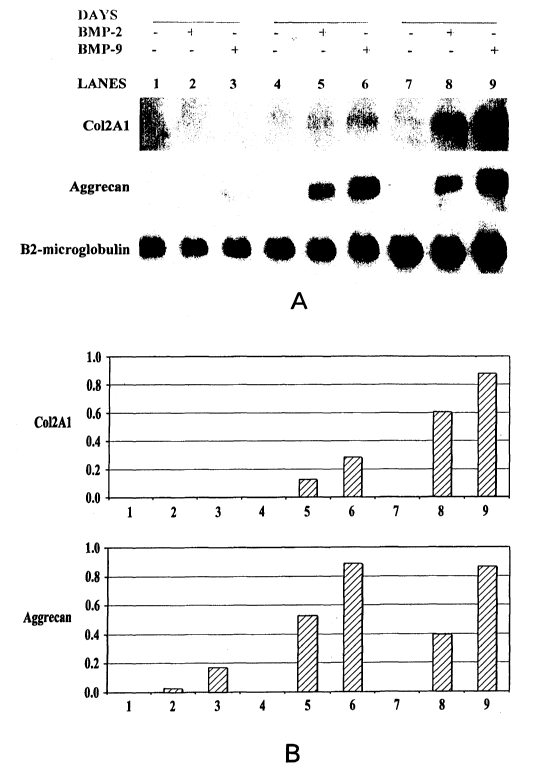
Figure 1 is directed to the induction of the expression of chondrogenic
markers in a time-dependant manner by BMP-2 and BMP-9. Figure 1A, total
RNA was isolated and subjected to Northern analysis with Col2Al, aggrecan
and Sox-9 probes as well as a (32-microglobulin probe as a loading control.
Figure 1.B, quantitation of Col2Al, aggrecan and Sox-9 signals by scanning
densitometry is shown. Lanes 1, 4 and 7-untreated cells; lanes 2,, 5 and 8-
rhBMP-2 treated cells; lanes 3, 6 and 9-rhBMP-9 treated cells.
Figure 2 indicates that BMP-2 and BMP-9 are able to reverse the
expression of chondrogenic markers after IL-1 withdrawal. Figure 2A, total
RNA was isolated and subjected to Northern analysis with Col2Al, aggrecan
and Sox-9 probes as well as a (32-microglobulin probe as a loading control.
Expression of Col2A1, aggrecan and Sox-9 after 14 days in culture are
demonstrated (lanes 1-3). Cell aliquots from the cultures were removed,
washed and cultured for 72 h in media with IL-1 at 200 pg/ml (lanes 4-6).
Cell aliquots of IL-1 treated cells were removed, washed and cultured with or
without BMPs for an additional 96 h (lanes 7-9). Parallel cultures with or
without BMPs were also maintained for the total culture period of 21 days
(lanes 10-12). Figure 2B, quantitation of Col2Al , aggrecan and Sox-9 signals
by scanning densitometry is shown.
Figure 3 indicates the ability of BMP-2 and BMP-9 to overcome the
inhibitory effect of IL-1. Figures 3A and 3C, total RNA was isolated and
subjected to northern analysis with Col2Al and Sox-9 probes as well as a (32-
9
CA 02438934 2003-08-21
WO 02/067978 PCT/US02/04880
microglobulin probe as a loading control. Cells untreated for 21 days (lane 1)
and untreated cells cultured for 14 days were treated with IL-1 for the next 7
days (lanes 2-4). Cells treated with BMP-2 for 14 days (lanes 5), aliquots of
the BMP-2 treated cells were either cultured for an additional 7 days in
increasing concentrations of BMP-2 (lanes 6-8) or in BMP-2 and IL-1
together (lanes 9-17). Cells treated with BMP-9 for 14 days (lanes 18),
aliquots of the BMP-9 treated cells were either cultured for an additional 7
days in increasing concentrations of BMP-9 (lanes 19-21), or in BMP-9 and
IL-1 together (lanes 22-30). Figures 3B and 3D, quantitation of Col2Al and
Sox-9 signals by scanning densitometry is shown.
Figure 4: Gene expression of cartilage specific markers by CD105+ cells
in alginate cultures. CD105+ cells isolated from human bone marrow were
encapsulated in alginate and cultured in a serum-free media (untreated)
supplemented with BMP-2 or BMP-9 for 3 weeks. RT-PCR elisa for type II
collagen, aggrecan and link protein was performed on RNA extracted from the
cells. The bars represent the mean (+ SEM) from 3 donors.
Detailed Description of the Invention
The invention is directed to compositions comprising BMPs which
promote chondrogenic differentiation. These compositons are able to
maintain the expression of chondrocyte specific extracellular matrix
molecules in the presence of osteoarthritis-related physiological levels of IL-
1. The invention is further directed to methods utilzing these compositions.
Preferred BMPs for the compositions and methods are BMP-2 and BMP-9.
The DNA encoding and amino acid sequences of BMP-2 and methods for
preparing the same are described for example in US 5,013,649, the disclosure
of which is incorporated herein by reference. The DNA encoding and amino
acid sequences of BMP-9 are disclosed in W093/00432, the disclosure of
which is incorporated herein by reference.
The present invention is also directed to compositions comprising non-
tissue culture expanded cells isolated from bone marrow which have
CA 02438934 2003-08-21
WO 02/067978 PCT/US02/04880
chondrogenic potential. In a preferred embodiment the non-tissue culture
expanded cells are CD105+ cells. In a further embodiment the composition
of the invention comprises non-tissue culture expanded cells isolated from
human bone marrow and a protein which induces the formation of cartilage
andlor bone. These cells isolated from bone marrow and non-tissue culture
expanded demonstrate chondrogenic potential when treated with BMP.
By the present invention, Applicant have shown these cells have the potential
to be the source of precursor cells important for clinical treatments of
connective tissue diseases including cartilage repair. The isolated cells may
therefore be treated with the BMP proteins. In further embodiments the
sequences encoding the BMPs may be incorporated into the cells.
The compositions and methods of the present invention find
application in the induction of cartilaginous tissue or other tissue formation
in circumstances where such tissue is not normally formed, and has
application in the healing of cartilage, for example articular cartilage
tears,
deformities and other cartilage defects in humans and other animals. Such a
preparation employing a cartilaginous tissue inducing protein may have
prophylactic use in preventing damage to cartilaginous tissue, as well as use
in the improved fixation of cartilage to bone or other tissues, and in
repairing
defects to cartilage tissue. L~e novo cartilaginous tissue formation induced
by
a composition of the present invention contributes to the repair of
congenital,
trauma induced, or other cartilage defects of other origin, and is also useful
in
surgery for attachment or repair of cartilage. The compositions of the
invention may also be useful in the treatment of arthritis and other cartilage
defects. The compositions of the present invention can also be used in other
indications wherein it is desirable to heal or regenerate cartilage tissue.
Such
indications include, without limitation, regeneration or repair of injuries to
the
articular cartilage. The compositions of the present invention may provide an
environment to attract cartilage-forming cells, stimulate growth of cartilage-
forming cells or induce differentiation of progenitors of cartilage-forming
cells.
11
CA 02438934 2003-08-21
WO 02/067978 PCT/US02/04880
The proteins useful in the methods of the present invention are capable
of inducing the formation of cartilaginous tissue. By cartilaginous tissue, it
is
meant chondrocytes, and tissue which is formed by chondrocytes, which
demonstrate the histological and compositional characteristics of cartilage.
These proteins may be further characterized by the ability to demonstrate
cartilaginous tissue formation activity in the assays described below. It is
contemplated that these proteins may have ability to induce the formation of
other types of tissue, such as tendon and ligament.
The compositions for inducing cartilaginous tissue formation of the
present invention may comprise an effective amount of a cartilaginous tissue
inducing protein. In preferred embodiments, the active agent is one or more
proteins selected from the group of proteins known as the Transforming Growth
Factors-Beta (TGF-(3) superfamily of proteins, preferably selected from the
Bone
Morphogenetic Proteins (BMPs), the Growth and Differentiation Factors (GDFs),
as well as other proteins, as described more fully herein. Osteogenic
proteins,
DNA sequences, compositions and methods for producing them, useful in the
present invention, are those comprising the BMP proteins BMP-2, BMP-3,
BMP-4, BMP-5, BMP-6 and BMP-7, disclosed for instance in United States
Patents 5,108,922; 5,013,649; 5,116,738; 5,106,748; 5,187,076, 5,459,047,
5,849,880; and 5,141,905; BMP-8, disclosed in PCT publication W091/18098;
and BMP-9, disclosed in PCT publication WO93/00432, BMP-10, disclosed in
PCT application W094/26893; BMP-11, disclosed in PCT application
W094/26892, or BMP-12 or BMP-13, disclosed in PCT application
WO95l16035, or BMP-15, disclosed in PCT application W096/36710 or BMP-
16, disclosed in co-pending patent application serial number 08/715/202, filed
September 18, 1996. Preferred BMPs for the compositions and methods are
BMP-2 and BMP-9. The DNA encoding and amino acid sequences of BMP-
2 and methods for preparing the same are described for example in US
5,013,649, the disclosure of which is incorporated herein by reference. The
DNA encoding and amino acid sequences of BMP-9 are disclosed in
W093/00432, the disclosure of which is incorporated herein by reference.
12
CA 02438934 2003-08-21
WO 02/067978 PCT/US02/04880
Other DNA molecules and the proteins which they encode which may
also be useful include those encoding Vgr-2, and any of the growth and
differentiation factors [GDFs], including those described in PCT applications
W094/15965; W094/15949; W095101801; W095/01802; W094/21681;
W094/15966; and others. Also useful in the present invention may be BIP,
disclosed in W094/01557; and MP52, disclosed in PCT application
W093/16099. The disclosures of all of the above applications are hereby
incorporated by reference for the disclosure contained therein.
It is expected that the proteins act in concert with or perhaps
synergistically with other related proteins and growth factors. Other DNA
molecules and the proteins which they encode which may be useful, in addition
to
DNA encoding a BMP protein, include DNA molecules encoding other
therapeutically useful agents including growth factors such as epidermal
growth
factor (EGF), fibroblast growth factor (FGF), FGF-4, transforming growth
factor
(TGF-cx and TGF-(3),leukemia inhibitory factor (LIF/HILDA/DIA), insulin-
like growth factors (IGF-I and IGF-II), interleukins such as IL-1 l, hedgehog
proteins such as sonic, Indian and desert hedgehog, parathyroid hormone and
parathyroid hormone related peptide, cadherins, activins, inhibins, and IGF,
FSH,
frizzled, frzb or frazzled proteins, PDGF and other endothelial growth
factors,
BMP binding proteins such as chordin and fetuin, estrogen and other steroids
as
well as truncated versions thereof, and transcription factors such as wrct
proteins,
mad genes and cbfa. Portions of these agents may also be used in
compositions of the present invention. Such a composition may be useful for
treating defects of the junction between cartilage, and bone form
simultaneously at contiguous anatomical locations, and may be useful for
regenerating tissue at the site of cartilage attachment to bone.
The disclosures of the above identified applications are hereby
incorporated herein by reference. The unique inductive activities of these
proteins, along with their presence in bone, suggests that they are important
regulators of bone and cartilage repair processes, and may be involved in the
normal maintenance of bone tissue.
13
CA 02438934 2003-08-21
WO 02/067978 PCT/US02/04880
The cartilaginous tissue-inducing proteins provided herein also include
factors encoded by the sequences similar to those of the naturally-occurring
protein, but into which modifications are naturally provided (e.g. allelic
variations in the nucleotide sequence which may result in amino acid changes
in the polypeptide) or deliberately engineered. For example, synthetic
polypeptides may wholly or partially duplicate continuous sequences of the
amino acid residues of the proteins. These sequences, by virtue of sharing
primary, secondary, or tertiary structural and conformational characteristics
with cartilaginous tissue growth or maintenance factor polypeptides of
naturally-occurring proteins may possess cartilaginous or other tissue growth
or maintenance factor biological properties in common therewith. Thus, they
may be employed as biologically active substitutes for naturally-occurring
cartilaginous tissue inducing polypeptides, and cartilaginous tissue
maintenance polypeptides in therapeutic compositions and processes.
Other specific mutations of the sequences of cartilaginous tissue
inducing proteins described herein involve modifications of glycosylation
sites. These modifications may involve O-linked or N-linked glycosylation
sites. For instance, the absence of glycosylation or only partial
glycosylation
results from amino acid substitution or deletion at asparagine-linked
glycosylation recognition sites. The asparagine-linked glycosylation
recognition sites comprise tripeptide sequences which are specifically
recognized by appropriate cellular glycosylation enzymes. These tripeptide
sequences rnay be asparagine-X-threonine, asparagine-X-serine or
asparagine-X-cysteine, where X is usually any amino acid except proline. A
variety of amino acid substitutions or deletions at one or both of the first
or
third amino acid positions of a glycosylation recognition site (and/or amino
acid deletion at the second position) results in non-glycosylation at the
modified tripeptide sequence. Additionally, bacterial expression of protein
will also result in production of a non-glycosylated protein, even if the
glycosylation sites are left unmodified.
24
CA 02438934 2003-08-21
WO 02/067978 PCT/US02/04880
The preparation and formulation of such physiologically acceptable
protein compositions, having due regard to pH, isotonicity, stability and the
like, is within the skill of the art. The therapeutic compositions are also
presently valuable for veterinary applications due to the lack of species
specificity in TGF-~3 proteins. Particularly domestic animals and
thoroughbred horses in addition to humans are desired patients for such
treatment with the compositions of the present invention.
The therapeutic method includes administering the composition
topically, systemically, or locally as an injectable and/or implant or device.
When administered, the therapeutic composition for use in this invention is,
of course, in a pyrogen-free, physiologically acceptable form. Further, the
composition may desirably be encapsulated or injected in a viscous form for
delivery to the site of tissue damage. Topical administration may be suitable
for wound healing and tissue repair. Therapeutically useful agents other than
the proteins which may also optionally be included in the composition as
described above, may alternatively or additionally, be administered
simultaneously or sequentially with the composition in the methods of the
invention. In addition, the compositions of the present invention may be used
in conjunction with presently available treatments for cartilage injuries,
such
as suture (e.g., vicryl sutures or surgical gut sutures, Ethicon Inc.,
Somerville,
NJ ) or cartilage allograft or autograft, in order to enhance or accelerate
the
healing potential of the suture or graft. For example, the suture, allograft
or
autograft may be soaked in the compositions of the present invention prior to
implantation. It may also be possible to incorporate the protein or
composition of the invention onto suture materials, for example, by freeze-
drying.
As described above, the compositions of the invention may be
employed in methods for treating a number of cartilage defects, such as the
regeneration of cartilaginous tissue in areas of cartilage damage, to assist
in
repair of tears of cartilage tissue, and various other types of tissue defects
or
wounds. These methods, according to the invention, entail administering to a
CA 02438934 2003-08-21
WO 02/067978 PCT/US02/04880
patient needing such cartilaginous tissue or other tissue repair, a
composition
comprising an effective amount of a cartilaginous tissue inducing protein,
such as described in W095/16035, the disclosure of which is hereby
incorporated by reference. These methods may also entail the administration
of a cartilaginous tissue inducing protein in conjunction with at least one of
the BMP proteins described above.
In another embodiment, the methods may entail administration of a
heterodimeric protein in which one of the monomers is a cartilaginous tissue
inducing BMP polypeptide and the second monomer is a member of the TGF-
~3 superfamily of growth factors. In addition, these methods may also include
the administration of a cartilaginous tissue inducing protein with other
growth
factors including EGF, FGF, TGF-cx, TGF-(3, and IGF.
Thus, a further aspect of the invention is a therapeutic method and
composition for repairing cartilaginous tissue, for repairing cartilage as
well
as treating arthritis and other conditions related to arthritis defects. Such
compositions comprise a therapeutically effective amount of one or more
cartilaginous tissue inducing proteins, such as BMP- 2 or BMP-9, in
admixture with a pharmaceutically acceptable vehicle, carrier or matrix.
Culture-expanded MMCs could be engineered to deliver
chondrogenic growth factors to the site of axticular cartilage repair.
Therefore, the combination of MMCs and BMPs may provide and
significantly improve clinical cartilage repair procedures.
The dosage regimen for embodiments of the invention will be
determined by the attending physician considering various factors which
modify the action of the composition, e.g., amount of cartilaginous tissue
desired to be formed, the site of cartilaginous tissue damage, the condition
of
the damaged cartilaginous tissue, the size of a wound, type of damaged tissue,
the patient's age, sex, and diet, the severity of any infection, time of
administration and other clinical factors. The dosage may vary with the type
of matrix used in the reconstitution and the types of additional proteins in
the
composition. The addition of other known growth factors, such as IGF-I
16
CA 02438934 2003-08-21
WO 02/067978 PCT/US02/04880
(insulin like growth factor I), to the final composition, may also affect the
dosage. In general, the amount of recombinant BMP protein useful for
inducing formation of cartilaginous tissue will be in an amount of about 1 to
about 100 ug for a defect of approximately 20 cc in volume. In general, the
amount of recombinant BMP protein useful for inducing maintenance of
cartilaginous tissue will be in an amount of about 1 to about 1000 ng per ml
of solution.
The identification of patients needing treatment for various conditions
including articular cartilage damage may be accomplished by procedures which
are well known in the art. These procedures include measurement of bone
mass/density using dual-energy X-ray absorptiometry (DEXA), Kilgus et al., J.
Bone & Joint Surge, 75-B:279-287 (1992); Markel et al., Acta Orthop Scand,
61:487-498 ( 1990); and quantitative computed tomography (QCT), Laval-Jeantet
et al., J Comt~ut Assist Tomo~r, 17:915-921 (1993); Market, Calcif Tissue Int,
49:427-432 (1991); single-photon absorptiometry, Market et al. Calcif Tissue
Int,
48:392-399 (1991); ultrasound transmission velocity (UTV); Heaney et al.,
JAMA, 261:2986-2990 (1989); Langton et al., Clin Phys Physiol Meas, 11:243-
249 (1990); and radiographic assessment, Gluer et al., J Bone & Mineral Res,
9:671-677 (1994). Other methods of identification are known to those skilled
in
the art. The above publications are hereby incorporated by reference herein.
Progress can be monitored by periodic assessment of cartilaginous
tissue formation, or cartilaginous tissue growth and/or repair. The progress
can be monitored by methods known in the art, for example, X-rays,
arthroscopy, histomorphometric determinations and tetracycline labeling.
Cells directly isolated from the bone marrow without expansion have
several therapeutic advantages. Selection based on adherence preferentially
chooses a subpopulation of cells demonstrating a characteristic which has
never
been shown to necessarily correlate with chondrogenic potential. The liklihood
of discarding a potential important subpopulation of cells with chondrogenic
capabilities based on their inability to adhere is diminished. In vitro
responses to
differentiation factors during culture expansion may alter cell surface
17
CA 02438934 2003-08-21
WO 02/067978 PCT/US02/04880
characteristics rendering the cells immunogenic to the host, and resulting in
a
graft versus host response after transplantation. Finally, by the present
invention
it has been demonstrated that the chondrogenic differentiation of CD105+ cells
is
not dependent on culture and/or expansion of the cells. Based on chondrogenic
differentiation of human bone marrow-derived CD 105+ cells in a 3-dimensional
matrix in the presence of BMPs in serum-free conditions the invention
therefore
features a clinical transplant protocol employing bone marrow-derived
autologous cells transplanted for the repair of articular cartilage. This
protocol
eliminates the extended, expensive and laborious culture expansion of the
cells.
The present invention further features non-tissue culture expanded
CD105+ cells isolated from human marrow- and directly encapsulated in a 3-
dimensional matrix of alginate and cultured in a serum-free medium. A further
embodiment therefore includes a suitable matrix.
The present invention includes methods for cartilaginous tissue healing
and tissue repair, for treating osteoarthritis, or other cartilage defects,
and for
inducing cartilaginous tissue formation in a patient in need of same,
comprising
administering to said patient an effective amount of a composition of the
invention comprising non-tissue culture expanded cells isolated from bone
marrow and a bone and/or cartilage inducing protein. In preferred embodiments
the composition comprises non-tissue culture expanded CD 105+ cells and BMP.
In a preferred embodiment, the present invention comprises compositions
comprising CD105+ cells and an effective amount of BMP-2 or BMP-9. This
method comprises administering to said patient simultaneously with the cells
or
subsequently an effective amount of a composition comprising BMP-2 or BMP-
9.
Various clinical applications have been proposed using primary stem
and progenitor cells [Fuchs et al Cell. 100:143-155 (2000)]. Mesenchymal cell
therapies have been proposed for vaxious tissue repair with culture-expanded
cells [Caplan Journal of Orthopaedic Research. 9:641-650(1991). The present
invention widens the clinical applications of cell-based tissue repair,
procedures which minimize the ira vitro manipulation of these cells is
18
CA 02438934 2003-08-21
WO 02/067978 PCT/US02/04880
advantageous. The differentiation potential of mesenchymal cells without
culture expansion as shown by the present invention provide for clinical
treatments of connective tissue diseases.
The compositions of the invention may include an appropriate matrix
and/or sequestering agent as a carrier. For instance, the matrix may support
the composition or provide a surface for cartilaginous tissue formation and/or
other tissue formation. The matrix may provide slow release of the protein
and/or the appropriate environment for presentation thereof. The
sequestering agent may be a substance which aids in ease of administration
through injection or other means, or may slow the migration of protein from
the site of application.
The choice of a carrier material is based on biocompatibility,
biodegradability, mechanical properties, cosmetic appearance and interface
properties. The particular application of the compositions will define the
appropriate formulation. Potential matrices for the compositions may be
biodegradable and chemically defined. Further matrices are comprised of pure
proteins or extracellular matrix components. Other potential matrices are
non-biodegradable and chemically defined. Preferred matrices include collagen-
based materials, including sponges, such as Helistat° (Integra
LifeSciences,
Plainsboro, N.J.), or collagen in an injectable form, as well as sequestering
agents, which may be biodegradable, for example hyaluronic acid derived.
Biodegradable materials, such as cellulose films, or surgical meshes, may also
serve as matrices. Such materials could be sutured into an injury site, or
wrapped
around the cartilage.
Another preferred class of carrier are polymeric matrices, including
polymers of poly(lactic acid), poly(glycolic acid) and copolymers of lactic
acid
and glycolic acid. These matrices may be in the form of a sponge, or in the
form
of porous particles, and may also include a sequestering agent. Suitable
polymer
matrices are described, for example, in W093/00050, the disclosure of which is
incorporated herein by reference.
19
CA 02438934 2003-08-21
WO 02/067978 PCT/US02/04880
Additional optional components useful in the practice of the subject
application include, e.g. cryogenic protectors such as mannitol, sucrose,
lactose,
glucose, or glycine (to protect the protein from degradation during
lyophilization), antimicrobial preservatives such as methyl and propyl
parabens
and benzyl alcohol; antioxidants such as EDTA, citrate and BHT (butylated
hydroxytoluene); and surfactants such as poly(sorbates) and
poly(oxyethylenes).
Preferred families of sequestering agents include blood, fibrin clot
and/or cellulosic materials such as alkylcelluloses (including
hydroxyalkylcelluloses), including methylcellulose, ethylcellulose,
hydroxyethylcellulose, hydroxypropylcellulose, hydroxypropyl-
methylcellulose, and caxboxymethylcellulose, the most preferred being
cationic salts of carboxymethylcellulose (CMC). Other preferred
sequestering agents include hyaluronic acid, sodium alginate, polyethylene
glycol), polyoxyethylene oxide, carboxyvinyl polymer and polyvinyl
alcohol). The amount of sequestering agent useful herein is 0.5-20 wt%,
preferably 1-10 wt% based on total formulation weight, which represents the
amount necessary to prevent desorbtion of the protein from the polymer
matrix and to provide appropriate handling of the composition, yet not so
much 'that the progenitor cells are prevented from infiltrating the matrix,
thereby providing the protein the opportunity to assist the activity of the
progenitor cells.
In particular embodiments of the invention, components of the
composition may be encapsulated in a resorbable polymer delivery system,
such as polylactic acid, polyglycolic acid or copolymers thereof,
polyorthoesters, polyorthocaxbonates, and other polymers. Suitable polymers
are disclosed for example in EP 0145240, the disclosure of which is hereby
incorporated by reference. Alternatively, the BMP may be encapsulated in
liposomes For example, liposome delivery of TGF-(3 protein is described in
United States Patent 5,206,023, 5,270,300; and 5,368,858, the disclosure of
each of which are hereby incorporated by reference. Both of these delivery
systems may be modified to provide for release of BMP at a later time, or
CA 02438934 2003-08-21
WO 02/067978 PCT/US02/04880
over a more sustained time period, allowing for the beneficial effects of the
BMP on chondrocyte and cartilage maintenance to act complementary to the
beneficial effects of the BMP on induction of chondrocytes and cartilaginous
tissue.
The proteins and compositions of the present invention may also be
useful for treating cell populations, such as embryonic cells or stem cell
populations, to enhance or enrich the growth, differentiation and/or
maintenance of the cells. The treated cell populations may be useful for gene
therapy applications.
The following examples illustrate practice of the present invention in
demonstrating BMP promotion of chondrogenic differentiation of human
mesenchymal precursor cells and the ability of these BMPs to overcome the
inflamatory effect of IL-1.
The following examples further illustrate practice of the present invention in
demonstrating BMP promotion of chondrogenic differentiation of non-tissue
culture expanded human mesenchymal precursor cells.
Example 1
Isolation and Culture Expansion of MMCs.
Human MMCs were isolated according to previously reported procedure
~Jouf-rcal of Cellular Physiology 176, 57-669(1998)]. Mononuclear cells
(MNCs) were isolated from human bone marrow samples according to a
modification of a previously reported method ~J Cell Physiol 185(1), 98-
106(2000). Total nucleated cells in the marrow sample was diluted to a
concentration of 7x10⁶ cells per ml with isolation buffer (calcium and
magnesium
free phosphate-buffered saline (PBS), 2% bovine serum albumin (BSA), 0.6%
sodium citrate and 1 % penicillin-streptomycin). Thirty to 35 ml of the
diluted
cell suspension was layered over 15 ml of Ficoll-Paque (Pharmacia, Piscataway,
NJ) and centrifuged at 800xg for 20 min. The MNCs were collected, counted,
washed with magnetic-activated cell sorting (MACS) buffer (PBS with 0.5%
BSA and 2mM EFTA, pH7.2) and resuspended in MACS buffer at 2-4X 10⁸
21
CA 02438934 2003-08-21
WO 02/067978 PCT/US02/04880
cells per ml. 1X 10$ MNCs were incubated with 0.2 ml of anti-human CD105
antibody-microbeads for 45 min at 4 °C and CD105~ cells were isolated
using the
MS+ columns (Miltenyi Biotec) according to the manufacturer's
recommendation. The CD105- cells were collected as the column eluate, while
the CD105+ cells remained attached to the column. CD105+ cells were recovered
from the column by removing it from the magnet and flushing out the cells with
MACS buffer. CD105+ cells were plated in 185 cmz Nunclon Solo flasks (Nunc
Inc., Naperville, IL) at a density of 5-7.5X10⁵ cells per flask and cultured
in
complete medium consisting of Alpha-MEM supplemented with 10°70 fetal
bovine serum (FBS, Hyclone, Logan, UT) and 1 % antimycotic-antibiotic at
37°C
in 5% COZ in air. Medium was changed after 48 h and thereafter every 3-4 days.
At day 14, cells were detached by incubation with 0.05% trypsin-EDTA and
designated primary (p0) and replated for expansion at a density of 1X10⁶ cells
per flask as passage 1 cells. The cells reached 90% of confluence in 6-7 days,
after which they were either passaged as mentioned, used in other assays or
stored in 90% FBS and 10% dimethyl sulphoxide in liquid nitrogen for future
use. The cells used for this study were derived from passage 2 or passage 3.
The
CD 105+ were designated MMCs as they are of mesenchymal origin and have
multipotential differentiation capability.
Example 2
Culture of MMCs in Alginate
The MMCs were encapsulated in alginate according to a previously
reported procedure ~J Cell Physiol 185(1), 98-106(2000)]. Briefly, MMCs were
detached and washed with wash buffer (0.15M NaCl, 25 mM Hepes, pH 7.0) and
resuspended at a density of 25X106 per ml in 1.2% alginate in wash buffer.
Individual beads of the cell suspension were expressed through a 20-gauge
needle
into a solution containing 102 mM CaCl2 and 25 mM Hepes (pH7.0). The beads
were allowed to polymerize for 10 min, washed once in wash buffer, three times
in complete medium and cultured overnight in the same medium at 37°C
with 5%
COZ in air. The next day, the medium was changed to chemically defined
22
CA 02438934 2003-08-21
WO 02/067978 PCT/US02/04880
medium consisting of DMEM with high glucose, 100nM dexamethasone
(Sigma), 50 ~,glm1 ascorbic acid-2-phosphate (WAKO Pure Chemicals, Tokyo,
Japan), 100 ~,glml of sodium pyruvate (Life Technologies), 50 ~,glml proline
(Sigma), 1 % TTS-Premix (Becton Dickenson, Bedford, MA) and 100 ng/ml
rhBMP-2 or rhBMP-9. The medium was changed twice a week for the next 2-3
weeks. RNA was isolated from the cells in order to examine the expression of
genes at various time intervals during the culture.
For 1L-1 studies, MMCs were cultured in alginate for 14-21 days in the
serum-free media with or Without BMPs for chondrogenic differentiation. At day
14, beads were washed and cultured in the media with 200 pg/ml of IL-1 (Roche
Biochemicals, Indianapolis, IN) for 72 h (dayl7) as reported in a previous
study
[Journal of CellularPlaysiology 176, 57-66(1998)]. The beads were again
washed and cultured in media with BMPs for 96 h (day 21). RNA was isolated
from the cells at days 14, 17 and 21. In the next study, MMCs were cultured in
alginate beads for 14 days and subsequently for an additional 7 days in BMPs
alone, IL-1 alone or in various combinations of IL-1 and BMPs together. RNA
was isolated from the cells at days 14 and 21.
In another study the expanded cells were encapsulated in a 3-dimensional
alginate matrix and cultured in serum free media with or without ILr 1 l and
BMP
9 to analyze their potential to undergo chondrogenic differentiation.
Example 3
RNA Preparation and Northern Analysis
Culture-expanded MMCs were encapsulated in alginate beads and
cultured in serum-free media. For RNA isolation, the beads were transferred to
cell recovery buffer (55mM Sodium Citrate, 0.15M NaCl and 25mM Hepes, pH
7.0), incubated for l Omin at 4° C to release the cells from the
alginate matrix and
centrifuged at 1400 X g for 15 min at 4°C to recover the cells. Total
RNA was
prepared from the cell pellet by a previously reported procedure [Jouf-ycal of
Cellular Physiology 176, 57-66(1998)]. Briefly, cell pellet was resuspended in
lysis buffer (4M guanidinium isothiocynate, 0.03M sodium acetate and 0.4 g/ml
23
CA 02438934 2003-08-21
WO 02/067978 PCT/US02/04880
of cesium chloride) and the lysate was layered over 5.7M cesium chloride and
centrifuged for 18 h at 155,000 X g in a SW40 rotor (Beckman, Palo Alto, CA).
The RNA pellet was dissolved in water at 0.5-1 mg/ml. For northern blot
analysis, 5 ~g of total RNA per sample was fractionated on 1 % formaldehyde-
agarose gels. Subsequent to electrophoresis, RNA was transferred onto a
positive
charged nylon membrane, BrightStar-Plus (Ambion, Austin, TX). The gene
probes for northern analysis was prepared as PCR amplified products using
specific oligonucleotide primers as listed in Table I and the amplified
products
Were confirmed by sequencing. These probes were radiolabeled by [cx 3zP]dCTP
(NEN Life Sciences Products) using the random primer method as recommended
by the manufacturer (Amersham Pharmacia Biotech Inc., NJ) and hybridized in
ultrahyb solution (Ambion) overnight. Col2Al hybridization was performed at
54°C and all others were performed at 42°C. The filters were
washed in
2xSSCl0.1%SDS at room temperature and then in 0.lxSSCl0.1%SDS at 65°C
for
30 min. The filter was exposed to X-ray film overnight. Hybridization signals
were quantified by scanning the x-ray image and utilizing Image Gauge (Fuji
Photo and Film Co, Japan). Col2Al, aggrecan, COMP and Sox-9 mRNA levels
were corrected for RNA loading by normalization with ~32-microglobulin. For
detection of Col2A1 gene expression by reverse transcriptase-polymerase chain
reaction (RT-PCR), RNA was prepared from cells isolated from 2-3 solubilized
beads by the RNeasy kit (Qiagen, Valencia, CA). RT-PCR was performed using
total RNA as a template, oligonucleotide primers, RNA PCR core kit (Perkin-
Elmer, Norfolk, CT). The amplified products were analyzed on a 1.2% E-gels
(Invitrogen, Caxlsbad,CA).
24
CA 02438934 2003-08-21
WO 02/067978 PCT/US02/04880
Table I Oligonucleotide primers used for PCR amplification of probes
Oligonucleotide primers / Size
Reference /
(bp)
Human BETA2-MICROGLOBUL1N 270
Majumdar et al
Sense:5'-TCTGGCCTTGAGGCTATCCAGCGT-3' (1998)
Antisense: 5'-GTGGTTCACACGGCAGGCATACTC-3'
Human COL2A1 451 X16468
Sense:5'-AACCTGGACAGAGGGAAGC-3'
Antisense: 5' -GGGGCCAGGATTCCATTAC-3'
Human AGGRECAN 450 M55172
Sense: 5'-TACTCTGGGTTTTCGTGACTC-3'
Antisense: 5' -CGATGCCTTTCACCACGACTT-3'
Human Sox-9 381 246629
Sense: 5'-CCCGATCTGAAGAAGGAGAGC-3'
Antisense: 5' -GTTCTTCACCGACTTCCTCCG-3'
Human COMP 501 L32137
Sense: 5'-GCAGATGCTTCGGGAACTGCA-3'
Antisense:5'-TTGATGCACACGGAGTTGGGG-3'
Example 4
BMP-2 and BMP-9 Induce Chondrogenic Differentiation of MMCs
Mesenchymal stem and progenitor cells cultured in the presence of TGF-
~(3 undergo chondrogenic differentiation (Tissue Erzgif2eering 4, 415-
428(1998);The Jourlzat of Bone afzd Joifzt Surjery 80(12), 1745-1757(1998)].
MMCs cultured in alginate and stimulated by TGF-~33 express Col2Al and
differentiate along the chondrogenic lineage [J Cell Physiol 185(1), 98-
106(2000)]. To examine the effect of BMPs on chondrogenic differentiation,
monolayer culture-expanded MMCs from 3 donors were further cultured in a 3-
dimensional alginate matrix in the presence of 100 nglml of rhBMP-2 or rhBMP-
9. RT-PCR was performed on RNA extracted from cells at various time intervals
CA 02438934 2003-08-21
WO 02/067978 PCT/US02/04880
to detect the expression of Col2Al. The results showed that Col2A1 expression
was induced in cells in all the 3 donors between day 8 and day 14. At day 14,
total RNA was prepared from the cells in culture and northern blot analysis
was
performed. The results showed that all 3 donors responded to stimulation by
both
BMPs and expressed Col2A1, while the expression was undetected in untreated
cells. The results also showed that BMP-9 treatment induced a higher level of
Col~A1 expression than BMP-2.
It has been shown in mouse studies that BMP-2 upregulated the
expression of chondrogenic-related transcription factor Sox-9 which in turn
regulates the expression of Col2Al and aggrecan [Tlze Journal of Bone and
Joiht
Surgery 82-A(2), 151-160(2000)]. To evaluate this observation in human MMCs
and to further analyze the state of chondrogenic lineage progression, the
expression of chondrogenic specific markers including aggrecan, cartilage
oligomeric matrix protein (COMP) and Sox-9was investigated. MMCs from
multiple donors were analyzed and the results were from one donor,
representative of the group. The expression of both aggrecan and COMP
increased in response to BMPs in a manner similar to Col2Al expression. Sox-9
showed a basal level of expression in the untreated cells, but underwent an
observable upregulation in cells treated with BMPs. Therefore, it is
contemplated that MMCs in alginate cultures are induced to differentiate along
the chondrogenic lineage by BMP-2 and BMP-9.
In the Il-11 and BMP-9 study cells cultured without IL-11 or BMP-9 and
with IL-11 alone did not express Col2Al. Cells cultured with increasing
concentration of BMP-9 showed a significant level of Col2A1 expression when
compared to untreated cells. Cells cultured in combination of II,-1 l and BMP-
9
showed higher levels of Col2Al expression than BMP-9 alone. The ynergistic
effect of IL-11 and BBMP-9 was optimum at concentration of lOng/ml of Il-11
where the increase in Col2Al expression was over 5-fold greater than BMP-9
alone.
26
CA 02438934 2003-08-21
WO 02/067978 PCT/US02/04880
Example 5
BMP Induction of a Steady Increase in Expression of the Chondrogenic
Markers in MMCs in a Time-Dependant Manner
RNA isolated from MMCs cultured in alginate beads at day 5, 10 and 15
were subjected to northern analysis. The results showed (Figure 1) that Col2A1
gene expression was detected at day 10 with a sequential increase at day 15.
Aggrecan expression was detected at day 5 and showed a progressive increase
with days in culture. Again BMP-9 treated cells showed higher expression of
both Col2A1 and aggrecan than BMP-2 treated cells. The results indicate that
aggrecan expression responds earlier than Col2A1 expression when MMCs are
treated with BMP-2 and BMP-9.
Example 6
BMPs Overcome the Inflammatory Effect of IL-1
Previous studies have shown that IL-1 inhibits chondrogenic specific
genes including Col2Al and aggrecan by downregulation of the transcription
factor Sox-9 Baoclaif~a Biophys Acta 1052(3), 366-78(1990); J Cell Physiol
166(2), 351-9(1996); JBiol Chem 275(5), 3687-92(2000)]. The effect of IL-1 on
chondrogenic differentiated MMCs was examined by analyzing the expression of
these three genes. The results showed that at day 14, BMP-2 and BMP-9 induced
expression of Col2Al and aggrecan and, as mentioned before, BMP-9 treatment
caused a higher level of expression than BMP-2 treatment (Figure 2, lanes 1-
3).
Removal of BMPs and addition of IL,-1 for 72 h led to a reduced level of
expression of Col2Al, aggrecan and Sox-9 (lanes 4-6). Removal of IL,-1 and
addition of BMPs for an additional period of 96 h resulted in rebound
expression
of the three genes (lanes 7-9) with the expression level similar to cells
continuously exposed to BMPs for 21 days (lanes 10-12).
The effect of BMPs in the presence of IL-lwas then examined. Cells
were allowed to differentiate along the chondrogenic lineage for 14 days
followed
by 7 days in the presence of 1L,-1 alone, BMPs alone, and 1L-1 and BMPs
together. The results showed (Figure 3) that untreated cells (lane 1) and 14
days
27
CA 02438934 2003-08-21
WO 02/067978 PCT/US02/04880
untreated cells exposed to an increasing concentration of lL-1 for an
additional 7
days showed no Col2Al expression and no appreciable expression of Sox-9
(lanes 2-4) as expected. Cells treated with BMPs progressed through
chondrogenic differentiation during the first 14 days as demonstrated by
Col2A1
expression (lanes 5 and 18). Exposure of the chondrocytic-differentiated cells
to
an increasing concentration of BMP-2 for an additional 7 days (lanes 6-8)
increased expression of Col2Al and Sox-9. A similar effect was observed with
BMP-9 treatment (lanes 19-21), although maximal response to BMP-9 was
achieved at the lowest dose of 100 ng/ml. Both BMP-2 and BMP-9 were able to
partially prevent the IL-1 induced suppression of Col2A1 and Sox-9 (lanes 9-17
and 22-30 respectively) especially at the lowest concentration of IL-1 used
(20
pg/ml). In addition, BMP-9 was able to maintain a higher level of Col2Al
expression at all concentrations of IL,-1. These observations showed that BMPs
are potent molecules that have the ability to function as anabolic factors in
an
environment containing inflammatory cytokines.
Example 7
Isolation and Culture Expansion of CD105+ cells
Human MMCs were isolated according to a previously reported
procedure [Majumdar et al.,Journal of Cellular Ph s~gy. 185:98-106 (2000)].
Mononuclear cells (MNCs) were isolated from human bone marrow, washed
with magnetic-activated cell sorting (MACS) buffer consisting of phosphate
buffered saline with 0.5% BSA and 2mM EDTA, pH7.2. The cells were
resuspended in MACS buffer and 1X 10$ MNCs were incubated with 0.2 ml of
anti-human CD105 antibody-microbeads (Miltenyi Biotec, Auburn, CA) for 45
min at 4 °C. The cells were then washed and separated on a magnetic
column
MS+ (Miltenyi Biotec) according to the manufacturer's recommendation. The
column eluate consisted of the CD105-cells. The attached CD105'~ cells were
recovered by removing the column from the magnet and flushing out the cells
with MACS buffer. A small fraction (5-7.5X10⁵) of the CD105+ cells were
plated in 185 cm2 Nunclon Solo flasks (Nunc Inc., Naperville, IL) in complete
28
CA 02438934 2003-08-21
WO 02/067978 PCT/US02/04880
medium consisting of Alpha-MEM supplemented with 10% fetal bovine serum
(FBS, Hyclone, Logan, IJT) and 1 % antimycotic-antibiotic (Life Technologies,
Gaithersburg, MD) at 37°C in 5% COZ in air to analyze for the plating
efficiency
of the cells. At day 14, cells were detached by 0.05% trypsin-EDTA (Life
Technologies) treatment and stored in 90% FBS and 10% dimethyl sulphoxide in
liquid nitrogen for future use.
Example 8 Flow Cytometry
Analysis of cell surface molecules was performed according to a previously
reported procedure [Majumdar et al.,Journal of Cellular Physiology. 176:57-66.
(1998)]. Column-isolated CD105~ cells were washed in FACS buffer (2% BSA,
0.1 % sodium azide in PBS) and aliquots ( 1X 10⁵-1X 10⁶) of cells were
incubated with
anti-human CD45 fluorochrome-conjugated monoclonal antibodies (Pharmingen;
San Diego). Cells were washed and the cell pellet was resuspended in FAGS
buffer
with 1 % paraformaldehyde. Nonspecific fluorescence was determined using equal
aliquots of cell preparation that were incubated with mouse isotype monoclonal
antibodies. Data were collected by analyzing 10,000-50,000 events on a Becton
Dickson instrument (San Jose, CA) using Cell-Quest software.
Example 9 Culture of CD105+ cells in alginate
The CD 105+ cells were encapsulated in alginate by modification of a
previously reported procedure (Majumdar et al., 2000). Cells were washed with
wash buffer (0.15M NaCI, 25 mM Hepes, pH 7.0) and resuspended at a density of
10-20X106 per ml in 1.2% alginate in wash buffer. Individual beads of the cell
suspension were then slowly expressed through a 20-gauge needle into a
solution
containing 102 mM CaCl2 and 25 mM Hepes (pH7.0). The beads were allowed to
polymerize for 10 min, washed once in wash buffer, three times in complete
medium
and cultured overnight in the same medium at 37°C with 5% COZ in air.
The next
day, the medium was changed to chemically defined medium (Majumdar et al.,
2000). The alginate beads were cultured in the above medium (untreated) or
medium supplemented with 100 ng/ml BMP-2, or BMP-9 (treated). The medium
29
CA 02438934 2003-08-21
WO 02/067978 PCT/US02/04880
was changed twice a week for the next 3 weeks. MNCs as well as CD105- cells
were also encapsulated in alginate beads at the same cell concentration and
cultured
similarly..
Example 10 RNA Preparation and Analysis
CD 105+ cells from multiple donors were encapsulated in alginate beads and
cultured for 3 weeks. At the end of the culture period, cells were recovered
from the
beads, total RNA was extracted from the cell pellet by RNeasy kit (Qiagen,
Valencia, CA) and reverse transcriptase-polymerase chain reaction (RT-PCR)-
elisa
was performed according to a previously reported procedure (Majumdar et al.,
1998). Briefly, RT-PCR was performed using total RNA as a template,
oligonucleotide primers (Table IIJ, RNA PCR core kit (Perkin-Elmer, Norfolk,
CT),
and substituting the deoxy-nucleotides with digoxigenin-labeled nucleotides
(Roche
Biochemicals, Indianapolis, IN) to label the amplified products. Elisa was
performed as recommended by the manufacturer (Roche Biochemicals,
Indianapolis,
IN). The data for each untreated and treated sample from each donor were
normalized to b2-microglobulin. Progression was determined by cartilage
specific
markers including type II collagen, aggrecan and link protein. For each donor
the
expression of type II collagen, aggrecan and link protein was computed as fold
increase over untreated. The result shown is the mean fold increase for 3
donors.
RT-PCR elisa analysis (Figure 4) showed that in comparison to untreated cells,
BMP-2 and BMP-9 treated cells had a significant increase in gene expression
for
chondrogenic specific genes including Col2Al , aggrecan and link protein
suggesting
that the CD105+ cells were undergoing chondrogeic differentiatiton.. In
contrast,
MNCs as well as CD 105- cells did not show any evidence of chondrogenic
differentiation.
CA 02438934 2003-08-21
WO 02/067978 PCT/US02/04880
Table II Oligonucleotide primers for RT-PCR elisa.
Oligonucleotide primers / Size Reference /
5'-biotinylated probes (bp) Accession #
Human BETA2-MICROGLOBULIN 270 Majumdar et al.
Sense: 5'-TCTGGCCTTGAGGCTATCCAGCGT-3' (1998)
Antisense: 5'-GTGGTTCACACGGCAGGCATACTC-3'
Probe: 5'-Biotinylated CATCGATCCGACATTGAAGTTGAC-3'
Human COL2A1 451 X16468
Sense:5'-TCCCAAAGGTGCTCGAGGAGA-3'
Antisense: 5'-CTCACCACGATCACCCTTGAC-3' ,
Probe: 5'-Biotinylated GAGAGAGGATTCCCTGGCTT-3'
Human AGGRECAN 450 M55172
Sense: 5'-TACTCTGGGTTTTCGTGACTC-3'
Antisense:5'-CGATGCCTTTCACCACGACTT-3'
Probe: 5'-Biotinylated GAGAAGGAGGTAGTGCTGCT
Human LINK PROTEIN 361 X17405
Sense: 5'-GCTGATTTCAATCTGCTGGG -3'
Antisense: 5'-GTCTGTGATGACCAGAGAAGC -3'
Probe: 5'-Biotinylated AGCATTTGGCTCAGGAATCC-3'
Example 11 Immunohistochemistry
Immunohistochemistry was performed to detect the presence of type lI
collagen protein in the alginate according to a previously reported procedure
(Majumdar et al., 2000). Alginate beads from cultures were washed with water
and incubated in 100mM barium chloride for lOmin for irreversible
polymerization. The beads were then washed with water again and fixed in 10%
buffered formalin and embedded in paraffin. Sections of alginate beads were
31
CA 02438934 2003-08-21
WO 02/067978 PCT/US02/04880
incubated with goat anti-type II collagen antibody (Southern Biotechnology
Associates, Birmingham, AL). Immunoreactivity was detected by incubating
sections with biotinylated anti-goat antibody and horse radish peroxidase H
reagents (Vector Laboratories, Burlingame, CA). Signal was developed by
treating the sections with peroxidase substrate 3,3'-diaminobenzidine (DAB)
and
HZO2. Images were recorded on 35mm slide film and multipanel figures were
made with Photoshop (Adobe Systems, San Jose, CA). Experimental controls
consisted of alginate sections stained with nonimmune primary antibody
followed
by secondary antibody. The results indicate that in comparison to the
untreated
cells, BMP-2 and BMP-9 treated cells showed a significant presence of type II
collagen protein. Type IC collagen protein was present in the intercellular
region
due to the secretion of the protein by the differentiating cells and
subsequent
entrapment in the alginate matrix. Alginate sections stained with nonimmune
pri-many antibody did not show any immunoreactivity.
While this invention has been particularly shown and described with
references to preferred embodiments thereof, it will be understood by those
skilled in the art that various changes in form and details may be made
therein
without departing from the scope of the invention encompassed by the appended
claims.
32