Note: Descriptions are shown in the official language in which they were submitted.
CA 02439278 2003-08-26
WO 02/069818 PCT/USO1/25455
DEMINERALIZED BONE-DERIVED IMPLANTS
FIELD OF THE INVENTION
The invention is related to implants formed from bone. More particularly,
the invention is related to implants formed from partially demineralized or
demineralized
bone.
BACKGROUND OF THE INVENTION
Bone grafts have become an important and accepted means for treating bone
fractures and defects. In the United States alone, approximately half a
million bone grafting
procedures are performed annually, directed to a diverse array of medical
interventions for
complications such as fractures involving bone loss, injuries or other
conditions
necessitating immobilization by fusion (such as for the spine or joints), and
other bone
defects that may be present due to trauma, infection, or disease. Bone
grafting involves the
surgical transplantation of pieces of bone within the body, and generally is
effectuated
through the use of graft material acquired from a human source. This is
primarily due to
the limited applicability of xenografts, transplants from another species.
Orthopedic autografts or autogenous grafts involve source bone acquired
from the same individual that will receive the transplantation. Thus, this
type of transplant
moves bony material from one location in a body to another location in the
same body, and
has the advantage of producing miW mal immunological complications. It is not
always
possible or even desirable to use an autograft. The acquisition of bone
material from the
body of a patient typically requires a separate operation from the
implantation procedure.
Furthermore, the removal of material, oftentimes involving the use of healthy
material from
the pelvic area or ribs, has the tendency to result in additional patient
discomfort during
rehabilitation, particularly at the location of the material removal. Grafts
formed from
synthetic material have also been developed, but the difficulty in mimicking
the properties
of bone limits the efficacy of these implants.
As a result of the challenges posed by autografts and synthetic grafts, many
orthopedic procedures alternatively involve the use of allografts, which are
bone grafts from
other human sources (normally cadavers). The bone grafts, for example, are
placed in a
host bone and serve as the substructure for supporting new bone tissue growth
from the host
bone. The grafts are sculpted to assume a shape that is appropriate for
insertion at the
fracture or defect area, and often require fixation to that area as by screws
or pins. Due to
-1-
CA 02439278 2003-08-26
WO 02/069818 PCT/USO1/25455
the availability of allograft source material, and the widespread acceptance
of this material
in the medical community, the use of allograft tissues is certain to expand in
the field of
musculoslceletal surgery.
With respect to the overall structure of a given bone, the mechanical
properties vary throughout the bone. For example, a long bone (leg bone) such
as the femur
has both compact bone and spongy bone. Cortical bone, the compact and dense
bone that
surrounds the marrow cavity, is generally solid and thus carries the majority
of the load in
major bones. Cancellous bone, the spongy inner bone, is generally porous and
ductile, and
when compared to cortical bone is only about one-third to one-quarter as
dense, one-tenth to
one-twentieth as stiff, but five times as ductile. While cancellous bone has a
tensile strength
of about 10-20 MPa and a density of about 0.7, cortical bone has a tensile
strength of about
100-200 MPa and a density of about 2. Additionally, the strain to failure of
cancellous bone
is about 5-7%, while cortical bone can only withstand 1-3% strain before
failure. It should
also be noted that these mechanical characteristics may degrade as a result of
numerous
factors such as any chemical treatment applied to the bone material, and the
manner of
storage after removal but prior to implantation (z.e. drying of the bone). In
addition, bones
have a grain direction similar to the grain found in wood, and thus the
strength of the bone
varies depending on the orientation of the grain.
Notably, implants of cancellous bone incorporate more readily with the
surrounding host bone, due to the superior osteoconductive nature of
cancellous bone as
compaxed to cortical bone. Furthermore, cancellous bone from different regions
of the body
is known to have a range of porosities. For example, cancellous bone in the
iliac crest has a
different porosity from cancellous bone in a femoral head. Thus, the design of
an implant
using cancellous bone may be tailored to specifically incorporate material of
a desired
porosity.
Demineralization of cortical, cancellous, and corticocancellous bone of
autograft, allograft, and xenograft types is known. In one form, bone powder
or chips are
chemically processed using an acid such as hydrochloric acid, chelating
agents, electrolysis
or other treatments. The demineralization treatment removes the minerals
contained in the
natural bone, leaving collagen fibers with bone growth factors including bone
morphogenic
protein (BMP)
The use of expandable materials as a prosthetic element is disclosed in U.S.
Patent No. 5,545,222 to Bonutti. Materials disclosed which expand when they
come in
contact with water or other fluids include PEEK (polyether-etherketone), a
desiccated
_2_
CA 02439278 2003-08-26
WO 02/069818 PCT/USO1/25455
biodegradable material, or a desiccated allograft. As an example, a tendon can
be
compressed in a desiccated state, and as it imbibes water it expands and
creates a firmer
loclc or tighter fit in the host site.
A shaped, swollen demineralized bone and its use in bone repair is disclosed
in U.S. Patent No. 5,298,254 to Prewett et al. In general, cortical allogeneic
bone tissue is
preferred as the source of bone. Demineralized bone is contacted with a
biocompatible
swelling agent for a period of time sufficient to cause swelling of the piece.
A flexible implant using partially demineralized bone is disclosed in U.S.
Patent No. 6,206,923 to Boyd et al. The bone implant has a first substantially
rigid portion
and a second substantially rigid portion which are joined by an intermediate
portion that has
been at least partially demineralized to create an area of flexibility in the
bone implant. The
pair of rigid bone portions cooperate to provide support for spacing between
adjacent
vertebra.
Demineralized bone has been disclosed for use as artificial ligaments in U.S.
Patent No. 5,092,887 to Gendler. Completely or partially demineralized
cortical bone is
sliced in strips and rods of approximately 0.1-1.5 centimeters wide and 0.1-
1.5 centimeters
thick with compliant elasticity and longitudinal strength similar to natural
ligaments and
tendons. The strips or rods are used as artificial ligaments for in vivo
replacement, repair
and augmentation of damaged ligaments, tendons or other fibrous tissue that
permanently
connects first and second body members such as the femur and tibia. Disclosure
of a
segmentally demineralized bone implant is found in U.S. Patent No. 6,090,998
to Grooms
et al. The implant comprises a first mineralized portion or segment, and a
second, flexible,
demineralized portion or segment that are produced by machining a piece of
cortical bone.
A textured, demineralized, and unitary mammalian bone section for
providing a rigid, foraminous, collagen scaffold for allogenic skeletal
reconstruction is
disclosed in U.S. Patent No. 5,112,354 to Sires. Texturing or pore formation
is carried out
prior to demineralization to permit completeness of demineralization and
additionally
promote osteoinduction due to the increased surface area. Pores of between 200
~.m and
2000 ~m are created with a laser. The depth of the holes in the bone may be
varied.
Also disclosed in U.S. Patent No. 5,899,939 to Boyce et al. is a bone-derived
implant for load-supporting applications. The implant is formed of one or more
layers of
fully mineralized or partially demineralized cortical bone and, optionally,
one or more layers
of some other material such as fully demineralized bone or mineral substances
such as
hydroxyapatite. The layers constituting the implant are assembled into a
unitary structure to
-3-
CA 02439278 2003-08-26
WO 02/069818 PCT/USO1/25455
provide an implant with load-supporting properties. Superimposed layers are
assembled
into a unitary structure such as with biologically compatible adhesives.
U.S. Patent No. 5,556,430 discloses flexible membranes produced from
organic bone matrix for skeletal repair and reconstruction. Completely or
partially
demineralized organic bone is sliced into thin sheets. The bone may be
perforated prior to
demineralization, to increase the osteoinductivity of the final bone product.
Similarly, U.S.
Patent No. 5,298,254 to Prewett et al, discloses demineralized bone sliced
into a thin sheet
which can be used to patch an injury.
A cortical bone interference screw is disclosed in U.S. Patent No. 6,045,554
to Grooms et al. The interference screw has a cortical surface into which a
self tapping
thread is machined.
In addition, U.S. Patent No. 5,053,049 to Campbell discloses the use of
milling, grinding, and pulverizing to produce pulverized bone with the desired
particle size.
The pulverized bone can then be combined with any suitable biologically
compatible or
inert carrier substance, which should have a consistency that imparts the
desired flexible
texture to the pulverized bone/carrier suspension, or should solidify to the
desired
consistency after molding or casting.
Despite these developments, there exists a need for implants formed from
partially or fully demineralized cancellous bone. Furthermore, there exists a
need for
implants formed of bone that have been selectively masked during
demineralization so that
portions of the bone are at least partially demineralized while other portions
are
substantially remain in the mineralized state.
SUMMARY OF THE INVENTION
The present invention relates to a bone sheet for implantation, the sheet
including a demineralized field substantially surrounding at least one
mineralized region.
The sheet may be formed of cortical bone, and the at least one mineralized
region may
define at least one hole in the sheet. The at least one hole may be configured
and
dimensioned to receive at least one fastener. The sheet may have a thickness
of between
about 0.5 rnrn and about 3 mm. The sheet may have ribs or projections
providing localized
thiclmess.
The present invention also relates to a method of forming a flexible bone
sheet including: providing a sheet of cortical bone; creating at least one
hole in the cortical
sheet which is configured and dimensioned to receive a fastener; masking the
cortical sheet
proximate the at least one hole to create a masked region surrounding the at
least one hole;
-4-
CA 02439278 2003-08-26
WO 02/069818 PCT/USO1/25455
and demineralizing the cortical sheet around the masked region. A plurality of
maslcing
elements may be removably attached to the sheet to provide maslcing proximate
the at least
one hole. The masking may be provided by at least one of the group consisting
of tape,
paint, and a coating. The method further may include creating perforations in
the sheet that
are substantially smaller than the at least one hole. In addition, the method
may further
include cutting a bone section along a spiral path.
The present invention further relates to a sheet formed of bone including two
or more strips of bone each having a bone grain orientation, wherein the bone
grain
orientation of at least one strip is disposed transverse to the grain
orientation of another
strip. The strips may be interwoven, and may be selected from mineralized
bone,
demineralized bone, and partially demineralized bone. A portion of at least
one strip may
be demineralized. The strips may be interwoven to form a plurality of
generally parallel
rows and a plurality of generally parallel columns. The strips may have a
width between
about 1 mm and about 6 mm, a thickness of between about 0.5 mm and about 2 mm,
and a
width of about 5 mm and a tluckness of about 1 mm. The bone strips may be
unitary in
construction. At least one strip may be formed by braiding two or more bone
fibers. Each
bone strip may have a longitudinal axis and the bone grain orientation may be
substantially
parallel thereto.
The present invention also is related to a bone defect filler including a
demineralized cancellous bone section in a first geometry. The first geometry
is
compressible and dryable to a second geometry smaller than the first geometry,
and the
second geometry is expandable and rehydratable to a third geometry larger than
the second
geometry.
The present invention is further related to a method of filling an open region
with cancellous bone, the method including: demineralizing a section of
cancellous bone;
compressing the section; drying the compressed section; inserting the section
into the open
region; rehydrating the section; and allowing the section to expand to fill
the open region.
In addition, the present invention relates to a method of providing a bone
implant including: demineralizing a cancellous bone section having a first
geometry;
compressing the bone section from the first geometry to a second geometry
smaller than the
first geometry; drying the bone section while the bone section has
approximately the second
geometry; and inserting the bone section into a space. When the bone section
is inserted
into the space, the bone section may be at least partially surrounded by a
wall while having
approximately the second geometry. The method may further include expanding
the bone
section from the second geometry to a third geometry larger than the second
geometry, as
-5-
CA 02439278 2003-08-26
WO 02/069818 PCT/USO1/25455
well as allowing the bone section to expand to contact the wall. The bone
section may be
expanded by rehydrating.
The invention also relates to a method of maintaining a distance between
vertebral bodies including: demineralizing a bone section having a first
geometry; drying
the bone section; inserting the bone section in between vertebral bodies; and
expanding the
bone section to a second geometry. The bone section may be demineralized
cortical bone.
The drying step may include freeze drying the bone section, and the bone
section may be
expanded by hydrating. The method further may include compressing the bone
section.
The bone section may be cancellous bone. The method may be used to replace the
nucleus
of a vertebral disc.
The invention further relates to a method of replacing nucleus of a vertebral
disc including: providing a demineralized cortical bone section having a first
geometry;
inserting the bone section in between vertebral bodies; and expanding the bone
section to a
second geometry. The bone section may be expanded by hydrating, and may be
expanded
to a height in the second geometry which is larger than the height in the
first geometry. The
bone section in the second geometry may have a top surface and a bottom
surface, each of
the top and bottom surfaces being configured to approximately match a concave
vertebral
endplate. The top and bottom surfaces may be convex with a radius of between
about 50
mm and about 70 mm.
In addition, the invention relates to an implant including demineralized
cancellous bone capable of being softened and compressed into a smaller first
shape and
hardened in said first shape, and capable of expanding into a second shape
larger than said
first shape when resoftened and permitted to expand. The bone may be softened
by
hydration and may be hardened by dehydration. The bone may be configured and ,
dimensioned to by received in an anatomical void. The first shape may be
smaller than the
anatomical void, and the second shape may be about the same as the shape of
the
anatomical void. The second shape also may span a lateral dimension of the
anatomical
void. The implant may further include cortical bone.
In some embodiments, the implant is configured and dimensioned to be
disposed in a burr hole or void in the cranial region of the skull. The
implant may form a
burr hole cap including an upper cortical bone section and a Lower,
demineralized
cancellous bone section, at least the lower section being sized to fill the
burr hole when in
the expanded second shape. In some embodiments, the implant is generally T-
shaped and
includes an upper cortical bone section and a lower, demineralized cancellous
bone section.
In the expanded second shape, at least the lower section may be sized to
contact walls of the
-6-
CA 02439278 2003-08-26
WO 02/069818 PCT/USO1/25455
void. The upper section may have an arcuate portion for generally matching the
contour of
the skull. Slits may be included extending through at least a portion of one
or both of the
cortical bone and cancellous bone.
The present invention further is related to a bone sheet for implantation, the
sheet including a demineralized field surrounding mineralized regions.
The present invention also is related to a bone defect filler including a
demineralized cancellous bone section in a first geometry. The first geometry
is
compressible and dryable to a second geometry smaller than the first geometry,
and the
second geometry is expandable and rehydratable to a third geometry larger than
the second
geometry.
The present invention is further related to a method of filling an open region
with cancellous bone, the method including: demineralizing a section of
cancellous bone;
compressing the section; drying the compressed section; inserting the section
into the open
region; rehydrating the section; and allowing the section to expand to fill
the open region.
In addition, according to one embodiment, a cranial void filler is described
herein and comprises an upper mineralized cortical bone section, and a lower,
at least
partially demineralized cortical bone section, wherein the lower section is
adapted and
configured to contact walls of a cranial void. Preferably, the upper and lower
sections form
a T-shape. One or more slits may extend through the upper or lower sections,
or both, or
portions thereof. The slits in the lower section may be colinear with the
slits in the upper
section. The upper section may have a rounded upper surface portion and also
may have a
curved lower surface portion.
According to another embodiment, a plate is described. The plate of this
embodiment comprises a unitary body formed of cortical bone with a pair of
portions
having a first width and a central portion disposed therebetween having a
second width, the
first width being greater than the second width, and the body having at least
one partially
demineralized region wherein the at least one partially demineralized region
provides
flexibility to the plate. The plate further may comprise a plurality of
fastener holes. The
body may have a central longitudinal axis and a first at least partially
demineralized region
that is coaxial therewith. The first at least partially demineralized region
may extend
substantially across the entire length of the body. The fastener holes may be
disposed
proximate the ends of the body. The fastener holes may be disposed on a
central
longitudinal axis, and a first at least partially demineralized region may be
coaxial
therewith. One or more second at least partially demineralized regions may be
disposed
transverse to the first at least partially demineralized region. The one or
more second at
CA 02439278 2003-08-26
WO 02/069818 PCT/USO1/25455
least partially demineralized regions may be generally perpendicular to the
first at least
partially demineralized region. The one or more second at least partially
demineralized
regions may intersect one or more fastener holes. The body may be relatively
thin compared
to its length or width. The at least partially demineralized region may extend
substantially
across the second width. The body may have a central longitudinal axis and the
at least
partially demineralized region may extend transverse to the central
longitudinal axis.
The plate may further comprise a plurality of fastener holes and the body
may be generally dog-bone-shaped. The body may have a central longitudinal
axis, and the
length of the body along the central axis may be between about 10 mm and about
20 mm.
The first width may be between about 4 mm and about 7 mm and the body may have
a
thickness between about 1 mm and about 3 mm. The body may have a length of
about 15
mm, a first width of about 5 mm, and a thickness of about 2 mm.
In a further embodiment, an implant comprising a unitary section of cortical
bone having a first portion that is mineralized and a second portion that is
at least partially
demineralized, wherein the mineralized portion includes a plurality of slits
to facilitate
bending of the unitary section is described.
A method of forming an implant also is described. The method comprises
the steps of obtaining cortical fibers; at least partially demineralizing the
fibers; allowing the
fibers to clump together; and allowing the fibers to dry in a clumped state.
The fibers may
be allowed to dry in a mold and may be pressed while they are clumped
together. The fibers
may be obtained by milling or other processes.
A still further embodiment describes an implant for maintaining a space in a
bisected vertebrae. The implant according to this embodiment comprises a
cortical bone
cord having first and second free ends adapted for engaging exposed portions
of the lamina,
and a region positioned between the first and second ends, wherein the region
is at least
partially demineralized to provide flexibility. The free ends may be
mineralized. The cord
may comprise a pair of at least partially demineralized regions with a
mineralized region
therebetween. The at least partially demineralized region may be centrally
located between
mineralized free ends.
$RIEF DESCRIPTION OF THE DRAWINGS
Preferred features of the present invention are disclosed in the accompanying
drawings, wherein:
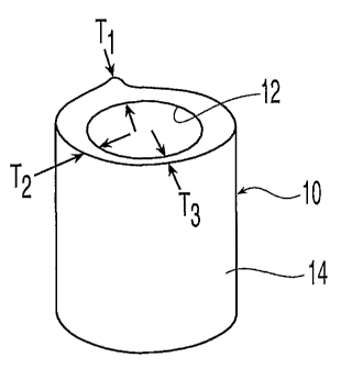
FIG. 1 shows a bone section of a femur;
FIGS. 2-3 show a cortical shell of the present invention;
_g_
CA 02439278 2003-08-26
WO 02/069818 PCT/USO1/25455
FIG. 4 shows a cortical sheet formed from the cortical shell of FIG. 2;
FIG. 4A shows a spiral cortical sheet formed from the cortical shell of FIG.
2;
FIG. 4B shows a cortical sheet according to an alternative embodiment;
FIGS. 5-7 show various forms of cancellous bone of the present invention;
FIG. 8 shows a cage for filling with cancellous bone of FIG. 7;
FIG. 8A shows a cage filled with cancellous bone of FIG. 7;
FIG. 9 shows a femur section for filling with demineralized cancellous bone
of FIG. 6;
FIG. 9A shows a femur section filled with demineralized cancellous bone of
FIG. 6;
FIG. 10 shows a partially demineralized cancellous bone cylinder of the
present invention;
FIG. 11 shows a woven bone implant of the present invention;
FIG. 12 shows a demineralized cortical bone implant for nucleus
replacement according to the present invention;
FIGS. 13-15 show ligament replacements using bone implants of the present
invention;
FIGS. 16-18 show the use of partially demineralized bone struts for disc
replacement according to the present invention;
FIGS. 19-21 show a bendable implant of the present invention;
FIGS. 22-23 show bone cords of the present invention;
FIG. 24 shows a cortico-cancellous demineralized bone of the present
invention;
FIGS. 25-27 show cranial flap void and burr hole filling according to the
present invention;
FIGS. 28-29 show dogbone-shaped plates of the present invention;
FIG. 30 shows a cortical tack or suture anchor of the present invention; and
FIG. 31 shows an embodiment of a ribbed bone sheet.
DETAILED DESCRIPTION OF THE PREFERRED EMBODIMENT
The present invention in one embodiment is directed to an implantable bone
sheet that exhibits semi-pliable properties over portions of the sheet, while
exhibiting semi-
rigid properties over other portions. The variation in properties is achieved
by the selective
demineralization of bone preferably selected from a femur, tibia, humerus,
fibula, ulna, and
radius. The terms "demineralization," "demineralized" and "at least partially
-9-
CA 02439278 2003-08-26
WO 02/069818 PCT/USO1/25455
demineralized" as used herein are intended to refer to fully demineralized
bone or partially
demineralized bone. The term "fully demineralized" refers to bone where the
minerals have
been substantially completely removed from the bone whereas the term
"partially
demineralized" refers to bone where at least some portion of the minerals have
been
removed. As will become apparent, the degree of demineralization will depend
upon the
characteristics sought to be achieved in the implant.
Turning to FIG. 1, a bone section 10 of a femur has an inner surface 12, and
an outer surface 14 which initially conforms to the natural shape of the bone.
The wall
thickness of bone section 10 varies, as indicated by thicknesses Tl, T2, and
T3. As shown in
FIG. 2, bone section 10 may be machined to have a relatively uniform wall
thickness T4,
forming a cortical shell 16. Initially, cortical shell 16 is generally rigid,
and holes 18 are
formed from machined inner surface 20 to machined outer surface 22. Holes 18
may be
provided in repeating patterns, or as desired.
In order to selectively screen areas of cortical shell 16 from direct contact
with treatments such as hydrochloric acid, chelating agents, electrolysis, or
other suitable
treatments, a pair of masking elements 24 are disposed proximate each hole 18,
with one
masking element 24 disposed on machined inner surface 20 and the other
disposed on
machined outer surface 22. When tightly retained against surfaces 20, 22,
masking
elements 24 seal portions of cortical shell 16 from surrounding treatment
fluids and
reactions. In one preferred embodiment, masking elements 24 are toroidal in
shape and
have some flexibility such that the toroidal shape may be compressed to bear
against the
surface of cortical shell 16. Suitable masking elements include rubbery
washers, o-rings,
and grommets, which preferably have resistance to chemical attack from the
treatments to
which cortical shell 16 will be subjected. In order to create a secure seal,
masking elements
24 are retained in place, using screws 26, the heads 28 of which bear against
one masking
element 24 and the threaded shafts 30 of which extend through the aligned pair
of masking
elements 24 and hole 18. Preferably, the screws are formed of a material that
does not react
with or otherwise contaminate cortical shell 16, such as a suitable polymer.
Pressure is
applied to masking elements 24 by threadably receiving a nut 32 on each
threaded shaft 30
to bear against the other of the masking elements 24 in each pair that is not
in contact with a
head 28 of screw 26. A partial side view of a pair of masking elements 24
retained against
cortical shell 16 are shown in FIG. 3. Although heads 28 of screws 26 are
shown disposed
inside cortical shell 16 adjacent machined inner surface 20 and nuts 32 are
shown disposed
outside cortical shell 16 adjacent machined outer surface 22, the reverse
configuration is
also contemplated.
-10-
CA 02439278 2003-08-26
WO 02/069818 PCT/USO1/25455
Other masking elements 24 are also suitable for the present invention. For
example, press-fit elastic rings with outer circumferential grooves may be
used to seal the
regions of cortical shell 16 around each hole 18, as long as adequate surface
contact and/or
pressure can be applied by the rings to prevent leakage of treatment liquids
therebetween.
Alternatively, tapes or paints may be applied to serve as masking elements 24
to seal
particular regions. For example, an air dry synthetic rubber coating may be
used by dipping
or otherwise painting select regions of an implant to mask the regions from
treatment.
Preferably, the aforementioned masking techniques are not only resistant to
the bone
treatments, but are readily removed following treatment.
Various configurations of masking elements 24 can be chosen to provide the
desired amount of protection from treatments. As will now be explained, the
present
configuration is useful for providing limited regions of mineralized bone
surrounded by a
field of demineralized bone. Such a configuration is particularly useful, for
example, in
permitting the production of a generally flexible sheet of demineralized
cortical bone with
mineralized, rigid regions bordering holes for use in receiving fasteners.
Thus, surgical
procedures necessitating the attachment of demineralized cortical bone for
eventual
assimilation into neighboring tissue may make use of a flexible sheet of the
present
invention that includes regions, for example, for receiving bone screws, with
the regions
being resistant to tearing or other damage during installation and stressing
of the bone sheet.
After suitable masking procedures have been completed, cortical shell 16 is
immersed or otherwise treated with a demineralizing agent. While the untreated
cortical
shell 16 initially possessed rigid properties, the selectively demineralized
cortical shell 16
exhibits rubbery, elastic-like properties. Turning to FIG. 4, the treated
cortical shell 16 has
been cut across its length, such that a sheet 34 is formed. Sheet 34 includes
a demineralized
field 36 surrounding mineralized regions 38 which are disposed about holes 18.
Although
not shown, a near mirror image is present on both surfaces 20, 22, and
generally extends
across the thickness of sheet 34.
Because the selectively demineralized cortical sheet is malleable, and thus
generally can be made to conform to the shape of a given anatomical region,
such a cortical
sheet may also find use in orthopaedic procedures as a "wrapping" material to
surround
areas requiring surgical intervention, or as a sealing material over defect
areas such as
regions excised due to tumors. In one embodiment, the cortical sheet may be
used as a
bridging agent for a bad fracture, and in another embodiment it may be used to
encapsulate
bone inside a barrier to retain blood and other products in a localized area.
Furthermore, the
sheet may serve as a patch, such as to cover regions of the skull temporarily
removed to
permit surgical access to the cranial area. Also, if the sheet is perforated
sufficiently, it may
-11-
CA 02439278 2003-08-26
WO 02/069818 PCT/USO1/25455
serve as a mesh. Preferably, the perforations are substantially smaller than
fastener holes
provided in the sheet. In addition, the demineralized cortical sheet may be
used to surround
an iliac crest harvest, instead of the polymer sheet otherwise used.
Preferably, the cortical
sheet has a thickness of between about 0.5 mm and about 3 mm.
Notably, the above selective demineralization process may be used with bone
portions already in sheet-like form prior to selective demineralization
treatment. For
example, strips of cortical bone may be precut from bone section 10, with
holes 18 drilled
accordingly. In the case of a cortical shell 16 as discussed above, however,
the shell-like
structure is preferably kept intact until after treatment due to its rigid and
thus fracture-prone
characteristics. Although the application of masking elements 24 is more
complicated with
a shell geometry than with a sheet or strip geometry, the production of
selectively
demineralized sheets of significantly greater area is possible with the shell-
like structures.
In an alternate embodiment shown in FIG. 4A, a bone section 10' may be cut
in spiral form 37 so that the overall outer and inner geometry of the bone
need not be
extensively machined to achieve a uniform wall thickness. Longitudinal cuts 39
also may
be made such that individual sheets may be produced from the spiral. The cuts
can be
formed at regular distances through the spiral form 37 so that sheets of
desired sizes can be
produced. Thus, demineralized or partially demineralized sheets may be formed
using this
technique.
The mineralized regions 38 which are formed in the demineralized field 36
of sheet 34 may have a configuration other than shown in FIG. 4. For example,
mineralized
regions 38 may be laxger or smaller than shown and may have a different
configuration as
shown in FIG. 4B. In addition, mineralized regions 38 may be connected
together for
example by a connecting strip or strut 38' of mineralized bone. The struts 38'
may be
configured to be directed substantially along parallel axis to provide the
sheet with different
characteristics in different directions. In this manner, the connecting struts
may provide the
sheet with a preferred orientation. Struts 38" also may be provided and may be
oriented in
an orthagonal or other direction from strut 38' to provide the desired
properties for sheet 34
in the direction of strut 38". By changing the shape and size of mineralized
regions 38 and
sits 38' and 38", a sheet having desired directional properties may be
designed.
The present invention is also directed to selectively demineralized cancellous
bone for filling voids, bone defects, or other regions such as the cavities
inside spinal cages.
While mineralized cancellous bone may function in some load bearing capacity
in wet and
dry conditions, demineralized cancellous bone acts like a sponge when it is
wet and exhibits
"memory" properties when dried and subsequently rehydrated. For example,
turning to
FIGS. 5-9, a block 40 of cancellous bone may initially be provided in a
demineralized state,
-12-
CA 02439278 2003-08-26
WO 02/069818 PCT/USO1/25455
with an initial geometry and volume Vl. Block 40 may be submersed in water,
and
permitted to assume a soft, hydrated state in which it may be compressed to a
smaller
configuration such as pellet 42 with new volume VZ < Vl. The compressed pellet
42 is then
allowed to dry, and it hardens in the pellet-like configuration instead of the
block-like
configuration. It should also be noted the when demineralized bone dries, it
further shrinks,
but it will re-expand when rehydrated. To regain the block-like configuration
of block 40,
pellet 42 is subsequently rehydrated and permitted to expand back to its
original shape and
regain soft, spongy properties. Because of this "memory" effect, the
demineralized,
cancellous bone may be supplied in standard geometries that can be used to
fill
correspondingly sized cavities, or in geometries that are used to expand and
fill any given
shape smaller than or equal to their expanded size. In addition, the degree of
expansion
from compression (i. e., as a function of the volume of void to be filled) may
be used to
produce a demineralized cancellous body with particular porosity. Swelling
agents other
than or in addition to water may also be employed.
In one embodiment, a bone section such as femur section 50 shown in FIG. 9
with an internal channel 52 may be loaded with a pellet 42, and when the
pellet 42 is
permitted to rehydrate, pellet 42 expands to fill the channel 52 as shown in
FIG. 9A. This is
particularly useful for irregularly shaped volumes as shown with channel 52.
In another embodiment, block 40 may also be compressed to a cylindrical
configuration such as a cylinder 44. Cylinder 44 is particularly well adapted
for use with a
hollow cage 46 with internal cavity 47 and perforations 48, shown in FIG. 8.
When a
suitably sized cylinder 44 is placed within cage 46 and rehydrated, cylinder
44 expands to
fill internal cavity 47 and perforations 48 as shown in FIG. 8A. The cage 46
may or may
not be provided with perforations 48 but expansion of the pellet 42 or
cylinder 44 or other
dried cancellous bone section in perforations 48 helps to retain the bone
section within the
cage or shell.
In yet another embodiment, a pellet 42 or cylinder 44 may be delivered to a
defect region in the body, and rehydrated to fill the defect. Other geometries
and degrees of
compression are contemplated as well, including a flat, pancake-like
configuration, a donut
like configuration, and a dumbell configuration which may be used to expand
within a
defect such as a through-hole and plug either end of the through-hole. Based
on the degree
of compression, as well as the degree of demineralization, control of the
degree of porosity
of the demineralized cancellous bone insert may be achieved.
With reference to FIG. 10, a partially demineralized cancellous bone cylinder
60 is shown. Cylinder 60 includes mineralized, rigid portions 62, 64 and a
demineralized,
sponge-like section 66 therebetween. As discussed above with respect to
selectively
-13-
CA 02439278 2003-08-26
WO 02/069818 PCT/USO1/25455
screening areas of a bone portion from direct contact with chemical
treatments, portions 62,
64 are preferably maslced during treatment of cylinder 60. In addition, while
section 66 is
exposed to demineralization treatment, the degree of demineralization can be
controlled as a
function of the duration of treatment (i.e., submersion time in demineralizing
agent) and the
strength of the treatment medium (i.e., dilute or strong acid). Thus, the
degree of
"sponginess" or resiliency may be selected to meet a particular clinical
application. Fully or
partially demineralized cylinders such as cylinder 60 may be used, for
example, to fill bony
defects caused by the removal of bone screws during subsequent surgical
procedures, to fill
bony defects resulting from the removal of diseased bone, or as burr hole
covers
necessitated by cranial surgery.
Turning again to demineralized cortical bone, the ligament-like, pliable
properties of the bone resulting from the demineralization treatment
advantageously may be
used. Because the properties of bone vary as a function of direction with
respect to the bone
grains, sheets of pliable bone may be woven together from strips of bone cut
at particular
orientations with respect to the grains. Woven bone implant 70 is shown in
FIG. 11. Strips
72 running generally parallel to each other along a first direction form
columns which are
woven together with strips 74 that are running generally parallel to each
other along a
second grain direction forming rows. By disposing the strips in this manner,
the properties
of woven bone implant advantageously may be tailored to a particular need, for
example
through the selective orientation of the grains of criss-crossing bone strips.
In some
embodiments, strips 72, 74 of woven bone implant 70 may each be mineralized,
demineralized, or partially demineralized. Also, each strip 72, 74 may include
mineralized
regions and demineralized regions. The orientation of the grain direction of
each of the
strips may further be used to tailor the properties of the woven bone implant
70.
As an illustrative, non-limiting example, bone strips 72, 74 may have an
overall length less than or equal to the maximum length of a bone from which
the strips are
produced. Thus, bone strips 72, 74, for example, may be 12 inches in length if
a bone has
such an overall length. Moreover, the bone strips 72 may be much shorter than
an overall
bone length, and thus, for example one-inch bone strips 72 may be used. Bone
strips 72, 74
may have a width of between about 1 mm and about 6 mm, and a thickness of
between
about 0.5 mm and about 2 mm. In another embodiment, bone strips 72, 74 may
have a
width of about 5 mm and a thickness of about 1 mm. The bone strips 72, 74 may
be woven
in a similar fashion to a basket, as shown for example in FIG 11. The
resulting sheets may
have the same uses and applications, for example, as the sheet described in
FIG. 4.
In another exemplary embodiment, bone strips preferably at least about 1
mm in thickness and width may be braided, similar to carbon fiber, in uni-
directional, bi-
-14-
CA 02439278 2003-08-26
WO 02/069818 PCT/USO1/25455
directional, two-dimensional, and three-dimensional braid configurations. In
yet another
exemplary embodiment, individual bone fiber strands, preferably with a
thickness of less
than about 0.5 mm, may be braided andlor woven to create a bone cloth. An
increase in
strength may be realized by alternating grain directions, thereby also
permitting larger
overall implants to be produced. Braids additionally may incorporate other
materials, such
as laminations, bonding agents, and/or bone inducing substances.
Demineralized bone may also be used in nucleus replacement. The nucleus
pulposus is the inner gel-like portion of an intervertebral disc consisting of
proteoglycans
and a collagen meshwork. lounger individuals possess water in this region, but
older
individuals lose water resulting in disc degeneration and deydration. Such
difficulties are
commonly known as disc herniation - the nucleus pulposus herniates through the
annulus
when this occurs. In one preferred embodiment, as shown in FIG. 12, a
demineralized
cortical bone implant 80 having an initial height Hl is freeze-dried so that
it shrinks to a
second height H2, with Hl>HZ. In the smaller configuration, implant 80 is
loosely inserted
into a degenerated disc region to provide support, and subsequently rehydrated
so that it
expands to provide rubber-like structural support so that proper disc height
is regained. An
implant 80 used in nucleoplasty preferably has an initial height Hl at its
largest dimension
between about 3 mm and about 17 mm. Top and bottom surfaces 81a,'81b
preferably may
be radiused to approximate the concavity of the vertebral endplates, and
preferably have a
radius of between about 50 mm and about 70 rmn. In one exemplary embodiment,
top and
bottom surfaces 81a, 81b are protruding and convex with a radius of about 60
mm.
Referring to FIGS. 13-15, the use of demineralized and partially
demineralized cortical bone in ligament replacements is shown. A demineralized
cortical
bone, generally rectangular plate 82 may be fastened in place using fasteners
84 located in
corners of the plate. In other embodiments, alternate shapes of plate 82 may
be used. The
plate may be used, for example, to replace the anterior longitudinal ligament
(ALL) that
extends over the length of the lumbar spine anterior to the vertebral bodies,
or the
interspinous ligament (ISL) that attaches adjoining spinous processes and
serves, for
example, to limit forward bending. As shown for example in FIG. 14, partially
demineralized cortical bone for use in ALL may include a demineralized section
86
bordered above and below by mineralized sections 88. The mineralized sections
retain
rigidity, and thus are most suitable for containing fastener holes 90.
Refernng to FIG. 15, a
lateral view of the spine is shown with a partially demineralized cortical
bone 92 used to
replace an ISL disposed adjacent the spinous process.
Turning to FIGS. 16-18, the use of demineralized or partially demineralized
femoral struts for disc replacement is shown. The pertinent spinal structures
are shown in
-15-
CA 02439278 2003-08-26
WO 02/069818 PCT/USO1/25455
FIG. 16, with a pair of vertebral bodies 100 disposed adjacent a disc 102. A
generally
cylindrical femoral strut 104 with teeth 106 and a central hole l OS, includes
a demineralized
central portion 110 and mineralized portions 112. Once femoral strut 104 is
implanted
between vertebral bodies 100, mineralized portions 112 advantageously fuse
with vertebral
bodies 100, while demineralized central portion 110 mimics the behavior of
disc-like
collagen.
Another demineralized cortical bone implant 120 is shown in FIGS. 19-21.
Implant 120 preferably includes a partially demineralized layer 122 and a
mineralized,
mechanically stronger layer 124. Slits 126 are cut in mineralized layer 124,
and the
pliability of layer 122 permits implant 120 to be bent as shown in FIGS. 20-
21.
Referring to FIGS. 22-23, demineralized cortical bone may also be used in
laminoplasty, the replacement of bone at the site of a previous excision in
order to re-
establish structural support and protection of the spinal cord. In
laminectomy, the lamina
and spinous process have been removed, while in laminotomy only a portion of
the lamina
is removed. A demineralized cortical bone cord 130 with mineralized cortical
portions 132
and demineralized portions 134 to provide flexibility. Cord 130 may have free
ends suitable
for fixation, for example, to the exposed portions of the lamina following
removal of a
lamina section. Alternatively, a demineralized cortical bone cord 140 with
mineralized
cortical portions 142 and demineralized central portion 144 may similarly be
used. Cords
130, 140 are used to bridge the gap created by the tissue excision. As
discussed above with
respect to other embodiments of the present invention, fastener holes may be
located in the
mineralized portions of the cortical bone cords.
Turning to FIG. 24, a section 150 of cortico-cancellous demineralized bone
taken, for example, from the wall where the transition from the midshaft to
the condyle of a
bone occurs. A layer of cancellous bone 152 and a layer of cortical bone 154
may be jointly
demineralized, resulting in a bone implant with two types of properties. Such
selectively
demineralized bone is particularly useful in maxillofacial procedures
including
reconstructive procedures as well as elective procedures such as face lifts,
chin
augmentations, cheek enhancements, and eye brow lifts. The demineralized
region is
relatively soft, while the mineralized region remains relatively hard and thus
better
accommodates implant fixation screws.
As shown in FIGS. 25-27, demineralized bone also can be used as a cranial
flap void filler. In particular, during craniotomies, which are surgical
procedures performed
in the treatment of various brain problems such as tumors, aneurysms, blood
clots, head
injuries, abscesses, and the like, access to the brain is achieved by the
creation of a hole in
the bone that defines the skull. The hole or "window" in the skull is usually
created by
-16-
CA 02439278 2003-08-26
WO 02/069818 PCT/USO1/25455
identifying the area of the brain to which access is needed, drilling several
holes into the
skull near the periphery of this area, inserting a cutting tool into one of
the holes, and
malting cuts from one hole to another. Removal of the cut-out area of the
skull, generally
referred to as a bone flap, allows the desired access to the brain. After the
desired medical
or surgical procedure on the brain has been performed, the bone flap must be
replaced and
held in a stable position to allow the skull to heal.
Typically, when the bone flap is replaced in the region from which it was
removed, gaps or voids remain between the bone flap and skull due to the
cutting operation.
To fill the gaps or voids, pliable, demineralized cortical bone may be used.
For example,
pliable, demineralized cortical bone may be inserted in the void 168 formed in
the cranial
region 166 of the skull. In one preferred embodiment, a generally T-shaped
bone implant
160 is inserted in void 168 so that first portion 162 fits in void 168, while
second portion
164 abuts the top of cranial region 166 of the skull. Preferably, first
portion 162 of bone
implant 160 is demineralized to provide flexibility, while second portion 164
remains
mineralized bone to provide stiffness. To provide flexibility, slits 165a may
extend through
parts of second portion 164. Similarly, slits 165b may extend through a part
of first portion
162, and may be aligned with slits 165a. In one exemplary embodiment, implant
160 is
provided with an upper side 169a of second portion 164 that may be arcuate in
cross-section
and preferably concave. In another exemplary embodiment, second portion 164 is
provided
with lower arcuate portions 169b that generally match the contour of the skull
in the region
of use. An arcuate, upper portion 169c also may be provided. Such a flexible
implant 160
thus permits the filling of a curved channel such as a void 168. In an
alternate embodiment,
demineralized cancellous bone may be used.
Burr holes 170 may be filled with covers formed of fully or partially
demineralized bone as well. A burr hole cap 172 is shown in FIG. 27, with an
upper portion
174 and a lower portion 176. Burr hole cap 172 may be formed of cortico-
cancellous bone,
with a cortical upper portion 174 and a lower cancellous portion 176. In
addition, a portion
of cap 172 may be demineralized, such as upper portion 174, while another
portion such as
lower portion 176 may be mineralized.
The "memory" properties of demineralized cancellous bone, as discussed
above, may also be used to provide selectively compressible portions of a bone
implant such
as T-shaped bone implant 160 or burr hole cap 172. For example, in one
preferred
embodiment, lower portion 176 of cap 172 is demineralized cancellous bone,
while upper
portion 174 is mineralized or demineralized cortical bone. The demineralized
cancellous
bone of lower portion 176 may be hydrated so that it assumes a soft state in
which it may be
compressed to a smaller configuration, and then subsequently allowed to dry
and harden in
-17-
CA 02439278 2003-08-26
WO 02/069818 PCT/USO1/25455
the compressed state. After insertion of the compressed lower portion 176 into
a burr hole
170, lower portion 176 may be rehydrated and permitted to expand back to its
original
shape, regaining soft, spongy properties, and filling burr hole 170.
In an alternate embodiment of T-shaped bone implant 160, first portion 162
is formed of demineralized cancellous bone and fits in void 168, while second
portion 164
is formed of cortical bone and is disposed proximate the top of cranial region
166 of the
skull. Thus, the aforementioned "memory" properties of demineralized
cancellous bone
may be used to provide a desired fit of T-shaped bone implant 160 in void 168.
In yet another alternate embodiment, T-shaped bone implant 160 and one or
more burr hole caps 172 may be provided as a unitary structure. The variable
dimensions of
the void 168 and burr holes 170 may be accommodated by the expandable "memory'
properties of the demineralized cancellous bone portion.
Turning to FIGS. 28-29, additional embodiments of implants produced from
partially demineralized cortical bone are shown. Preferably, dogbone-shaped or
dumbbell-
shaped plates 180, 186 are formed of a unitary body with a pair of generally
symmetrical
side portions having a first width Wl, and a central portion disposed
therebetween having a
second width W2 which is less than the first width. Plate 180 includes
mineralized portions
182 and demineralized portion 184. Portion 184 is disposed diagonally across
plate 180 to
facilitate movement. In the embodiment of plate 186, demineralized portions
188, 190,
which may be perpendicular or otherwise transversely disposed with respect to
each other,
permit angulation of plate 186 with more than one degree of freedom. Such
dogbone plates
may be used, for example, in thin areas of the face where fixation is
required. In one
embodiment, plates 180, 186 may have, for example, an overall length of
between about 10
mm and about 20 mm, as measured for example along the central longitudinal
axis defined
by demineralized portion 188 of plate 186. In addition, plates 180, 186
preferably may
have, for example, a maximum width Wl between about 4 mm and about 7 mm, as
measured for example along the axis deFned by demineralized portion 190 of
plate 186, and
may have, for example, a thickness between about 1 mm and about 3 mm. In one
exemplary embodiment, a dogbone-shaped plate 180, 186 has a length of about
l5mm, a
m~imum width of about Smm, and a thickness of about 2 mm.
Refernng to FIG. 30, a cortical tack or suture anchor 210 is shown, including
a head 212, eyelet 214, and shaft 215 with ribs 216. All areas of suture
anchor 210 except
ribs 216 may be masked and thereafter subjected to a demineralizing agent.
Following
treatment, head 212 remains hard, while demineralized ribs 216 are malleable.
Once
inserted into a hole in bone, the demineralized ribs 216 of suture anchor 210
permit an
interference fit, and may serve as resilient o-rings. Thus, when a suture
anchor 210 is
-18-
CA 02439278 2003-08-26
WO 02/069818 PCT/USO1/25455
pressed into a hole, the demineralized o-ring structure provides holding power
to resist
removal or backout of the suture anchor from the hole.
In FIG. 31 an implantable bone sheet 134 that exhibits selective directional
properties is disclosed. Bone sheet 134 maybe formed ofmineralized or
demineralized
bone, and may be produced from, and in a manner similar to, cylindrical tube
or shell 16 of
FIGS. 1 and 2. Sheet 134 has a longitudinal axis 150 and a cross axis 155
perpendicular to
longitudinal axis 150. A plurality of corrugations or ribs 165 extend along
the length L of
the sheet 134 parallel to longitudinal axis 150. The ribs 150 provide a
greater thickness and
stiffiiess to the sheet. W particular the ribs resist bending in the direction
along which they
extend while providing greater flexibility in the opposite direction. The
sheet is more
flexible in the direction opposite the direction of the ribs and may be formed
into a tube
similar to that shown in FIGS. 2 and 8 (but with the ribs, although the
perforations may or
may not be included).
The ribs may be of any shape, for example, square or triangle cross-section.
As shown in FIG. 31, the ribs may be formed having pointed or rounded peaks
166 and may
form troughs 168 therebetween. The troughs 168 may have a flat section 169
which
separates adjacent ribs 165. Instead of ribs 134, projections such as, for
example, teeth may
be used. By varying the thickness, height, shape, number and direction of the
ribs 165 or
projections, the sheet 134 can be tailor designed to have the desired
properties in the desired
directions.
The sheet 134 may be formed to have a mineralized bone section 170 and
demineralized section 175. The demineralized section provides flexibility to
the sheet while
the mineralized section provides stiffiiess. Alternatively, the sheet 134 may
be formed by
machining a bone section, whether it be in the form of a sheet or precursor
tube, to have the
ribs or other projections and then subjecting the sheet or tube to
demineralization agents.
The sheet or tube may be subjected to demineralization from one or both sides.
Where the
sheet or tube is subject to demineralization agents from side 185, the sheet
may take the
form shown in FIG. 31 where it has a demineralized section 175 and a
mineralized section
170. The demineralizing agents also may attack only the side 180, having the
ribs as shown
30 in FIG. 31, in which case because of the greater thickness at the ribs, the
demineralized
section of the sheet will take a shape that conforms more closely to the outer
configuration
of the ribbed side of the sheet. In other words, the interface between the
demineralized
section and the mineralized section may not have the straight planar
configuration as shown
in FIG. 31 but instead will approximate the shape of the ribs.
35 If the demineralizing agent were applied to both sides of the sheet or
tube,
the resulting sheet may have an interior mineralized section which corresponds
roughly to
-19-
CA 02439278 2003-08-26
WO 02/069818 PCT/USO1/25455
the ribs because of the greater thickness of the sheet where the ribs are
located. Depending
upon the time with which the demineralizing agent is applied to the bone
section, the
thickness of the mineralized section can be varied. If the mineralized agents
were applied to
both sides for a sufficient amount, the resulting sheet or tube may have a
plurality of interior
discrete mineralized sections between and dispersed in the field of
demineralized bone. As
a result of the ribs or projections which provide a greater localized
thickness, a mineralized
section may remain while its surrounding areas where the sheet may be less
thick has no
mineralized bone remaining. The ribs or projections are configured to provide
the desired
flexibility in the desired direction while retaining the desired stiffness in
the desired
direction. The sheet 134 is preferably formed of cortical bone and the grain
of bone material
may extend in the same or a different direction tha~z the ribs 165.
The side 185 may be substantially smooth, or may have ribs as illustrated for
side 180 in FIG. 31, or other projections. Side 185 may have a ribbed design
similar to or
different than side 180. For example, the ribs on side 185 may extend in the
same direction
as side 180 or may extend in a direction transverse or orthagonal to the ribs
of side 180. It
will be appreciated that while FIG. 31 has been illustrated with ribs, the
sheet may
alternatively have projections such as teeth on one or both sides. The sheet
also may be
provided with perforations or be subject to masking selective areas as
illustrated in FIGS. 1-
4.
As discussed herein, demineralized cortical, cancellous, and cortico-
cancellous bone may be used as a relatively soft substance for enhancing
anatomical areas
such as during plastic surgery, or for filling defect regions resulting from
disease, congenital
conditions, or surgical procedures. Demineralized bone of the present
invention may also
be formed into screws, which advantageously are less brittle than screws
formed of
mineralized bone. In particular, selective demineralization may be undertaken
for portions
of a screw structure so that a surgeon applying the screw receives tactile
feedback from the
pliable, demineralized portion when certain stress is reached. Angulation
control also is
possible by selectively demineralizing the screw.
Other processes of the present invention include the recovery of the minerals
removed from the demineralizing of the bones, and the reintroduction of these
minerals into
bone implants. In addition, the various machining operations for the
production of bone
implants produce different bone fibers, bone powder and particulates, bone
chips, or
combinations thereof. Milling of cortical bone can produce long and short
fibers. The
thickness and length of the fibers is a function of the blade design, milling
speed of the
milling operation, and the feed rate of the bone. Grinding can produce powder
or
particulates of varying sizes, which may be sieved to separate the powder or
particulates
-20-
CA 02439278 2003-08-26
WO 02/069818 PCT/USO1/25455
into desired size ranges. Moreover, bone chips may be produced by a lathe
operation. The
properties and usage of these by-products vary depending upon the degree of
any
demineralization. For example, cortical long fibers produced by milling of
bone may be
treated in hydrochloric acid for a~i extended period of time, and allowed to
demineralize to a
mushy consistency. The demineralized long fibers tend to clump together.
Additional
pressing means may be used to further encourage clumping. Demineralized
cortical fibers
may be pressed together in a wet or semi-wet state in a compression molding
operation to
produce a part of a desired geometry. Once dry, the solid part has significant
strength.
While various descriptions of the present invention are described above, it
should be understood that the various features can be used singly or in any
combination
thereof. Therefore, this invention is not to be limited to only the
specifically preferred
embodiments depicted herein.
Further, it should be understood that variations and modifications within the
spirit and scope of the invention may occur to those skilled in the art to
which the invention
pertains. For example, a demineralized cortical shell may be sized to behave
like a rubber
band, and used for a similar purpose. Accordingly, all expedient modifications
readily
attainable by one versed in the art from the disclosure set forth herein that
are within the
scope and spirit of the present invention are to be included as further
embodiments of the
present invention. The scope of the present invention is accordingly defined
as set forth in
the appended claims.
30
-21 -