Note: Descriptions are shown in the official language in which they were submitted.
CA 02439390 2003-08-25
WO 03/034893 PCT/USO1/20170
OPTIMIZED PULSATILE-FLOW VENTRICULAR-ASSIST DEVICE
AND TOTAL ARTIFICIAL HEART
BACKGROUND OF THE INVENTION
FIELD OF THE INVENTION
This invention relates generally to the field of mechanical cardiac pumping
devices, and, more particularly, to a ventricular assist device (VAD) and a
total
artificial heart (TAH) device and method of using same. More specifically,
this
invention relates to a VAD and a TAH that are optimized by the new method to
produce customized pulsatile blood flow mimicking that of the healthy native
heart
for each individual patient case.
2. DESCRIPTION OF RELATED ART
Introduction:
Some medical studies indicate: a) 400,000 new cases of congestive heart
failure are diagnosed annually in the United States; b) a mortality rate of 75
percent in
1 S men and 62 percent in women; c) standard medical therapies benefit only a
limited
percentage of patients with ventricular dysfunction; and d) from 17,000 to
66,000
patients per year, in the United States alone, may benefit from a permanent
implantable blood pump. Presently, potential cardiac transplant recipients
with
hemodynamic compromise (inadequate perfusion of the systemic circulation by
the
native heart) sometimes receive temporary mechanical circulatory support as a
"bridge" to permit them to survive until cardiac transplantation is possible.
It is
foreseen that some day mechanical blood pumps will provide a cost-effective
alternative to either cardiac transplantation or long term medical management
of
CA 02439390 2003-08-25
WO 03/034893 PCT/USO1/20170
patients. It is to this end that the devices and methods described herein have
been
developed.
It is to be understood that for purposes of this document a "ventricular-
assist
device (VAD)" is a mechanical blood pump that assists a diseased native heart
to
circulate blood in the body, and a "total artificial heart (TAH)" is another
type of
mechanical blood pump that replaces the native heart and provides all of the
blood
pumping action in the body.
In order for a VAD to function optimally, it must both complement the
diseased native heart and make the combined output of the VAD and native
diseased
heart emulate the pumping action of the natural healthy human heart. That is,
it
should provide pulsatile flow similar to that of the healthy heart. In order
for a TAH
to function optimally, it must mimic the pulsatile pumping action of the
natural
healthy human heart. In either case, the device must be sized such that it
fits within
the required areas in the patient's body. In order to minimize the size of the
power
supply portion of the device, each device (VAD or TAH) must use as little
energy and
as little power as possible to accomplish the required function. Thus, there
is a need
for bio-emulating efficient pump (BEEP) systems for VAD and TAH applications.
It is known that VADs can be implanted to assist a functioning heart that does
not have adequate pumping capability. Often, however, residual cardiac
function is
not taken into account in the design of such devices, resulting in less than
optimal
effects. What is needed is a bio-emulating efficient pump (BEEP) system, which
works in concert with the native human heart. The new VAD device and system
and
optimization procedure described herein utilize patient specific information
concerning residual cardiac output to optimize the pumping action provided for
each
2
CA 02439390 2003-08-25
WO 03/034893 PCT/USO1/20170
individual patient, thereby providing such a BEEP system. The TAH device and
optimization procedure described in this document optimize the pumping
function
provided for each individual patient, thereby providing such a BEEP system
which is
customized for each such patient.
Known Heart Pump Devices:
Previously, a number of devices were developed for blood pumping. Highly
specialized pumps have been used to completely replace a biological heart
which has
been surgically removed. Such known heart pumps may be temporary, or
permanently implantable. Temporary heart pump devices usually involve either:
1)
an attempt to augment a compromised native heart while it recovers from
surgery or
some other short-term problem; or 2) use of the device as a "bridge" to extend
the life
of a patient by temporarily replacing the native heart until a suitable donor
heart can
be found for cardiac transplantation.
Many types of permanently implantable heart pumps have been proposed and
several have been developed. Because the left ventricle of the heart, which
pumps
blood to the entire body except for the lungs, becomes diseased far more
commonly
than the right ventricle (which pumps blood only to the lungs), most heart
pumps have
been developed to assist or replace the left ventricle. Fewer pumps have been
proposed, tested, and used for bi-ventricular function (i.e. assisting both
the left and
right ventricles).
Known mechanical blood pumps can be roughly divided into three major
categories: a. pulsatile sacks; b. reciprocating piston-type pumps; and c.
pumps with
axial or centrifugal impellers. Each category has distinct advantages and
disadvantages.
3
CA 02439390 2003-08-25
WO 03/034893 PCT/USO1/20170
a. Pulsatile Sacks
Pulsatile sack devices are the most widely tested and used implantable blood
pumps. These devices employ flexible sacks or diaphragms which are compressed
and released in a periodic mariner to cause pulsatile flow of blood. Sack or
diaphragm
pumps are subject to fatigue failure of compliant elements. They are generally
used
as temporary heart-assist devices, and they are mechanically and functionally
different from the present invention described hereafter.
The intra-aortic balloon (IAB) counter-pulsation device, a pulsatile sack
device, is readily available. It is a catheter-mounted intra-vascular device
designed to
improve the balance between myocardial oxygen supply and demand. The first
successful clinical application of the balloon was reported by Kantrowitz et
al. in
1968. The IAB is positioned in the thoracic aorta and set to inflate at the
dicrotic
notch of the atrial pressure waveform when monitoring aortic pressure. The
diastolic
rise in aortic pressure augments coronary blood flow and myocardial oxygen
supply.
The IAB is deflated during the isovolumetric phase of left ventricular
contraction.
The reduction in the afterload component of cardiac work decreases peak left
ventricular pressure and myocardial oxygen consumption. These units are not
portable and are limited to in-hospital critical care use only. Use of the IAB
is now a
standard form of therapy for a variety of patients with cardiovascular
disease,
primarily reserved for patients with deteriorating heart function while
awaiting
revascularization procedure. In 1993, nearly 100,000 IABs were inserted in the
United States alone.
Another example of a pulsatile sack device is the AbiomedT"" BVS°
device
(Abiomed, Inc., Boston, MA). It is an externally placed dual-chamber device
that is
4
CA 02439390 2003-08-25
WO 03/034893 PCT/USO1/20170
capable of providing short term univentricular or biventricular support. It
has
pneumatically driven polyurethane blood sacks and it is not intended for long-
term
support. Also, U.S. Patent No. 4,888,011 to Kung and Singh discloses a
hydraulically driven dual-sack system; and U.S. Patent No. 5,743,845 to Runge
discloses a sack-operated bi-ventricular assist device that balances the flow
in the left
and right side of the circulatory system.
b. Reciprocating Piston-Type Pumps
Several types of implantable blood pumps containing a piston-like member
have been proposed to provide a mechanical device for augmenting or totally
replacing the blood pumping action of a damaged or diseased heart. For
example, the
HeartMate° (Thermo Cardiosystems, Inc., Woburn, MA) is a pneumatically
powered
device that is implanted in the left upper quadrant of the abdomen. A
pneumatic air
hose exits from the lower half of the abdominal wall and is attached to a
pneumatic
power unit. Blood from the cannulated left ventricular apex empties into a
pump, at
1 S which point an external control system triggers pumping. The blood chamber
is
pressurized by a pusher plate forcing a flexible plastic diaphragm upward.
This
motion propels the blood through an outflow conduit grafted into the aorta,
the main
artery supplying the body with blood. This device is unique in that the
textured,
blood-containing surface promotes the formation of a stable neointima, hence
full
anticoagulation is not necessary, only anti-platelet agents are required. This
device is
designed for left ventricular support only. It uses trileaflet polyurethane
valves.
There is an electrically powered version with percutaneous electric leads
connecting
the pump to external batteries.
5
CA 02439390 2003-08-25
WO 03/034893 PCT/USO1/20170
The Thoratec° VAD (Thoratec Laboratories, Pleasanton, CA) is a
pneumatically powered device that is placed externally on the anterior
abdominal
wall. Cannulas pass through the chest wall in a manner similar to that of a
conventional chest tube. The device takes blood from the left ventricular apex
and
returns it to the aorta. Full systemic anticoagulation is required with this
device. It
can be used to support either ventricle and uses tilting disc type mechanical
valves.
Novacor° (Cedex, France) produces an electrically driven device
that is
implanted in the left upper quadrant of the abdomen and the electric line and
vent tube
are passed through the lower anterior abdominal wall. This system also
incorporates a
polyurethane blood sac that is compressed by dual symmetrically opposed pusher
plates. Blood is taken from the left ventricular apex and returned to the
aorta. Full
anticoagulation is required.
U.S. Pat. No. 3,842,440 to Karlson discloses an implantable linear motor
prosthetic heart and control system containing a pump with a piston-like
member
1 S which reciprocates in a magnetic field. The piston includes a compressible
chamber in
the prosthetic heart which communicates with the vein or aorta.
U.S. Pat. Nos. 3,911,897 and 3,911,898 to Leachman, Jr. disclose heart assist
devices controlled in the normal mode of operation to copulsate and
counterpulsate
with the heart, respectively, and produce a blood flow waveform corresponding
to the
blood flow waveform of the assisted heart. The heart assist device is a pump
connected serially between the discharge of a heart ventricle and the vascular
system.
This pump has cylindrical inlet and discharge pumping chambers of the same
diameter and a reciprocating piston in one chamber fixedly connected with a
reciprocating piston of the other chamber.
6
CA 02439390 2003-08-25
WO 03/034893 PCT/USO1/20170
U.S. Pat. No. 4,102,610 to Taboada et al. discloses a magnetically operated
constant volume reciprocating pump which can be used as a surgically
implantable
heart pump or assist. The reciprocating member is a piston carrying a check
valve
positioned in a cylinder.
U.S. Pat. Nos. 4,210,409 and 4,375,941 to Child disclose a pump used to assist
the pumping action of the heart with a piston movable in a cylindrical casing
in
response to magnetic forces. A tilting-disk type check valve carried by the
piston
provides for flow of fluid into the cylindrical casing and restricts reverse
flow.
U.5. Pat. No. 4,965,864 to Roth discloses a linear motor using multiple coils
and a reciprocating element containing permanent magnets, driven by
microprocessor-controlled power semiconductors. A plurality of permanent
magnets
is mounted on the reciprocating member. U.5. Pat. No. 4,541,787 to DeLong
describes a pump configuration wherein a piston containing a permanent magnet
is
driven in a reciprocating fashion along the length of a cylinder by energizing
a
sequence of coils positioned around the outside of the cylinder.
U.5. Pat. No. 4,610,658 to Buchwald et al. discloses an implantable fluid
displacement peritoneovenous shunt system. The device is a magnetically driven
pump, which can be a reciprocating diaphragm, or piston type, or rotary pump.
U.S. Pat. No. 5,089,017 to Young et al. discloses a drive system for
artificial
hearts and left ventricular assist devices comprising one or more implantable
pumps
driven by external electromagnets. The pump utilizes working fluid, such as
sulfur
hexafluoride to apply pneumatic pressure to increase blood pressure and flow
rate.
Larson et al. in a series of patents (1997-1999, U.S. patents 5,879,375;
5,843,129; 5,758,666; 5,722,930; 5,722,429; 5,702,430; 5,693,091; 5,676,651;
7
CA 02439390 2003-08-25
WO 03/034893 PCT/USO1/20170
5,676,162) describe a piston-type pump for ventricular assist or total
replacement, and
associated driving equipment and power supply. The piston is an artificial
heart
valve, with valves that have at least two leaflets, acting as a check valve
and
reciprocating in a cylinder. The walls of the cylinder are a few millimeters
thick
because they contain the coils of a linear electric motor that must provide
pumping
power to the VAD. Around the artificial heart valve and inside the cylinder is
a
hollow cylindrical rare-earth permanent magnet, which is driven by the linear
electric
motor. In one embodiment one device is implanted in series to the aorta (left
VAD),
or another device is implanted in series to the pulmonary artery (right VAD),
or two
devices are used on both aorta and pulmonary artery (BI-VAD). In a second
embodiment one device replaces the left ventricle, or another device replaces
the right
ventricle, or two devices replace the whole heart.
Measurements on experimental devices made with hollow pump cores indicate
that such devices are too large to fit in the available space in the chest
cavity in the
1 S aorta or pulmonary artery, due to the size of the coils necessary to drive
the device.
For a given volume of blood pumped per stroke, if the length of the cylinder
is
restricted such that the device fits lengthwise in the human body, then the
diameter
must be increased until the desired volume is reached. The outer diameter of
the
device is severely restricted by the surrounding tissue, and this leaves
little room
available in the diameter for the linear magnet motor. In a bi-ventricular
application,
if the axes of the two cylinders are located in parallel, then even more space
is needed
due to the diameters required; and if they are not parallel the magnetic
fields of the
two motors introduce additional electromagnetic losses because the linear
magnet
motors are not parallel. Even if the volumetric displacement of the device is
reduced
8
CA 02439390 2003-08-25
WO 03/034893 PCT/USO1/20170
in order to fit in the available space at the expense of throughput, much of
the outside
diameter of the device must still be devoted to the linear motor. However, the
most
important disadvantage is that the linear motor is driving an annular magnet
containing a one-way valve, so that the ferromagnetic material can not be in
the core
(center) of the motor coils, leading to lower efficiency.
At the geometric center (axis) of the motor described by Larson et al. is the
artificial valve acting as the piston, and the blood itself. This structure
introduces
electromagnetic losses in the device that make it less desirable than devices
that have
ferromagnetic material in the geometric center (axis) of the motor coils. In
addition,
voltage propagates at constant velocity from coil to coil in the linear magnet
motor of
the Larson et al. device, and motion of the magnet carrying the artificial
heart valve is
coupled to this application of voltage, so that the application of current in
the Larson
et al. device is not optimized to minimize the power required to effect the
blood-
pumping action.
c. Pumps with Axial or Centrifugal Impellers
After pulsatile devices, rotary pumps, having either centrifugal or axial
impellers, are the most widely used and tested devices. In centrifugal pumps,
the
blood flow enters axially into a centrifugal impeller, centrifugal
acceleration increases
the blood flow velocity, the flow exits radially, and the flow is subsequently
decelerated to increase blood static pressure in the diffusion process. Most
such
centrifugal pumps provide continuous (non-pulsatile) flow; or flow with a
small
fluctuating pressure trace superimposed on a larger steady-pressure component,
such
as U.S. Patent No. 5,928,131 to Prem and U.S. Patent No. 6,179,773 to Prem and
Kolenik.
9
CA 02439390 2003-08-25
WO 03/034893 PCT/USO1/20170
Axial pumps direct blood flow along a cylindrical axis, which is in a straight
(or nearly straight) line with the direction of the inflow and outflow. The
impeller
looks like an axial fan, or propeller, inside a nozzle. The impeller imparts
acceleration to the fluid, and the subsequent deceleration (diffusion) process
increases
the blood pressure. Most such axial pumps provide continuous (non-pulsatile)
flow.
Some types of axial rotary pumps use impeller blades mounted on a center
axle, which is mounted inside a tubular conduit. As the blade assembly spins,
it
functions like a fan or an outboard motor propeller. Another type of axial
blood
pump, called the "haemopump" uses a screw-type impeller with a classic screw
(also
called an Archimedes screw; also called a helifoil, due to its helical shape
and thin
cross-section). In screw-type axial pumps, the screw spins at very high speed
(up to
about 10,000 rpm). The entire haemopump unit is usually less than one
centimeter
(approximately 0.4 inches) in diameter. The pump can be passed through a
peripheral
artery into the aorta, through the aortic valve, and into the left ventricle.
An external
motor and drive unit powers it.
Axial and centrifugal pumps provide mostly steady (continuous) flow with an
imperceptible high-frequency low-amplitude pulsatile component. Various
mechanisms have been proposed to convert this practically steady-flow output
into
pulsatile flow. However, both axial and centrifugal impeller pumps introduce
rapid
acceleration and deceleration forces and large shear stresses in the blood. As
is well
known to those with ordinary skill in the art (Balje, 1981), both types of
turbomachines (axial and centrifugal) are a balanced compromise between
diameter
and speed to provide the specified flow rate and pressure increase. Imposing
limits in
diameter in order to reduce shear stresses means that the optimum machine
requires a
CA 02439390 2003-08-25
WO 03/034893 PCT/USO1/20170
higher-speed axial component. Imposing speed limits in order to reduce shear
stresses means that the optimum machine requires a higher-diameter centrifugal
component. It is well know to those with ordinary skill in the art (Wilson and
Korakianitis, 1998) that small impellers that can fit inside the spaces
available in the
human body will result in high blood shear, due to the high operational speed
required.
The Jarvik 2000~ (registered trademark of R. Jarvik, New York, New York)
System consists of a small axial flow pump (about the size of a C-cell
battery) that is
placed in the left ventricular apex and pumps blood into the aorta. It is
still currently
being developed and will use external batteries and control electronics
utilizing
induction coils to carry the control signals through the skin. Power is also
delivered
transcutaneously.
Medical Complications:
According to several medical studies, the above devices are subject to a
number of complications. Insertion of a cannula to feed a pump can cause
damage to
the left ventricle. At least 50 percent of patients who are supported for
prolonged
periods develop infections, including those associated with pneumatic lines or
electrical leads. Septic emboli may occur, and the mortality rate is up to 50
percent.
VADs may also activate the coagulation cascade, resulting in thrombi
formation.
This occurs in the approximate range of nine to forty-four percent of
patients. Stasis
of blood within the pump may lead to thrombus deposition. Right ventricular
failure
may occur peri-operatively with placement of a left VAD. The right heart
failure rate
may be as high as 33 percent, with one-fifth of those patients dying from the
complication. Rapid recognition of this complication and implantation of a
right
11
CA 02439390 2003-08-25
WO 03/034893 PCT/USO1/20170
VAD may reduce the mortality rate resulting from right heart failure.
Hemorrhage
occurs in about 27 to 87 percent of patients who require mechanical
ventricular
assistance. Hemorrhage is also related to inflow and outflow cannulae and to
anticoagulation required with the devices.
S One of the most important problems in axial and centrifugal rotary pumps
involves the interface between the edges of the blades and the blood flow. The
outer
edges of the blades move at high speeds and generate high levels of shear. Red
blood
cells are particularly susceptible to shear stress damage, as their cell
membranes do
not include a reinforcing cytoskeleton to maintain cell shape. Lysis of red
blood cells
can result in the release of cell contents and trigger subsequent platelet
aggregation.
Lysis of white blood cells and platelets also occurs upon application of high
shear
stress. Even sublytic shear stress leads to cellular alterations and direct
activation and
aggregation of platelets. Rotary pumps generally are not well tolerated by
patients for
prolonged periods. In medical tests, animals placed on these units for a
substantial
length of time often suffer from strokes, renal failure, and other organ
dysfunction.
The device and method of optimization disclosed herein minimizes the above,
and other, known complications resulting from implantation of either a VAD or
a
TAH.
Desirable Pump Characteristics:
In many patients with end stage heart disease, there is enough residual
function left in the native heart to sustain life in a sedentary fashion, but
insufficient
reserve for even minimal activity, such as walking a short distance. This
residual
function of the diseased native heart is typically not considered in the
design of most
VADs. Most known VADs are designed to assume complete circulatory
12
CA 02439390 2003-08-25
WO 03/034893 PCT/USO1/20170
responsibility and to receive blood from the cannulated ventricular apex of
the
particular ventricle they are "assisting," in what is commonly called "fill to
empty"
mode. It generally takes one or more contractions of the diseased native
ventricle to
supply enough blood to the VAD. Once a pre-specified volume of blood is
accumulated in the VAD, then the ejection phase of the VAD is initiated. Thus,
most
known VADs operate in this "fill-to-empty" mode that is in random association
with
native heart contraction, and can be installed in parallel to the native
ventricle or in
series. These constructions are not considered to "complement" the native
heart, as
does the present invention.
1~0 At least some residual cardiac function is present in the majority of
patients
who would be candidates for mechanical circulatory assistance. It is
preferable for
the natural heart to continue contributing to the cardiac output even after a
mechanical
circulatory device is installed. This points away from the use of total
cardiac
replacements and suggests the use of assist devices whenever possible.
However, the
use of assist devices also poses a very difficult problem. In patients
suffering from
severe heart disease, temporary or intermittent crises often require
artificial pumps to
provide bridging support which is sufficient to entirely replace ventricular
pumping
capacity for limited periods of time. Such requirements arise in the hours or
days
following a heart attack or cardiac arrest, or during periods of certain life
threatening
arrhythmias. Therefore, there is an important need to provide a pump and
method that
can meet a wide spectrum of requirements by providing two different and
distinct
pumping functions, assisting the native heart and total substitute pump
support.
SUMMARY OF THE INVENTION
13
CA 02439390 2003-08-25
WO 03/034893 PCT/USO1/20170
The present invention provides a cardiac ventricular-assist device and method
of optimizing any design of VAD or TAH wherein the amount of power required by
the device is minimized to the extent necessary to complement the cardiac
output of
the native heart, and no more. In this manner, the weight and size of the
device are
kept within suitable reasonable ranges to permit placement of the VAD/TAH
within
the body of the subject patient using the new device.
The present invention further provides a VAD and method wherein the
principles of unsteady thermodynamics and fluid mechanics are used to provide
a
uniquely optimized pulsatile blood flow which complements the cardiac output
of the
individual native human heart. It is to be understood that throughout this
document,
when the terms "optimize" and "complement" are used in reference to the
devices and
systems of the present invention, it is meant that at each heart beat and
stroke of the
VAD (used here to mean either the L-VAD, R-VAD, BI-VAD or TAH as described
below), several actions are carefully timed such that:
a) the native heart is allowed to pump as much blood as it can on its own
before the VAD is activated;
b) as the blood-ejection phase of the native heart nears completion, the VAD
is
energized to provide additional pumping action;
c) the additional pumping action reduces the back pressure in that native
ventricle so that the native ventricle pumps more than it would have pumped
unaided;
d) the timing of the action, length of pumping stroke, and rate of pumping
(stroke displacement versus time and resulting power input versus time) of the
VAD
are related to the native heart ejected blood volume and rhythm in a manner
that
minimizes power input to the VAD while meeting physiological constraints;
14
CA 02439390 2003-08-25
WO 03/034893 PCT/USO1/20170
e) the optimization processes in d) take into account the dynamic interaction
between the native heart and the VAD; and
f) the optimization process and the control scheme are integrated with the
resulting changes in blood ejected per heart beat and heart rate (beats per
minute) by
the combined action of the native heart and the VAD.
Before turning to the Figures, it is considered useful to provide some
introductory material. The present invention, described below, is distinct
from each
of the three categories of mechanical circulatory support devices previously
described, and consolidates the advantages and avoids the disadvantages of
each
category. First, it is carefully noted that several of the devices described
in the known
art mention that the power input is "optimized", but they do not describe how
this is
accomplished. The optimization method described herein can be applied to all
existing VAD and TAH devise that have been devised to date, or will be devised
in
the future.
The pump of the present invention has ferromagnetic material as the solid
center of the motor coils, thus providing a more compact arrangement of the
electromagnetic fluxes than pumps with non-ferromagnetic centers, and
simultaneously permitting reduction of electromagnetic losses in use.
Ultimately this
permits placement of a device that can pump sufficient volume per stroke at
the outlet
of the native ventricles and allows the power supply to be smaller than was
possible
with previous cardiac pumping devices. The remote hydraulic drive and power
supply/controller assembly are located in the abdomen, thus allowing
practically all
available space in the vicinity of the heart for use by the device. Power is
transmitted
CA 02439390 2003-08-25
WO 03/034893 PCT/USO1/20170
hydraulically from the abdomen to the blood pump in the vicinity of the heart.
Also,
electromagnetic losses are not introduced by the location of the two pumping
devices
(artificial heart valves) in non-parallel configuration in the vicinity of the
aorta and
pulmonary artery.
S Details of the dynamics of the pumping action of the human heart have been
incorporated for the first time into the design of the VADs and TAHs in the
present
invention. Understanding these details:
1) is essential for optimization of the timing of unsteady-flow events in the
heart-
pumping cycle;
2) directly impacts the optimum geometric shape of the artificial devices; and
3) identifies prerequisite means to minimize shear stresses on the blood
(reducing
blood-cell lysis) and optimizing energy flows (reducing the power input
required to produce the required blood flow and pressure characteristics).
The adult heart is located between the lungs and is about the size of a large
grapefruit, weighing 0.2 to 0.5 kg (0.44 to 1.1 pounds), depending on the size
of the
individual. The cardiovascular system performs two major tasks: it delivers
oxygen
and nutrients to body organs; and removes waste products of metabolism from
tissue
cells. Its major components are: the heart (a two-sided biological pump); and
the
circulatory system of elastic blood vessels (veins and arteries) that
transport blood.
As an example, the heart of a 70 kg (154 pounds) human circulates about 6 kg
(13.2
pounds), or 6 L (6.34 qt.s), of blood.
The human heart is divided into four chambers: the right atrium and right
ventricle; and the left atrium and left ventricle. The walls of the chambers
are made
of a special muscle called the myocardium that contracts rhythmically under
electric
16
CA 02439390 2003-08-25
WO 03/034893 PCT/USO1/20170
stimulation. The left and right atria are separated from each other by the
atrial
septum; and the left and right ventricles are separated from each other by the
ventricular septum.
In the circulatory system, blood returns by the venous system from the body
S and enters the heart through the right atrium, then subsequently blood
enters the right
ventricle. Each time the right ventricle contracts, it propels this blood (low
in oxygen
content) into the lungs, where it is enriched with oxygen. Pulmonary veins
return the
blood to the left atrium, then subsequently the blood enters the left
ventricle. The left
ventricle, which traditionally has been considered as the main pumping
instrument of
the heart, ejects the blood through the main artery, the aorta, to supply
oxygenated
blood to the various organs of the body. The organs use the oxygen and with
capillary action between the arterioles and the venules return the blood to
the venous
system and the right atrium. The pumping action of the left and right side of
the heart
generates pulsatile flow and pressure on the aorta and pulmonary artery,
discussed
further below.
Blood is kept flowing in this pulsatile cycle by a system of four one-way
valves in the heart, each closing an inlet or outlet in one of the heart's
four chambers
at the appropriate time in the cardiac cycle. The valve system helps maintain
a
pressure difference between the right and left sides of the heart. The aortic
valve and
the pulmonary valve each have three tissue cusps (leaflet flaps), referred to
as
"semilunar valves" because of the crescent shape of these cusps. The tricuspid
and
mural valves separate the atria from the ventricles. The mitral valve has two
cusps
and the tricuspid valve has three cusps. In addition, the cusps have thin
chords of
fibrous tissue (chordae tendineae), which tether the valves to the ventricular
walls.
17
CA 02439390 2003-08-25
WO 03/034893 PCT/USO1/20170
When the ventricles contract, small muscles in their walls (papillary muscles)
restrict
closure of the mitral and tricuspid valve leaflets, preventing them from
overextending.
Electric currents control the pumping motion of the heart. The currents
originate in the sinus node (the heart's natural pacemaker), a microscopic
bundle of
specialized cells located in the superior portion of the atria. The currents
travel
through a network of conducting fibers to the atrioventricular or AV node, the
bundle
of His, and the Purkinje fibers. The electric currents cause impulses that are
transmitted and propagate in a wave fashion through the muscle fibers of the
left and
right atria to the atrioventricular node, located on the juncture between the
right and
left sides of the heart where the right atrium and right ventricle meet. From
the
atrioventricular node, they travel along the bundle of His and the Purkinje
fibers
through the muscles of the right and left ventricles. Most currents in the
heart are less
than a millionth of an Ampere, but they exert a powerful influence on the
heart
muscle.
1 S The new VAD utilizes electromagnetic coils to drive a high-ferromagnetic-
constant driving magnet in a reciprocating fashion so as to act as a piston
for
hydraulic fluid. The resultant movement of hydraulic fluid through the system
in turn
moves another magnet, which is annular, and which also drives in a
reciprocating
fashion. The movement of the driven annular magnet in turn moves still another
magnet, an annular valve seat magnet, which supports a one-way valve. This
valve
seat magnet is located inside the annular driven magnet, the two magnets
sharing a
common center axis, hence coupling them together. The one-way valve pushes
blood
through the ascending aorta of the heart when the valve is pushed forward, and
allows
blood to flow freely past when the one-way valve is moved backward.
18
CA 02439390 2003-08-25
WO 03/034893 PCT/USO1/20170
The present invention provides a ventricular-assist device and method for
optimizing same that can be utilized to assist either the left ventricle (L-
VAD) or right
ventricle (R-VAD) of the native human heart or, if necessary, to assist both
cardiac
ventricles (BI-VAD). The L-VAD, R-VAD and BI-VAD devices all utilize
principles
of unsteady fluid mechanics to provide a uniquely individualized optimized
pulsatile
blood flow for each particular patient.
In an alternative embodiment, a total artificial heart (TAH) device that
utilizes
the principles of unsteady fluid mechanics provides a uniquely individualized
optimized pulsatile blood flow for each particular patient. The optimized
pulsatile
blood flow mimics that of the native heart while simultaneously minimizing the
power required to drive the TAH device.
Accordingly, it is among the goals of the present invention to provide a
cardiac pump (VAD or TAH) device and system, and method for controlling and
operating same which permit customized, optimized "assist" or "total"
("complete")
cardiac pumping support for an indefinite period of time. Under appropriate
conditions, the new VAD acts synergistically with the native heart to provide
a
seamless augmentation to the otherwise suboptimal output of the diseased
native
heart. This allows the new pump device (VAD) to take advantage of the natural,
non-
hemolytic pumping action of the native heart to the fullest extent possible to
minimize
red blood cell lysis, and to reduce mechanical stress on the VAD system pump,
requiring less volume, less energy, and hence allowing longer pump life and
longer
battery life.
Accordingly, in furtherance of the above objects and goods, the present
invention is, briefly, a method of optimizing a mechanical cardiac pumping
device
19
CA 02439390 2003-08-25
WO 03/034893 PCT/USO1/20170
includes modeling the physical system, or portions thereof, of the patient who
will
receive the mechanical cardiac pumping device and identifying an operating
condition
of the native heart to which the device will respond. The model is used to
determine
the required blood volume to be ejected from the device and an initial
estimate of the
power required to be provided to the mechanical cardiac pumping device is
provided
in order to provide the required ejected blood volume. The resultant ejected
blood
volume is evaluated with data obtained from the model and the estimate of the
power
requirement is then updated. The above steps are iteratively performed until
the
power required to obtain the necessary ejected blood volume is identified.
Possible
variations of power and pumping rate that allow the mechanical cardiac pumping
device to provide the required volume are determined and the variation that
best
matches the physiological constraints of the patient and minimizes the power
required
by the mechanical cardiac pumping device is selected. The steps are
iteratively
performed until the mechanical cardiac pumping device is optimized to respond
to
each desired operating condition of the native heart.
The mechanical system for accomplishing the new method is, briefly, a system
for assisting cardiac ventricular function, the system including a hydraulic
pumping
assembly and a cardiac ventricular assist device (VAD) in fluid communication
with
the hydraulic pumping assembly, wherein the hydraulic pumping assembly
includes
an encapsulated hydraulic pump having a pumping chamber for retaining
hydraulic
fluid therein. The pumping chamber has opposed first and second ends and at
least
one electromagnetic coil surrounding the pumping chamber. A substantially
solid
high ferromagnetic-constant magnet is disposed longitudinally, slideably and
reciprocally within the pumping chamber to act as a piston for driving
hydraulic fluid
CA 02439390 2003-08-25
WO 03/034893 PCT/USO1/20170
within the pumping chamber in response to signals from a battery/controller
assembly. A fluid line has a first end and a second end. The first end of the
fluid line
is connected to and in fluid communication with the first end of the pumping
chamber
and the second end of the fluid line is connected to and in fluid
communication with
the second end of the pumping chamber. The VAD is in fluid communication with
the fluid line at a point on the fluid line after the point of connection of
the check
valve and before the connection of the second end of the fluid line and the
second end
pump chamber. A battery/controller assembly is operatively connected to the
check
valve and to the at least one electromagnetic coil to provide electric power
and control
signals to the pump. The battery controller assembly is in electrical
communication
with the native heart of the patient using the system, to thereby receive
signals
corresponding to physiological parameters from the native heart for transfer
to the
VAD.
The new VAD device is, briefly, a device to assist the function of a cardiac
ventricle, the device having a first magnet with an open center and formed of
high
ferromagnetic-constant material. A first vessel of the device surrounds the
first
magnet and defines a space in fluid communication with the blood flow output
great
vessel associated with the diseased ventricle of a patient using the device,
the first
magnet being movable within the first vessel in substantially fluid-tight
relation
thereto. A second magnet is formed of high ferromagnetic-constant material in
magnetic communication with the first magnet, so that the magnetic fluxes of
the first
magnet and the second magnet affect each other, so that the first magnet and
the
second magnet are biased toward and tend to lock to one another, to thereby
move in
the same direction as one another. A second vessel encases the second magnet
and
21
CA 02439390 2003-08-25
WO 03/034893 PCT/USO1/20170
defines a space and is movable within the space in substantially fluid-tight
relation to
the second vessel, the space being defined by the second vessel being in fluid
communication with a hydraulic pump for actuation the second magnet. A one-way
valve is connected to the first magnet, the one-way vale being movable with
the first
magnet, and adapted to cause movement of blood from the diseased ventricle to
and
into the great vessel associated with the diseased ventricle.
These and other advantageous features of the present invention will be in part
apparent and in part pointed out herein below.
BRIEF DESCRIPTION OF THE DRAWINGS
FIG. 1 is a schematic view generally identifying a bio-emulating efficient
pump (BEEP) system. The Figure specifically illustrates the left ventricular-
assist
device (L-VAD) embodiment of a BEEP system at the beginning of the blood-
pumping stroke.
FIG. 2 is a schematic view of the L-VAD embodiment of a BEEP system of
Figure 1, wherein the system is near the middle of the blood-pumping stroke.
FIG. 3 is a schematic view of the L-VAD embodiment of a BEEP system of
Figure 1, wherein the system is at the beginning of the return stroke.
FIG. 4 is a schematic view of the L-VAD embodiment of a BEEP system of
Figure 1, wherein the system is near the middle of the return stroke.
FIG. S is a cross-sectional view of the hydraulic pump of the BEEP system of
Figure 1, along line 5-5.
FIG. 6 is a cross-sectional view of the L-VAD of the BEEP system of Figure
1, along line 6-6.
22
CA 02439390 2003-08-25
WO 03/034893 PCT/USO1/20170
FIG. 7 is a schematic concept illustration of the human heart illustrating the
location of an L-VAD in place of at least part of the ascending aorta.
Figure 8 a schematic sectional view of a human torso O, illustrating the
location of the main components of an L-VAD embodiment of BEEP system 35 in
the
human body. The L-VAD is shown in place of the ascending aorta, and the
hydraulic
pump and battery/controller assembly are illustrated in the abdominal cavity.
FIG. 9 is a concept illustration of the human heart illustrating location of
proximity sensors embedded in the endocardial surface of the left and right
ventricles,
and mounted on the pericardium.
FIG. 10 is a concept illustration of the human heart illustrating the KG
diaphragm in late diastole.
FIG. 11 is a concept illustration of the human heart illustrating the KG
diaphragm in early systole.
FIG. 12 is a concept illustration of the human heart illustrating the KG
diaphragm in late systole.
FIG. 13 is a concept illustration of the human heart illustrating the KG
diaphragm in early diastole.
FIG. 14 is a graph illustrating typical pressure-volume diagrams of a native
healthy heart and a native diseased heart.
FIG. 15 is a left-ventricle pressure versus time diagram of a native healthy
heart and a native diseased heart.
FIG. 16 is a graph illustrating the relationship between the travel of the
piston
of the present device and the residual cardiac output provided by the native
diseased
heart.
23
CA 02439390 2003-08-25
WO 03/034893 PCT/USO1/20170
FIG. 17 is a series of graphs comparing the position and power requirements
of a prior art pumping system and the present BEEP system with respect to a
typical
electro cardio gram (ECG) trace.
FIG. 18 illustrates the location of three coils of one embodiment of the BEEP
system and the corresponding current flow sequence in the coils.
FIG. 19 illustrates the location of three electromagnetic coils in one
embodiment of the BEEP system and the corresponding current flow sequence in
the
coils when only two of the coils are used to move the piston.
FIG. 20 is a concept illustration of the human heart illustrating the location
of
a right ventricular-assist device (R-VAD) embodiment of the BEEP system.
FIG. 21 a schematic view of the human torso illustrating the location of the
main components of an R-VAD embodiment of a BEEP system in the human body.
FIG. 22 is a concept illustration of the human heart illustrating the location
of
a bi-ventricular-assist device (BI-VAD) embodiment of the BEEP system.
FIG. 23 a schematic view of the human torso illustrating the location of the
main components of a BI-VAD embodiment of a BEEP system in the human body
FIG. 24 is a concept illustration of a total artificial heart (TAH) embodiment
of the BEEP system.
FIG. 25 a schematic view of the human torso illustrating the location of the
main components of a TAH embodiment of a BEEP system in the human body
FIG. 26 is a schematic view generally identifying an alternative-component
configuration of an L-VAD embodiment of a BEEP system.
FIG. 27 is a concept illustration of the human heart illustrating the design
and
location of an alternative ejection volume measuring apparatus.
24
CA 02439390 2003-08-25
WO 03/034893 PCT/USO1/20170
FIG. 28 is a diagrammatic illustration of the main components of the
circulation system in the BI-VAD embodiment.
FIG. 29 is a flow chart schematically illustrating the development of the
mathematical model (equations 7 and 8) for the dynamic system including the
new
VAD, in this case the L-VAD.
FIG. 30 is a flow chart schematically illustrating application of the power
optimization process in a system including the new ventricular assist device
(VAD),
in this case the L-VAD.
Figure 31 is a flow chart schematically illustrating the multi-input, multi-
output control system for performing the new process, and the controller
optimization
process.
FIG. 32 is a flow chart schematically illustrating application of the new
process in a system including the new VAD in an L-VAD arrangement.
FIG. 33 is a flow chart schematically illustrating application of the new
process in a system including the new VAD in a BI-VAD arrangement.
FIG. 34 is a flow chart schematically illustrating application of the new
process in an alternative system including the new total artificial heart
(TAH).
DETAILED DESCRIPTION OF THE INVENTION
Figures 1 through 4 are schematic illustrations of the BEEP system of the
present invention, and the structural elements thereof. For the convenience of
the
reader, the unique power-optimizing and controller-optimizing methods, which
are
major aspects of the invention, and they are incorporated in the new BEEP
system, are
illustrated schematically by flow charts in Figures 28-34, to be described
further, later
herein.
CA 02439390 2003-08-25
WO 03/034893 PCT/USO1/20170
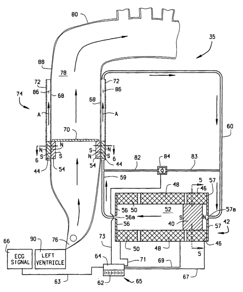
The elements of the new BEEP system, as shown in Figure 1, for example,
and generally designated 35, compose three primary components: a ventricular
assist
device (VAD), which in this embodiment is an L-VAD, generally designated 74
(shown on the left side of Fig. 1). L-VAD 74 is actuated by a hydraulic pump,
generally designated 42, and controlled by a battery/controller assembly,
generally
designated 65. It is to be understood that the new BEEP system 35 will be
referred to
throughout this document by the same reference numeral, in relation to a
variety of
embodiments. Thus, BEEP system 35 may include a L-VAD, R-VAD, BI-VAD, or
TAH, all of which are described further herein, or the system may include
alternative
embodiments of any of the VADs or the TAH described below. The BEEP system is
only a vehicle for the other aspects of the invention, the optimization
process
described in Figures 28 - 34. The optimization process can be applied to any
current
or future apparatus design of L-VAD, R-VAD, BI-VAD or TAH. The BEEP system
per se, however, is nonetheless considered to be another important aspect of
the
invention, regardless of which embodiments of the various components are
included
in the system. Further in regard to the various embodiments of the system, if
certain
aspects of the overall system are not described in detail as being different
or
distinguishable from the other embodiments, they are considered to be the same
or
equivalent to those previously or later described.
BEEP system 35 utilizes electromagnetic coils 46, 48, and 50 to drive a high
ferromagnetic-constant solid cylindrical driving magnet 40 in reciprocating
fashion
along the length of hydraulic pump 42. While three such coils are preferred,
it is to be
understood that the new system 35 and the alternative embodiments thereof can
operate adequately with more than or fewer than three electromagnetic coils on
pump
26
CA 02439390 2003-08-25
WO 03/034893 PCT/USO1/20170
42. Driving magnet 40 is acting as a piston in a hydraulic pump. The interior
vessel
of the hydraulic pump may or may not incorporate end caps 56 and 57 as part of
its
hydraulic-vessel design. However, the presence of the end caps made of
ferromagnetic material assist in directing the flux lines from the surrounding
coils to
the driving magnet 40. It will be obvious to those skilled in the art that
several
alternative embodiments can be contemplated by changing the cross sectional
areas of
the components, which may be circular, rectangular, or a number of other
closed
shapes.
Figure 5 shows pump 42 in cross section and illustrates the external
cylindrical
surface of driving magnet 40 mating with the interior cylindrical surface of
electromagnetic coils 46, 48, and 50. These surfaces, whether shaped as the
preferred
cylinders, or otherwise, are nevertheless sized and shaped to slidingly
interact as well
as to minimize leakage of hydraulic fluid therebetween. It is understood that
the
function of pump 42 is to use one or more electromagnetic coils to drive one
or more
magnets in a way to provide motion to driven magnet 44; and several
alternative
embodiments can be used to accomplish this function. It will be obvious to
those with
ordinary skill in the art that there can be many variations on the cross-
sectional view
of the components, on the exact orientation of the electromagnetic fields, on
the exact
orientation of the magnets, on the type of hydraulic or pneumatic fluid, and
on the
details of the design of the vessel containing the hydraulic or pneumatic
fluid etc, and
these alternative embodiments are herein included. It is understood that
several
alternative embodiments to minimize leakage from the high to the low pressure
of the
hydraulic fluid and blood, and alternative embodiments to minimize friction
between
27
CA 02439390 2003-08-25
WO 03/034893 PCT/USO1/20170
sliding components, are conceived and considered acceptable alternative
designs in
the present invention.
End caps 56, 57 are also made of ferromagnetic material, and are disposed on
opposite ends of pump 42. End caps 56, 57 are provided with central openings
56a,
57a, so that the interior space defined by the electromagnetic coils is in
fluid
communication with main hydraulic line 60 at each end of the pump cylinder,
permitting the hydraulic fluid to flow in and out of the pump cylinder, as
described
further hereafter.
End caps 56, 57 also serve to concentrate the magnetic flux of electromagnetic
coils 46, 48 and SO in a smaller combined area, thereby improving pump
efficiency.
The shape of end caps 56 and 57 assists in the optimal placement and
concentration of
magnetic flux lines and minimization of the weight and dimensions of system
components. While end caps 56, 57 act as "stops" for the piston, there may
also be
provided with separate "stops" of known construction, for example, as
illustrated in
Figure 19.
Magnet 40 is preferably entirely solid and thus is sometimes referred to
herein
as "solid magnet 40" for convenience of the reader. However, magnet 40 may be
only substantially solid; i.e., there could be a small through- hole plugged
with plastic,
for example, or other conceivable interruptions to the integrity of the magnet
40
which would not prohibit system 35 from working sufficiently in the present
system.
However, for most efficient, ideal operation, magnet 40 is entirely solid.
Solid magnet 40 acts as a piston to apply force to hydraulic fluid 52 to
thereby
ultimately move a driven annular magnet 44 (as indicated by arrows A, in
Figure 1 )
along the length of L-VAD 74. The magnetic flux of annular driven magnet 44
and
28
CA 02439390 2003-08-25
WO 03/034893 PCT/USO1/20170
annular valve-seat magnet 54 are along the axial length of L-VAD 74 so that
they are
biased toward and tend to lock to one another. Movement of annular driven
magnet
44 in turn moves high-ferromagnetic-constant annular valve-seat magnet 54.
Blood
78 is therefore pumped by L-VAD 74 in the direction of the flow arrows B
through
aortic arch 80, as shown in Figure 1.
Except for hydraulic fluid leakages, the reciprocating motion of solid driving
magnet 40 is in phase with the reciprocating motion of annular driven magnet
44, but
is slightly out of phase with the reciprocating motion of valve-seat magnet 54
due to
flood, hydraulic fluid, and electromagnetic inertia effects. The out-of phase
separation of driven magnet 44 from valve-seat magnet 54 varies throughout the
reciprocating cycle. These delays are accounted for in the time {t}
expressions in
equation (7) of the new process described later herein.
The reciprocating movement of driving magnet 40 along the length of
hydraulic pump 42 is controlled by power, voltage and current from battery 62
to
electromagnetic coils 46, 48 and 50 in the sequence depicted in Figures 17, 18
and 19
and described below. The timing and magnitude of the current from battery 62
is
controlled by controller 64 in response to ECG signals initiated from the ECG
signal
66, signals from measurements of ejected blood volume, and as a result of the
optimization process explained below. Battery 62 and controller 64 can be
connected
as a battery/controller assembly 65, as illustrated, or utilized as separate
components.
The movement of driving magnet 40 is slightly out of phase with the magnetic
field
along electromagnetic coils 46, 48 and SO due to electromagnetic hysteresis
effects,
which are also accounted for in the time f t} expression of equation (7)
described
herein below. The out-of phase separation of driving magnet 40 from the
magnetic
29
CA 02439390 2003-08-25
WO 03/034893 PCT/USO1/20170
field of electromagnetic coils 46, 48 and 50 varies throughout the
reciprocating cycle
and is mathematically accounted for by the optimization method described later
herein to minimize the power required for operation of the new system.
Inside an inner sleeve 68 is contained an annular valve-seat magnet 54 which
contains one-way valve 70. The sliding facing surfaces of annular valve-seat
magnet
54 and the inner sleeve 68 are sized and shaped to be substantially fluid-
tight to
minimize leakage of blood therebetween. Similarly, the mating surfaces of
annular
driven magnet 44 and the inner sleeve 68 and outer sleeve 72 (shown in Figure
6) are
designed to minimize leakage between sliding facing surfaces thereof.
It is to be understood that several alternative embodiments to minimize
leakage from the various mating elements are conceived. It is further to be
understood that all elements of the new pumping device and the entire system
for
operation thereof are formed of suitable biocompatible, surgical grade
materials.
Such materials may be appropriately selected from materials that are now
known, as
well as new materials, which may yet be developed.
Hydraulic pump 42 drives hydraulic fluid 52 in the direction of the flow
arrows through hydraulic line 59 and into annular space 86, located between
inner
sleeve 68 and outer sleeve 72. The reciprocating motion of hydraulic fluid 52
moves
annular driven magnet 44, also located between inner sleeve 68 and outer
sleeve 72.
By reversing the direction of current flow in electromagnetic coils 46, 48 and
50, the
direction of driving magnet 40 is reversed, hence the direction of hydraulic
fluid 52 is
also reversed; it follows that the direction of driven magnet 44 is reversed
as well. As
annular driven magnet 44 is moved by the flow of hydraulic fluid 52, magnetic
interaction with valve-seat magnet 54 causes valve-seat magnet 54 to move
along
CA 02439390 2003-08-25
WO 03/034893 PCT/USO1/20170
with annular driven magnet 44. Because one-way heart valve, for example, as
indicated schematically at 70, is secured to valve-seat magnet 54, one-way
valve 70
moves in the same direction as valve-seat magnet 54 and annular driven magnet
44.
When one-way valve 70 moves in a direction away from aortic valve 76, it is
closed
and pushes blood 78 through aortic arch 80.
One-way valve 70 can be any artificial or natural heart valve. Some known
valves are mechanical, some are biological and some are made with compliant
man-
made materials. Some one-way valves may also eventually be made with stem cell
research. Depending upon the particular type of valve selected for the one-way
valve
70, limits may be imposed on the optimization process of equation (7), due to
the
pressure differences the particular valves can withstand (e.g. prolapse may
occur with
some compliant heart valves). Such differences are taken into account in the
selection
for a particular system as may be necessary.
Figure 1 depicts the state of BEEP system 35 at late systole of the native
human heart, when valve-seat magnet 54 is at the beginning of its pumping
stroke
along the length of L-VAD 74. At the stage of the cycle shown in Figure 1 the
ECG
signal 66 and other volume and pressure signals have been transmitted along
wire 63
to controller 64. (Wire 63 may also be inside of conductor 410 in the
embodiment
shown in Fig. 27 and discussed hereafter.) In response to these signals and
the new
optimization process, controller 64 discharges electrical power, voltage and
current to
hydraulic pump 42 along wires 67, 69 (and 71, in some cases). Specifically,
current
from battery 62 has energized electromagnetic coils 46 and 48. In response to
the
energization of coils 46 and 48, driving magnet 40 has begun to move away from
its
position within electromagnetic coil 46 and has moved partially within the
walls of
31
CA 02439390 2003-08-25
WO 03/034893 PCT/USO1/20170
electromagnetic coil 48 (a cross-sectional view of driving magnet 40 and
electromagnetic coil 46 is shown in Figure 5).
Further with reference to Figure 1, the movement of driving magnet 40 has
forced hydraulic fluid 52 to move through main hydraulic line 60 and secondary
hydraulic line 82. The motion of hydraulic fluid 52 places pressure on both
driven
magnet 44 and check valve 84. Check valve 84 is closed, as is normally the
case,
securing the required pressure gradient between the high-pressure and low-
pressure
imposed by the motion of driving magnet 40 within hydraulic pump 42. Due to
pressure from hydraulic fluid 52, annular driven magnet 44 has just begun to
move
along the length of L-VAD 74, within annular space 86. Magnetic interactions
have
caused valve-seat magnet 54 to move in a corresponding manner. Driven magnet
44
is located slightly ahead of valve-seat magnet 54 due to electromagnetic and
fluid
inertia. A cross-sectional view of L-VAD 74, through outer sleeve 72, annular
driven
magnet 44, inner sleeve 68, and valve-seat magnet 54 is shown in Figure 6. The
function of the two magnets, 44 and 54, is to magnetically "lock" to each
other so that
the movement of magnet 54 is affected by the movement of magnet 44. By "lock"
it
is meant that the motion of one magnet affects the motion of the other magnet
via
their magnetic interaction, even though the dynamics of the system may dictate
that
the motions of the two magnets may be out of phase. Hydraulic vessel (or
"sleeve"
in some cases) 72 for magnet 54 and blood vessel 68 for magnet 44 may be
concentric
or not, parallel or not, and may have any cross-section. It will be obvious to
those
with ordinary skill in the art that there are several alternative embodiments
for the
cross-sectional view of the hydraulic and blood vessels (parallel axes or not,
concentric axes or not, circular, rectangular or other cross section etc) and
the exact
32
CA 02439390 2003-08-25
WO 03/034893 PCT/USO1/20170
location and orientation of north and south poles of the magnets, and these
are
included herein.
Aortic valve 76, located at the outlet of the left ventricle 90, has been
retained
open by the beginning of the movement of one-way valve 70, which is closed and
is
being moved upward by driven magnet 44. The difference in axial location of
driven
magnet 44 and valve-seat magnet 54 is due to fluid inertia, but also due to
magnetic
inertia. Neither fluid inertia nor magnetic inertia is accounted for in the
prior art.
Although in this embodiment it is preferred that one-way valve 70 is an
artificial
valve of known or newly developed variety, valve 70 may also, if desired or
necessary, be a natural heart valve or a one-way valve formed of tissue (human
or
other animal).
The movement of closed one-way valve 70 is beginning to pump blood along
the length of the ascending aorta 88 and into the aortic arch 80.
Figure 2 depicts the state of BEEP system 35 halfway through the pumping
motion of L-VAD 74. In this figure driving magnet 40 has moved within the
walls of
electromagnetic coil 48, approximately halfway through its motion along the
length of
hydraulic pump 42, and electromagnetic coil 50 has been energized by current
from
battery 62. The continued motion of driving magnet 40 has placed further
pressure,
via hydraulic fluid 52, on annular driven magnet 44. Due to magnetic
interactions
with annular driven magnet 44, valve-seat magnet 54 has moved approximately
halfway through its motion along the length of L-VAD 74. Still closed, one-way
valve 70 has pumped more blood, that would otherwise not have been pumped by
the
native heart, out of the left ventricle along the length of the ascending
aorta 88 and
33
CA 02439390 2003-08-25
WO 03/034893 PCT/USO1/20170
into aortic arch 80. Aortic valve 76 remains open, allowing the flow of blood
from
the left ventricle 90 into ascending aorta 88.
Figure 3 depicts the state of BEEP system 35 at the beginning of the return
stroke of valve-seat magnet 54. As driving magnet 40 reverses its previous
motion
along the length of hydraulic pump 42, the flow of hydraulic fluid 52 through
main
hydraulic line 60 is reversed as well, as indicated by the flow arrows in the
Figure.
The reverse flow of hydraulic fluid 52 places pressure on annular driven
magnet 44,
pushing it back along the length of the L-VAD 74, in the direction of aortic
valve 76.
As annular driven magnet 44 moves back along the length of L-VAD 74, valve-
seat
magnet 54 and one-way heart valve 70 move in a corresponding manner. One-way
valve 70 is open as it moves toward aortic valve 76, allowing blood to flow
freely
through one-way valve 70 as it moves. Aortic valve 76 is closed at this time,
preventing blood from flowing out of the L-VAD 74 and into left ventricle 90.
Figure 4 depicts the state of BEEP system 35 halfway through the return
stroke of valve-seat magnet 54. Driving magnet 40 has moved back within the
walls
of electromagnetic coil 48, approximately halfway through its return motion
along the
length of hydraulic pump 42. The continued motion of driving magnet 40 has
placed
further pressure, via hydraulic fluid 52, on annular driven magnet 44, pushing
it back
down along the length of L-VAD 74. Valve-seat magnet 54 has moved
approximately halfway through its return motion along the length of L-VAD 74.
One-way valve 70 is still open, allowing blood to flow freely through it as it
moves.
Aortic valve 76 remains closed, preventing the flow of blood from L-VAD 74
into left
ventricle 90.
34
CA 02439390 2003-08-25
WO 03/034893 PCT/USO1/20170
Pulsatile Flow and the Present Approach:
The principles of fluid dynamics require a measurable work per cycle (and
power output) from the heart to overcome the pressure difference in the
passages of
the circulatory system. Providing pulsatile instead of steady flow,
accelerating and
S decelerating blood and muscle, consumes significant measurable additional
work
(and power) from that required for steady flow. If the natural heart provided
continuous flow under constant pressure, then thrombi would tend to form and
gradually enlarge in relatively stagnant or low-velocity flow regions. In
steady flow
conditions these thrombi would tend to become larger with time. Eventually the
larger thrombi could potentially be dislodged by the surrounding flow causing
blockage in narrower passages downstream. The results would be disastrous. The
human body would not provide more pulsatile flow than that required for
physiological reasons.
The human body requires pulsatile blood flow for survival, and a successful
artificial heart pump or VAD should emulate the type of pulsatile blood flow
provided
by the native heart. Unlike known art devices, the present invention produces
an
optimized pulsatile flow. The VAD of the present invention provides the
"vector" or
"matrix" difference between the unsteady flow required by the human body and
the
unsteady flow provided by the native diseased heart, hence supplying only the
required deficit. By "vector" or "matrix" difference we imply that this is not
a simple
subtraction of two quantities, as it will become evident in the following. In
a total
replacement configuration (TAH) the invention provides the total unsteady flow
required by the human body. While other inventions purport to optimize the
flow, the
CA 02439390 2003-08-25
WO 03/034893 PCT/USO1/20170
present invention illustrates the actual requirements (engineering principles)
for this
optimization.
The physical dimensions of the VAD or total replacement heart must be
optimized to each application (i.e. to each patient). The moving mass, damping
and
stiffness of the combined system (moving parts of the VAD plus native heart,
if any,
plus driven blood flow through the vessels plus hydraulic fluid, surrounding
tissue,
electromagnetic dynamic phenomena, etc.) must be optimized to the dynamic
response of the system (which is a form of the natural frequencies and damping
of the
overall system). If these conditions are not met, then the VAD or total
replacement
heart will be inefficient; it will require more power than the minimum to
obtain the
desired unsteady-flow output to the body. A good physical example of this is a
yo-yo.
If the string is pulled with the right forces at the right times (which
corresponds to the
optimized forcing function for the yo-yo), it requires minimum effort for
maximum
periodic travel and produces spectacular results. If either the forcing
function or the
timing are not exactly right, then it takes more effort to obtain any travel,
and the
results are not as good. Another equally important aspect of the invention is
that the
physical arrangement and dimensions of the invention are optimized to the
desired
amplitude and frequency of the unsteadiness in blood flow required by the
circulatory
system.
Thus the power input to TAHs and VADs must be optimized to the dynamic
response of the system, otherwise the efficiency will be low (they will
require a lot of
power to drive them). One of the claims of the proposed VAD is that its
driving
force-time and force-distance relationships are optimized for minimum power
input to
the desired unsteady-flow characteristics, via a prescribed procedure, thus
increasing
36
CA 02439390 2003-08-25
WO 03/034893 PCT/USO1/20170
its efficiency. This is done via a mathematical method described below. A pre-
requisite for the use of this method is a deeper understanding of details of
the flow
and pressure conditions in the cardiovascular system than that in present
medical and
bioengineering practice. In other words, one needs to understand the details
of the
S pressure and flow traces in the native as well as the artificial systems in
order to
design an efficient VAD or TAH.
While it is understood that the pressure trace changes phase and amplitude
downstream from the aorta, there is no acknowledgement as to whether the
measured
pressure traces are static, stagnation or total pressures (defined in most
fluids
engineering texts). While it is clear that during most of systole the left
ventricular
pressure must be higher than the aortic pressure (otherwise flow would be in
the
reverse direction from the aorta to the ventricle), some texts indicate
otherwise. The
premise of this disclosure is that any TAH or VAD must be optimized around the
details of the pumping system and match the requirements of the human body.
Static pressureps~ is the pressure one would feel while traveling along with
the
velocity of the fluid in a channel. Stagnation pressure po is the pressure one
would
feel with the fluid coming to rest against the measuring device. Total
pressure pT is
the stagnation pressure plus the static head of a column of fluid above the
measuring
point. For a perfect incompressible fluid of constant density p (which is one
of many
frequently used mathematical models for blood) moving with velocity C the
governing equations are:
ho = Psr + PCZ ~2
Pr=Psr+PCz~2+pgz
37
CA 02439390 2003-08-25
WO 03/034893 PCT/USO1/20170
The distinction between these three pressures in the blood flow is important
in
the design of the optimal VAD, as is the choice of measurement devices that
are
specialized to distinguish measurement of static, stagnation and total
pressures, and
the location of these measuring devices in the system. Optimum design of the
present
device is integrally related to the fundamental laws of fluid mechanics
applied for
unsteady flow conditions to the thermodynamic system enclosing the heart and
circulatory system. Those skilled in the art of unsteady thermofluid dynamics
will
recognize that the system definition is of paramount importance to the
solution of the
problem, and must be defined with the accuracy and detail suggested in the
text by
Gyftopoulos and Beretta (1981); i.e. the system definition will require
amounts and
range of valves for: matter, parameters or constraints, and interacting forces
between
system elements. These fundamental laws are usually expressed as one equation
for
conservation of mass, three equations for conservation of momentum, for
example,
along (x,y,z), and a fifth equation for the energy balance (the first law of
thermodynamics).
The following equations (1-5) are valid for any fluid continuum (compressible
or incompressible, Newtonian or non-Newtonian), and they are general in
nature. The
nomenclature used is as follows:
E~ , e~ - energy and energy per unit volume,
including internal, kinetic, and
potential energy, etc.
- specific internal energy
38
CA 02439390 2003-08-25
WO 03/034893 PCT/USO1/20170
f 6d - external body forcing function per
unit
volume (gravity, electromagnetic,
etc.)
f Sf - surface forcing function per unit
volume
(resulting in stress tensor i)
F"h~t~ - force as a function of time from
the
native heart
Fvad~t~ - force as a function of time from
the VAD
h - specific enthalpy
m - mass
p - pressure
Q, qx,qy, qZ - heat into control volume, and heat
per
unit volume in (x, y, z)
coordinates
t - time
(x, y, z) - Cartesian coordinates
u, v, w - velocity components alone (x, y, z)
coordinates
W - work into the control volume (from
surface, from shaft, etc.)
x - axial direction
p - density
p - dynamic viscosity
39
CA 02439390 2003-08-25
WO 03/034893 PCT/USO1/20170
z - stress tensor
z,~ - element of stress tensor (includes
pressure) along (i,j) coordinates
O - divergence operator
s D - total derivative operator
a - partial derivative operator
The mass balance (continuity) is given by:
a
+ v(p~) = o
_~ +a(au)+a(pv)+a(pw)=o 1
ay az ( )
where the equation can be further simplified using certain assumptions such as
incompressible fluid (but here we consider the general form of the equation
with no
restrictions other than continuous fluid).
The vector form of the equation for conservation of linear momentum can be
written as the (x, y, z) momenta equations:
is a(p'')+v(~v)= ff+ fbd =OZ + fbd
ac
a(,~) a(~2) a(~v) a(puw) a(z~) a(z~) a(zx=)
+ + + - + + + f , 6d (2)
ac ax ay az ax ay az
a(a~) a(~u) a(~Z ) a(p~) a(Zyx> a(zyy) a(zy=>
+ + + - + + +f,bd
ar ax ay az ax ay az
a(~) a(~u) a(PWV) a( pwz ) a(z=x
a(z=y) a(z==) .f (4)
ac + ax + ay + az - ax + ~, + aZ + z, bd
CA 02439390 2003-08-25
WO 03/034893 PCT/USO1/20170
where the body forces are exerted on the whole body of fluid (such as by
gravity,
when fnd = pg ; or by external electromagnetic fields); and the surface forces
are
exterted by the interior surface of the control volume of fluid. Some texts
choose to
separate the pressure terms from the stress tensor, but here the pressure
terms are
included in the stress tensor i.
The energy balance equation is given by:
DE~ - DQ + DW
Dt Dt Dt
a( pe~) + 7( peer) + a(Aver) + a( pwet) -
at ax ay aZ
a(qx+ZlZxx+V'Cxy+WZxz) + a(qy+ZlZyx+VZyy+WZyz) + a(qz+2lTzx+VTzy+W'tzz)
ax ay aZ ( )
where qx, qy, qZ are the external heat transfers (Q) in each direction, the
work (W)
terms are given by tensor times velocity applied to the surface of the control
volume,
and Et includes all the energy terms. For example, if these include only
internal
energy, kinetic energy, and potential energy, then Er = per = P(e + Iv Z l 2 +
g ~ i-) .
However, in the general case, Et includes all energy terms affecting the
solution of the
equations.
The above five equations can be written in vector form as:
aG+aA+aB+aC - aX +aY+aZ (6)
at ax ay aZ ax ay aZ
where
G = ~P~ Pu~ Pv~ Pi'~'~ P~
A = ~pu, pu 2 , puv, puw, puer~
41
CA 02439390 2003-08-25
WO 03/034893 PCT/USO1/20170
B = ~Pv, puv, pV Z , pwv, Pver
C = ~P~'~ P~'u~ P~'v~ PW Z ~ Pu'e
t1' = L0, Txr + fx, bd, Txy, Txz, C~x + Zl Txr + V Txy + W Txz
~' _ [0, Tyx, Tyy + f , bd, Tyz, Cjy + Zl Tyx + V Tyy + W Tyz]
Z = L0, Tzx, Tyx, Tzz + f , bd , C~z + Zt Tzx + V Tzy + W Tzz ]
(the native heart and VAD forcing function are terms fx, bd in X, Y, Z).
The only restriction in the above equations 1-6 is that blood behaves as a
continuous fluid (if it didn't, e.g. if there is cavitation, severe lysis, or
severe clotting,
then the model is inadequate, but the resulting VAD is also useless). These
equations
are valid for steady and unsteady flow (periodic and transient), with any
external force
field or surface forcing function, with heat transfer, with Newtonian or non-
Newtonian fluids etc.
The resulting instantaneous equations of fluid motion (1-6) have instantaneous
eigenvalues and eigenvectors that can be computed, and those must be matched
with
the combined forcing function from the native heart F,~n{r} and the VAD
F~d~{r}, i.e. the
dynamic system of equations for the native cardio-rheology as modified by the
presence of the operating VAD. The resulting instantaneous system of dynamic
equations are of the form:
[M~{x} + ~C~{x} + [K]{x} = F{t} = F~rb {t} + Fad {t} (7)
where [M], [C], [K] are the instantaneous non-linear mass, damping and
stiffness
matrices respectively of the dynamic model. They are non-linear because they
change
with time and with mathematical or experimental data model, because the human
tissue and mechanical components are not linear, and because they change with
42
CA 02439390 2003-08-25
WO 03/034893 PCT/USO1/20170
instantaneous position and geometry (for example with open or closed valves),
and
also with daily condition of the patient. In any case the procedures to model
the
dynamic system are well established, and the fidelity of the dynamic model is
improving with time as better experimental data and theoretical or numerical
models
become available for each component of the dynamic system.
The forcing function of the native heart F"t, {t} to the dynamic system is
provided by the human and can be measured (though it also can be modeled with
basic physiological interactions). The forcing function of the VAD system is
provided
by the magnetic field to the coils, which is generated by the current and
voltage to the
coils, so that for a discretized dynamic system the instantaneous power (at
any time t)
by the VAD is balanced with:
W (t) = F ad {t} ~ {x} + losses = V {t} ~ i {t} (8)
where W(t) is the instantaneous power at any instant in time t, {x} are the
elemental
velocities at the displacements where elemental forces F"ad{t} are acting, and
the
product V {t} i {t} represents the sum of the electric power (voltage times
current)
supplied to the coils. In one embodiment of the optimization procedure the
physical
dynamic systems are linked so that the left-hand side of equation (7) is
linked directly
in the optimization process to the right-hand side of equation (8). The losses
are
electromagnetic losses of transmitting magnetic flux from the coils to the
magnets,
and friction losses until this power reaches the elemental displacements {x}
on which
forces F,,aa{t} act, and other similar losses. These losses can be measured or
modeled
mathematically with techniques available in mechanics, fluid dynamics,
electromagnetism, and other engineering texts. Thus the model includes muscle,
tissue, blood, hydraulic fluid, and electromagnetic and mechanical effects of
mass,
43
CA 02439390 2003-08-25
WO 03/034893 PCT/USO1/20170
damping and stiffness. For example, these include friction and leakage in the
mechanical components and fluid passages, the hysteresis loop of the
electromagnetic
drive of the VAD (a condition commonly called "latching"), other
electromagnetic
losses, and the stress tensors in equations 1-6, so that the resulting fluid-
structure
system is driven in an optimal manner.
In general engineering systems, the power optimization and control-scheme
optimization (such as those described later for the mechanical blood pumping
device
and patient) would be best applied to the actual system itself. In this
particular
system, namely the patient with the mechanical pump surgically implanted, it
would
be extremely difficult to perform the steady-state power optimization scheme,
and
difficult to perform the control-optimization scheme, as this may endanger the
life of
the patient. These best preferred embodiments of using the physical dynamic
should
eventually be possible after clinical trials. Alternative embodiments
(alternative
models) of the physical dynamic system are likely to be used for scientific
1 S development of the BEEP system. These are likely to use analytic,
numerical, or
experimental, etc., expressions, or their combination, to represent the
physical
dynamic system. These models can be of various degrees of complexity. Some of
these models may represent the whole dynamic system, and others may represent
only
portions of the whole system. From the above it is easy to foresee that one
group of
such possible dynamic models may include the mechanical blood pump only, while
others may, in addition, incorporate portions of the native heart and the
circulatory
system, etc. Similarly, one set of such models may concentrate on finding the
optimum F,,ad{t~, represented, for example, by forces and velocities acting on
one of:
a) valve seat magnet 54; b) driven magnet 44; or c)driving magnet 40, etc.
With
44
CA 02439390 2003-08-25
WO 03/034893 PCT/USO1/20170
similar dynamic models of the electromagnetic and hydraulic systems, this
forcing
function, F~aa{t~, can be correlated with the instantaneous electric power to
the coils
(resulting in a form of equation (8)). The preferred embodiment of the dynamic
system model directly correlates the forces on the left-hand side of equation
(7) with
the power on the right-hand side of equation (8). Alternative embodiments of
the
optimization schemes may use simplified portions of the whole dynamic system,
such
as those that find the force, F,,~a f t} on: a) valve-seat magnet 54; b)
driven magnet 44;
or c) driving magnet 40.
In this invention the forcing function F~t~ in equation (7) consists of two
parts,
one provided by the native heart and the other provided by the VAD. For this
purpose, "optimize" means minimizing the power required to drive the VAD while
minimizing the shear stress imposed on the blood.
Again, the terms "optimize" and "complement" are used in reference to the
devices and systems of the present invention, it is meant that at each heart
beat and
stroke of the VAD (used here to mean either the VAD or TAH as described
below),
several actions are carefully timed such that:
a) the native heart is allowed to pump as much blood as it can on its own
before the VAD is activated;
b) as the blood-ejection phase of the native heart nears completion, the VAD
is
energized to provide additional pumping action;
c) the additional pumping action reduces the back pressure in that native
ventricle so that the native ventricle pumps more than it would have pumped
unaided;
d) the timing of the action, length of pumping stroke, and rate of pumping
(stroke displacement versus time and resulting power input versus time) of the
VAD
CA 02439390 2003-08-25
WO 03/034893 PCT/USO1/20170
are related to the native heart ejected blood volume and rhythm in a manner
that
minimizes power input to the VAD while meeting physiological constraints;
e) the optimization processes in d) take into account the dynamic interaction
between the native heart and the VAD; and
f) the optimization process and the control scheme are integrated with the
resulting changes in blood ejected per heart beat and heart rate (beats per
minute) by
the combined action of the native heart and the VAD.
Specifically, the combination of the patient's native cardiovascular system
and
the VAD at any condition of flow rate and beating frequency supplied by the
native
heart will result in an optimal shape (function of location and time as shown
in the
Figure) for the forcing function provided by the VAD. The forcing function and
frequency of the VAD are controlled as explained elsewhere in these documents.
The
equations presented above are general and they are not dependent on the
details of the
mathematical models. Some research teams will choose to simplify these
equations
using the incompressible fluid approximation, Newtonian fluid approximation,
linear
models in finite element method programs or linearized equations in
computational
fluid dynamics (CFD) approaches. All of these simplifications are fully
included in
the general equations (1-7).
The L-VAD is intended to be placed between the aortic root and the aortic
arch. Thus, for VAD applications the length L and overall outside diameter Do
of L-
VAD 74 are limited by human physiology. There is a desire to directly wrap
coils 46,
48 and 50 around the length of travel of driven magnet 54, but this not always
possible, due to geometric constraints. For example, for an adult male L ~ 10
cm and
Do ~ 4 cm, the overall force that can be carned by a hollow magnet 54 is a
function of
46
CA 02439390 2003-08-25
WO 03/034893 PCT/USO1/20170
the volume of the magnet, among other factors, for example, if cylindrical,
approximated by ~(Dz -D; )l l4 , where D2 and Dt are the outside and inside
diameters of the magnet and l the length, the magnetic properties of the
materials
(factor k1), geometry (factor k2) and technology of components (e.g. leakage
and
S friction characteristics, coil packing, heat transfer constraints (factor
k3)).
f = F(kl, k2, k3)
where clearly, other factors being equal, the force is increased by increasing
D2, the outside magnet diameter. Thus the outside diameter of electromagnetic
coils
used in the linear magnet motor of prior art becomes too big for VAD to fit
into the
human body in the vicinity of the aortic arch. This maximum-diameter issue is
resolved with the use of driving magnet 44, the hydraulic fluid, and solid (or
substantially solid) magnet 40. The following table is an indication for
several
distinct sizes of VADs, assuming that the diseased native heart provides SO%
of the
cardiac output required by the human body:
Weight Height Area Normal Output Required
of
(kg) (m) (m2) Cardiac Diseased VAD Output
Output Native (cc)
(cc) Heart
(cc)
Child 50 1.3 1.3 58 29 29
Teen 55 1.65 1.6 72 36 36
Avg. Adult55 1.75 1.7 76 38 38
Female
Avg. Adult75 1.85 2.0 90 45 45
Male
Large 110 ~ 175 2.3 102 51 51
Adult
~
1S
As can be seen by the equations above, several standard sizes of L-VAD 74
can be designed. As a guide, the smallest would be a pediatric device and the
largest
would be for a large adult. What follows is an example of calculations
performed on
47
CA 02439390 2003-08-25
WO 03/034893 PCT/USO1/20170
a hypothetical individual, and is not intended to in any way limit the present
invention.
The height and weight features of a person can be converted to body surface
area by using the approximate formula below. Once body surface area is known,
normal cardiac output for a given individual can be calculated. Normal cardiac
output
volume per body surface area is 45cc/m2.
Body Surface Area (m2) _ [ht (cm)]°'"g [wt (kg)]°~42~
[0.007449]
Using a 75kg 185cm adult male as an example, the body surface area
calculation results in a value of 2m2. Given the body surface area value of
2m2
calculated above, the normal stroke volume for the individual is 90cc. In end-
stage
cardiomyopathy, the native heart provides approximately 50% of the required
cardiac
output. In the example above the native heart would provide approximately
45cc.
Therefore, the L-VAD would have to provide an additional 45cc.
What follows is a general description of the approximate sizes of an L-VAD
for the above patient at one ejection volume (45cc) and one heart rate. The
procedure
must be repeated several times for different ejection volumes and heart rates
before
the optimum L-VAD dimensions for the patient are decided. The procedure is
also
affected by the size of available one-way valves 70, especially if these are
of the
standard artificial heart valves available commercially, which are available
in several
standard diameters, usually measured in millimeters (mm).
The maximum L-VAD displacement required in this example is 45cc. A
standard 29 mm valve nomenclature is used for one-way valve 70. This choice
affects the length of L-VAD 74 as well as the force that must drive driven
magnet 44
and valve-seat magnet 54. The axial length of driven magnet 44 and valve-seat
48
CA 02439390 2003-08-25
WO 03/034893 PCT/USO1/20170
magnet 54 is 13 mm. The wall thickness between driven magnet 44 and valve-seat
magnet 54 is lmm, as is the thickness of outer sleeve 72.
For certain illustrative example cases the steady state and acceleration force
required to pump blood through L-VAD 74 is 30 to 36 N (kg m sec 2). This is
based
on an initial estimate of the pressure that will be supplied by L-VAD 74,
multiplied by
the area of the pumping diameter. This initial estimate accounts for the
acceleration
of fluids (blood and hydraulic) pumped in the system (about 6L in
circulation), and
the initial masses of the moving components. The volume of rare earth magnet
in
driven magnet 44 and valve-seat magnet 54 required to provide the 30 to 36 N
is 3.83
x 103 mm3. The resulting thickness of valve-seat magnet 54 with a length of 13
mm is
about 3.2 mm. Thus, the inside diameter of valve-seat magnet 54 is 29 mm and
the
outside diameter is 35.4 mm. The inside diameter of driven magnet 44 is 37.4
mm.
The thickness of driven magnet 44, with a length of 13 mm and an inside
diameter of
37.4 mm, must be around 2.8 mm to achieve the desired 30 - 36 N. Thus, the
outside
diameter of driven magnet 44 is 43 mm. Therefore, the outside diameter of L-
VAD
74 is 45 mm.
The stroke length required if one-way valve is to give the required 45 cc
volume is 45.7 mm. Adding this to the axial length of valve-seat magnet (as
required
by the geometry of the device) the overall axial length of the pumping portion
of L-
VAD 74 becomes 58.7 mm. This length will be increased to allow for the cuffs
for
hydraulic fluid and blood. It is understood that several alternative
embodiments for
the cross-sectional shape of the heart valve 70, magnets 54, 44 and 40, and
cuff (or
"capsule") designs for the hydraulic connections for hydraulic fluid and blood
can be
49
CA 02439390 2003-08-25
WO 03/034893 PCT/USO1/20170
used and will be apparent to one skilled in the art and thus they are hereby
incorporated in this disclosure.
The above dimensions are used to provide geometric inputs for the models
used in equation (7). The inputs result in elements for mass matrix [M],
damping
matrix [C], and stiffness matrix [K]. Elements of [M] are evaluated using
material
densities. Elements of [C] are evaluated using fluid dynamics for the flow
passages,
structural damping for tissues and electromagnetic properties for magnets,
coils and
other components, as needed. Elements of [K] are evaluated using material and
surrounding tissue properties and electromagnetic properties for magnets,
coils and
other components, as needed. The surrounding tissue must extend to the control
volume of the system where the tissue geometry is not moving. This means a
little
further out of the pericardium (to fully include pericardium tremors) and a
little
further out of the blood and hydraulic fluid vessels (to include stiffness and
compliance, providing elements for [C] and for [K]).
For example, the pressure drops in hydraulic lines 60 and 82 initially can be
estimated using analytic calculations available in standard textbooks, and
later
evaluated by discretized mathematical models as elements of matrices in
equation (7).
Continuing the above example, with certain engineering assumptions driving
magnet 40 could be 3800mm3 grade 37 rare earth magnet. In one embodiment this
magnet could have radius 9.7 mm, and length 12.86 mm. The hydraulic volume
displaced by 45.7 mm of stroke length of driven magnet 44 is 54.7 mm. Thus,
the
overall length of hydraulic pump 42 is about 81 mm (this length will be
increased by
the length of the hydraulic cuffs).
CA 02439390 2003-08-25
WO 03/034893 PCT/USO1/20170
Additional secondary calculations are made to evaluate the geometries of
auxiliary components such as hydraulic lines, and other components such as
tissue in
the myocardium and blood flow system. The inputs result in elements for
matrices
[M], [C], and [K] (some measured in clinical trials, others measured for
individual
patients). The elements of vector of displacements {x} and its derivatives { x
} and
f x } in equation (7) are elemental displacements. The equation is nonlinear
and can
be decomposed in a few or infinitely many degrees of freedom, depending on the
fidelity of the dynamic model.
F"t,{t] in equation (7) is measured for each condition (heart rate, ECG
signal,
volumes ejected from right and left ventricles, and pressures) of the patient.
For L-
VAD 74 this is given by the total pressure (static + dynamic + elevation
components)
provided by the diseased heart inside the left ventricle as a function of time
(measured
during the heartbeat) integrated over the inside surface area of the four
chambers of
the heart. This surface area is also measured with magnetic resonance imaging
(MRI), echocardiography, or other similar technique. These give pressure-
volume-
time traces for the diseased heart as illustrated in Figures 14 and 15. The
volume
information is correlated with data from proximity sensors, such as 406 and
416 in
Figure 9, which may be, for example, proximity sensors. This information
changes as
the condition of the patient worsens or improves. This means that the data
needs to be
calibrated before surgery, and again soon after surgery, and monitored
periodically so
that the data provided by proximity sensors 406 and 416 reflect the forcing
function
provided by the native heart, where the mathematical expressions are:
dF,",, {t} = p(t)dA(t)
51
CA 02439390 2003-08-25
WO 03/034893 PCT/USO1/20170
F~tn f t~ = J P(t)dA(t)
A
The resulting pressure-volume-time traces of the native heart have a phase
associated
with the timing of the forcing function F"h f t} during the beat. This can be
modeled
with Fourier series analysis of the pressure and volume signals of the native
heart over
time. Again, these vary with the rate (beats per minute) of the native heart
and with
the condition of the patient (i.e. the information changes as a function of
time and
needs periodic updating).
Figure 6 is a cross-sectional view of L-VAD 74, along line 6-6 of Figure 1.
By contrast, Figure 7 is a concept illustration of the human heart showing the
location
of L-VAD 74 in place of ascending aorta 88. It is understood that the L-Vad
may
replace a portion, not necessarily the entire ascending aorta. Further in some
embodiments the aorta may simply be transected to place the L-VAD outside the
body, with blood conduits connecting the ends of the transected aorta to the L-
VAD.
Corresponding alternative embodiments are possible for the R-VAD, BI-VAd.
Blood
moves from left ventricle 90, through aortic valve 76 and into L-VAD 74,
situated in
place of the ascending aorta 88, pumps blood into aortic arch 80.
Figure 8 shows the placement of an entire BEEP system 35 within the human
torso. The illustration depicts the spatial relationship between
battery/controller
assembly 65 and L-VAD 74. Figures 7 to 13 and 20 to 25 are schematic
illustrations,
not cross-sectional views, and the location of L-VAD 74 in Figure 8 is at a
different
plane from the location of R-VAD 58 in Figure 21. (The LVAD in Figure 8 is
correctly shown more to the right of the patient's chest than RVAD in Figure
21, but
Figures 8 and 21 are anatomically correct, while Figures 7 and 20 are simple
arrangement illustrations).
52
CA 02439390 2003-08-25
WO 03/034893 PCT/USO1/20170
Figure 9 is a concept illustration of the human heart, illustrating a
technique
to measure the volumes of the left and right ventricles, which is used in the
control
algorithm. Rare earth (or similar material) magnets 402 and 404 are embedded
in the
endocardial surface of the right ventricle, and their relative motion changes
the
magnetic field between them. These changes are measured by proximity sensor
406,
mounted on the pericardium. The signal is transferred by electrical lead 408
to wire
bundle 410. Rare earth or similar material magnets 412 and 414 are embedded in
the
endocardial surface of the left ventricle, and their relative motion changes
the
magnetic field between them. These changes are measured by proximity sensor
416,
mounted on the pericardium. The signal is transferred by electrical lead 418
to wire
bundle 410. The signals from wire bundle 410 are transmitted to controller 64
and
used as described later.
The proximity sensors are currently available devices that may operate on the
resistive, capacitative or inductive principles, or combinations, or other
similar
distance-measuring technology. Auxiliary (parallel horizontal) lines 1 through
6 in
Figures 10 through 13 represent the motion of the ventricles and the KG
diaphragm
(see below) during a cardiac cycle. It has traditionally been thought that the
valves of
the heart open to let the blood through when the chambers contract, and snap
shut to
prevent it from flowing backward as the chambers relax. While this is correct,
the
valves also act as pumping pistons for at least a portion of the cardiac
cycle, a fact not
known to be previously recognized in the literature. The plane of the valves
and the
supporting tissue on the perimeter of the valves form an internal diaphragm,
approximately in the horizontal plane, which buckles and moves in 3D, which is
also
not known to be named in the existing literature. For the purposes of this
document
53
CA 02439390 2003-08-25
WO 03/034893 PCT/USO1/20170
this diaphragm will be the Korakianitis-Grandia (KG) diaphragm, illustrated in
Figures 10 to 13, and generally designated at 92. KG diaphragm 92 has four
quadrants with a valve in each quadrant. It is activated by the surrounding
cardiac
muscle, which forces the diaphragm into a periodically-changing three-
dimensional
surface. (Thus Figures 10 - 13 are illustrations rather than cross-sections of
the
human heart). During the cardiac cycle the aortic valve and pulmonary valve
stay
nearly immobile (which allows one to place the VAD on the outlet side of these
two
valves); but the mitral and tricuspid valves move substantially, contributing
at least in
part to the pumping action of the ventricles. The mitral valve movement is
comparable to the movement of the inside wall of the left ventricle. The
tricuspid
valve exhibits an even greater excursion and corresponding pumping action and
is
actually used in current medical practice as a measure of right ventricular
ejection
fraction.
While the exterior surface of the heart moves slightly during the cardiac
cycle,
the volume of the four-chamber heart does not change appreciably in time.
However,
the known art does not recognize that the total overall volume of each of the
two
sides, left and right, of the heart does not change appreciably during the
cardiac cycle,
even though the ventricular and atrial septa move. In operation of the native
heart,
during left ventricular ejection, the left atrium concurrently expands (while
filling for
the next cycle), and KG diaphragm 92 begins to move towards the apex of the
heart,
with complementing motions of the atrial septum and of the ventricular septum,
thus
keeping the overall volume of the left side of the heart about constant. Apex
of the
heart is a common term for the tip of the left ventricle. Similar arguments
keep the
right-side volume approximately constant, while the right and left sides of
the heart
54
CA 02439390 2003-08-25
WO 03/034893 PCT/USO1/20170
expel blood to the lungs and the aorta about simultaneously. Minor deviations
from
these equal-volume considerations on each side, right and left, occur due to
one side
of the heart beating slightly before the other, heart-muscle and blood-vessel
elasticity,
transient accelerations or decelerations of the overall cardiac cycle
(governed by the
body's demand), and blood compressibility (which at circulatory system
pressures is
practically negligible).
The human cardiac cycle consists of two phases, conventionally called
diastole and systole. During diastole (Figures 10 and 13) the ventricular
muscle is
relaxing, KG diaphragm 92 moves toward the base of the heart while the aortic
and
pulmonary valves are closed, and the mitral and tricuspid valves are open and
moving
towards the base of the heart, thus increasing the volume inside the
ventricles while
concurrently decreasing the volumes inside the atria. Base of the heart is a
common
term for the posterior aspect of the heart, behind the atria in the heart's
anatomical
position. The open mural and tricuspid valves move upwardly (when the body is
upright) to engulf blood from what was volume inside the atria (thus
concurrently
increasing the volume inside the ventricles while decreasing the volumes
inside the
atria). In a model of ventricular flow this volume exchange would affect the
thermodynamic system definition, mentioned previously. A scrutinizing review
of
echocardiography tapes reveals that radial volume changes around the vertical
axes of
the ventricles account for roughly 75% of the volume change, with the
corresponding
movement of the KG diaphragm accounting for the remaining 25% volume change.
There is heart-muscle work associated with these changes in volume that must
be
accounted with the correct mathematical model in any attempt to model flow in
the
heart or in VAD mimicking the function of the heart.
CA 02439390 2003-08-25
WO 03/034893 PCT/USO1/20170
By the end of diastole, the relaxing ventricle allows KG diaphragm 92 to move
toward the base of the heart. During initial systole (Figure 11) the aortic
and
pulmonary valves remain closed while pressure is building up inside the
ventricles.
Subsequently the blood pressure inside the left ventricle becomes higher than
the
pressure in the ascending aorta, and the aortic valve is opened by blood
flowing out of
the ventricle. Substantially concurrently the blood pressure inside the right
ventricle
becomes higher than the pressure in the pulmonary trunk, and the pulmonary
valve
opens. The motion of the KG diaphragm carrying the mitral valve toward the
apex of
the heart, along with the simultaneous concentric contraction of the
ventricle, ejects
blood into the ascending aorta.
Correspondingly, the movement of KG diaphragm 92 carrying the tricuspid
valve towards the apex of the heart, along with the simultaneous contraction
of the
right ventricle, ejects blood into the pulmonary trunk. This same motion of
the KG
diaphragm with the tricuspid and mitral valves closed increases the volumes
inside the
atria, hence refilling the atria with blood from the pulmonary veins (left
atrium) and
the vena cava (right atrium). Towards the end of systole the aortic and
pulmonic
valves close, then the mitral and tricuspid valves open and the cycle starts
anew.
Each cycle takes approximately one second.
The double throb ("lub dub") of the beating heart is generated by the snapping
of the closing valves, but also from the accompanying vibrations of the
surrounding
heart muscle and contained blood.
The three-dimensional motion of KG diaphragm 92 forces each one of the four
one-way valves to act as pumping pistons for at least part of the cardiac
cycle. The
56
CA 02439390 2003-08-25
WO 03/034893 PCT/USO1/20170
blood flow must be optimized around artificial heart valves to provide the
desirable
flow and pressure pattern while minimizing shear stresses on blood cells.
Figure 10 represents late diastole. At this point KG diaphragm 92 is at its
uppermost position (horizontal lines 1-3). Pulmonary valve 94 and aortic valve
76 are
closed, and mural valve 96 and tricuspid valve 98 are open, completing filling
of
ventricles 106 and 90 following contraction of right atrium 100 and left
atrium 102.
Ventricular myocardium 104 is in its relaxed state.
Figure 11 represents early systole. At this point ventricular myocardium 104
is thickened concentrically, and KG diaphragm 92 is moving downward
(horizontal
lines 2-4), the tricuspid valve side more so than the mural valve side. Due to
this
motion, the volume of ventricles 106 and 90 is decreased while that of atria
100 and
102 is increased. Hence, the total volume of the heart remains essentially
constant.
Figure 12 represents late systole. At this point ventricles 106 and 90 have
maximally thickened concentrically, and KG diaphragm 92 has been pulled
maximally downward (lines 4-6). 'This completes the emptying of the
ventricles.
Figure 13 represents early systole. At this point KG diaphragm 92 is
beginning to return upward (lines 3-5) toward atria 100 and 102, while
ventricular
myocardium 104 relaxes concentrically. As a result of this motion, the volume
of
atria 100 and 102 decreases while that of ventricles 106 and 90 increases,
hence the
overall volume of the heart remains essentially constant.
There are minor variations to the basic steps outlined above, due to damping
and elasticity of the heart tissues, and small amounts of native heart valve
leakage,
which can be accounted for in the thermodynamic system definition mentioned
earlier.
57
CA 02439390 2003-08-25
WO 03/034893 PCT/USO1/20170
Figure 14 represents pressure-volume loops for healthy (solid lines) and
diseased (broken lines) hearts, with pressure plotted along the Y axis and
volume
plotted along the X axis. Line 112 represents a healthy heart. Point 118 to
point 120
represents ventricular filling. Point 120 to point 122 represents
"isovolumetric
contraction". Point 122 to point 124 represents ejection during systole. Point
124 to
point 118 represents isovolumetric relaxation.
Line 114 represents the normal response in accordance with the Frank Starling
law to an increase in volume. Points 126, 128, 130 and 132 correspond to
points 118,
120, 122, and 124, on line 112, respectively, and represent the corresponding
phases
of the cardiac cycle. Notice that points 124 and 132 lie on the same line,
commonly
referred to as the End Systolic Pressure Volume Relationship (ESPVR).
Line 116 represents a diseased heart which has exceeded the limits of the
Frank Starling curve. In these hearts, the end diastolic pressure and volume
are
elevated but the end systolic pressure is decreased from that associated with
normal
1 S myocardium, as noted by a lesser slope of the ESPVR line. Points 134, 136,
138 and
140 correspond with points 118, 120, 122 and 124 of line 112, respectively.
Figure 15 represents ventricular pressure over time of the healthy and
diseased
hearts. Again, the end diastolic ventricular pressure is greater in the
diseased heart
than in the healthy heart, and because the ejection fraction is decreased in
the diseased
heart, the heart rate is increased so that the total cardiac output is
maintained.
VADs are activated by either ECG signal, or via a fill-to-empty mode. The
control algorithm of the present device utilizes inputs from the ECG of the
native
heart, as well as a measurement of the ejection volume and pressures of both
the right
58
CA 02439390 2003-08-25
WO 03/034893 PCT/USO1/20170
and left ventricles. As a result, the prior art complication of mismatch of
ejection
volume between the right and left ventricles is eliminated.
Figure 16 is a concept illustration of the pumping travel of new BEEP system
35, as compared to that of known cardiac pumping devices. The abscissa is the
S number of beats per minute of the native heart. The ordinate is the length
of travel of
driving magnet 40 along the length of hydraulic pump 42. The known devices are
either on or off (travel X is always equal to maximum travel Xmax), as shown
by the
dotted line. In the present device, the length of travel of driving magnet 40
along
hydraulic pump 42 varies, following solid line 142, depending on the number of
beats
per minute of the native heart. As the number of beats per minute of the
native heart
increase, they reach high threshold dotted line 144 at which point controller
64 signals
for driving magnet 40 to start moving along the length of hydraulic pump 42
smoothly
increasing the stroke travel, approaching solid line 142.
Over time, due to augmentation of ejected volume by the VAD, the end
diastolic volume of the native heart decreases. This allows the internal
volume of the
ventricle to become smaller, which subsequently allows the muscle of the
native heart
to begin to recover, and hence eject a greater volume of blood per stroke. As
cardiac
output is the product of ejected blood volume times heart rate, the increased
ejected
volume allows the heart rate to decrease. These changes are sensed by
controller 64,
which reduces the stroke length of the VAD as a greater portion of the cardiac
output
is now being supplied by the native heart. The stroke length of the VAD
progresses to
the left, from point 148 toward point 146. When the beats per minute reach the
lower
threshold point 146 (reflecting at least partial recovery of the native
myocardium),
this will cause a decrease in stroke length along line 150, which effectively
reduces
59
CA 02439390 2003-08-25
WO 03/034893 PCT/USO1/20170
the stroke length of the VAD. If recovery of the native heart continues, the
stroke
volume of the L-VAD is reduced to zero along line 150. At this point the
native heart
is once again providing the total cardiac output on its own, without the
assistance of
the VAD. The shape of line 142 is actively manipulated by controller 64 using
additional inputs for the measurement of the ejection volume and pressures of
the
right and left ventricles provided by the measurement system shown in Figure 9
and
described in relation thereto.
A critical factor in the success of a newly-installed L-VAD is satisfactory
operation of the right ventricle during the immediate peri-operative period.
The
ejection fractions of the right and left ventricles are monitored by
mechanisms such as
those illustrated in Figure 9 and alternative embodiments thereof. This allows
the
volumetric outputs of the right ventricle and the assisted left ventricle to
be the same
by manipulating the shape of line 142 up or down. For example, suppose that a
short
time after activation, the right ventricle is measured to give SOcc per beat
and the left
ventricle gives 25cc per beat. In this case, the stroke length of L-VAD 74
will be
adjusted to give 25cc per beat. If, a short time later, the right ventricle
starts to fail
and now only ejects 40cc per beat (while the left ventricle still gives 25cc
per beat),
this discrepancy in the ejection fractions will be detected by proximity
sensors 406
and 416. In response, controller 64 will lower the level of line 142,
resulting in a
shorter stroke length of L-VAD 74, to give l5cc per beat, for a total of 40cc
from the
assisted left ventricle. The human body will compensate with a corresponding
increase~in heart rate (beats per minute). In this fashion, controller 64
matches the
ejection volumes of the right and left sides. Should the right ventricle
continue to fail,
CA 02439390 2003-08-25
WO 03/034893 PCT/USO1/20170
a decision will have to be made as to the appropriateness of installing an R-
VAD,
making this a BI-VAD system.
Assuming that a patient is supported on an L-VAD alone, the length of the
pumping stroke of the L-VAD is determined as a function of beats per minute,
as
shown in Figure 16, and manipulated by matching the ejection volumes of the
left and
right sides of the heart. For example, starting from point 148, if the beats
per minute
continue to increase, then the piston stroke also continues to increase
smoothly to
point 178. Starting from any operating point to the right of point 178, as
beats per
minute decrease, at point 178 the device reduces the travel of driving magnet
40 from
its maximum travel along hydraulic pump 42. If the number of beats of the
native
heart is reduced sufficiently to reach low threshold line 150, then the travel
of driving
magnet 40 is reduced smoothly until it becomes zero following low threshold
line
150. Lines 150 and 142 may coincide over the length of line 150. Line 142 to
the left
of point 146 in Figure 16 can represent the initial activation of the L-VAD
after
surgical installation thereof.
The locations, magnitudes and exact shape of lines 142, 150 and 144 shown in
Figure 16 are for purposes of illustration and will vary from patient to
patient, and
device to device. In addition, during normal operation of the L-VAD, small up
or
down variations of the level of line 142 are made by controller 64 in order to
match
the ejection volumes, measured as described in Figure 9, of the left and right
sides of
the system. The control algorithm has several input variables; among others,
beats
per minute measured by the ECG (as described below), the ejection volume and
pressures from the right and left side of the system as illustrated in Figure
9. For
61
CA 02439390 2003-08-25
WO 03/034893 PCT/USO1/20170
clarity, in the remaining Figures the concepts are illustrated using beats per
minute to
represent the function of controller 64.
Figure 17 is a chart comparing the activation sequence of the known Larson et
al. device and that of the present BEEP system, as well as the corresponding
power
requirements, in relation to the ECG trace of the native heart. The solid
lines
represent the present device and the dotted liens represent the known device
of Larson
et al. The common abscissa is the time period required for two beats of the
native
heart. The ECG trace has characteristic spikes Q, R, S (commonly known as the
QRS
complex), and waves T and P, whose physiological function and importance is
described in detail in medical texts. The beginning and end of the stroke of
Larson's
device occurs at or near point R of the QRS signal.
The graph shown in portion A of Figure 17 shows non-dimensional stroke
distance traveled by driving magnet 40 (X to Xmax) from 0.0 to 1.0, according
to
Figure 16. Portion B of Figure 17 illustrates a typical ECG voltage trace
during
cardiac operation. The beginning of the stroke of the present system is at or
about the
beginning of the T wave, allowing the rapid ejection phase of the native heart
to
precede the augmentation of the VAD. The pumping phase (points 154 to 156) of
the
present system occurs between the end of the T wave and the beginning of the P
wave. The return stroke begins at this time (point 156) and ends at or just
after the
QRS complex (point 158).
Driving magnet 40 rests at the center of electromagnetic coil 46 (X=0) during
the time period between the end of the return stroke (point 158) and the
beginning of a
new stroke (point 160). The resting period between the end of the return
stroke (point
158) and the beginning of the next stroke (point 160 corresponding to 154) is
62
CA 02439390 2003-08-25
WO 03/034893 PCT/USO1/20170
important for a number of reasons. For example, acceleration at the beginning
154
and end 156 points of the stroke is minimal. This resting period allows time
for
depolarization of electromagnetic coils 46, 48 and 50 between strokes. When
necessary, it also allows driving magnet 40 to be centered within
electromagnetic coil
46, thereby allowing driven magnet 44 and valve seat magnet 54 to return to
the
beginning of the stroke of L-VAD 74. This return function is accomplished by
opening and closing check valve 84, as necessary.
Due to leakage of hydraulic fluid around magnets 40 and 44 it is possible that
one of the two magnets is stopped at one of the two ends of its travel while
the other
magnet is somewhere in the middle of its stroke. For example, if annular
driven
magnet 44 is at the end of its travel at the pump inlet (by the aortic valve
as shown in
Figure 1) but driving magnet 40 is not yet all the way back to the beginning
of its
stroke (as shown in Figure 4), then high hydraulic pressure will arise between
driving
magnet 40 and end cap 57. This condition would be sensed by the large increase
in
the power required by the coils. At that time the controller would open check
valve
84 and would pull driving magnet 40 by the coils towards end cap 57, until it
touches
end cap 57, thus bringing the two magnets back into phase, and normal
operation
would resume. The procedure is similar if driving magnet 40 reaches the end of
its
pumping travel (as shown in Figure 3) while driven magnet 44 is near the
middle of
its travel (as shown in Figure 2). It is also possible to correct for these
leakages at the
end of every pumping stroke, or ever few pumping strokes. A similar procedure
can
be used for initial activation of the device, to start the device after it has
been stopped,
and to re-lock magnets 54 and 44 if they are not locked relative to each other
at any
63
CA 02439390 2003-08-25
WO 03/034893 PCT/USO1/20170
time during operation. The latter condition is sensed by a large decrease in
the power
required by the coils.
The shape of line 164 in Fig. 17 (portion A) is determined by the optimization
procedure described later herein. Acceleration begins smoothly (point 154 on
line
164) so that less power is required than if the device started with a constant
velocity.
Maximum acceleration is achieved somewhere in the middle of the stroke, based
on
the optimization procedure. The VAD of the present system approaches maximum
stroke travel with minimum velocity at point 156 so that it does not impact
against the
mechanical stop at the end of travel and no energy is lost due to impact.
Thus, less
energy is required to start the return stroke. The return stroke is less
critical than the
pumping stroke because one-way valve 70 is open and less energy is required to
return the new VAD (e.g. L-VAD 74) to its starting position. Even so, the
shape of
line 162 is optimized by the procedure. Velocity is zero at X=Xmax, requiring
less
power than if there was a change in velocity at Xmax. The shape of lines 164
and
1 S 162, and resting period between points 158 and 160 is optimized by the
procedure.
Figure 17C shows the power requirements of the present device in Watts
during usage. The maximum power requirement (point 166) occurs somewhere along
line 164, as optimized by the procedure. During the return stroke, power peak
at point
168 occurs slightly before X=Xmax at point 156, which corresponds to the power
requirement at point 170. This occurs because of the sequence of energization
of
electromagnetic coils 46, 48, and 50 as explained further in Figure 18.
In comparison, the known device, illustrated by the dotted lines in Figure 17,
begins the pumping stroke at or near R of the QRS complex, with constant
velocity,
until the point of maximum travel, which occurs at or near the end of the T
wave. The
64
CA 02439390 2003-08-25
WO 03/034893 PCT/USO1/20170
return stroke is with constant velocity from the point of maximum travel until
the R
peak of the next QRS complex, requiring large acceleration at the two ends 172
and
174 of its stroke. This requires correspondingly large power input. In
addition, the
velocities are not optimized for the unsteady flow and the time varying
magnetic
fluxes, requiring large power input at all points during the stroke. Points
172 and 174
correspond to power peaks 176 and 178, respectively. As a result, the present
device
will take less power (solid line 180) than the prior art device (dotted line
182), and the
peaks occur at different times.
Further with reference to Figure 17, power peak 166 of the present device
occurs during the T wave of the native heart, allowing the native heart to
finish its
rapid ejection. This increases the volume of blood pumped due to the
combination of
the native heart and the VAD with respect to the prior art (due to summation
of
volume), requires less power than the prior art device, and allows the native
heart a
chance to recover by decreasing left ventricular volume. These combinations
make
the BEEP system bio-compatible.
Figure 18 is a schematic illustration of the embodiment of BEEP system 35
shown in Figure 1, using three electromagnetic coils 46, 48 and 50 along the
length of
hydraulic pump 42 showing the corresponding magnetic flux of the driving
magnet
and the electromagnetic coils. Vector 184 represents the magnetic flux of
driving
magnet 40. Position X=0 is at the center of electromagnetic coil 46. Position
X/Xmax is at the center of electromagnetic coil 50. The top portion of the
Figure
shows electromagnetic coils 46, 48 and 50 and positive stops 186 and 188. The
middle portion of the Figure is an illustration of the typical periodic
representation of
the magnetic fluxes of electromagnetic coils 46, 48 and 50 during the pumping
stroke
CA 02439390 2003-08-25
WO 03/034893 PCT/USO1/20170
from X=0 to X=Xmax. The bottom portion of the Figure is an illustration of the
typical periodic representation of the magnetic fluxes of electromagnetic
coils 46, 48
and 50 during the return stroke from X=Xmax to X=0.
With reference to the middle portion of Figure 18, the abscissa is the axial
position of driving magnet 40 from the center of coil 46 (point 190) to the
center of
electromagnetic coil 50 at X=Xmax (point 192).
With reference to the bottom portion of Figure 18, the abscissa is axial
position of driving magnet 40 from the center of coil 50 at X=Xmax (point 192)
to the
center of electromagnetic coil 46 at X=0 (point 190). Starting from X=0 in
Figure
18(b) (point 190, which corresponds to point 154 in Figure 17), positive stop
186
ensures that activation of magnetic fields 194 and 196 in electromagnetic
coils 46 and
48, respectively, will force driving magnet 40 from the center of
electromagnetic coil
46 towards the center of electromagnetic coil 48. Magnetic field 194 is
reduced to
zero soon after driving magnet 40 is a little outside electromagnetic coil 46.
As
driving magnet 40 approaches the center of coil 48, magnetic field 196 in coil
48 is
reversed in direction to magnetic field 198. The reversal in magnetic field
196 does
not necessarily coincide with the point in time when magnet 40 is at the
center of coil
48. The exact location of reversal is dependent upon an optimization
procedure.
Magnetic field 200 is initiated just before driving magnet 40 enters
electromagnetic coil S0. The combination of magnetic fields 198 and 200 die
out by
point 192 (corresponding to point 156 in Figure 17) and smoothly bring the
magnet to
position X=Xmax, against positive stop 188. At that position, the magnetic
fields in
coils 48 and 50 are reversed as shown at point 192 in Figure 18(c). Positive
stop 188
ensures that magnetic fields 202 and 204 from coils 50 and 48, respectively,
push
66
CA 02439390 2003-08-25
WO 03/034893 PCT/USO1/20170
driving magnet 40 from X=Xmax (point 192) towards the center of coil 48.
Magnetic
field 202 is reduced to zero soon after driving magnet 40 is a little outside
electromagnetic coil 50. As driving magnet 40 approaches the center of coil
48,
magnetic field 204 in coil 48 is reversed in direction to magnetic field 206.
The
S reversal in magnetic field 204 does not necessarily coincide with the center
of coil 48.
The exact location of reversal is dependent upon the optimization procedure.
Magnetic field 208 is initiated just before driving magnet 40 enters
electromagnetic
coil 46. The combination of magnetic fields 206 and 208 die out by point 190
(corresponding to point 158 in Figure 17) and smoothly bring the magnet to
position
X=0, against positive stop 186. At that position, from point 158 to point 160
in
Figure 17, magnetic field 208 (or its residual effects) will retain driving
magnet 40 at
X=0, whereupon the cycle repeats itself. All of the magnetic fields 194-208
will be
optimized to give Fvaa~t~. The power to obtain magnetic fields 196-208 will be
minimized based on the resistance, inductance, and capacitance of the
electromagnetic
system, including the coils and magnets, and voltage source, using
constitutive
relations or experimental data for the dynamic representation of the systems
in
equation (7), established in electromagnetic theory.
In general, the magnitudes of magnetic fields 194-200 will be greater than
those of magnetic fields 202-208, because the former occur during the pumping
phase
with one-way valve 70 closed and pushing blood, while the latter occur during
the
return stroke with one-way valve 70 open. Even though the above embodiment
utilizes three electromagnetic coils, it is contemplated that the present
device may
contain more or fewer electromagnetic coils. In the limit, a linear stepper
motor may
be used.
67
CA 02439390 2003-08-25
WO 03/034893 PCT/USO1/20170
Figure 19 is a schematic illustration of the embodiment of Beep System 35
illustrated in Figure 1, wherein only two electromagnetic coils 46 and 48 are
activated
and used to move driving magnet 40. This is done in order to obtain some
stroke
length X less than Xmax, as shown in Figure 16. Similar relative functioning,
as
described with respect to Figure 18, is found in the displacements and
magnetic fluxes
of Figure 19 as well.
Figure 20 is a concept illustration of the human heart depicting the placement
of R-VAD 58 in place of a portion of the pulmonary trunk 210. Blood moves from
right ventricle 106 through pulmonary valve 94 and into the pulmonary trunk
210
before its bifurcation point 212.
Figure 21 shows the placement of an R-VAD embodiment of BEEP system 35
within the human torso O. The illustration depicts the spatial relationship
between
battery battery/controller assembly 65, hydraulic pump 42, and R-VAD 58. As
mentioned previously, Figures 7, 9-13 and 20, 22, 24 and 27 are simple
arrangement
1 S illustrations, not anatomically-correct views.
Figure 22 shows the placement of a BI-VAD, generally designated 77, which
consists of a combined assembly of L-VAD 74 and R-VAD 58 in the same system.
In
this embodiment L-VAD 74 is located in place of at least part of the ascending
aorta
88. In use of BI-VAD 77 blood moves from left ventricle 90 through the aortic
valve
76 and into ascending aorta 88; i.e. in this system L-VAD 74 pumps blood into
the
aortic arch 80, just as in use of the L-VAD alone. R-VAD 58, as part of the BI-
VAD
77, is located in place of at least part of the pulmonary trunk 210, just as
it is used in
the embodiment (R-VAD alone) shown in Figure 20. Blood moves from the right
ventricle 106 through pulmonary valve 94 and into pulmonary trunk 210 as R-VAD
68
CA 02439390 2003-08-25
WO 03/034893 PCT/USO1/20170
58 portion of BI-VAD 77 pumps blood into the bifurcation 212 (hidden from
view) of
the pulmonary arteries.
Figure 23 shows the general placement of the BI-VAD 77 embodiment of the
BEEP system in the human torso O. The illustration depicts the relative
spatial
relationship of BI-VAD 77 in the chest and of battery/controller assembly 65
and
hydraulic pump 42 in the abdomen. (The LVAD in Figure 23 is correctly shown
more to the right of the patient's chest than RVAD, but Figure 23 is
anatomically
correct, while Figures 22 is a simple arrangement illustration).
Figure 24 is a schematic illustration of the Total Artificial Heart (TAH)
embodiment, generally designated 95, for use in a variation of the new BEEP
system
35. In this TAH embodiment, atria 100 and 102, along with ECG signal 66, are
retained from the native heart. The TAH is comprised of a BI-VAD system with a
greater stroke volume than the assist embodiment, as the total cardiac output
is now
being supplied by the BEEP system. In some TAH embodiments it is possible to
use
the mitral valve 96 and tricuspid valve 98, shown in previous Figures,
provided their
papillary muscles and chordae tendineae are functioning, and insert he L-VAD
and/or
R-VAD portions of the BI-VAD into the respective ventricles. However, in other
alternative TAH embodiments artificial valves may be necessary or preferred.
In
addition, all, some or non of the native ventricle may be retained. In the
embodiment
illustrated in Figure 24 L-VAD 74 completely takes the place of left ventricle
90 (seen
in Figure 7, for example), and hence its inlet is grafted to an artificial
valve (not
shown) and its outlet is grafted into the ascending aorta 88. Right ventricle
106 is
replaced with an R-VAD 58, which has its inlet grafted to an artificial heart
valve (not
shown) and its outlet grafted into the pulmonary trunk 210.
69
CA 02439390 2003-08-25
WO 03/034893 PCT/USO1/20170
Figure 25 shows the general placement of the TAH embodiment 95 of the new
BEEP system within a human torso O. The illustration depicts the spatial
relationship
among battery 62, controller 64, in the abdomen, and TAH 95, in the chest
cavity. In
various embodiments of the TAH a portion or all of the native heart may be
removed.
If the chordae tendinae and papillary muscles are intact, then the TAH would
consist
of an LVAD and an RVAD placed inside the respective ventricles. If the mural
and
tricuspid valves of the native heart are not utilized, then the TAH would
require a one-
way valve at the inlet of the LVAD and a one-way valve at the inlet of the
RVAD,
and the combination that would make the overall TAH. If the entire native
heart
(including the sino-atrial node) is removed, then the LVAD and RVAD system
that
would comprise the TAH will be triggered by an electrical signal driven by
sensors
that indicate the level of oxygen in the blood stream and other sensors of
body
functions.
Figure 26 represents an alternative embodiment, generally designated 35', of
an L-VAD version of BEEP system 35. In this version, two or more
electromagnetic
coils, 300, 302 and 304 are used to drive valve seat magnet 54 in a reciprocal
fashion.
In this alternative embodiment, no hydraulic pump is required. The Figure
represents
the beginning of the pumping stroke, at which time one-way valve 70 is closed
and
electromagnetic coils 300, 302 and 304 are energized in a manner similar to
that
illustrated in Figure 18b or 19b to drive valve seat magnet 54 down the length
of L-
VAD 306, pumping blood into aortic arch 80. At the end of the pumping phase,
the
direction of current in electromagnetic coils 300, 302 and 304 is changed,
again in a
manner similar to that depicted in Figures 18c or 19c, driving valve seat
magnet 54
(this time with one-way valve 70 open) back down the length of L-VAD 306 to
its
CA 02439390 2003-08-25
WO 03/034893 PCT/USO1/20170
original position. Electromagnetic coils 300, 302 and 304 are energized by
controller
64, in response to the ECG signal 66, and the ejection volumes and pressures
of the
right and left sides of the system as illustrated in Figure 9, in a similar
manner as
described in the preferred embodiment.
Figure 27 represents an alternative embodiment of the ejection volume
measuring apparatus for use in an alternative BEEP system, generally
designated 35"
(only a portion of which is shown). In embodiment 35", instead of rare earth
magnets
402, 404, 412, and 414 (shown in Figure 9), any proximity sensor 502, 504, 520
and
522, can be substituted. Such proximity sensors may be included in any or all
of the
four chambers of the heart. These proximity sensors may or may not need to be
coupled with conductors 506, 508, 524, and 526. Proximity sensor output can be
fed
to signal converters 512 and 530 directly, or with conductors 510 and 528. The
output from signal converter 512 is directed with lead 408 into wire bundle
410, and
the output from signal converter 530 is directed with lead 418, also into wire
bundle
410. Wire bundle 410 can, but does not necessarily, include inside it
conductor 63,
shown in Figures 1 - 4, which transmits the ECG signal to controller 64. The
signals
from these conductors become inputs to controller 64.
Optimization procedure and control sequence:
The following optimization process can be applied to any VAD or TAH
device. During normal operation the native heart interacts with the VAD so
that the
overall system/control responds to X/Xmax, the ECG signal, and ~ of the
combined
system of the native heart and VAD.
Figs. 28-34 illustrate the basic procedure of the optimization and control
process for new BEEP system 35, 35' or 35". It is to be understood throughout
the
71
CA 02439390 2003-08-25
WO 03/034893 PCT/USO1/20170
discussion herein that, unless otherwise specified, "VAD" shall be interpreted
to mean
either the L-VAD, R-VAD, BI-VAD or TAH.
The main steps of the process are the following:
(a) Develop a mathematical model of the dynamic behavior of the desired part
of the physical system
(b) Identify the system inputs, outputs and desired constraints for the
physical
part of the system in (a) above
(c) Optimize power input to the VAD to complement the action of the
diseased native heart
(d) Develop an optimized control scheme for the inputs and outputs
(e) Perform tests on the individual patient
(f) Maintenance: updating the dynamic optimization and control schemes as
the condition of the patient changes with time.
The inlet and outlet boundary conditions, and other engineering inputs to the
model, and corresponding dynamic inputs, will depend on the dynamic system
definition (Gyftopoulos and Beretta, 1991). Several alternative embodiments of
the
definition of the dynamic system that will perform the optimization can be
defined,
and the following examples are given to illustrate the flexibility and
potential of the
power optimization method and control optimization method.
Figure 28 illustrates the main components of the circulation system with the
main BI-VAD components in place, with the right and left side forcing
functions from
the native heart and the VAD. The level of detail of system components shown
in
Figure 28 is for illustration purposes only, and several alternative models
with more
or fewer details of system components can be drawn. One could choose to obtain
the
72
CA 02439390 2003-08-25
WO 03/034893 PCT/USO1/20170
dynamic response of the whole system, including theoretical, numerical or
experimental models for the pulmonary system (lungs) and oxygenation dynamic
systems. A continuous model (differential equations expressing the dynamic
response
at any position and time of the component) for all components of the system
would be
of enormous complexity, though still theoretically possible. Continuous models
can
also lead to distributed parameter analyses. With current (2001 ) technology
those
skilled in the art would likely (and straining computational resources) choose
to
model: the L-VAD system from points M 1 to M2; the "right side" of the heart
system
from points M3 to M4 without R-VAD; and from M3 to MS with the R-VAD. These
M1 to MS points (or other similar points that may be used for the
optimization) are
used to separate suitable portions of the physical system in order to develop
a
dynamic model of portions of the system rather than the whole system shown in
Figure 28. Points such as M1 to MS (or other similar points) used in order to
simplify
the model, may represent physical cross-sections of the flow passages, usually
in the
main blood vessels. Those with ordinary skill in the art may choose to develop
the
dynamic model using combinations of
(a) discretized finite element method programs (FEM, for example Szabo and
Babuska, 1991; Bathe, 1995). For example, FEM models may be used for the
cardiac
muscle, for structural mechanical components, and other components;
(b) computational fluid dynamics models (CFD, for example Anderson et al.,
1984; Kiris et al., 1997). For example, CFD models may be used for the blood
and
hydraulic fluid flows, and other components of the system;
(c) analytic solutions for some dynamic elements. For example, analytic
models may be used for some of the fluid leakage in narrow passages, using
73
CA 02439390 2003-08-25
WO 03/034893 PCT/USO1/20170
lubrication theory, and other boundary-layer techniques. Samples of such
methods
for different sub-components of the system are presented by (Nichols and
O'Rourke,
1998; Panton, 1984; White, 1991; Schlichting, 1979; Hinze, 1987);
(d) specialized information for select parts of the cardiovascular system. For
example, select such models are described by (Fung, 1984; Braunwald, 1984;
Verdonck, 2000; Peskin and McQueen, 1997);
(e) lumped parameter models for some of the dynamic components. For
example, some of the mechanical components may be represented with techniques
described by (Meirovich, 1975); and
(f) experimental data (which are usually the most reliable models) can be used
for any aspect of the dynamic components of the system.
Thus the overall dynamic model can be based on continuum mechanics (or
their variant of distributed parameter models), can be discretized, can be
based on
lumped parameters, rely on experimental data, or any combination thereof. Some
of
the component models will be linear, others will be non-linear, and some will
be
discrete or piecewise continuous (for example valve 70 is sometimes open,
sometimes
closed, and sometimes in the process of opening or closing). The overall
dynamic
model is likely to be complex, requiring significant computational resources.
In
alternative embodiments useful information can also be derived from simpler
piecewise-continuous lumped-parameter non-linear dynamic models for the main
components. For example, such extremely simplified models can consist of
several
masses, springs and dampers for each of the main components shown in Figure
28, so
that the overall dynamic model could be run on a conventional desktop personal
computer.
74
CA 02439390 2003-08-25
WO 03/034893 PCT/USO1/20170
Once a suitable part of the physical prototype has been defined with points
such as M1 to MS for development of the dynamic model, the suitable inputs,
outputs,
boundary conditions, and other system constraints must be carefully defined
(this is
refernng to the correct system definition, mentioned above). Figure 29 is an
example
of one possible dynamic-system representation of the main components of the
left
side of the diseased native heart plus the L-VAD. Alternative embodiments of
the
model may include portions of the right side of the native heart and/or the R-
VAD. If
the sample model for Figure 29 extends between points M1 and M2 in Figure 28,
then
inlet and outlet boundary conditions would involve combinations of blood
pressures
and velocities at M1 and M2 as functions of time. In Figure 29 the center
diagram
illustrates the dynamic model of the physical system. The dynamic model can be
developed with experimental data or with equations or a combination thereof.
There
are two basic physical components to the center block of Figure 29. One
physical
component is the diseased native heart and surrounding tissue to the native
heart; and
the other physical component is the VAD or BI-VAD or TAH system. The two
physical components interact and dynamically affect each other during normal
operation as shown by the dashed line R. The combination of the two main
components of the system, whether presented mathematically or with
experimental
data, results in a dynamic representation of the physical system from M1 to M2
that
can be described by a form of equation (7). The engineering definitions of the
thermodynamic system (dynamic, thermodynamic, fluid dynamic, mechanical, etc.,
as
discussed earlier) are crucial to the analysis and must be such that the
patient's tissue
surrounding both the native heart and the VAD or TAH are sufficiently removed
from
the components so that the dynamic operation does not affect the boundary
surface
CA 02439390 2003-08-25
WO 03/034893 PCT/USO1/20170
that separates the dynamic system from the surrounding tissue. This is an
imaginary
boundary surface typical of the boundary surfaces in thermofluid dynamics
texts that
define the boundary of the engineering system being analyzed. The input for
the
dynamic representation of subsystems and components (shown on the left side of
Figure 29) can come from different sources. Some of the input can be
experimental
data, which is usually the preferred source of data, but other input can come
from
other adequate mathematical representations. Such inputs may come from the
constitutive relationships of cardiac muscle or other muscle, or of the
surrounding
tissue, or the constitutive relationships for blood flow, whether it is
modeled as
Newtonian or non-Newtonian, incompressible or compressible. Several
illustrations
of these models are published in the above references. The mathematical
representation of the physical system depicted in Figure 29 from M1 to M2 can
use
constitutive relationships for the mechanical components, or constitutive
relationships
for electromagnetic components, or experimental data, or any combination
thereof, or
experimental data and mathematical expressions of constitutive relations, as
shown on
the left side of Figure 29.
Other components of the physical system can also be incorporated, as needed,
depending upon how many or how few of the system components are used in the
dynamic model represented by the final form of "process equations" (7) and
(8). The
level of detail of the dynamic model will affect the complexity of the
required
solution. It will also affect the fidelity and thus accuracy of the results.
In general,
the higher the complexity and the fidelity the more accurate the results, but
at some
point there is a limit; i.e., a point of diminishing returns, where the
increased
complexity does not justify the higher accuracy. A judgment must be made on
the
76
CA 02439390 2003-08-25
WO 03/034893 PCT/USO1/20170
fidelity of the dynamic model required for the particular application of the
invention.
The final decision on this issue will also depend on the sophistication of
engineering
tools available, (CFD, FEM etc) and the level of accuracy required of the
results.
There are several alternative types of controllers suitable for the
application. In
the simplest case the controllers may give constant (battery) voltage, and
vary the
currents as a function of time to the three coils of Figure 18 (for example i,
{t}, i2 {t}
and i3 {t}). Other controllers may give constant current but varying voltages.
Other
controllers may vary both the voltage and the current. The latter is the most
likely
embodiment. The third solution is likely to give the least power required than
the
other two controllers, as the electrical resistance, inductance and impedance
of the
coils, driving magnet 40, and surrounding ferromagnetic material impose non-
linear
effects on the unsteady flows of voltage and current, and the third type of
controller
allows one to take full advantage of the "natural frequencies" of the dynamic
magnetic system in relation to the forcing function required by the patient-
VAD
system.
The dynamic model developed above is used in the power optimization
method, an example of which is illustrated in Figure 30. The purpose of this
optimization method is to minimize power requirements and maximize battery
life
between recharges. This is accomplished by identifying the minimum electrical
power required to the coils for each operating condition of the VAD. Since the
diseased native heart and VAD affect each other's dynamic performance (broken
line
U in Figure 29), the optimization process must be repeated separately for each
initial
operating condition of the unaided native heart. The inputs for the specific
illustration
example are the initial condition of the diseased native heart, comprised of
the ECG
77
CA 02439390 2003-08-25
WO 03/034893 PCT/USO1/20170
trace and the ejected blood volumes and pressures of the right and left side
(atria and
ventricles) as functions of time over the period of the heart rate. In one
embodiment
of the optimization process an intermediate output of the power optimization
method
is X/Xmax f t} and F,,aa f t} of the R-VAD and L-VAD. In an alternative
embodiment
of the optimization process the output is the voltage and current fed to each
coil as a
function of time as shown in Figure 30. This optimization process must be
repeated
for each initial condition of the diseased native heart identified above.
One or more of the several potential optimal solutions V f t} and i f t} are
stored
in controller 64. The choice of optimal solution to insert in the controller
is illustrated
with an example below.
The example power optimization method of Figure 30 searches for shapes of
X/Xmax{t} that require the minimum power. In one example embodiment of this
optimization process, the ejection volume of the native left ventricle is
evaluated
using MRI, echocardiography, or other similar techniques such as correlation
with the
movement of proximity sensors as described earlier. The desired additional
volume
that must be provided by L-VAD 74 is evaluated by methods illustrated in the
earlier
table of potential sizes of VAD. This dictates the required travel X/Xmax of L-
VAD
74 in Figure 16. Next, an initial estimate for the trace of line 162 in Figure
17
(starting with an initial shape resembling that of line 162 ) is input into
the power
optimization method. This line shape may also be modeled with Fourier series
analysis, and the amplitudes and phases in these Fourier series have a phase
difference
from the ECG trace of the native heart in F"h{t}. One measure of these phases
is
graphically reflected in the phase difference ~ from the phase of R in the QRS
complex (phase zero) to point 154 in the trace of X/Xmax in Figure 17.
78
CA 02439390 2003-08-25
WO 03/034893 PCT/USO1/20170
This concept is commonly referred to as "phase" in dynamic systems. The
pressures of the four chambers as functions of time are required at least for
the
optimization sequence, and it may also be required during the normal running
of the
device. However, the pressures and the volumes can also be correlated by other
means in normal running of the device (for examples the ECG signal alone, or
the
ECG signal plus volume traces, or ECG signal plus volume plus pressure
traces).
The forcing function of the native heart can be measured (for example with
measurements of ventricular and atrial pressures and volumes, or their
correlations) as
described elsewhere in the text. The forcing function of the VAD is an input
to the
optimization process as described below. There are several alternative
combinations
of specifying this forcing function as an input to the optimization process.
For
example, one way is to prescribe the displacement X/Xmax of driving magnet 40
as a
function of time, (Figures 16 and 17), evaluate the required force on driving
magnet
40 from the coils, and then evaluate the electrical power required from the
controller
(voltages and/or currents to the coils) to accomplish this motion. This is
further
elaborated below. Then in an iterative process the displacement versus time
(of
driving magnet 40) can be changed until the electrical power to the coils is
minimized
while the displacement of driving magnet 40 provides corresponding
displacements of
driven magnet 54 that result in acceptable ranges of volumetric blood
throughput and
heart rate.
This initial estimate of the forced motion of X/Xmax{t} results in changes in
the pressure supplied by the combined diseased left ventricle plus L-VAD 74.
The
result is that the pressure and volume traces of the diseased heart shown in
Figures 14
and 15 are modified, because the dynamic response of the native heart system
and L-
79
CA 02439390 2003-08-25
WO 03/034893 PCT/USO1/20170
VAD system affect each other. (In simple terms, the motion of the one-way
valve 70
from the aortic valve to the aortic arch sucks additional blood per heart beat
from that
accomplished by the diseased native ventricle alone, thus increasing ejected
blood
volume per beat, so that the whole system would tend to operate at lower heart
rates).
The initial estimate of the shape of X/Xmax{t} results in a required forcing
function F~aa {t} that must be provided by driving magnet 40 to the hydraulic
fluid and
from there to the blood, and this corresponds to the distribution of voltage
and current
over time that the coils must provide to the driving magnet 40. The forcing
function
on driving magnet 40 is computed using the dynamic model described above in
equation (7). This forcing function of the optimized design is compared with
the 30 -
36 N maximum force estimated in the discussion of Figures 1-4, above. The
electrical
power (V {t} and i {t}) required to provide this force (Figure 17, line 180)
is computed
using dynamic models of the transmission of power from the coils to the
magnet, or
measured experimentally, or with a similar technique, reflecting equation (8).
The
shape of Xmax {t} versus time is iteratively manipulated until the electrical
power
required is minimized.
In alternative embodiments of the power optimization method this
minimization can be done numerically or experimentally, or by neural networks
to
handle the volume of data and computations required. The optimization of the
transmission of electromagnetic power can be done for at least three different
cases,
depending on the type of controller 64: (a) the coils are supplied with
constant voltage
and power changes are obtained with changes in the electrical current; (b) the
coils are
supplied with constant current and power changes are obtained with changes in
the
CA 02439390 2003-08-25
WO 03/034893 PCT/USO1/20170
voltage; and (c) the controller can vary both the current and voltage applied
to each
coil as a function of time.
It is expected that for a given initial diseased heart condition (for example,
a
heart rate of 100 beats per minute and ejected volume from the unaided left
ventricle
50 cc) the power optimization process will result in several combinations of
modified
heart rates and ejected volumes (from the combined left ventricle and L-VAD)
with
slightly different power requirements. For example, three potential L-VAD
solutions,
each with a different shape of X/Xmax in Figures 16 and 17 to the above
diseased-
heart condition, may be:
(a) 80 beats per minute, 80 cc per beat, 7.0 Joules per beat (560
Joules/minute);
(b) 130 beats per minute, 60 cc per beat, 3.0 Joules per beat (465
Joules/min);
(c) 60 beats per minute, 90 cc per beat, 8.0 Joules per beat (480 Joules/min).
In the last step of Figure 30, for most practical applications a cardiologist
1 S would choose to store in controller 64 either solution (a) or solution (c)
rather than the
lower-energy solution (b). These "optimal" solutions are obtained in an
"external
optimization process" shown in Figure 28 for a wide range of diseased heart
conditions and stored in controller 64. The combined output of the diseased
native
heart and the new VAD/TAH is optimized both for the individual diseased native
heart of the patient and for the power required to drive the artificial
device. The
output of the power optimization method is the electrical power, and
combinations of
voltage and current, that must be applied by the controller to the coils.
In alternative embodiments this power optimization method can be carried out
mathematically (equations (7) and (8) for the chosen system), or
experimentally (with
81
CA 02439390 2003-08-25
WO 03/034893 PCT/USO1/20170
patient and VAD) in clinical trials for groups of patients, or individuals
patients. In
either case these optimization processes would benefit by the use of neural
networks.
The VAD installed in the patient must be able to adapt to dynamic changes
from one condition to the other, as the patient with the VAD implanted in
normal
operative condition goes through normal daily activities requiring changes in
heart
rates and ejected blood volumes. The optimal design of the mufti-input mufti-
output
dynamic system of the patient is illustrated in Figure 31. In one alternative
embodiment of the control optimization method the physical dynamic system in
Figure 31 is the patient with the VAD installed, or in other words the control
optimization method is experimental and is done clinically. In another
alternative
embodiment the control optimization method is done with the dynamic model of
the
physical prototype, reflected in expressions of "process equations" (7) and
(8).
Examples of the dynamic system shown in Figure 31 are dynamic models such
as those shown in Figures 28 and 29, and incorporate the results of the power
optimization method of Figure 30 (that defines the steady-state, non-
dynamically
changing conditions of the system). In control-system terminology this is a
multivariate control scheme (as opposed to more usual control schemes for
simpler
linear mechanical systems). For purposes of this document, "multivariate"
means that
the output state of the dynamic system is characterized by several input
variables and
several output variables, illustrated by the incoming and outgoing arrows on
the left
and right side of Figure 31.
Examples of these output-state variables are the ECG trace, the blood volumes
ejected from the ventricles, the flow rates through points such as Ml, M2, M3
and
M5, the blood pressures or hydraulic-fluid pressures at various points in the
flow
82
CA 02439390 2003-08-25
WO 03/034893 PCT/USO1/20170
system, other similar quantities, rates of change of these variables with
time, or
combinations thereof. The state of these variables is measured by various
pressure,
velocity, position, etc., transducers. Information about the output state
variables is fed
back to the control node (x in circle) via the feedback transfer matrices
G(s). The
purpose of the control optimization method is:
(a) to find the optimum output variables for the control scheme;
(b) to find the types and values of these feedback transfer matrices G(s),
which
feed back signals to the control node; and
(c) to find the optimum input state variables for the control scheme.
This optimization method is a multivariate input-output control method with
several
input-output state variables. The input state can be defined with variables
such as
beats per minute, the phase ~ of the distance from point Q to point 154 in
Figure 17
(in units of degrees or in units of time), the value of X/Xmax, the shape of
X/Xmax,
several other similar quantities, or their rates of change with time, and
combinations
thereof. In general, these input-state control variables would be different
from any
similar quantities that were computed in the power optimization process. This
does
not preclude using the quantities from the power optimization process, but
this may
lead to system instabilities during transients.
Thus the inputs to the dynamic system are manipulated by functions of the
outputs of the dynamic system (which can be the physical patient and VAD, or
the
dynamic model developed above) as affected by the feedback transfer matrices
G(s).
The modified inputs are fed into the dynamic system and affect its output
state. This
optimization of the control method can be done analytically, experimentally,
or with
83
CA 02439390 2003-08-25
WO 03/034893 PCT/USO1/20170
various heuristic methods. Essentially these control methods ensure that
corrections
to the dynamic system to accommodate transient operations do not become
unstable.
Simplified versions of this control sequence can be analyzed with linear
control theory. However it is more likely that development of the control
method will
require well established analytic and experimental techniques of non-linear,
discrete
or continuous systems control, as several of the elements of the dynamic
system (e.g.
in Figure 29) are non-linear.
Suitable analytic optimal control techniques have been published in the open
literature (Brogan, 1990; Glad and Ljung, 2000; Fradkov et al., 1999;
Schroder,
2000). However, it is envisioned that in alternative embodiments neural
networks,
adaptive control techniques, and observer-based methodologies will be suitable
alternative embodiments of the new control optimization method. The final step
in
the control optimization sequence is to store the optimized feedback transfer
matrices
G(s), and associated control scheme, into controller 64.
An additional way to illustrate the flow of information flow in the new device
during normal operation is shown in the flow charts of Figures 32, 33 and 34,
all of
which include points Ml, M2, M3 and MS that are also in Figure 28 and 29.
Figure
32 shows the application to an L-VAD. The figure shows at the top the native
heart
as right and left sides, atria and ventricles, and in the center there is an
illustration of
the ECG signal, which feeds information to the controller. In this case, shown
in
Figure 32, the information would be the ECG trace, or heart rate and phase
entering
the L-VAD controller. (Alternative embodiments may include measures of volumes
and pressures in atria and/or ventricles). The left ventricle provides some
output that
goes into the control junction, shown by an X in a circle, which is also fed
into the
84
CA 02439390 2003-08-25
WO 03/034893 PCT/USO1/20170
controller. The controller, using information provided by the power
optimization
method and control optimization method that were shown and discussed with
reference to Figures 30 and 31, provides electric power, V {t} and i {t} to
the coils,
that dictates the movement of driving magnet 40. Driving magnet 40 in turn
dictates
the movement of driven magnet 44, and that in turn dictates the movement of
valve-
seat magnet 54, which affects the output of the left ventricle (point M2). A
similar
arrangement could be drawn for the R-VAD, but it is exactly symmetrical to the
one
shown here so this is not drawn or described further.
The flow chart of Figure 33 illustrates the application of the new
optimization
process to a bi-ventricular assist device (BI-VAD, described above). The flow
chart
is split into two parts, for the L-VAD and the R-VAD, right and left
respectively. The
components themselves and the logic flow paths are similar to those shown in
Figure
32, and thus are not discussed further herein.
Figure 34 shows the application of the new optimization process to the total
artificial heart (TAH) embodiment, in which the left and right ventricles are
removed,
but signals are still received from the sinoatrial node. These signals are
provided to
the L-TAH and R-TAH controllers 64. In response, the controllers drive magnets
40,
44 and 54, and finally these magnets provide the overall volumetric throughput
for the
cardiac system, corresponding to points M2 and MS in Figures 30 and 31, as
previously discussed.
These mathematical and engineering techniques will be augmented by clinical
trials on groups of patients, and standard-sized or unique VAD devices sized
for
individual patients devices may be optimized to the individual patients. As
the
condition of the patient changes with time, the control variables and control
scheme
CA 02439390 2003-08-25
WO 03/034893 PCT/USO1/20170
stored on controller 64 will need to be updated to the new condition of the
patient.
The power and control optimization sequences are identical to the sequences
described above. The new data for controller 64 can be transmitted to the
controller
inside the patient's body using established infra-red data transmission
techniques, or
other similar techniques.
Several alternative embodiments to the described systems and methods are
also conceived. For example, one alternative embodiment entails the use of
neural
networks, or comparable technology, to optimize the displacements shown in
Figure
28 (X/Xmax and their shape), with the ejected blood volume. For example, with
reference to Figure 29, dynamic measurement of volume ejected and phase may be
eliminated, because volume and phase can be correlated (with neural networks)
with
the motion of two or more proximity sensors, as shown in Figure 27.
Alternative embodiment of the above-described methods are conceived in
which the optimization processes for Figures 30 and 31 is not done
mathematically,
1 S but it largely depends on clinical trials with extensive use of neural
networks to
expedite the computation process.
Another enhancement of the new system is that the controller can detect the
presence of certain arrhythmias, such as ventricular tachycardia, for example.
In this
event, the electromagnetic pump coils would be de-energized and no current
would be
supplied to such coils, as it would be undesirable for the VAD to be
activated. As a
"fail-safe", if the VAD was in fact de-energized, in such a case, the one-way
valve 70
inside of valve-seat magnet 54 would respond to the pressure gradient of the
blood
flowing past it, and would open and close via the forces applied to it by the
flowing
blood.
86
CA 02439390 2003-08-25
WO 03/034893 PCT/USO1/20170
In view of the foregoing, it will be seen that the several objects of the
invention are achieved and other advantages are attained. Although the
foregoing
includes a description of the best mode contemplated for carrying out he
invention,
various modifications are conceivable.
As various modifications could be made in the constructions and methods
herein described and illustrated without departing from the scope of the
invention, it is
intended that all matter contained in the foregoing description or shown in
the
accompanying drawings shall be interpreted as illustrative rather than
limiting.
87