Note: Descriptions are shown in the official language in which they were submitted.
CA 02439654 2003-08-28
WO 02/072028 PCT/US02/07649
COMPOSITIONS AND METHODS FOR MODIFYING THE CONTENT OF
POLYUNSATURATED FATTY ACIDS IN MAMMALIAN CELLS
This application claims priority from U.S.S.N. 60/275,222, filed March 12,
2001,
the contents of wluch are incorporated herein by reference in their entirety.
TECHNICAL FIELD
This invention relates to compositions and methods for altering the content of
polyunsaturated fatty acids in mammalian cells.
BACKGROUND
Some of the work presented herein was supported by a grant from the National
Institutes of Health (CA79553). The United States government may, therefore,
have
certain rights in the invention.
Polyunsaturated fatty acids (PUFAs) are fatty acids having 18 or more carbon
atoms and two or more double bonds. They can be classified into two groups, n-
6 or n-3,
depending on the position (n) of the double bond nearest the methyl end of the
fatty acid
(Gill and Valivety, Trends Biotecl2nol. 15:401-409, 1997; Broun et al., Annu.
Rev. Nuts.
19:197-216, 1999; Napier et al., Cur. Opifa. Plant Biol. 2:123-127, 1999). The
n-6 and
n-3 PUFAs are synthesized through an alternating series of desaturations and
elongations
beginning with either linoleic acid (LA, 18:2n6) or oc-linolenic acid (ALA,
18:3n3),
respectively (Gill and Valivety, supra; Broun et al., supra; Napier et al.,
supra). The
major end point of the n-6 pathway in mammals is arachidonic acid (AA, 20:4n6)
and
major end points of the n-3 pathway are eicosapentaenoic acid (EPA, 20:5n3)
and
docosahexaenoic acid (DHA, 22:6n3).
An important class of enzymes involved in the synthesis of PUFAs is the class
of
fatty acid desaturases. These enzymes introduce double bonds into the
hydrocarbon
chain at positions detemnined by the enzyme's specificity. Although, in most
cases,
animals contain the enzymatic activity to convert LA (18:2n6) and ALA (18:3n3)
to
longer-chain PUFA (where the rate of conversion is limiting), they lack the 12-
and 15-
CA 02439654 2003-08-28
WO 02/072028 PCT/US02/07649
desatuxase activities necessary to synthesize the precursor (parent) PUFA, LA
and ALA
(I~nutzon et al., J. Biol. Chen2. 273:29360-29366, 1998). Furthermore, the n-3
and n-6
PUFA are not interconvertible in mammalian cells (Goodnight et al., Blood 58:
880-885,
1981). Thus, both LA and ALA and their elongation, desaturation products are
considered essential fatty acids in the human diet. The PUFA composition of
mammalian
I O cell membranes is, to a great extent, dependent on dietary intake
(Clandinin et al., Can. J.
Playsiol. Pharmacol. 63:546-556, 1985; McLennan et al., Am. Heaf°t J.
116:709-717,
1988).
To the contrary, some plants and microorganisms are able to synthesize n-3
fatty
acids such as ALA (18:3n-3) because they have membrane-bound 12- and 15- (n-3)
I S desatur aces that act on glycerolipid substrates in both the plastid and
endoplasmic
reticulum (Browse and Somerville, Anrau. Rev. Plant Plzysiol. Plant Mol. Biol.
42: 467-
506, 1991). Genetic techniques have led to the identification of the genes
encoding the
12- and 15-desaturases from Arabidopsis thaliana and other higher plant
species (Okuley
et al., Plant Cell 6:147-158, 1994; Arondel et al., Science 258:1353-1355,
1992).
20 Recently, a fat-1 gene encoding an n-3 fatty acid desaturase was cloned
from
Caeno~°Izabditis elegans (Spychalla et al., P~oc. Natl. Acad. Sci. USA
94:1142-1147,
1997; see also US Patent No. 6,194,167).
SI7MMARY
25 The present invention is based, in part, on the discovery that the C.
elegans n-3
desaturase gene, fat-1, can be successfully introduced into other types of
animal cells
(e.g., mammalian cells), where it quickly and effectively elevates the
cellular n-3 PUFA
content and dramatically balances the ratio of n-6:n-3 PUFAs. More
specifically,
heterologous expression of the fat-1 gene in rat cardiac myocytes rendered
those cells
30 capable of converting various n-6 PUFAs to the corresponding n-3 PUFA and
changed
the n-6:n-3 ratio from about I5:1 (an undesirable ratio) to I :1 (a desirable
ratio). In
addition, an eicosanoid derived from n-6 PLTFA (i. e. arachidonic acid) was
significantly
reduced in the transgenic cells (as described further below, levels of
arachidonic acid can
2
CA 02439654 2003-08-28
WO 02/072028 PCT/US02/07649
be assessed to determine whether a given nucleic acid encodes a biologically
active
desaturase; similarly, one can assess the levels of n-6 PUFA; the levels of n-
3 PLTFA; and
the ratio of n-6:n-3 PLTFAs). Accordingly, the present invention features
compositions
(e.g., nucleic acids encoding fat-1, optionally and operably linl~ed to a
constitutively
active or tissue-specific promoter) and methods that can be used to
effectively modify the
content of PUFAs in animal cells (i. e., cells other than those of C. elegans,
for example,
mammalian cells such as myocytes, neurons (whether of the peripheral or
central nervous
system), adipocytes, endothelial cells, and cancer cells). More generally, a
fat-1
sequence or a biologically active variant thereof can be operably linlced to a
regulatory
sequence. Regulatory sequences encompass not only promoters, but also
enhancers or
other expression control sequence, such as a polyadenylation signal, that
facilitates
expression of the nucleic acid. The modified cells (whether in vivo or ex vivo
(e.g., in
tissue culture)), transgenic animals containing them, and food products
obtained from
those animals (e.g., meat or other edible parts of the animals (e.g., liver,
l~idney, or
sweetbreads)) are also within the scope of the present invention.
In one embodiment, the invention features mammalian cells that contain a
nucleic
acid sequence encoding the C. elegans n-3 desaturase or biologically active
variants (e.g.,
fragments or other mutants) thereof. Biologically active variants of the n-3
desaturase
enzyme are variants that retain enough of the biological activity of a wild-
type n-3
desaturase to be therapeutically or clinically effective (i.e., variants that
are useful in
treating patients, producing transgenic animals, or conducting diagnostic or
other
laboratory tests). For example, variants of n-3 desaturase can be mutants or
fragments of
that enzyme that retain at least 25% of the biological activity of wild-type n-
3 desaturase.
For example, a fragment of an n-3 desaturase enzyme is a biologically active
variant of
the full-length enzyme when the fragment converts n-6 fatty acids to n-3 fatty
acids at
least 25% as efficiency as the wild-type enzyme does so under the same
conditions (e.g.,
30, 40, 50, 75, 80, 90, 95, or 99% as efficient as wild-type n-3 desaturase).
Variants may
also contain one or more amino acid substitutions (e.g., 1%, 5%, 10%, 20%, 25%
or more
of the amino acid residues in the wild-type enzyme sequence can be replaced
with
CA 02439654 2003-08-28
WO 02/072028 PCT/US02/07649
another amino acid residue). These substitutions can constitute conservative
amino acid
substitutions, which are well lrnown in the art. Cells that express a fat-I
sequence
(optionally, operably linked to a constitutively active or tissue-specific
promoter) are
valuable aids to research because they provide a convenient system for
characterizing the
functional properties of the fat-1 gene and its product (cells in tissue
culture are
particularly convenient, but the invention is not so limited). They also allow
one to study
any cellular mechanism mediated by n-3 fatty acids without the lengthy feeding
procedures of cells or animals that are currently required, and they serve as
model
systems that can be used, for example, to evaluate existing methods and to
design new
methods for effectively transferring sequences encoding an n-3 desaturase into
cells
1 S ifz vivo. In any of these contexts (e.g., whether the compositions of the
invention are
being used to treat patients, to generate transgenic animals, or in cell
culture assays),
nucleic acids encoding fat-1 or a biologically active variant thereof can be
co-expressed
(by way of the same or a separate vector) with a heterologous gene. The
heterologous
gene can be, for example, another therapeutic gene (e.g., a receptor for a
small molecule
or chemotherapeutic agent) or a marker gene (e.g., a sequence encoding a
fluorescent
protein, such as green fluorescent protein (GFP) or enhanced (EGFP)).
The nucleic acids of the invention can be formulated for administration to a
patient. For example, they can be suspended in sterile water or a
physiological buffer
(e.g., phosphate-buffered saline) for oral or parenteral administration to a
patient (e.g.,
intravenous, intramuscular, intradermal, or subcutaneous injection (in the
event the
patient has a tumor, the compositions can be injected into the tumor or
adminstered to the
tissue surrounding the site from which a tumor was removed) or by inhalation).
The invention also features transgenic animals (including any animal lcept as
livestock or as a food source) that express the C. elegaf2s n-3 desaturase
gene or a
biologically active variant thereof. Given the discovery that a C. elegans fat-
1 gene can
be efficiently expressed when delivered to a mammalian cell, this gene can be
used to
generate transgenic mice or larger transgenic animals (such as cows, pigs,
sheep, goats,
rabbits or any other livestock or domesticated animal) according to methods
well known
4
CA 02439654 2003-08-28
WO 02/072028 PCT/US02/07649
in the art. Depending on whether the construct used contains a constitutively
active
promoter or a tissue-specific promoter (e.g., a promoter that is active in
skeletal muscle,
breast tissue, the colon, neurons, retinal cells, pancreatic cells (e.g.,
islet cells) etc.) the
fat-1 gene can be expressed globally or in a tissue-specific manner. The cells
of the
transgenic animals will contain an altered PUFA content that, as described
further below,
is more desirable for consumption. Thus, transgenic livestoclc (or any animal
that is
sacrificed for food) that express the desaturase enzyme encoded by the fat-1
gene will be
superior (i. e., healthier) sources of food. Food obtained from these anmals
can be
provided to healthy individuals or to those suffering from one or more of the
conditions
described below.
As noted, the invention features methods of treating patients (including
humans
and other mammals) who have a condition associated with an insufficiency of n-
3 PUFA
or an imbalance in the ratio of n-3:n-6 PLTFAs by administering a nucleic acid
encoding
an n-3 desaturase or a biologically active variant thereof (e.g., a fragment
or other
mutant). Alternatively, one can administer the protein encoded. The methods
can be
carried out with patients who have an arrhythmia or cardiovascular disease (as
evidenced,
for example, by high plasma triglyceride levels or hypertension), cancer
(e.g., breast
cancer or colon cancer), inflammatory or autoimmune diseases (such as
rheumatoid
arthritis, multiple sclerosis, inflammatory bowel disease (IBD), asthma,
chronic
obstructive pulmonary disease, lupus, diabetes, Sjogren's syndrome
transplantation,
ankylosing spondylitis, polyarteritis nodosa, reiter's syndrome, and
scleroderma), a
malfornation (or threatened malformation, as occurs in premature infants) of
the retina
and brain, diabetes, obesity, skin disorders, renal disease, ulcerative
colitis, Crohn's
disease, chronic obstructive pulmonary disease, or who are at risk of
rejecting a
transplanted organ. Given that fat-1 expression can also inlubit cell death
(by apoptosis)
in neurons, the methods of the invention can also be used to treat or prevent
(e.g., inhibit
the lilcelihood of, or the severity of) neurodegenerative diseases.
Accordingly, the
invention features methods of treating a patient who has (or who may develop)
a
neurodegenerative disease such as Parlcinson's disease, Alzheimer's disease,
CA 02439654 2003-08-28
WO 02/072028 PCT/US02/07649
Huntington's disease (HD), spinal and bulbar muscular atrophy (SBMA; also
known as
Kennedy's disease), dentatorubral-pallidoluysian atrophy, spinocerebellar
ataxia type 1
(SCAI), SCA2, SCA6, SCA7, or Machado-Joseph disease (MJD/SCA3) (Reddy et al.
Ts°erzds Neurosc. 22:248-255, 1999). As a balanced n-6:n-3 ratio is
essential for normal
growth and development, and as noted above, the methods of the invention can
be
advantageously applied to patients who have no discernable disease or
condition.
Abbreviations used herein include the following: AA for arachidonic acid
(20:4n-
6); DHA for docosahexaenoic acid (22:6n-3); EPA for eicosapentaenoic acid
(20:5n-3);
GFP for green fluorescent protein; Ad.GFP for adenovirus carrying GFP gene;
Ad.GFP.fat-1 for adenovirus carrying both fat-1 gene and GFP gene; and PUFAs
for
polyunsaturated fatty acids.
Unless otherwise defined, all technical and scientific terms used herein have
the
same meaning as commonly understood by one of ordinary skill in the art to
which this
invention belongs. Although methods and materials similar or equivalent to
those
described herein can be used in the practice or testing of the present
invention, useful
methods and materials are described below. All publications, patent
applications,
patents, and other references mentioned herein are incorporated by reference
in their
entirety. In case of conflicting subject matter, the present specification,
including
definitions, will control. In addition, the materials, methods, and examples
are
illustrative only and not intended to be limiting.
Other features and advantages of the invention will be apparent from the
following detailed description, the cliawings, and the Examples.
BRIEF DESCRIPTION OF THE DRAWINGS
Fig. 1 is a collection of four photomicrographs showing gene transfer
efficiency.
Rat cardiac myocytes were infected with Ad.GFP (left panels; control) or
Ad.GFP.fat-1
(right panels). Forty-eight hours after infection, cardiomyocytes were
visualized with
bright light (upper panels) and at 510 nm of blue light (lower panels).
Coexpression of
CA 02439654 2003-08-28
WO 02/072028 PCT/US02/07649
GFP demonstrates visually that the transgene is being expressed in cells with
a high
efficiency.
Fig. 2 is an autoradiogram of a ribonuclease (RNase) protection assay of fat-1
transcript levels in cardiac myocytes infected with Ad.GFP (control) and
myocytes
infected with Ad.GFP.fat-1. Total RNA (10 ~,g) isolated from the
cardiomyocytes was
hybridized with anti-sense RNA probes, digested with RNase and resolved by
electrophoresis through a denaturing polyacrylamide gel. The fat-1 mRNA was
visualized by autoradiography. A probe targeting (3-actin gene was used as
control.
Fig. 3. is a pair of partial gas chromatograph lxaces showing fatty acid
profiles of
total cellular lipids extracted from control cardiomyocytes infected with
Ad.GFP and
cardiomyocytes infected with Ad.GFP.fat-1.
Fig. 4 is a bar graph depicting prostaglandin E2 levels in control
cardiomyocytes
and cardiomyocytes expressing the fat-1 gene (as determined by enzyme
immunoassay).
Values are means ~ SDs of three experiments and are expressed as % of control.
*p<0.01.
Fig. 5 is a Table showing the polyunsaturated fatty acid composition of total
cellular lipids from control cardiomyocytes and the transgenic cardiomyocytes
expressing
a C. elegans fat-1 cDNA.
Fig. 6 is a flowchart of an experimental protocol.
Fig. 7 is a flowchart of an experimental protocol.
Fig. 8 is a flowchart of an experimental protocol.
Fig. 9 is a pair of partial gas chromatograph traces showing fatty acid
profiles of
total cellular lipids extracted from control neurons and neurons infected with
Ad-GFP-
fat-1. Fig. 10 is a Table comparing the PUFA composition of total cellular
lipids from
rat cortical neurons (control) and transgenic cells expressing a C. elegans
fat-1 cDNA
(fat-1 ).
Fig. 11 is a bar graph showing the results of an enzyme immunoassay of
prostaglandin E2 levels in control neurons and neurons expressing the fat-1
gene. Ad-
GFP fat-1 infected neurons have lower levels of PGEZ relative to control.
Values are
means ~ SD of three experiments and expressed as a percentage of control. *P <
0.01.
7
CA 02439654 2003-08-28
WO 02/072028 PCT/US02/07649
Fig. 12 is a bar graph representing the results of an MTT assay of cell
viability in
control and fat-1 expressing cultures. After 24 hours of growth factor
withdrawal, the cell
viability of neurons expressing the fat-1 gene is 50% higher than control
cells (p< 0.01).
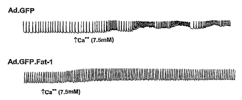
Fig. 13 is a pair of tracings showing differential responses of myocytes
infected
with Ad.GFP and myocytes infected with Ad.GFP.fat-1 to 7.SmM extracellular
calcium.
Fig. 14 is a line graph showing tumor volume over time (0-4 weeks after viral
injection) and thus, the effect of gene transfer on tumor growth. Breast
cancer cells
(MDA-MB-231) were implanted subcutaneously on the back of nude mice. Three
weelcs
later, the mice were treated with Ad.GFP fat-1 or Ad.GFP (control; 50 ~1, 1012
VP/m) by
intratumoral injection.
Fig. 15 is a table showing PUFA compositions of total cellular lipids from
control
MCF-7 cells and the transgenic MCF-7 cells expressing a C. elegaras fat-1
cDNA.
Fig. 16 is a bar graph depicting the results of an enzyme immunoassay of
prostaglandin EZ levels in control MCF-7 cells and MCF-7 cells expressing fat-
I gene.
Values are means ~ SE of three experiments and expressed as a percentage of
control.
(~P < 0.05).
Figs.l7A and 17B are representations of the nucleotide sequence of the C.
elegahs
fat-1 cDNA (Fig. 18A) and the deduced amino acid sequence of the Fat-1
polypeptide
(Fig. 18B).
DETAILED DESCRIPTION
The studies described below demonstrate that, hater alia, a nucleic acid
molecule
encoding an n-3 desaturase can be eff ciently expressed in a variety of
mammalian cell
types and, as a consequence, those cells produce significant amounts of n-3
PUFA from
endogenous n-6 PUFA and have a more balanced ratio of n-6 to n-3 PUFA (1:1).
The
studies were carried out using recombinant adenoviral expression vectors,
which can
mediate gene transfer i~2 vivo or in vity°o. Adenoviral vectors
expressing fat-1, or
biologically active variants thereof, as well as other types of viral and non-
viral
expression vectors are within the scope of the invention now claimed. Other
viral vectors
8
CA 02439654 2003-08-28
WO 02/072028 PCT/US02/07649
that can be employed as expression constructs in the present invention include
vectors
derived from viruses such as vaccinia virus (e.g., a pox virus or a modified
vaccinia virus
anlcara (MVA)), an adeno-associated virus (AAV), or a herpes viruses. These
viruses
offer several attractive features for various mammalian cells. For example,
herpes
simplex viruses (e.g., HSV-1) can be selected to deliver fat-1 or a
biologically active
variant thereof, to neuronal cells (and thereby treat patients with
neurodegenerative
conditions).
Other retroviruses, liposomes, and plasmid vectors are also well lcnown in the
art
and can also be used (e.g., the expression vector pUR278 can be used when one
wishes to
fuse a fat-1 sequence to the lacZ gene; lacZ encodes the detectable marker (3-
galactosidase (see, e.g., Ruther et al., EMBO J., 2:1791, 1983). A fat-1
sequence can also
be fused to other types of heterologous sequences, such as a sequence that
encodes
another therapeutic gene or a sequence that, when expressed, improves the
quantity or
quality (e.g., solubility or circulating half life) of the fusion protein. For
example, pGEX
vectors can be used to express the proteins of the invention fused to
glutathione S-
transferase (GST). In general, such fusion proteins are soluble and can be
easily purified
from lysed cells by adsorption to glutathione-agarose beads followed by
elution in the
presence of free glutathione. The pGEX vectors (Pharmacia Biotech Inc; Smith
and
Johnson, Gene 67:31-40, 1988) are designed to include thrombin or factor Xa
protease
cleavage sites so that the cloned target gene product can be released from the
GST
moiety. Other fusion partners include albumin and a region (e.g., the Fc
region) of an
immunoglobulin molecule (e.g., IgG, IgA, IgM, or IgE). Other useful vectors
inchtde
pMAL (New England Biolabs, Beverly, MA) and pRITS (Pharmacia, Piscataway, NJ),
which fuse maltose E binding protein and protein A, respectively, to an n-3
desaturase.
Transgene expression can be sufficiently prolonged from episomal systems, so
that readministration of the vector, with its transgene, is not necessary.
Alternatively, the
vector can be designed to promote integration into the host genome, preferably
in a site-
specific location, which would help ensure that the transgene is not lost
during the cell's
9
CA 02439654 2003-08-28
WO 02/072028 PCT/US02/07649
lifetime. Whatever the means of delivery, transcriptional control, exerted by
the host
cell, would promote tissue specificity and regulate transgene expression.
The expression vector will be selected or designed depending on, for example,
the
type of host cell to be transformed and the level of protein expression
desired. For
example, when the host cells are mammalian cells, the expression vector can
include viral
regulatory elements, such as promoters derived from polyoma, Adenovirus 2,
cytomegalovirus and Simian Virus 40. The nucleic acid inserted (i. e., the
sequence to be
expressed; here, fat-1 ) can also be modified to encode residues that are
preferentially
utilized in E, coli (Wada et al., Nucleic Acids Res. 20:2111-2118, 1992).
These
modifications can be achieved by standard recombinant techniques. More
generally, the
expression vectors of the invention can be designed to express proteins in
prokaryotic or
eukaiyotic cells. For example, polypeptides of the invention can be expressed
in
bacterial cells (e.g., E. coli), fungi, yeast, or insect cells (e.g., using
baculovirus
expression vectors). For example, a baculovirus such as Autographa californica
nuclear
polyhedrosis virus (AcNPV), which grows in Spodoptera frugiperda cells, can be
used as
a vector to express foreign genes.
As noted elsewhere, the expression vectors and nucleic acids used to express
fat-1
can also contain a tissue-specific promoter. Such promoters are known in the
art and
include, but are not limited to liver-specific promoters (e.g., albumin;
Miyatalce et al.,
1997), muscle-specific promoters (e.g., myosin light chain 1 (Shi et al.,
1997) a-actin),
pancreatic-specific promoter (e.g., insulin or glucagon promoters), neural-
specific
promoters (e.g., the tyrosine hydroxylase promoter or the neuron-specific
enolase
promoter), endothelial cell-specific promoters (e.g., von Willebrandt; Ozalci
et al., 1996),
and smooth muscle-cells specific promoters (e.g., 22a; Kim et al., 1997).
Tumor-specific
promoters are also being used in developing cancer therapies, including
tyrosine lcinase-
specific promoters for B16 melanoma (Diaz et al., 1998), DF3/MUC1 for certain
breast
cancers (Wen et al., 1993; for breast cancer, an adipose-specific promoter
region of
human aromatase cytochrome p450 (p450arom) can also be used (see U.S. Patent
No. 5,446,143; Mahendroo et al., .I. Biol. Claem. 268:19463-19470, 1993; and
Simpson et
CA 02439654 2003-08-28
WO 02/072028 PCT/US02/07649
al., Clin. Claem. 39:317-324, 1993). An a-fetoprotein promoter can be used to
direct
expression in hepatomas (Chen et al., 1995). The vectors and other nucleic
acid
molecules of the invention (e.~g., the fat-1 cDNA per se) can also include
sequences that
limit the temporal expression of the transgene. For example, the transgene can
be
controlled by drug inducible promoters by, for example including cAMP response
element enhancers in a promoter and treating the transfected or infected cell
with a cAMP
modulating drug (Suzuki et al., 1996). Alternatively, repressor elements can
prevent
transcription in the presence of the drug (Hu et al., 1997). Spatial control
of expression
has also been achieved by using ionising radiation (radiotherapy) in
conjunction with the
ergl gene promoter (Hallaham et al., 1995). Constructs that contain such
regulatory
sequences are within the scope of the present invention.
In the examples that follow, RNA analysis and enzymatic assays were performed
to assess gene expression, and gas chromatography-mass spectrometry were used
to
determine fatty acid profiles (these are standard techniques that one of
ordinary skill in
the art could use to assess any variant of the fat-1 sequence for biological
activity; or
incorporate in any method of assessing a sample obtained from a patient for
fat-1
expression).
Some of the studies described below were conducted using cortical new-ons.
Fat-1 expression not only modified the cellular n-6:n-3 fatty acid ratio and
eicosanoid
profile in these neurons, but also protected the cells from apoptosis, thereby
increasing
cellular viability. More specifically, fat-1 expression modified the fatty
acid ratio and
protected rat cortical neurons against growth factor withdrawal-induced
apoptotis in the
absence of supplementation with exogenous n-3 PLTFAs. Accordingly, the nucleic
acid
molecules (and other compositions) described herein taxi be used as
neuroprotectants,
which can be administered to premature infants and to older patients having
any
neurodegenerative disease (alternatively, the molecules or other compositions
can be
delivered to an animal, parts of which are then consumed by the patient). The
protective
effect of gene transfer on neuronal apoptosis mimics the protective effects of
n-3 fatty
acid supplementation.
11
CA 02439654 2003-08-28
WO 02/072028 PCT/US02/07649
S The positive results obtained with neurons are especially encouraging
because n-3
PUFA deficiency leads to abnormal development of the retina and the brain,
particularly
in premature infants (Uauy et al., Lipids 36:885-895, 2001), and animals
deficient in n-3
PUFA show deficits in memory, spatial and context-dependent learning, and loss
of
visual acuity (Carne et al., Neurosci. Lett. 266:69-72, 1999; Yehuda et al.,
J. Nem~osci.
Res. S6:S6S-70, 1999). There are also indications that various neurological
disease states
in humans are associated with an n-3 deficient status (Vancassel et al.,
P~~ost. Leuk. Ess.
Fatt. Acids 65:1-7, 2001; Hoffman and Birch, World Rev. Nuts-. Diet 83:52-69,
1998).
The biological functions of PUFAs are described further here, as these
functions
bear on the types of conditions amenable to treatment with the nucleic acid
molecules
1 S (and other compositions) described herein. PUFAs are important structural
components
of membrane phospholipids and are precursors of families of signaling
molecules
(eicosanoids) including prostaglandins, thromboxanes, and leukotrienes
(Needleman
et al., Ann. Rev. Bioclzem. SS:69-102, 1986; Smith and Borgeat, In
Biocl2etnistfy ofLipids
and Menzbf~anes, D.E. Vance & J.E. Vance, Eds., Benjamin/Cummings, Menlo
Parlc, CA,
00 325-360, 1986). The eicosanoids derived from PUFAs play a leey role in
modulating
inflammation, cytolcine release, the immune response, platelet aggregation,
vascular
reactivity, thrombosis and allergic phenomena (Dyerberg et al., Lancet 2:117-
119, 1978;
Cyerberg and Bang, Lasacet 2:433-435, 1979; James et al., Ana. J. Clin Nutr.
7:3435-
34385, 2000; Calder; Ann. Nutr. Metab. 41:203-234, 1997). The principal fatty
acid
2S precursors of these signaling compounds are arachidonic acid (AA, 20:4n6),
providing an
n-6 substrate that is responsible for the major synthesis of the series 2
compounds, and
eicosapentaenoic acid (EPA, 20:Sn3), an n-3 substrate that is responsible for
the parallel
synthesis of many series 3 eicosanoids with an additional double bond. The n-
6:n-3 ratio
W phospholipids modulates the balance between eicosaniods of the 2 and 3
series derived
from AA and EPA. The eicosanoids derived from AA (series 2) and EPA (series 3)
are
functionally distinct and some have important opposing physiological functions
(Dyerberg et al., Lancet 2:117-119, 1978; Cyerberg and Bang, Lancet 2:433-435,
1979;
James et al., Am. J. Clin Nutr. 7:3435-34385, 2000; Calder, Ann. Nuts. Metab.
41:203-
12
CA 02439654 2003-08-28
WO 02/072028 PCT/US02/07649
234, 1997). Series 3 eicosanoids are weak agonists or, in some cases,
antagonists of
series 2 eicosanoids. For example, eicosanoids of the 2 series promote
inflammation and
platelet aggregation, and activate the immune resoponse, whereas series 3
eicosanoids
tend to ameliorate these effects. In addition, PLTFAs, in the form of free
fatty acids, are
involved in gene expression and intercellular cell-to-cell communication
(Price et al.,
Cm°~. Opin. Lipidol 11:3-7, 2000; Sellmayer et al. Lipids 31 Supp1:S37-
540, 1996;
vonSchacky, J. Lab. Clin. Med. 128:5-6, 1996). Thus, PLTFA can exhibit many
diverse
biological effects.
The compositions and methods described herein can be used to treat a variety
of
specific conditions as well as to improve general health. Any condition that
is amenable
to treatment by administration of n-3 PUFAs is amenable to treatment by way of
the
methods of the present invention, which comprise administration of a gene
encoding an
n-3 desaturase (e.g., the C. elegans fat-1 gene). Some of the conditions
amenable to
treatment are described below.
n-3 PUFAs have attracted considerable interest as pharmaceutical and
nutraceutical compounds (Connor, Afn. J. Clisz Nutr~. 70:5605-5695, 1999;
Simopoulos,
Am. J. Clin. Nutr°. 70:5625-5695, 1999; Salem et al., Lipids 31:51-
5326, 1996). During
the past 25 years, more than 4,500 studies have explored the effects of n-3
fatty acids on
human metabolism and health (e.g., cardiovascular health). From epidemiology
to cell
culture and animal studies to randomized controlled trials, the
cardioprotective effects of
omega-3 fatty acids have been recognized (Leaf and Fang, Wo~ld Rev.
Nuts°. Diet. 83:24-
37, 1998; De Caterina et al., Eds., n-3 Fatty Acids and T~asculaf~ Disease,
Springer-
Verlag, London, 1999, pp 166; O'Keefe and Harris, Mayo Clin. Proc. 75:607-614,
2000).
The predominant beneficial effects include a reduction in sudden death (Albert
et al.,
JAMA 279:23-28, 1998; Siscoviclc et al., JAMA 274:1363-1367, 1995), decreased
risk of
arrhythmia (Fang and Leaf, Circulation 94:1774-1780, 1996), lower plasma
triglyceride
levels (Harris, Am. J. Clin. Nutf°. 65:16455-16545, 1997), and a
reduced blood-clotting
tendency (Agren et al., P~ostagland. Leukot. Essent. Fatty Acids 57:419-421,
1997; Mori
et al., Af~te~ioscler~. Tlarona. Basc. Biol. 17:279-286, 1997). Evidence from
13
CA 02439654 2003-08-28
WO 02/072028 PCT/US02/07649
epidemiological studies shows that another n-3 fatty acid, a-linolenic acid,
reduces risk of
myocardial infarction (Guallar et al., Artef°ioscler. Thromb. T~asc.
Biol. 19:1111-11 I8,
1999) and fatal ischemic heart disease in women (Hu et al., Arra. J. Clin.
Nutr. 69:890-
897, 1999). Several randomized controlled trials recently have demonstrated
beneficial
effects of both oc-linolenic acid (de Lorgeril et al., Circulation 99:779-785,
1999) and
marine c~-3 fatty acids (Singh et al., Cardiovasc. drugs then. 11:485-491,
1997; Von
Schaclcy et al., Ann. Intern. Med. 130:554-562, 1999; GISSI-Prevenzione
Investigators,
Lancet 354:447-455, 1999) on both coronary morbidity and mortality in patients
with
coronary disease. The n-3 fatty acid, EPA, exerts anticancer activity in vitro
and
in animal models of experimental cancer (Bougnoux, Cur°r. Opifa. Clin.
Nutr. Metab.
Care 2:121-126, 1999; Cave, Breast Cancer Res. Treat. 46:239-246, 1997). Human
studies show that populations whose diets are rich in EPA exhibit a
remarlcably low
incidence of cancer (Rose and Connolly, Plaaf°macol. Ther. 83:217-244,
1999).
Supplementation with n-3 PUFAs shows therapeutic effects on inflammatory and
autoimmune diseases such as arthritis (I~remer, Ana. J. Clin. Nutr. 71:3495-
3515, 2000;
Ariza-Ariza et al., Semin. Arthritis Rheum. 27:366-370, 1998; James et al.,
Am. J. Clin.
Nuts°. 71:3435-3485), and studies with nonhuman primates (Neuringer et
al., Pr°oc. Natl.
Acad. Sci. USA 83:4021-4025, 1986) and human newborns (Uauy et al., Proc.
Nutr. Soc.
59:3-15, 2000; Uauy et al., Lipids 31:5167-176, 1996) indicate that the n-3
fatty acid,
DHA, is essential for the normal functional development of the retina and
brain,
particularly in premature infants. Fm-thennore, n-3 PUFA have been shown to
have
beneficial effects on many other clinical problems, such as hypertension
(Appel et al.,
Arch. Inter°n. Med. 153:1429-1438, 1993), diabetes (Raheja et al.,
Anya. N. Y. Acad. Sci.
683:258-271, 1993), obesity (Clarlce, Br. J. Nutr. 83:559-66, 2000), skin
disorders
(Ziboh, World Rev. Nutr. Diet. 66:425-435, 1991), renal disease (De Caterina
et al.,
Kidney Int. 44:843-850, 1993), ulcerative colitis (Stenson et al., An sa.
Intern. Meal
116:609-614, 1992), Crohn's disease (Belluzzi et al., N. Engl. J. Med.
334:1557-1560,
1996), chronic obstructive pulmonary disease (Shahar et al., N. Engl. J. Med.
331:228-
233, 1994), and transplanted organ rejection (Otto et al., Transplantation
50:193-198,
14
CA 02439654 2003-08-28
WO 02/072028 PCT/US02/07649
1990). In general, a balanced n-6:n-3 ratio of the body lipids is essential
for normal
growth and development and plays an important role in the prevention and
treatment of
many clinical problems. The diseases, disorders, and conditions described
above are
amenable to treatment with the nucleic acid molecules (and other compositions)
described herein.
According to recent studies (Simopoulos, Poultry Science 79:961-970, 2000),
the
ratio of n-6 to n-3 essential fatty acids in today's diet is around 10-20:1.
This indicates
that present Western diets are deficient in n-3 fatty acids compared with the
diet on which
hmnans evolved and their genetic patterns were established (n-6/n-3 = 1:1)
(Leaf and
Weber, Afn. J. Clifa Nuts°. 45:1048-1053, 1987). Since the n-6 and n-3
fatty acids are
metabolically and functionally distinct and have important opposing
physiological
functions, their balance is important for homeostasis and normal development.
However,
n-3 and n-6 PUFAs are not interconvertible in the human body because mammalian
cells
laclc the enzyme n-3 desaturase. Therefore, the balance between n-6 and n-3
PUFA in
biological membranes is regulated based on dietary supply. Elevating the
tissue
concentrations of n-3 fatty acids in human subjects or animals relies on
increased
consumption of n-3 PUFA-enriched foods or n-3 PUFA supplements. Given the
potential therapeutic actions of n-3 PUFAs, an international scientific
working group has
recommended diets in which the intake of n-6 fatty acids is decreased and the
intake of n-
3 fatty acids is increased (Simopoulos, Food Australia 51:332-333, 1999). The
American
Heart Association has also recently made such a dietary recommendation (AHA
Dietary
Guidelines: Revision 2000, Circulation 102:2284-2299, 2000).
Although dietary supplementation with n-3 PUFA is a safe intervention, it has
a
number of limitations. For example, to achieve a significant increase in
tissue
concentrations of n-3 PUFA ira vivo requires a chronic intalce of high doses
of n-3 PUFA
for a period of at least 2-3 months. Bioavailability of fatty acids to cells
from the diet
involves a series of physiological processes including digestion, absorption,
transport and
metabolism of fat. Thus, the efficacy of dietary intervention depends on the
physiological
and health status of an individual. A patient in critical condition or who has
a
CA 02439654 2003-08-28
WO 02/072028 PCT/US02/07649
gastrointestinal disorder is unlikely to be able to ingest or absorb fatty
foods or n-3 PUFA
supplements. In addition, encapsulated fish oil supplements are unlilcely to
be suited to
daily use over a person's lifetime because of their high caloric content.
Moreover,
ingestion of some species of fish from costal waters and lakes may carry toxic
amounts of
mercury or organic toxins, and effective dietary intervention requires a
disciplined
change in dietary habits that some people may not be able to sustain. In view
of the
foregoing, there is a great need for the means to quiclcly and effectively
increase cellular
n-3 PLIFA content and balance the n-6:n-3 ratio without resorting to long-term
intake of
fish or fish oil supplements. This need is met by the methods of the present
invention,
which create an alternative food source (via transgenic livestoclc whose cells
contain
substantially more n-3 PUFAs than in non-transgenic animals) or which provide
for
administration of a gene encoding an n-3 desaturase enzyme to patients (e.g.,
human
patients). A particular advantage of the present methods is that they not only
elevate
tissue concentrations of n-3 PUFAs, but also simultaneously decreases the
levels of
excessive endogenous n-6 PUFA.
16
CA 02439654 2003-08-28
WO 02/072028 PCT/US02/07649
EXAMPLES
Example 1: Construction of a recombinant adenovirus.
A recombinant adenovirus caiTying the fat-I gene was constructed following
procedures similar to those described by He et al. (P~°oc. Natl. Acad.
Sci. USA 95:2509-
2514, 1998). The n-3 fatty acid desaturase cDNA (fat-1 gene) in pCE8 was
kindly
provided by Dr. J. Browse (Washington State University) (but can be
synthesized or
cloned using information and techniques available to those of ordinary skill
in the art; see
Spychalla et al., Proc. Natl. Acad. Sci. USA 94:1142-1147, 1997; US Patent
No. 6,194,167; and Fig. 17A and 17B). The cDNA insert of pCE8 was excised from
the
plasmid with an EcoRIlKphI double digest, inserted into a shutter vector, and
then
recombined with an adenoviral backbone according to the methods of He et al.
(supra).
Two, first-generation type 5 recombinant adenoviruses were generated: Ad.GFP,
which
carries the green fluorescent protein (GFP, as reporter gene) under control of
the
cytornegalvirus (CMV) promoter, and Ad.GFP.fat-1, which carries both the fat-1
and
GFP genes, each under the control of separate CMV promoters. The recombinant
viruses
were prepared as high titer stocks through propagation in 293 cells, as
described
previously (Hajjar et al. Circulation 95:423-429, 1997). The constructs were
confirmed
by enzymatic digestion and by DNA sequence analysis. See also Hajjar et al.,
Ci~culatiofa 95:4230429, 1997 and Hajjar et al., Circ. Res. 81:145-153, 1997.
Wild-type adenovirus contamination can be assessed and shown to be excluded
by the absence of both PCR-detectable El sequences and cytopathic effects on
the
nonpermissive A549 cell line. Alternative adenoviral vectors with other
promoters or
adeno-associated viral (AAV) vectors can be constructed if necessary or
desired.
Example 2: Culture and infection of cardiac myocytes witli adenovirus.
Cardiac myocytes were isolated from one-day-old rats using the National
Cardiomyocyte Isolation System (Worthington Biochemical Corp., Freehold, NJ).
The
isolated cells were placed in 6-well plates and cultured in F-10 medium
containing S%
fetal bovine serum and 10% horse serum at 37°C in a tissue culture
incubator with 5%
17
CA 02439654 2003-08-28
WO 02/072028 PCT/US02/07649
COZ and 98% relative humidity. Cells were used for experiments after 2-3 days
of
culture. Viral infection was caiTied out by adding viral particles at
different
concentrations (5x1 O~ - 101° pfu) to culture medium containing 2%
fetal bovine serum
(FBS). After a 24 hour incubation, the infection medium was replaced with
normal (15%
serum), culture medium supplemented with 10 ~,M of 18:2n-6 and 20:4n-6. About
48
hours after infection, the cells can be used (e.g., one can then analyze gene
expression,
fatty acid composition, viability, or growth (e.g., proliferation or rate of
division)).
Example 3: Detecting fat-1 expression with fluorescence microscopy and RNA
analysis.
Gene expression can be assessed by many methods lrnown in the art of molecular
biology. Here, expression of fat-I in cardiac myocytes, infected as described
above, was
assessed by visual examination of infected cells and a ribonuclease (RNase)
protection
assay.
More specifically, the coexpression of GFP allowed us to identify the cells
that
were infected and expressed the transgene. About 48 hours after infection,
almost all of
the cells (>90%) exhibited bright fluorescence, indicating a lugh efficiency
of gene
transfer and a high expression level of the transgene (see Fig. 1). Expression
of fat-1
transcripts was also determined by RNase protection assay using a RPA IIITM
lcit
(Ambion). Briefly, total RNA was extracted from cultured cells using an RNA
isolation
lcit (Qiagen) according to the manufacturer's protocol. The plasmid containing
the fat-1
gene, pCE8, was linearized and used as a transcription template. Anti-sense
RNA probes
were transcribed in vitf°o using 33P-UTP, hybridized with the total RNA
extracted from
the myocytes, and digested with RNase to remove non-hybridized RNA and probe.
The
protected RNA:RNA was resolved by electrophoresis through a denaturing gel and
subjected to autoradiography. A probe targeting the [3-actin gene was used as
a control.
Fat-1 mRNA was not detected in cells infected with AD.GFP (also used as a
control), but
was abundant in cells infected with Ad.GFP.fat-1 (Fig. 2). Tlus result
indicates that
18
CA 02439654 2003-08-28
WO 02/072028 PCT/US02/07649
adenovirus-mediated gene transfer confers very high expression of fat-1 gene
in rat
cardiac myocytes that normally lack the gene.
Example 4: Lipid analysis; the effect of n-3 desaturase on fatty acid
composition
By lipid analysis, one can determine whether the expression of a fat-1 gene in
cardiac myocytes (or any other cell type) converts n-6 fatty acids to n-3
fatty acids and,
thereby, changes the fatty acid composition of the cell. Following infection
with the
adenoviruses described above, cells were incubated in medium supplemented with
n-6
fatty acids (10 ~.M 18:2n-6 and 10 pM 20:4n-6) for 2-3 days. After the
incubation, the
fatty acid composition of total cellular lipids was analyzed as described
previously (Fang
et al., Biochifn. BioplZys. Acta. 1128:267-274, 1992; Weylandt et al., Lipids
31:977-982,
1996).
Lipid was extracted with chloroform/methanol (2: l, v/v) containing 0.005%
butylated hydroxytoluene (as antioxidant). Fatty acid methyl esters were
prepared using
14% BF3/methanol reagent. Fatty acid methyl esters are quantified by GC/MS
using a
HP5890 Series II gas chromatograph equipped with a Supelcowax SP-10 capillary
column attached to a HP-5971 mass spectrometer. The injector and detector are
maintained at 260°C and 280°C, respectively. The oven program is
initially maintained
at 150°C for 2 minutes, then ramped to 200°C at 10°C/min
and held for 4 minutes,
ramped again at S°C/min to 240°C, held for 3 minutes, and
finally ramped to 270°C at
10°C/min and maintained for 5 minutes. Carrier gas flow rate is
maintained at a constant
0.8 mL/min throughout. Total ion monitoring is performed, encompassing mass
ranges
from 50-550 amus. Fatty acid mass is determined by comparing areas of various
analyzed fatty acids to that of a fixed concentration of internal standard.
The fatty acid profiles were remarlcably different between the control cells
infected with Ad.GFP and the cells infected with Ad.GFP.fat-1 (Fig. 3).
Moreover, cells
infected with Ad.GFP showed no change in their fatty acid profiles when
compared with
non-infected cells. In the cells expressing the fat-1 gene (n-3 desaturase),
almost all
binds of n-6 fatty acids were largely converted to the corresponding n-3 fatty
acids,
19
CA 02439654 2003-08-28
WO 02/072028 PCT/US02/07649
namely, 18:2n-6 to 18:3n-3, 20:2n-6 to 20:3n-3, 20:3n-6 to 20:4:n-3, 20:4n-6
to 20:5n-3,
and 22:4n-6 to 22:5n-3. As a result, the fatty acid composition of the cells
expressing fat-
1 was significantly changed with respect to that of the control cells infected
with Ad.GFP
(Fig. 5). Importantly, the ratio of n-6:n-3 was reduced from 15:1 in the
control cells to
1:1.2 in the cells expressing the n-3 fatty acid desaturase.
Example 5: Measuring eicosanoids following fat 1 expression
Since 20:4n-6 (AA) and 20:5n-3 (EPA) are the precursors of 2-series and 3-
series
of eicosanoids, respectively, differences in the contents of AA and EPA may
lead to a
difference in production of eicosanoids in the cells. Thus, we measured the
production of
eicosanoids in the infected cells following stimulation with calcium ionophore
A23187
by using a EIA lcit that specifically detect prostaglandin EZ with a 16% cross-
reactivity
with prostaglandin E3. More specifically, Prostaglandin EZ was measured by
using
enzyme immunoassay kits (Assay Designs, Inc) following the manufacturer's
protocol.
(The cross-reactivity with PGE3 is 16%). Cultured cells were washed and serum-
free
medium containing calcium ionophore A23187 (5 p,M). After a 10 minute
incubation,
the conditioned medium was recovered and subj ected to eicosanoid measurement.
The
amount of prostaglandin EZ produced by the control cells was significantly
higher than
that produced by cells expressing the n-3 desaturase encoded by fat-1 (Fig.
4).
Example 6: Analysis of animal cells in culture
In this example and the two that follow, we set out three different
experimental
models: cultured cells (other types of cultured cells are tested further
below), adult rats,
and transgenic mice. As shown above, the cultured cell model can be used to
characterize the enzymatic properties and biochemical effects of the n-3
desaturase when
expressed in mammalian cells in vitro; the adult rat model can be used to
evaluate the
efficacy with which a transferred fat-1 gene can elevate tissue concentrations
of n-3
PUFA ira vivo, and the transgenic mouse model can be used to assess the long-
teen and
systematic effects of the transgene on lipid composition of various tissues or
organs ifz
CA 02439654 2003-08-28
WO 02/072028 PCT/US02/07649
vivo. For the first two models, the introduction of the fat-1 gene into
mammalian
cells/tissues will be carried out by mean of adenoviral gene transfer
(mediated by
recombinant adenoviruses). For the last model, gene transfer will be carried
out by
microinjection of the transgene into fertilized mouse eggs. Following gene
transfer, the
expression profile of the transferred gene can be characterized by mRNA and/or
protein
analysis (see, e.g., Example 3, above), and the biochemical effects, mainly
the fatty acid
composition of the cells or tissues, will be detennined by GC-MS technology
(see, e.g.,
Example 4, above). Eicosanoids will be measured by enzyme immunoassay (see,
e.g.,
Example 5). Changes are identified by comparing the data obtained from fat-1-
expressing cells with data obtained from control cells or tissues infected
with the same
(or a similar) virus, but not transfected with fat-1. The end point of these
studies is the
biochemical changes in cellular fatty acid composition and eicosanoid profile.
Cultures of virtually any animal cells (including hmnan cell lines) can be
infected
with recombinant adenovirus (Ad.GFP.fat-1 or Ad.GFP), after which expression
of the
transferred gene can be assessed by RNA or protein analysis. The experimental
procedures and related methods are described in the Examples above and
outlined in
Fig. 6. Various cell types including cardiac myocytes, neurons, hepatocytes,
endothelial
cells, and macrophages have been used in studies of n-3 fatty acids.
Cardiac myocytes can be isolated and cultured as described above (see
Example 2), and other cell types, such as cerebellar granule neurons and
hepatocytes can
be prepared from 1-5 day-old rats following the method described by Schousboe
et al. (Ifa
A Dissectiofa ayad Tissue CultuT a Mayaual of the Nervous System, Shahar et
al., Eds., Alan
R. Liss, New York, N.Y., pp. 203-206, 1989). Human cell lines, including
breast cancer
cell lines and leukemia cell lines can be cultured in MEN medium or RPMI 1640
supplemented with 10% fetal boviile serum (FBS) in a 37°C/5% COZ
incubator.
Viral infection can be carried out by adding viral particles at various
concentrations (e.g., 2 x 10~ - 2 x 101° pfu) to culture medium
containing no FBS or 2%
FBS (see also Example 2). After a 24-hour incubation, the infection medium is
replaced
with normal (10% FBS) culture medium. Forty-eight hours after infection, cells
can be
21
CA 02439654 2003-08-28
WO 02/072028 PCT/US02/07649
used for analysis of gene expression or fatty acid composition. Transgene
expression can
be assessed by fluorescence microscopy when a fluorescent tag is included in
the
transgene (see Example 1 and Fig. 1; similarly, the tag can be an antigenic
protein
detected by a fluorescent antibody) or by a standard RNA assay (e.g. a
Northern blot or
RNase protection assay). Since the fat-1 gene normally does not exist in
control cells, it
is not difficult to identify the difference in fat-1 mRNA between the control
cells and
cells expressing fat-1.
n-3 desaturase catalyzes the introduction of an n-3 double bond into n-6 fatty
acids, leading to formation of n-3 fatty acids with one more double bond than
their
precursor n-6 fatty acids (e.g., 18:2n-6 -318:3n-3, 20:4n-6 -~20:Sn-3). The
rate of
conversion of substrates to products (the amount of products formed within a
given time
period) is thought to be directly proportional to the expression/activity of a
desaturase.
Thus, the functional activity of this enzyme can be determined, from a sample
obtained
from an animal (e.g., a tissue sample) or in cultured cells by measurement of
the
conversions (the quantity of products) using the following methods.
Fatty acid desaturation assay usifag radiolabeled n-6 fatty acids as
subst~°ates:
The assay can be performed following the protocol described by Fang et al.
(Biochim.
Biophys Acta. 1128:267-274, 1992). Briefly, various labeled n-6 fatty acids
(e.g.,
[iaC] 18:2n-6, [14C]20:4n-6) bound to BSA are added to serum-free cultua-e
medium and
incubated with cells for 4-6 hours. After that, cells and culture medium will
be harvested.
Lipids are extracted and methylated (see below). The labeled fatty acid methyl
esters are
separated according to degree of unsaturation (i. e., the number of double
bond) on silica-
gel TLC plates impregnated with AgN03, Bands containing fatty acids with
different
double bonds can be identified by comparison with reference standards.
Quantity of the
labeled fatty acids is determined by scintillation counting, and data are
compared
between control cells and the cells expressing the fat-1 gene.
Fatty acid analysis by gas chromatogf°ap7zy: Conversion of fatty acids
can be
determined more accurately by analysis of fatty acid composition using gas
chromatography-mass spectrometry (see below). Using this method, no
radiolabeled
22
CA 02439654 2003-08-28
WO 02/072028 PCT/US02/07649
fatty acid is required. Fatty acid contents of cultured cells expressing the n-
3 desatw-ase
gene, in the presence of various substrates, can be analyzed. The conversion
of each fatty
acid can be determined by comparison of fatty acid profiles between control
cells and the
cells expressing the fat-1 gene.
The fatty acid composition of total cellular lipids or phospholipids can be
analyzed as described previously (Kang et al., Bioclzizzz. Biophys. Acta.
1128:267-274,
1992; Weylandt et al., Lipids 31:977-982, 1996). The procedures are as
follows:
Lipid extraction (see also Exazzzple 4): Five ml of chloroformlmethanol (2:1,
v/v)
containing 0.005% butylated hydroxytoluene (as antioxidant) is added to washed
cell
pellets and vortexed vigorously for 1 minute then left at 4°C
overnight. One ml of 0.88%
NaCI is added and mixed again. 'The chloroform phase containing lipids is
collected.
The remains are extracted once again with 2 ml chloroform. The chloroform is
pooled
and dried under nitrogen and stored in sealed tubes at -70°C.
Separation of lipids by thin-layer chz°onaatog>"aplzy (TLC): TLC
plates are
activated at 100°C for 60 minutes. TLC tanks are equilibrated with
solvent for at least
one hour prior to use. Total phospholipid and triacyglycerol are separated by
running the
sample on silica-gel G plates using a solvent system comprised of petroleum
ether/diethyl
ether/acetic acid (80:20:1 by vol.) for 30-35 muiutes. Individual
phospholipids are
separated by TLC on silica-gel H plates using the following solvent system:
chloroform/methanol/2-propanol/0.25% I'Cl/ triethylamine (30:9:25:6:18 by
vol.).
Bands containing lipids are made visible with 0.01% 8-anilino-1-
naphthalenesulfonic
acid, and gel scrapings of each lipid fraction are collected for methylation.
23
CA 02439654 2003-08-28
WO 02/072028 PCT/US02/07649
Fatty acid methylation: Fatty acid methyl esters are prepared using 14%
BF3/methanol reagent. One or two ml of hexane and 1 ml of BF3/methanol reagent
are
added to lipid samples in glass tubes with Teflon-lined caps. After being
flushed with
nitrogen, samples are heated at 100°C for one hour, cooled to room
temperature and
methyl esters are extracted in the hexane phase following addition of 1 ml
H20. Samples
are allowed to stand for 20-30 minutes, the upper hexane layer is removed and
concentrated under nitrogen for GC analysis.
Gas ch~onaatography-mass spect~°ornetyy. Methylated samples are
reconstituted
in 100-200 ~,1 hexane or isooctane of which 1-2 p,1 will be analyzed by gas
chromatography. An Omegawas colmnn (30 m; Supelco, Bellefonte, PA) will be
used in
a Hewlett-Packard 5890A gas chromatograph (Hewlett-Paclcard, Avondage, PA).
Carrier
gas is hydrogen (2.39 ml/min), inj ected with a split ratio of 1:31. The
temperature is
initially 165°C for 5 minutes, then is increased to 195°C at
2.5°C/min and, from there, to
220°C at 5°C/min. The temperature is held for 10.5 minutes and
then decreased to 165°C
at 27.5°Chnin. Peaks will be identified by comparison with fatty acid
standards (Nu-
Chek-Prep, Elysian, MN), and area percentage for all resolved peaks will be
analyzed
using a Perlcin-Elmer M1 integrato (Perkin-Elmer, Norwood, CT). These
analytical
conditions separates all saturated, mono, di- and polyunsaturated fatty acids
from C14 to
C25 carbons in chain length. The sample size will be calculated based on
external
standards when added. In addition, the gas chromatography-mass spectrometry
(GC-MS)
will be carried out using a Hewlett-Packard mass selective detector (model
5972)
operating at an ionization voltage of 70 eV with a scan range of 20-S00 Da.
The mass
spectrum of any new peals obtained will be compared with that of standards (Nu
Chelc
Prep, Elysian, MN) in the database NBS75I~.L (National Bureau of Standards).
Example 7: Evaluation of n-3 desaturase gene transfer in vivo. The
experiments described here allow intTOduction of the fat-1 gene into animal
tissues or
organs (e.g., heart), where the enzyme product can quiclcly optimize fatty
acid profiles by
increasing the content of n-3 PUFAs and decreasing the content of n-6 PLTFAs.
The heart
24
CA 02439654 2003-08-28
WO 02/072028 PCT/US02/07649
is selected as an experimental target for the gene transfer because it has
been well studied
in relation to n-3 fatty acids, and it is a vital organ.
Adult rats, fed a normal diet or a diet high in n-6 PUFA for two months, will
be
randomized to receive either an adenovirus carrying the fat-1 gene (Ad.GFP.fat-
1) or an
adenovirus carrying the reporter gene GFP (Ad.GFP, as control). The
adenoviruses will
be delivered to the heart of a living animal using a catheter-based technique,
which can
produce an expression pattern that is grossly homogeneous throughout the heart
(Hajjar et
al., Proc. Natl. Acad. Sci. USA 95:525105256, 1998). Two days, 4 days, 10
days, 30
days and 60 days after infection (gene transfer), animals will be sacrificed,
and their
hearts will be harvested and used for determination of the transgene
expression and
analysis of fatty acid composition. Another group of rats will be fed a diet
rich in n-3
fatty acids (low n-6/n-3 ratio) for two months without gene transfer and used
as
references. These experiments (in which animals are on different diets and
samples
harvested at different time points) are designed to determine whether transfer
of the fat-1
gene can bring about a desired biochemical effect (n-6/n-3 ratio, eicosanoid
profile)
similar to or even superior to that induced by dietary intervention (i.e., n-3
FA
supplementation), how quickly a significant change in fatty acid composition
can be
achieved, and how long the change can last. Rats injected with the reporter
(GFP) gene
will be used as controls (our preliminary studies showed that gene transfer of
GFP has no
effect on fatty acid composition). The experimental flow chart is shown in
Fig. 7.
Afzi~nals and Diets: weight-matched adult Sprague-Dawley rats will be randomly
assigned to three groups. Each group is fed with one of three different diets:
normal
(basal) diet, a high n-6 diet, or a high n-3 diet. These diets are prepared as
follows.
Basal diet: a commercial rat fat-free diet (Agway Inc. C.G., Syracuse, NY) to
which 2% (w/w) corn oil is added; High fa-6 diet: the basal diet supplemented
by
addition of a fiuther 13% (w/w) corn oil or safflower oil (high in n-6 fatty
acids),
bringing the final diet to a total of 15% fat; High ra-3 diet: the basal diet
supplemented
with 13% (w/w) fish oil (30% EPA, 20% DHA, 65% total n-3 PUFA) (Pronova
Biocare
A/S, Oslo), bringing the final diet to a total of 15% fat. This group will
serve as a control
CA 02439654 2003-08-28
WO 02/072028 PCT/US02/07649
group for this study.
The diets will be prepared in small batches weeldy, lcept at -20° C and
thawed
daily in the aanounts required. Vitamin E (100mg/100g fat) and butylated
hydroxy
toluene (final concentration 0.05%) will be added to prevent oxidation of long-
chain
polyunsaturated fatty acids (The BHT should serve to prevent autooxidation of
the
unsaturated fatty acids during preparation and storage). To ensure animals are
receiving
adequate nutrition, the rats in all groups will be weighted weekly. After 8
weelcs on the
diets, the animals will be subjected to gene transfer.
Aclefzovi~al Delivef y Protocol. The delivery of adenoviruses to the heart
will be
performed by using a cathether-based technique similar to that described by
Hajjar et al
(supy-a). Briefly, rats will be anesthetized with infra peritoneal
pentobarbital (60 mgllcg)
and placed on a ventilator. The chest is entered from the left side through
the third
intercostals space. The pericardium is opened and a 7-0 suture placed at the
apex of the
left ventricle. The aorta and pulmonary artery are identified. A 22-gauge
catheter
containing 200 ~,L adenovirus (9-10 x 101° pfu/ml) is advanced from the
apex of the left
ventricle (LV) to the aortic root. The aorta and pulmonary arteries are
clamped distal to
the site of the catheter, and the solution is injected. The clamp
is.maintained for 10
seconds while the heart pumped against a closed system (isovolumically). After
10
seconds, the clamp on the aorta and pulmonary artery is released, the chest is
closed, and
the animals are extubated and transferred back to their cages.
At day 2, 4, 10, 30 and 60 after gene transfer, animals will be sacrificed,
their
hearts infected with the viruses will be removed, perfused or rinsed with
saline to
removed all blood and a portion of the tissues will be promptly frozen at -
80°C for lipid
analysis and eicosanoid measurement. The remaining tissues will be used for
determination of the mRNA levels and/or protein levels of the n-3 desaturase.
It is possible that other organs such as brain and liver may also be infected
at high
levels by the adenoviruses entering the blood stream. Thus, other organs, in
addition to
the heart, will be also harvested for analyses of transgene expression and
lipid profile.
Other methods, including assessment of transgene expression (by Northern blot,
26
CA 02439654 2003-08-28
WO 02/072028 PCT/US02/07649
lRNase protection assay, or i~a situ hybridization), analysis of fatty acid
composition,
measurement of eicosanoids, and statistical analysis will be carried out, as
described
above in the context of cultured cells.
Example 8: Transgenic animals.
The studies described here are designed to create transgenic mice that
globally
express the fat-1 gene and to characterize the tissue and organ lipid profiles
of these
animals. Transgenic mice have become a valuable model for evaluation of
physiological
significance of a gene irZ vivo. Availability of transgenic mice allows us to
study the
effect of a transgene in a variety of cell types at different stages of an
animal's lifespan.
This n-3 transgenic mouse model will provide new opportunities to elucidate
the roles of
n-3 PUFA and compounds derived from them in the development and cell biology.
To generate transgenic animals that can globally express the fat-1 gene, one
can
use an expression vector that contains a fat-1 gene and the chiclcen beta-
actin promotor
with the CMV enhancer (CAG promotor), which is highly active in a wide range
of cell
types and therefore allows high-level and broad expression of the transgene
(Niwa et al.,
Gene 108:193-199, 1991; Okabe et al., FEBSLett. 407:313-319, 1997). The
expression
construct will be microinjected into the pronuclei of one-cell embryos of
C57BL/6 X
C3H mice to produce transgenic nuce. They will be bred and transgenic mouse
line is
established. Weanling mice are fed either a normal diet or a diet high in n-6
PUFA.
Various tissues will be harvested from these animals at different ages
(neonate, wean --1
month, adult --6 ms and aging -12 ms, 3-5 mice per time point will be used)
for
assessment of the expression levels of the transgene and determination of
fatty acid
composition. The levels of eicosanoids in plasma and various tissues will also
be
measured. A group of wild-type mice (C57BL/6) fed with the same diet (either a
normal
diet or a high n-6 diet) will be used as controls. The results will be
compared with those
from wild type animals fed the same diet. The procedure is illustrated in Fig.
8.
The transgene will be prepared by methods similar to those described by Okabe
et
al. (supra). Briefly, a cDNA encoding the fat-1 gene is amplified by PCR with
primers,
27
CA 02439654 2003-08-28
WO 02/072028 PCT/US02/07649
5-agaattcggcacgagccaa gtttgaggt-3' (SEQ ID NO:l) and 5'-
gcctgaggctttatgcattcaacgcact-
3' (SEQ ID N0:2), using pCEB-fatl (provided by Dr. J. Browse, Washington State
University) as a template. No additional amino acid sequence is added on
either side of
the fat-1. The PCR product will be confirmed by DNA sequencing. The EcoRl and
Bgl-
II sites included in the PCR primers are used to introduce the amplified fat-1
cDNA into a
pCAGGS expression vector containing the chicken beta-actin promoter and
cytomegalovirus enhancer, beta-actin intron and bovine globin poly-adenylation
signal
(provided by Dr. J Miyazalci, Osalca University Medical School). The entire
insert with
the promoter and coding sequence will be excised with BamHI and Sall and gel-
purified.
Transgenic mouse lines will be produced by injecting the purified BanaHI and
SaII
fragment into C57BL/6 X C3H fertilized eggs. The DNA-injected eggs are
transplanted
to pseudo-pregnant mice (B6C3F1) to produce transgenic mice. The founder
transgenic
mice will be identified by PCR and Southern blot analyses of tail DNA and bred
with
C57BL/6J mice. Offspring (either heterozygote or homozygote) will be used
dependent
on the expression levels of the transgene or phenotype.
Weanling transgenic mice will be fed either a normal diet or a diet high in n-
6
PUFA (see above). Animals will be sacrificed at different ages (neonate, wean
to 1
month, adult to 6 mos and aging -12 mos, 3-5 mice per time point will be used)
and
various tissues will be harvested for assessment of the expression level of
transgene and
determination of fatty acid composition. The results will be compared with
those from
wild type animals fed the same diet.
Other methods, including assessment of transgene expression (Northern blot,
RNase protection assays, or in situ hybridization), analysis of fatty acid
composition,
measurement of eicosanoids, and statistical analyses will be carried out as
described
above.
Example 9: Inhibition of neuronal cell death
Constnuctiora of Recombinant Aderaovir~us (Ad): A recombinant Ad carrying the
fat-1 gene was constructed as described previously (Kang et al., Proc. Natl.
Acad. Sci.
28
CA 02439654 2003-08-28
WO 02/072028 PCT/US02/07649
USA 98;4050-4054, 2001; see also, above). The n-3 fatty acid desaturase cDNA
(fat-1
gene) used was that described above, provided in plasmid pCEB. . The fat-1
cDNA was
excised from the plasmid with an EcoRIlKpuI digestion, and inserted into
pAdTraclc-
CMV vector. The construct was subsequently recombined homologously with an
adenoviral backbone vector (pAdEasy 1) to generate two clones: Ad-GFP, which
expresses GFP as a reporter or marlcer, and Ad-GFP-fat-I, which carries both
the fat-1
and the GFP genes, each under the control of separate CMV promoters.
Recombinant
adenoviral vector DNA was digested with PacI. The linerized vector DNA was
mixed
with SuperFectTM (QIA.GEN) and used to infect 293 cells. The recombinant
viruses were
prepared as high-titer stocks through propagation in 293 cells. The integrity
of the
constructs was confirmed by enzymatic digestion (i. e., restriction mapping)
and by DNA
sequencing. Purified virus was checked and its sequence confirmed again by PCR
analysis.
Tissue Culture and Ihfectioya with Ad: Rat cortical neurons were prepared
using
standard techniques. Briefly, prenatal embryonic day 17 (E17) rat cortical
neurons were
dissociated and plated in poly-lysine-coated wells at 2 x 10~ cells/well. The
cells were
grown in NeurobasalT"" Medium (NBM, Life Technologies) supplemented with 25 mM
glutamic acid (Sigma Chemical Co., St. Louis, MO), 0.5 mM glutamine, 1 %
antibiotic-
antimycotic solution, and 2% B27 (Life Technologies). Cultures were kept at
37°C in air
with 5% C02 and 98% relative humidity. The culture medium u~as changed every
four
days. After 8-10 days in culture, cells were transfected with either the Ad-
GFP (control)
or the Ad-GFP-fat-1 plasmids. Viral infections were carried out by adding
viral particles
to the culture medium. After a 48-hour incubation, cells were used for
analyses of gene
expression, fatty acid composition, eicosanoid production, and induction of
apoptosis.
RNA Analysis: The Level of fat-1 expression was determined by probing for
mRNA transcripts in an RNAse protection assay using the RPA IIIT"" Icit
(Ambion,
Austin, TX). Briefly, total RNA was extracted from cultured cells using a
total RNA
isolation reagent (TRIzoI, GIBco BRL) according to the manufacturer's
protocol. The
plasmid containing the fat-1 gene, pCEB, was linearized and used as a
transcription
29
CA 02439654 2003-08-28
WO 02/072028 PCT/US02/07649
template. Antisense RNA probes were transcribed in nits°o using [33P]-
IJTP, T7
polymerase (Riboprobe SystemT"" T7 lcit, Promega), hybridized with total RNA
(15 pg)
extracted from neurons, and digested with ribonuclease to remove nonhybridized
RNA
and probe. The protected RNA~RNA hybrids were resolved in a denaturing 5%
sequence
gel and subjected to autoradiography. A probe targeting the (3-actin gene was
used as an
internal control. fat-1 mRNA was not detected in cells infected with Ad-GFP
(control),
but was highly abundant in cells infected with Ad-GFP fat-1.
The cells were also examined by fluoresence microscopy. Infected cells that
expressed the fat-1 gene were readily identifiable because they co-expressed
GFP.
Forty-eight hours after infection, 30-40% of the neurons were infected and
expressed
GFP. These results demonstrate that Ad-mediated gene transfer confers high
expression
of fat-1 gene in rat cortical neurons, which normally lacy the gene.
Lipid Analysis: The fatty acid composition of total cellular lipids was
analyzed as
described in Fang et al. (Py°oc. Natl. Acad. Sci. USA 98:4050-4054,
2001). Lipid was
extracted with chloroform:methanol (2:1, vol:vol) containing 0.005% butylated
hydroxytoluene (BHT, as an antioxidant). Fatty acid methyl esters were
prepared using a
14% (wt/vol) BF3/methanol reagent. Fatty acid methyl esters were quantified
with
GC/MS by using an HP-5890 Series II gas chromatograph equipped with a
SupelcowaxTM SP-10 capillary column (Supelco, Bellefonte, PA) attached to an
HP-5971
mass spectrometer. The injector and detector are maintained at 260°C
and 280°C,
respectively. The oven program is maintained initially at 150°C for 2
minutes, then
camped to 200°C at 10°C/minute and held for 4 minutes, camped
again at 5°C/minute to
240°C, held for 3 minutes, and finally camped to 270°C at
10°C/minute and maintained
for 5 minutes. Carrier gas-flow rate is maintained at a constant 0.8 ml/min
throughout.
Total ion monitoring is performed, encompassing mass ranges from 50-550 atomic
mass
units. Fatty acid mass is deterniined by comparing areas of various analyzed
fatty acids
to that of a fixed concentration of internal standard.
The expression of fat-1 resulted in conversion of n-6 fatty acids to n-3 fatty
acids,
and thus a significant change in the ratio of n-6:n-3 fatty acids. The fatty
acid profile
CA 02439654 2003-08-28
WO 02/072028 PCT/US02/07649
obtained from control cells is significantly different from that of cells
infected with Ad-
GFP fat-1 (Fig. 9; see also Fig. 10). Cells infected with Ad-GFP show no
change in fatty
acid composition when compared with non-infected cells. In cells expressing
the n-3
desaturase, almost all types of n-6 fatty acids were converted to the
corresponding n-3
fatty acids, namely, 18:2n-6 to 18:3n-3, 20:4n-6 to 20:5n-3, 22:4n-6 to 22:5n-
3, and
22:5n-6 to 22:6n-3. The change in fatty acid composition of the cells
expressing the fat-I
gene resulted in reduction of the n-6:n-3 ratio from 6.4:1 in the control
cells to 1.7:1 in
the cells expressing the n-3 desaturase. Expression of the C. elegans n-3
fatty acid
desaturase resulted in a significant increase in the levels of DHA in
transfected cells. An
increase in levels of EPA and ALA is observed with a concomitant decrease in
AA and
LA suggesting that the decrease in production of PGEZ resulted from both the
shift in the
n-6:n-3 fatty acid ratio and from DHA-mediated inhibition of AA hydrolysis.
Measu~~e~raent of Eicosanoids: 2-series eicosanoids may be associated with
neuronal apoptosis in age-associated neurodegenerative diseases and acute
excitotoxic
insults such as ischemia (Sanzgiri et al., J. Neu~obiol. 41:221-229, 1999;
Drachman and
Rothstein, Aran. Neurol. 48:792-795, 2000; Bezzi et al., Nature 391:281-285,
1998).
Arachidonic acid (AA, 20:4n-6) and eicosapentaenoic acid (EPA, 20:5n-3) are
the
precursors of 2- and 3-series of eicosanoids, respectively. To detei~nine
whethex the gene
transfer-mediated alteration in the contents of AA and EPA may lead to a
difference in
the production of eicosanoids in the cells, we measured the production of
prostaglandin
E2, one of the major eicosanoids derived from AA, in infected cells after
stimulation with
calcium ionophore A23187. More specifically, prostaglandin E~ was measured by
using
enzyme immunoassay lcits (Cayman Chemical, Ann Arbor, MI) following the
manufacturer's protocol. (The crossreactivity with prostaglandin E3 is 16%.)
Cultured
cells were washed with LH buffer (with 1% BSA) and incubated with the same
buffer
containing the calcium ionophore A23187 (5 ~,M). After a 10-minute incubation,
the
conditioned buffer was recovered and subjected to eicosanoid measurement. The
amount
of prostaglandin E2 produced by fat-1 expressing cells was 20% lower than that
produced
by control cells (Fig. 11).
31
CA 02439654 2003-08-28
WO 02/072028 PCT/US02/07649
IfZductiort of apoptosis afad deter°miraatioya of cell growth afad
viability: Apoptosis
was induced by growth factor withdrawal. Forty-eight hours after neurons were
transfected, the culture media was changed to NeurobasalTM Medium supplemented
with
25 rnM glutamic acid (Sigma Chemical Co., St. Louis, MO) and 0.5 mM glutamine.
Cytotoxicity was measured 24 hours after growth factors were withdrawn using
the
VybrantTM Apoptosis Assay (Molecular Probes, Eugene, OR). Briefly, cells were
washed with ice-cold phosphate buffered saline (PBS) and subsequently
incubated on ice
for 20-30 min in ice-cold PBS containing Hoechst 33342 solution (1 ml/ml) and
PI
solution (1 ml/n~I). A photograph was taken at the end of the incubation
period.
Cell gy°owth ai2d viability: Cell growth and viability were determined
using the
MTT cell proliferation kit (Ruche Diagnostic Corporation). MTT labeling
reagent (100
w1) was added to each well. After 4 hours of incubation, 1.0 m1 of the
solubilization
solution was added into each well. The cells were then incubated overnight at
37°C and
the spectrophotometrical absorbency of the solution at 600 nm was measured.
Expression of the fat-1 gene provided strong protection against apoptosis in
rat
cortical neurons. Hoest 33625 and PI staining of cortical cultures 24 hours
after the
induction of apoptosis, show that cultures infected with Ad-GFP fat-1
underwent less
apoptosis than those infected with Ad-GFP. MTT analysis indicated that the
viability of
Ad-GFP fat-1 cells was significantly (p<0.05) higher than that of cells
infected with Ad-
GFP (Fig. 12). These results indicate that the expression of fat-1 can inhibit
neuronal
apoptosis and promote cell viability. The ability of the C. elegaTZS n-3 fatty
acid
desaturase to inhibit apoptosis of neuronal cells highlights the importance of
the n-6:n-3
fatty acid ratio in neuroprotection. Accordingly, techniques that deliver a
fat-1 sequence,
or a biologically active variant thereof, to neurons provide the means to
quicldy and
dramatically balance cellular n-6:n-3 fatty acid ratio, alter eicosanoid
profile (and thereby
exert an anti-apoptotic effect on neuronal cells) without the need for
supplementation
with exogenous n-3 PLTFAs. Compared to dietary intervention, this approach is
more
effective in balancing the n-6:n-3 ratio because it simultaneously elevates
the tissue
concentration of n-3 PUFAs and reduces the level of endogenous n-6 PUFAs. This
32
CA 02439654 2003-08-28
WO 02/072028 PCT/US02/07649
method is a novel and effective approach to modifying fatty acid composition
in neuronal
cells, and it can be applied as a stand-alone gene therapy or as an adjuvant
therapy or
chemopreventive procedure (in, for example, apoplexy patients).
Data analysis, statistical a~aalysis: Cell viability data (MTT), as well as
fatty acid
composition and eicosanoids levels were compared using the Student t-test. The
analysis
included 6 wells/group (except lipid analyses; 4 wells/group) and each
experiment was
repeated 3 times. The level of significance was set at p<0.05.
Example 10: fat 1 expression in human endothelium and inhibition of
inflammation
To determiile whether the conversion of n-6 to n-3 PUFA can be genetically
conferred to primary human vascular endothelium and to study its potential
protective
effects against endothelial activation after cytokine stimulation, a first
generation (type 5)
recombinant adenoviral vector (Ad) was constructed which contained the fat-1
transgene
in series with a GFP expression cassette under the control of the CMV promoter
(Ad.fat-
1). A GFP/[3-gal adenovirus seined as the control vector (Ad.GFP/(3-gal).
Monolayers of
primary human umbilical vein endothelial cells (HUVECs) were infected
withAd.fat-1 or
the control Ad for 36 hours, exposed for 24 hours to 10 mM arachidonic acid,
and
subjected to lipid analysis by gas chromatography, surface adhesion molecule
analysis by
immunoassay, and videomicroscopy to study endothelial interactions with the
monocytic
cell line, THP-1, under laminar flow conditions.
Expression of fat-I dramatically altered the lipid composition of human
endothelial cells and changed the overall ratio of n-6 to n-3 PUFA from 8.5 to
1.4.
Furthermore, after cytokine exposure (TNF-a, 5 E.t/ml applied for 4 hours) fat-
1
expression significantly reduced the surface expression of the adhesion
molecules and
markers of inflammation (E-Selectin, ICAM-l, and VCAM-I by 42%, 43%, and 57%,
respectively (p < 0.001)).
We then aslced whether changes in the adhesion molecule profile were
sufficient
to alter endothelial interactions with monocytes, the most prevalent white
blood cell type
found in atherosclerotic lesions. Under laminar flow and a defined shear
stress of ~ 2
33
CA 02439654 2003-08-28
WO 02/072028 PCT/US02/07649
dynes/cm2, fat-1-infected HUVEC, compared to control vector-infected HUVEC,
supported -~ 50% less fine adhesion with almost no effect on the rolling
interactions of
THP-1 cells. Thus, heterologous expression of the C. elegaf2s desaturase, fat-
l, confers
on human endothelial cells the ability to convert n-6 to n-3 PUFA . This
effect
significantly repressed cytolcine induction of the endothelial inflammatory
response and
firm adhesion of the monocytic cell line, THP-1, under simulated physiological
flow
conditions. Accordingly, expression of fat-1 represents a potential
therapeutic approach
to treating inflammatory vascular diseases, such as atherosclerosis.
Example 11: n-3 desaturase as an anti-arrhythmic agent
To determine whether fat-1 expression could provide an anti-arrhythmic effect,
myocytes expressing the n-3 desaturase were examined for their susceptibility
to
arrhythrnias induced by anhythmogenic agents. Neonatal rat cardiac myocytes,
gTOwn
on glass coverslips and able to spontaneously beat, were infected with
Ad.GFP.fat-1 or
Ad.GFP. Two days after infection, cells were transferred to a perfusion system
and
perfused with serum free medium containing high concentrations (5-10 mM) of
calcium.
These media are anhythmogenic. During the perfusion process, myocyte
contraction was
monitored using a phase contrast microscope and video-monitor edge-detector.
Following the high [Ca2+] (7.5 mM) challenge, the control cells infected with
Ad.GFP
promptly exhibited an increased beating rate followed by spasmodic
contractions or
fibrillation whereas the cells infected with Ad.GFP.fat-1 could sustain
regular beating.
Thus, myocytes expressing the n-3 desaturase show little, if any,
susceptibility to
arrhythmogenic stimuli (Fig. 13).
Example 12: fat-1 expression and inhibition of tumor growth
To test the effect of the gene lxansfer on tumor growth iya vivo, we have
carried
out a pilot experiment in two nude mice bearing human breast cancer xenografts
(MDA-
MB-231). One mouse was injected intratumorally with SOmI of Ad.GFP.fat-1 (1012
particles/ml) twice every other day. The other was injected with the control
vector
34
CA 02439654 2003-08-28
WO 02/072028 PCT/US02/07649
(Ad.GFP). The growth rate of the tumors was monitored for four weeks. The
growth
rate of the tumor treated with Ad.GFP.fat-1 appeared to be slower than that of
the control
tumor (Fig. 14).
Example 13: The effect of fat 1 expression on fatty acid composition and
growth of
human breast cancer cells in culture
Constf uctiora of Reconabii2aht AdefZOVirus (Ad) : A recombinant Ad carrying
the
fat-1 cDNA was constructed as described previously (Fang et al., Proc. Natl.
Acad. Sci.
USA 98:4050-4054, 2001). Briefly, the fat-1 cDNA in pCE8 (as described above)
was
excised from the plasmid with an EcoRIlKphI double digest, inserted into a
shutter vector
and then subjected to homologous recombination with an adenoviral backbone
according
to the methods of He et al. (P~oc. Natl. Acad. Sci. USA 95:2509-2514, 1998).
Two first-
generation type 5 recombinant adenoviruses were generated: Ad.GFP, which
caiTies
GFP as a reporter gene under control of the CMV promoter, and Ad.GFP.fat-1,
which
carries both the fat-1 and GFP genes, each under the control of separate CMV
promoters.
The recombinant viruses were prepared as high titer stocks through propagation
in 293
cells, as described previously (Fang et al., Pt°oc. Natl. Acad. Sci.
USA 98:4050-4054,
2001). The integrity of the constructs was confirmed by enzymatic digestion
and by
DNA sequence analysis.
Cell Cultuf°es and If2fectioh wit72 Ad.: MCF-7 cells were routinely
maintained in
1:1 (v/v) mixture of DMEM and Ham's F12 medium (JRH, Bioscience) supplemented
with 5% fetal bovine serum (FBS) plus antibiotic solution (penicillin, 50
U/ml;
streptomycin, 50 ~,ghnl) at 37°C in a tissue culture incubator with 5%
COZ and 98%
relative humidity. Cells were infected with Ad for experiments when they were
grown to
about 70% confluence by adding virus particles to medium without serum (3-5
x10$
particles/ml). Initially, optimal viral concentration was determined by using
Ad.GFP to
achieve an optimal balance of high gene expression and low viral titer to
minimize
cytotoxicity. After a 24-hour incubation, the infection medium was replaced
with normal
culture medium supplemented with 10 ~M 18:2n6 and 20:4n6. Forty-eight hours
after
CA 02439654 2003-08-28
WO 02/072028 PCT/US02/07649
infection, cells were used for analyses of gene expression, fatty acid
composition,
eicosanoid production, and cell proliferation and apoptosis.
RNA Analysis: The fat-1 transcripts were examined by ribonuclease protection
assay using a RPA IIITM lcit (Ambion, Austin, TX). Briefly, total RNA was
extracted
from cultured cells using a RNA isolation lcit (Qiagen) according to the
manufacturer's
protocol. The plasmid containing fat-I, pCEB, was linearized and used as
transcription
template. Antisense RNA probes were transcribed ih vitf°o using 33P-UTP
and T7
polymerase (RiboprobeT"" System T7 lcit, Promega), hybridized with the total
RNA
extracted from the cancer cells, and digested with RNase to remove non-
hybridized RNA
and probe. The protected RNA:RNA was resolved in denaturiilg sequence gel and
subjected to autoradiography. A probe targeting the GAPDH gene was used as an
internal control.
The cells that were infected and expressed the transgene could be readily
identified by
fluorescence microscopy since they co-expressed the GFP (which exhibites
bright
fluorescence). Three days after infection, it was observed that about 60-70
percent of the
cells were infected and expressed the transgene. Analysis of mRNA using a
ribonuclease
protection assay showed that fat-1 mRNA was highly abundant in cells infected
with
Ad.GFP.fat-1, but was not detected in cells infected with Ad.GFP (control).
This result
indicates that the Ad-mediated gene transfer could confer a high expression of
fat-1 gene
in MCF-7 cells, which normally Iaclc the gene.
Lipid Ayaalysis: To examine the efficacy of the gene transfer in modifying the
fatty acid composition of the human MCF-7 cells, total cellular lipids were
extracted and
analyzed by gas chromatograph after infection with the Ads and incubation with
n-6 fatty
acids for 2-3 days. The fatty acid composition of total cellular lipids was
analyzed as
described (Kang et al., supra). Lipid was extracted with chloroform/methanol
(2:1,
vol/vol) containing 0.005% butylated hydroxytoluene (BHT, as antioxidant).
Fatty acid
methyl esters were prepared by using a 14% (wt/vol) BF3/methanol reagent.
Fatty acid
methyl esters were quantified with GC/MS by using an HP-5890 Series II gas
chromatograph equipped with a Supelcowax SP-10 capillary column (Supelco,
36
CA 02439654 2003-08-28
WO 02/072028 PCT/US02/07649
Bellefonte, PA) attached to an HP-5971 mass spectrometer. The injector and
detector are
maintained at 260°C and 280°C, respectively. The oven program is
maintained initially at
150°C for 2 min, then camped to 200°C at 10°C/min and
held for 4 min, camped again at
5°C/min to 240°C, held for 3 min, and finally camped to
270°C at 10°C/min and
maintained for 5 min. Carrier gas-flow rate is maintained at a constant 0.8
ml/min
throughout. Total ion monitoring is performed, encompassing mass ranges from
50-550
atonuc mass units. Fatty acid mass is determined by comparing areas of various
analyzed
fatty acids to that of a fixed concentration of internal standard.
The expression of fat-1 cDNA in MCF-7 cells resulted in conversions of n-6
fatty
acids to n-3 fatty acids, and a significant change in the ratio of n-6/n-3
fatty acids. The
fatty acid profiles are remarkably different between the control cells
infected just with the
Ad.GFP and the cells infected with the Ad.GFP.fat-1 (Fig. 15). Cells infected
with
Ad.GFP had no change in their fatty acid profiles when compared with
noninfected cells.
In the cells expressing the fat-1 cDNA (n-3 fatty acid desaturase), various n-
6 fatty acids
were converted largely to the corresponding n3 fatty acids, for example,
18:2n6 to
18:3n3, 20:4n6 to 20: Sn3, and 22:4n6 to 22:5n3. As a result, the fatty acid
composition
of the cells expressing fat-1 gene was changed significantly when compared
with that of
the control cells infected with Ad.GFP (Fig. 15), with a large reduction of
the n-6/n-3
ratio from 12 in the control cells to 0.8 in the cells expressing the n-3
fatty acid
desaturase.
Measu~efnent of Eicosanoids: It has been shown previously that prostaglandin
E2
(PGE2), one of the major ecosanoids derived from 20:4n6 (arachidonic acid), is
associated with cancer development (Rose and connolly, Pharmacol. then 83:217-
244,
1999; cave, Breast Cancer Res. Treat. 46:239-246, 1997). To determine whether
the gene
transfer-induced alteration in the contents of arachidonic and
eicosapentaenoic acids can
change the production of eicosanoids in the cells, we measured the production
of PGE2
in the infected cells after stimulation with calcium ionophore A23187 by using
an
enzyme immunoassay kit that specifically detects prostaglandin E2 derived from
AA
with a 16% crossreactivity with prostaglandin E3 from EPA. More specifically,
37
CA 02439654 2003-08-28
WO 02/072028 PCT/US02/07649
prostaglandin EZ was measured by using enzyme immunoassay lcits (Assay
Designs, Inc)
following the manufacturer's protocol. (The cross-reactivity with PGE3 is
16%). Cultured
cells were washed with PBS containing 1% BSA and incubated with serum-free
medium
containing calcium ionophore A23187 (5 ~,M). After a 10-minute incubation, the
conditioned medium was recovered and subjected to eicosanoid measurement. The
amount of prostaglandin EZ produced by the fat-1 cells was significantly lower
than that
produced by the control cells (Fig. 16).
Analysis of Cell Pf°oliferation and Apoptosi.s: To determine the
effect of
expression of the fat-1 gene on MCF-7 cell growth, cell proliferation and
apoptosis
following gene transfer were assessed. Routinely, cell morphology was examined
by
microscopy (dead cells appear to be detached, round and small) and total
number of cell
in each well was determined by counting the viable cells using a
hemocytometer. In
addition, cell proliferation was assessed using a MTT Proliferation Kit I
(Roche
Diagnostics Corporation). Apoptotic cells were determined by nuclear staining
with
Vybrant TM Apoptosis Kit #5 (Molecular Probes) following the manufacturer's
protocol.
A large number of the cells expressing fat-1 gene underwent apoptosis, as
indicated by morphological changes (small size with round shape or
fragmentation) and
nuclear staining (bright blue). Statistic analysis of apoptotic cell counts
showed that 30-
50% of cells infected with Ad.GFP.fat-1 were apoptotic whereas only 10% dead
cells
found in the control cells (infected with Ad.GFP). MTT analysis indicated that
proliferative activity of cells infected with Ad.GFP.fat-1 was significantly
lower than that
of cells infected with Ad.GFP. Accordingly, the total number of viable cells
in the cells
infected with Ad.GFP.fat-1 was about 30% less than that in the control cells.
These
results are consistent with the proposition that fat-1 expression can serve as
an anti-
cancer agent.
38
CA 02439654 2003-08-28
WO 02/072028 PCT/US02/07649
Data analyses, statistical analyses: Data were pf~ese~2tecl as mean ~SE.
Student s T test was used to evaluate the d~ef°ence betweeTa two
values. The level of
significance was set at p<O.OS.Results
A number of embodiments of the invention have been described. Neveutheless, it
will be understood that various modifications may be made without departing
from the
spirit and scope of the invention.
39