Note: Descriptions are shown in the official language in which they were submitted.
CA 02440244 2003-09-05
WO 02/073519 PCT/US02/07346
SYSTEM AND METHOD FOR FUSION-ALIGNED
REPROJECTION OF INCOMPLETE DATA
Field of the Invention
This invention relates generally to radiation therapy and radiology, and more
particularly to a method for reconstructing incomplete patient data for
radiation therapy set-up
and treatment verification.
Background of the Invention
Medical equipment for radiation therapy treats tumorous tissue with high
energy
radiation. The amount of radiation and its placement must be accurately
controlled to ensure
both that the tumor receives sufficient radiation to be destroyed, and that
the damage to the
surrounding and adjacent non-tumorous tissue is minimized.
In external source radiation therapy, a radiation source external to the
patient treats
internal tumors. The external source is normally collimated to dixect a beam
only to the
tumorous site. The source of high energy radiation may be x-rays, or electrons
from linear
acceleratoxs in the range of 2-25 MeV, or gamma rays from highly focused
radioisotopes such
as a Co60 source having an energy of 1.25 MeV.
One form of external radiation therapy uses the precision of a computed
tomography
(CT) scanner to irradiate cancerous tissue because it acquires CT scans (e.g.
mega-voltage CT
or kilo-voltage CT) immediately before, immediately after, or during radiation
delivery, with
the patient on a treatment apparatus and in the treatment position. This
therapy technique
uses intensity modulated beams that enter the patient's body at a greater
number of angles and
positions than conventional therapies, thereby lessening the amount of
radiation that healthy
tissues are subjected to and concentrating the radiation where it is needed
most, at the cancer
site(s). Essentially, the radiation field is "sculpted" to match the shape of
the cancerous tissue
to keep the dose of radiation to healthy tissue near the cancer low.
CA 02440244 2003-09-05
WO 02/073519 PCT/US02/07346
A radiation treatment plan may be based on a computed tomography ("CT") image
of
the patient. As is known in the art, a CT image is produced by a mathematical
reconstruction
of many projection images obtained at different angles about the patient. In a
typical CT scan,
the projections are one-dimensional line images indicating the attenuation of
the beam by a
"slice" of the patient. The actual CT data is held in a matrix wherein each
row represents an
angle and each column represents a distance. The matrix of data obtained in a
CT scan can be
displayed as a sinogram as shown in FIG. 1, or reconstructed into a two-
dimensional image,
as shown in FIG. 2.
In some radiotherapy systems, the oncologist views the cancerous areas on the
CT
image and determines the beam angles and intensities (identified with respect
to the tumor
image) which will be used to treat the tumor. In an automated system, such as
that disclosed
in U.S. Patent No. 5,661,773, and hereby incorporated by reference, a computer
program
selects the beam angles and intensities after the physician identifies the
tumorous region and
upper and lower dose limits for the treatment.
More specifically, the planning images are used to create a 3-D treatment plan
of a
region of interest. This region of interest is broken down into units called
voxels, which are
defined as volumetric pixels. Each voxel is then assigned a particular
radiation dose
depending on what type of tissue or other matter it contains, e.g. cancerous
tissue, air, etc.
Normally, the CT image of the patient is acquired substantially before the
radiation
treatment to allow time for the treatment plan to be prepared. However, the
position of
organs or other tissue to be treated can change from day-to-day because of a
variety of factors.
Further, patients move during treatment because of breathing, muscle twitching
or the lilee.
Uncertainty in the positioning of the patient with respect to the original CT
image can
undermine the conformality of the radiation delivery.
2
CA 02440244 2003-09-05
WO 02/073519 PCT/US02/07346
Thus, it is highly preferable to verify the treatment plan based on data
obtained just
prior to the time of treatment. The verification process can be done by
techniques that
compare the planning image to an image of the patient at the time of
treatment.
Unfortunately, the data sets obtained on the day of treatment to be used for
preparing
the patient model are often incomplete. Patients that are large in size may
not fit within the
field-of view (FOV) of the CT machine attached to the therapeutic equipment
applying the
radiation dose, and may yield an image such as that shown in FIG. 3, which
shows only a
portion of the image shown in FIG. 1. Not oniy is there a limited field of
view, the data
around the edges contains significant artifacts so that the image has an
irregular white border
and internal values are distorted. Alternatively, only a limited sample size
of slices may have
been obtained. There may be other limitations that result in the collection of
incomplete data
sets.
To resolve the problem of limited data sets in which only a portion of an
image can be
obtained, several scans of the patient may be made at various detector or
patient positions,
and then combined into a complete set. This has been done by adding together
sinogram data,
but requires that the imaging apparatus or patient position can be reliably
modified
accordingly, which is not always possible. Further, the problem of developing
artifacts is still
present due to the significant degree of mismatch between such data sets, and
the additional
handling of the patient is more costly, time intensive and can be difficult
for frail patients.
Moreover, the patients receive a higher dose of radiation with multiple scans
than with one
single scan.
Reconstruction of incomplete data sets using available techniques results in
images
that do not show the complete extent of the patient's body, can have artifacts
and incorrect
voxel values, and thus, limit the extent to which the images can be used for
delivery
verification, dose reconstruction and patient set-up, deformable patient
registration and
3
CA 02440244 2003-09-05
WO 02/073519 PCT/US02/07346
deformable dose registration. Accordingly, a need exists for a system and
method that can
solve the problems caused by limited data sets.
Summary of the Invention
The present invention relates to a method by which an incomplete CT patient
data set
can be combined with an existing CT patient data set to create an image of a
patient that is
complete and without significant artifacts.
The method includes the steps of obtaining a first sinogram data set from a
patient and
a second sinogram data set or image from a patient. Both data sets are
converted to images,
and aligned together so that statistically, there is optimal registration
between the two images.
The aligned or "fused" image is reprojected as a sinogram. This reprojected
sinogram is
compared to either the first or second sinogram to determine what data exists
beyond the
scope of the first or second sinogram. This additional data is added to the
sinogram to which
the fused sinogram was compared to obtain an augmented sinogram The augmented
sinogram is converted to an image, referred to as a fusion-aligned
reprojection image.
The method of the present invention is advantageous in that the availability
of only
one limited data sinogram/image will not affect the ability to perform
accurate delivery
verification, dose reconstruction, patient set-up or the lilce. The limited
data image or "first
image" is fused to a previously taken complete image or "second image." The
sinogram
representing the fused image is compared to the limited data sinogram, and the
augmented
limited data sinogram is prepared therefrom. From the augmented limited data
sinogram the
fusion-aligned reprojected (FAR) image is obtained. The FAR image is used to
accurately
apply radiation to the treatment area, which may be positioned differently
than as shown in
the previously obtained complete image.
The advantages of obtaining current data at the time of treatment or even
dosage
verification are many. Damage to healthy tissue will be reduced, and the
cancerous or
4
CA 02440244 2003-09-05
WO 02/073519 PCT/US02/07346
diseased tissue will be more accurately targeted. These differences are
especially critical in
areas that have frequent internal anatomy changes, such as the torso or
prostate.
While the present invention is particularly useful in the medical field, other
applications are possible and references to use in cancer therapy should not
be deemed to
limit the application of the present invention. The present invention may be
advantageously
adapted for use where similar performance capabilities and characteristics are
desired. These
and other objects and advantages of the present invention will become apparent
from the
detailed description, claims, and accompanying drawings.
Brief Description of the Drawings
FIG. 1 an example of a sinogram obtained from the CT scan of a patient;
FIG. 2 is an example of a planning CT image obtained from a CT-scan sinogram
similar to that shown in FIG. l;
FIG. 3 is an example CT image with a limited field of view;
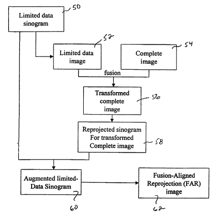
FIG. 4 is a flowchart showing the process steps of the present invention.
FIG. 5 is a schematic example of a patient CT scan;
FIG. 6 is a limited schematic view of FIG. 6, showing the limited scan portion
in the
center of the object, and the remaining nonscanned portion in phantom;
FIG. 7 demonstrates how the limited image of FIG. 6 is aligned with the full
image of
FIG. 5 through the process of fusion;
FIG. 7A show the actual alignment or "fusion" of the images from FIG. 5 and 6;
FIG. 8 is a schematic view of a fusion aligned reprojection image;
FIG. 9 is a schematic view of a full image corresponding to that in FIG. 6;
FIG. 10 is a reconstructed image of FIGS. 2 and 3 fused and aligned in
accordance
with the method of the present invention.
Detailed Description
5
CA 02440244 2003-09-05
WO 02/073519 PCT/US02/07346
A preferred method in accordance with the present invention is shown in the
flowchart
of FIG. 4. A limited data sinogram 50 representing the treatment area is
obtained from a
patient. In one preferred embodiment of the present invention, the limited
data sinogram 50
is prepared near the time that the patient is receiving his or her radiation
treatment. However,
the limited data sinogram 50 may be obtained at any time.
The limited data sinogram 50 is reconstructed to a limited data image 52, as
seen in
the example of FIG. 3, and represented schematically in FIG. 6 as limited
object 156. FIG. 3
contains a significant amount of artifacts such as the white irregular border
53, and some
distortion of image values. By way of example, the treatment area targeted in
FIG. 3 is a
prostate gland. The method can be applied to images of any part of the body,
or be used in
veterinary or radiological applications.
A complete image 54 of the same patient and same treatment area is seen in
FIG. 2,
and represented schematically in FIG. 5 as object 154. Typically, this
complete image 54 will
have been made prior to obtaining the limited data image 52 for the purpose of
treatment
planning. Even if limited image 52 were taken only minutes after the complete
data image
S4, there are almost always inherent differences between the location of
certain organs or
tissue due to patient motion or other bodily functions. If enough time has
elapsed between
images, weight loss or growth of certain tissue can occur.
It is noted that complete image 54 or limited image 52 need not be from CT
scans, and
that this technique can be generally applied to matching images from different
projection
imaging modalities such as magnetic resonance imaging, positron emission
tomography, and
single photon emission tomography. Thus, there may be misalignment or
disagreement
between the two images because of differing methods of data collection.
The two images shown in FIGS. 2 and 3 and represented schematically by objects
154
and 156, in FIGS. 5 and 6 have differences between them. In the actual image
example of
6
CA 02440244 2003-09-05
WO 02/073519 PCT/US02/07346
FIGS. 2 and 3, intestinal gas is shown in FIG. 3, thereby displacing the
treatment target. In
the schematic example, object 154 is composed of diagonals 158a and 160a and
an inclusion
161a, within a frame 162a. Limited object 156 shows only corresponding
diagonals 160b and
158b, and part of the inclusion designated as 161b. Thus, there is a change
between diagonal
158a and 158b and only partial data for inclusion 161b.
Referring to FIG. 7, "fusion" or image registration techniques are used to
align limited
data image 52 with complete image 54. In the schematic example, limited object
156 is fused
with complete object 154 so that statistically, there is optimal registration
between the objects
154 and 156. FIG. 7 shows how the orientation of object 154 is aligned to
closely match that
~ of object 156. FIG. 7A shows diagonal 160c as the perfect registration
between diagonals
160a and 160b. There is less than perfect registration between diagonals 158a
and 158b.
Both lines are superimposed only by way of example to show that fusion is not
perfect as
evidenced by the double edge 163.
Image registration or fusion may be achieved by several techniques. One such
technique is known as mutual information (MI), for which a well-known
algorithm has been
developed. One such example of this algorithm being used to register mufti-
modal images is
described in the following publication, incorporated herein by reference:
Frederik Maes,
Andre Collignon, Dirk Vendermeulen, Guy Marchal, and Paul Suetens,
Multimodality Image
Regist~~ation by Maximization of Mutual Informatiov~, Vol. 16, No. 2, IEEE
Transactions on
Medical Imaging, 187 (April 1997).
Extracted Feature Fusion (EFF) is another registration technique providing
numerous
advantages over prior art techniques. EFF is a voxel-based image registration
method,
wherein only extracted features of images are registered or fused. For
example, a patient's
bone structure usually stays the same even when a patient loses a substantial
amount of
weight. Therefore, the bones can in effect be extracted from each image
subject to alignment,
7
CA 02440244 2003-09-05
WO 02/073519 PCT/US02/07346
and then registered using statistical methods. In the simple example of FIG.
5, diagonal 160a
and frame 162 may represent bone or tissue that remains relatively unchanged
over time.
Therefore, only these relatively static features might be selected for fusion,
while other
features that are more dynamic, perhaps diagonals 158a,b and inclusion 161a,b,
need not be
included in the registration calculations.
The benefits of registering only an extracted portion of an image are reduced
calculation times, improved accuracy, and more clearly defined goals for
alignment in cases
where the patient has significantly changed in shape. The benefits arise from
the registration
of fewer data points, which in this case are voxels. The total processing time
is generally
proportional to the number of points selected, so reducing that number from
the size of the
entire three-dimensional image set to a subset of points meeting certain
criteria (e.g. voxels
that represent bone or do not represent air) will typically reduce calculation
times. This
reduction of voxels can provide more accurate results than other methods of
reducing the
n~unber of voxels for MI techniques, such as regular down-sampling.
, Other image registration techniques include manual fusion, alignment using
geometric
features (e.g. surfaces), gradient methods, and voxel-similarity techniques.
Referring back to FIG. 4, the aligned or transformed complete image 56 is
reprojected
as a sinogram 58. The data for sinogram 58 is once again in a matrix wherein
each row
represents an angle, and each column represents distance. The data matrix of
the reprojected
sinogram is compared to the data matrix for limited data sinogram 50 to
determine what data
is missing from the limited sinogram. This is now possible because the
complete sinogram is
in alignment with the limited sinogram.
The approximation of the missing sinogram data from the reprojected, fusion
aligned
version of image 154 is added to the limited sinogram 50 to create an
augmented limited data
sinogram, or augmented sinogram 60. The augmented sinogram 60 is reconstructed
to a
8
CA 02440244 2003-09-05
WO 02/073519 PCT/US02/07346
fusion aligned reprojection image (FAR image) 62 that is an approximation of
what the
complete image would have looked like at the time the limited data image was
obtained. The
FAR image 62 is represented schematically in FIG. 8. Frame 162 is the same as
in FIG. 5;
and~diagonals 158c, 160c and inclusion 161c are now complete. This can
compared to the
object 168 in FIG. 9, which represents the image that would have been taken at
the time of
treatment if it were possible to obtain a complete image. The fact that the
outer regions 170
of diagonal 158d are not the same as diagonal 158c is not critical to the
invention. FIG. 10
represents a reconstructed image obtained by combining FIGS. 2 and 3 in
accordance with the
method of the present invention. It can be seen that slight artifacts such as
the faint ring 180
can result. However, such artifacts are insignificant because they do not
impair the
conspicuity of the important structures in the field of view, nor do they
noticeably detriment
dose calculations or other processes that utilize these images.
The reconstructed image obtained from method of the present invention can then
be
used for patient setup (positioning the patient prior to delivery), dose
registration (changing
delivery patterns to compensate for patient position or tumor shape changes),
delivery
verification (using a signal measured at an exit detector to compute energy
fluence directed
toward a patient), deformable patient registration and deformable dose
registration (using
anatomical, biomechanical and region of interest data to map changes in the
patient's
anatomy between each fraction, a reconstructed dose is mapped to a reference
image to obtain
a cumulative dose).
It will be understood to those of ordinary skill in the art that other methods
of
comparing images may be used including, for example, those which would
recognize changes
beyond rigid body translation or rotation.
Although the invention has been herein shown and described in what is
perceived to
be the most practical and preferred embodiments, it is to be understood that
the invention is
9
CA 02440244 2003-09-05
WO 02/073519 PCT/US02/07346
not intended to be limited to the specific embodiments set forth above. It is
recognized that
modifications may be made by one of skill in the aut of the invention without
departing from
the spirit or intent of the invention and therefore, the invention is to be
taken as including all
reasonable equivalents to the subject matter of the appended claims.