Note: Descriptions are shown in the official language in which they were submitted.
CA 02440309 2003-09-09
WO 02/080783 PCT/US01/11224
VESSEL SEALER AND DIVIDER
BACKGROUND
The present disclosure relates to an electrosurgical instrument and
method for performing endoscopic surgical procedures and more particularly,
the
present disclosure relates to an open or endoscopic bipolar electrosurgical
forceps and method for sealing and/or cutting tissue.
Technical Field
A hemostat or forceps is a simple plier-like tool which uses
mechanical action between its jaws to constrict vessels and is commonly used
in
open surgical procedures to grasp, dissect and/or clamp tissue.
Electrosurgical
forceps utilize both mechanical clamping action and electrical energy to
effect
hemostasis by heating the tissue and blood vessels to coagulate, cauterize
and/or
seal tissue.
Over the last several decades, more and more surgeons are
complimenting traditional open methods of gaining access to vital organs and
body cavities with endoscopes and endoscopic instruments which access organs
through small puncture-like incisions. Endoscopic instruments are inserted
into
the patient through a cannula, or port, that has been made with a trocar.
Typical
sizes for cannulas range from three millimeters to twelve millimeters. Smaller
cannulas are usually preferred, which, as can be appreciated, ultimately
presents
1
CA 02440309 2003-09-09
WO 02/080783 PCT/US01/11224
a design challenge to instrument manufacturers who must find ways to make
surgical instruments that fit through the cannulas.
Certain endoscopic surgical procedures require cutting blood
vessels or vascular tissue. However, due to space limitations surgeons can
have
difficulty suturing vessels or performing other traditional methods of
controlling
bleeding, e.g., clamping and/or tying-off transected blood vessels. Blood
vessels,
in the range below two millimeters in diameter, can often be closed using
standard electrosurgical techniques. However, if a larger vessel is severed,
it
may be necessary for the surgeon to convert the endoscopic procedure into an
open-surgical procedure and thereby abandon the benefits of laparoscopy.
Several journal articles have disclosed methods for sealing small
blood vessels using electrosurgery. An article entitled Studies on Coagulation
and
the Development of an Automatic Computerized Bipolar Coagulator, J.
Neurosurg., Volume 75, July 1991, describes a bipolar coagulator which is used
to seal small blood vessels. The article states that it is not possible to
safely
coagulate arteries with a diameter larger than 2 to 2.5 mm. A second article
is
entitled Automatically Controlled Bipolar Electrocoagulation - "COA-COMP",
Neurosurg. Rev. (1984), pp.187-190, describes a method for terminating
electrosurgical power to the vessel so that charring of the vessel walls can
be
avoided.
As mentioned above, by utilizing an electrosurgical forceps, a
surgeon can either cauterize, coagulate/desiccate and/or simply reduce or slow
bleeding, by controlling the intensity, frequency and duration of the
electrosurgical
energy applied through the jaw members to the tissue. The electrode of each
2
CA 02440309 2003-09-09
WO 02/080783 PCT/US01/11224
jaw member is charged to a different electric potential such that when the jaw
members grasp tissue, electrical energy can be selectively transferred through
the
tissue.
In order to effect a proper seal with larger vessels, two predominant
mechanical parameters must be accurately controlled - the pressure applied to
the vessel and the gap distance between the electrodes - both of which are
affected by the thickness of the sealed vessel. More particularly, accurate
application of pressure is important to oppose the walls of the vessel; to
reduce
the tissue impedance to a low enough value that allows enough electrosurgical
energy through the tissue; to overcome the forces of expansion during tissue
heating; and to contribute to the end tissue thickness which is an indication
of a
good seal. It has been determined that a typical fused vessel wall is optimum
between 0.001 and 0.005 inches. Below this range, the seal may shred or tear
and above this range the lumens may not be properly or effectively sealed.
With respect to smaller vessel, the pressure applied to the tissue
tends to become less relevant whereas the gap distance between the
electrically
conductive surfaces becomes more significant for effective sealing. In other
words, the chances of the two electrically conductive surfaces touching during
activation increases as the vessels become smaller.
Electrosurgical methods may be able to seal larger vessels using an
appropriate electrosurgical power curve, coupled with an instrument capable of
applying a large closure force to the vessel walls. It is thought that the
process of
coagulating small vessels is fundamentally different than electrosurgical
vessel
sealing. For the purposes herein, "coagulation" is defined as a process of
3
CA 02440309 2003-09-09
WO 02/080783 PCT/US01/11224
desiccating tissue wherein the tissue cells are ruptured and dried. Vessel
sealing
is defined as the process of liquefying the collagen in the tissue so that it
reforms
into a fused mass. Thus, coagulation of small vessels is sufficient to
permanently
close them. Larger vessels need to be sealed to assure permanent closure.
U.S. Patent No. 2,176,479 to Willis, U.S. Patent Nos. 4,005,714 and
4,031,898 to Hiltebrandt, U.S. Patent Nos. 5,827,274, 5,290,287 and 5,312,433
to
Boebel et al., U.S. Patent Nos. 4,370,980, 4,552,143, 5,026,370 and 5,116,332
to Lottick, U.S. Patent No. 5,443,463 to Stern et al., U.S. Patent No.
5,484,436
to Eggers et al. and U.S. Patent No. 5,951,549 to Richardson et al., all
relate to
electrosurgical instruments for coagulating, cutting and/or sealing vessels or
tissue. However, some of these designs may not provide uniformly reproducible
pressure to the blood vessel and may result in an ineffective or non-uniform
seal.
Many of these instruments include blade members or shearing
members which simply cut tissue in a mechanical and/or electromechanical
manner and are relatively ineffective for vessel sealing purposes. Other
instruments rely on clamping pressure alone to procure proper sealing
thickness
and are not designed to take into account gap tolerances and/or parallelism
and
flatness requirements which are parameters which, if properly controlled, can
assure a consistent and effective tissue seal. For example, it is known that
it is
difficult to adequately control thickness of the resulting sealed tissue by
controlling
clamping pressure alone for either of two reasons: 1) if too much force is
applied,
there is a possibility that the two poles will touch and energy will not be
transferred through the tissue resulting in an ineffective seal; or 2) if too
low a
force is applied the tissue may pre-maturely move prior to activation and
sealing
and/or a thicker, less reliable seal may be created.
4
CA 02440309 2003-09-09
WO 02/080783 PCT/US01/11224
As mentioned above, in order to properly and effectively seal larger
vessels, a greater closure force between opposing jaw members is required. It
is
known that a large closure force between the jaws typically requires a large
moment about the pivot for each jaw. This presents a challenge because the jaw
members are typically affixed with pins which are positioned to have a small
moment arms with respect to the pivot of each jaw member. A large force,
coupled with a small moment arm, is undesirable because the large forces may
shear the pins. As a result, designers must compensate for these large closure
forces by either designing instruments with metal pins and/or by designing
instruments which at least partially offload these closure forces to reduce
the
chances of mechanical failure. As can be appreciated, if metal pivot pins are
employed, the metal pins must be insulated to avoid the pin acting as an
alternate
current path between the jaw members which may prove detrimental to effective
sealing.
Increasing the closure forces between electrodes may have other
undesirable effects, e.g., it may cause the opposing electrodes to come into
close
contact with one another which may result in a short circuit and a small
closure
force may cause pre-mature movement of the issue during compression and prior
to activation.
Typically and particularly with respect to endoscopic electrosurgical
procedures, once a vessel is sealed, the surgeon has to remove the sealing
instrument from the operative site, substitute a new instrument through the
CA 02440309 2003-09-09
WO 02/080783 PCT/US01/11224
cannula and accurately sever the vessel along the newly formed tissue seal. As
can be appreciated, this additional step may be both time consuming
(particularly
when sealing a significant number of vessels) and may contribute to imprecise
separation of the tissue along the sealing line due to the misalignment or
misplacement of the severing instrument along the center of the tissue sealing
line.
Several attempts have been made to design an instrument which
incorporates a knife or blade member which effectively severs the tissue after
forming a tissue seal. For example, U.S. Patent No. 5,674,220 to Fox et al.
discloses a transparent vessel sealing instrument which includes a
longitudinally
reciprocating knife which severs the tissue once sealed. The instrument
includes
a plurality of openings which enable direct visualization of the tissue during
the
sealing and severing process. This direct visualization allows a user to
visually
and manually regulate the closure force and gap distance between jaw members
to reduce and/or limit certain undesirable visual effects known to occur when
sealing vessels, thermal spread, charring, etc. As can be appreciated, the
overall success of creating an effective tissue seal with this instrument is
greatly
reliant upon the user's expertise, vision, dexterity, and experience in
judging the
appropriate closure force, gap distance and length of reciprocation of the
knife to
uniformly, consistently and effectively seal the vessel and separate the
tissue at
the seal along an ideal cutting plane.
6
CA 02440309 2003-09-09
WO 02/080783 PCT/US01/11224
U.S. Patent No. 5,702,390 to Austin et al. discloses a vessel sealing
instrument which includes a triangularly-shaped electrode which is rotatable
from
a first position to seal tissue to a second position to cut tissue. Again, the
user
must rely on direct visualization and expertise to control the various effects
of
sealing and cutting tissue.
Thus, a need exists to develop an electrosurgical instrument which
effectively and consistently seals and separates vascular tissue and solves
many
of the aforementioned problems known in the art.
SUMMARY
The present disclosure relates to a bipolar electrosurgical forceps for
clamping, sealing and dividing tissue. More particularly, the present
disclosure
relates to a bipolar electrosurgical forceps which effects consistency in the
overall
clamping pressure exerted on tissue between opposing jaw members, regulates
the gap distances between opposing jaws members, reduces the chances of
short circuiting the opposing jaw members during activation, includes non-
conductive stop members which assist in manipulating, gripping and holding the
tissue prior to and during activation and division of the tissue, and provides
a
uniquely-designed electrical cable path through the body of the instrument and
to
the opposing jaw members to reduce the chances of activation irregularities
during the manipulation, sealing and dividing of tissue.
7
CA 02440309 2003-09-09
WO 02/080783 PCT/US01/11224
The presently disclosed electrosurgical instrument includes a
housing having a shaft attached thereto which connects a pair of first and
second
jaw members in an opposing manner relative to one another. The jaw members
are movable relative to one another from a first open position wherein the jaw
members are disposed in spaced relation relative to one another to a second
closed position wherein the jaw members cooperate to grasp tissue
therebetween. The instrument also includes a handle assembly which
cooperates with a drive assembly to impart movement to the first and second
jaw
members from the first and second positions.
The handle assembly includes a four-bar mechanical linkage having
a handle which is movable relative to the housing and which cooperates with a
cam-like piston to effect movement of the drive assembly. Preferably, the
handle
and the cam member cooperate with a spring to create a uniform closure
pressure against tissue grasped between the jaw members.
In one embodiment, the handle is preferably lockable within the
housing to selectively lock the jaw members relative to one another. For
example, the handle may include a flange which is reciprocated into a channel
having predefined internal dimensions disposed within the housing. The flange
is
preferably dimensioned to cooperate with the predefined internal dimensions of
the channel to selectively lock the jaw members relative to one another.
8
CA 02440309 2003-09-09
WO 02/080783 PCT/US01/11224
In one embodiment, the electrosurgical instrument further includes a
knife assembly for separating tissue. Preferably, the knife assembly is
variable
from a locked configuration to an unlocked configuration upon movement of the
four-bar mechanical linkage. For example, the flange of the handle may be
dimensioned to cooperate with the predefined internal dimensions of the
channel
to selectively lock the jaw members relative to one another and unlock the
knife
assembly upon reciprocation of the flange into the channel.
In yet another embodiment, at least one jaw member includes a
longitudinal channel at least partially defined therethrough which permits
reciprocation of the knife assembly along an ideal cutting plane to separate
tissue. Preferably, the knife assembly is independently operable from the
handle
assembly when the jaw members are disposed in the second closed position. In
one embodiment, the knife assembly includes a leading edge which is
substantially blunt.
BRIEF DESCRIPTION OF THE DRAWINGS
Various embodiments of the subject instrument are described herein
with reference to the drawings wherein:
Fig. 1A is a left, perspective view of an endoscopic bipolar forceps
showing a housing, a shaft and an end effector assembly according to the
present
disclosure;
9
CA 02440309 2003-09-09
WO 02/080783 PCT/US01/11224
Fig. 1 B is a left, perspective of an open bipolar forceps according to
the present disclosure;
Fig. 2 is a top view of the forceps of Fig. 1;
Fig. 3 is a right, side view of the forceps of Fig. 1;
Fig. 4 is a right, perspective view of the forceps of Fig. I showing
the rotation of the end effector assembly about a longitudinal axis "A";
Fig. 5 is a front view of the forceps of Fig. 1;
Figs. 6 is an enlarged view of the indicated area of detail of Fig. 5
showing an enhanced view of the end effector assembly detailing a pair of
opposing jaw members;
Fig. 7 is an enlarged, left perspective view of the indicated area of
detail of Fig. 1 showing another enhanced view of the end effector assembly;
Fig. 8 is an enlarged, right side view of the indicated area of detail of
Fig. 3 with a pair of cam slots of the end effector assembly shown in phantom;
Fig. 9 is a slightly-enlarged, cross-section of the forceps of Fig. 3
showing the internal working components of the housing;
Fig. 10 is an enlarged, cross-section of the indicated area of detail
of Fig. 9 showing the initial position of a knife assembly disposed within the
end
effector assembly;
CA 02440309 2003-09-09
WO 02/080783 PCT/US01/11224
Fig. 11 is an enlarged, left perspective view showing the housing
without a cover plate and the internal working components of the forceps
disposed therein;
Fig. 12 is an exploded, perspective view of the end effector
assembly, the knife assembly and the shaft;
Fig. 13. is an exploded, perspective view of the housing and the
internal working components thereof with the attachment of the shaft and end
effector assembly to the housing shown in broken line illustration;
Fig. 14 is greatly-enlarged, top perspective view of the end effector
assembly with parts separated showing a feed path for an electrical cable
through
the top jaw member;
Fig. 15 is a longitudinal, cross-section of the indicated area of detail
of Fig. 9;
Fig. 16 is an enlarged, top perspective view of the end effector
assembly showing the feed path for the electrical cable through the opposing
jaw
members and the proximal attachment of the knife assembly to a longitudinally-
reciprocating knife tube disposed within the shaft;
Fig. 17 is an enlarged, top perspective view of the end effector
assembly showing the feed path for the electrical cable along a longitudinally-
disposed channel defined within the outer periphery of the shaft;
11
CA 02440309 2003-09-09
WO 02/080783 PCT/US01/11224
Fig. 18A is a greatly-enlarged, side perspective view of the housing
without the cover plate showing the feed path for the electrical cable through
a
rotating assembly adjacent to a distal end of the housing;
Fig. 18B is a greatly-enlarged, side perspective view of the housing
without the cover plate showing the feed path for the electrical cable through
a
rotating assembly with the shaft mounted within the housing;
Fig. 19 is a greatly-enlarged, rear view of the rotating assembly
showing an internally-disposed stop member;
Fig. 20 is a perspective view of the forceps of the present disclosure
shown in position to grasp and seal a tubular vessel or bundle through a
cannula;
Fig. 21 is a slightly-enlarged, cross-section of the internal,
cooperative movements of a four-bar handie assembly disposed within the
housing which effects movement of the jaw members relative to one another;
Fig. 22 is a greatly-enlarged, cross-section showing the initial
movement of a flange upon activation of the four-bar handle assembly shown in
phantom illustration;
Fig. 23 is a greatly-enlarged, side view showing the resulting
compression movement of a coil spring in reaction to the movement of the four-
bar handle assembly;
Fig. 24 is a greatly-enlarged, side view showing the proximal
movement of a cam-like drive pin of the end effector assembly as a result of
the
12
CA 02440309 2003-09-09
WO 02/080783 PCT/US01/11224
proximal compression of the coil spring of Fig. 23 which, in turn, moves the
opposing jaw members into a closed configuration;
Fig. 25 is a greatly-enlarged, cross-section showing the knife
assembly poised for activation within a cannula;
Fig. 26 is a top perspective view showing the opposing jaw
members in closed configuration with a tubular vessel compressed therebetween;
Fig. 27 is an enlarged perspective view of a sealed site of a tubular
vessel showing a preferred cutting line "B-B" for dividing the tubular vessel
after
sealing;
Fig. 28 is a longitudinal cross-section of the sealed site taken along
line 28-28 of Fig. 27;
Fig. 29 is a side view of the housing without a cover plate showing
the longitudinal reciprocation of the knife tube upon activation of a trigger
assembly;
Fig. 30 is a greatly-enlarged, cross-section of the distal end of the
instrument showing longitudinal reciprocation of the knife assembly upon
activation of the trigger assembly;
Fig. 31 is a longitudinal cross-section of the tubular vessel after
reciprocation of the knife assembly through the sealing site along preferred
cutting line "B-B" of Fig. 28; and
13
CA 02440309 2003-09-09
WO 02/080783 PCT/US01/11224
Fig. 32 is a greatly-enlarged, side view showing movement of the
flange upon re-initiation of the handle assembly along a predefined exit path
which, in turn, opens the opposing jaw members and releases the tubular
vessel.
DETAILED DESCRIPTION
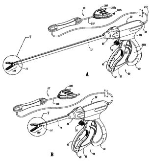
Referring now to Figs. 1-6, one embodiment of a bipolar forceps 10
is shown for use with various surgical procedures and generally includes a
housing 20, a handle assembly 30, a rotating assembly 80, a trigger assembly
70
and an end effector assembly 100 which mutually cooperate to grasp, seal and
divide tubular vessels and vascular tissue 420 (Fig. 20). Although the
majority of
the figure drawings depict a bipolar forceps 10 for use in connection with
endoscopic surgical procedures, an open forceps 10' is also contemplated for
use
in connection with traditional open surgical procedures and is shown by way of
example in Fig. 1 A. For the purposes herein, the endoscopic version is
discussed
in detail, however, it is contemplated that open forceps 10' also includes the
same
or similar operating components and features as described below.
More particularly, forceps 10 includes a shaft 12 which has a distal
end 14 dimensioned to mechanically engage the end effector assembly 100 and a
proximal end 16 which mechanically engages the housing 20. Preferably, shaft
12 is bifurcated at the distal end 14 thereof to form ends 14a and 14b which
are
dimensioned to receive the end effector assembly 100 as best seen in Figs. 7
and
12. The proximal end 16 of shaft 12 includes notches 17a (See Figs. 23 and 29)
14
CA 02440309 2003-09-09
WO 02/080783 PCT/US01/11224
and 17b (See Figs. 11, 12 and 13) which are dimensioned to mechanically
engage corresponding detents 83a (Fig. 18A) and 83b (Fig. 13 shown in
phantom) of rotating assembly 80 as described in more detail below. In the
drawings and in the descriptions which follow, the term "proximal", as is
traditional, will refer to the end of the forceps 10 which is closer to the
user, while
the term "distal" will refer to the end which is further from the user.
As best seen in Fig. 1A, forceps 10 also includes an electrical
interface or plug 300 which connects the forceps 10 to a source of
electrosurgical
energy, e.g., a generator (not shown). Plug 300 includes a pair of prong
members 302a and 302b which are dimensioned to mechanically and electrically
connect the forceps 10 to the source of electrosurgical energy. An electrical
cable 310 extends from the plug 300 to a sleeve 99 which securely connects the
cable 310 to the forceps 10. As best seen in Figs. 9, 11 and 18A, cable 310 is
internally divided into cable lead 310a and 310b which each transmit
electrosurgical energy through their respective feed paths through the forceps
10
to the end effector assembly 100 as explained in more detail below.
Handle assembly 30 includes a fixed handle 50 and a movable
handle 40. Fixed handle 50 is integrally associated with housing 20 and handle
40 is movable relative to fixed handle 50 as explained in more detail below
with
respect to the operation of the forceps 10. Rotating assembly 80 is preferably
attached to a distal end 303 (Fig. 18A) of housing 20 and is rotatable
approximately 180 degrees in either direction about a longitudinal axis "A".
As best seen in Figs. 2 and 13, housing 20 is formed from two (2)
housing halves 20a and 20b which eacli include a plurality of interfaces 307a,
307b and 307c (Fig. 13) which are dimensioned to mechanically align and engage
CA 02440309 2003-09-09
WO 02/080783 PCT/US01/11224
one another to form housing 20 and enclose the internal working components of
forceps 10. As can be appreciated, fixed handle 50 which, as mentioned above
is integrally associated with housing 20, takes shape upon the assembly of the
housing halves 20a and 20b.
It is envisioned that a plurality of additional interfaces (not shown)
may disposed at various points around the periphery of housing halves 20a and
20b for ultrasonic welding purposes, e.g., energy direction/deflection points.
It is
also contemplated that housing halves 20a and 20b (as well as the other
components described below) may be assembled together in any fashion known
in the art. For example, alignment pins, snap-like interfaces, tongue and
groove
interfaces, locking tabs, adhesive ports, etc. may all be utilized either
alone or in
combination for assembly purposes.
Likewise, rotating assembly 80 includes two halves 80a and 80b
which, when assembled, enclose and engage the proximal end 16 of shaft 12 to
permit selective rotation of the end effector assembly 100 as needed. Half 80a
includes a pair of detents 89a (Fig. 13) which are dimensioned to engage a
pair
of corresponding sockets 89b (shown in phantom in Fig. 13) disposed within
half
80b. Movable handle 40 and trigger assembly 70 are preferably of unitary
construction and are operatively connected to the housing 20 and the fixed
handle 50 during the assembly process.
As mentioned above, end effector assembly 100 is attached to the
distal end 14 of shaft 12 and includes a pair of opposing jaw members 110 and
120. Movable handle 40 of handle assembly 30 is ultimately connected to a
drive
rod 32 which, together, mechanically cooperate to impart movement of the jaw
members 110 and 120 from an open position wherein the jaw members 110 and
120 are disposed in spaced relation relative to one another, to a clamping or
16
CA 02440309 2003-09-09
WO 02/080783 PCT/US01/11224
closed position wherein the jaw members 110 and 120 cooperate to grasp tissue
420 (Fig. 20) therebetween. This is explained in more detail below with
respect
to Figs. 9 -11 and 20-29.
It is envisioned that the forceps 10 may be designed such that it is
fully or partially disposable depending upon a particular purpose or to
achieve a
particular result. For example, end effector assembly 100 may be selectively
and
releasably engageable with the distal end 14 of the shaft 12 and/or the
proximal
end 16 of shaft 12 may be selectively and releasably engageable with the
housing
20 and the handle assembly 30. In either of these two instances, the forceps
10
would be considered "partially disposable" or "reposable", i.e., a new or
different
end effector assembly 100 (or end effector assembly 100 and shaft 12)
selectively replaces the old end effector assembly 100 as needed .
Turning now to the more detailed features of the present disclosure
as described with respect to Figs 1A - 13, movable handle 40 includes an
aperture 42 defined therethrough which enables a user to grasp and move the
handle 40 relative to the fixed handle 50. Handle 40 also includes an
ergonomically-enhanced gripping element 45 disposed along the inner peripheral
edge of aperture 42 which is designed to facilitate gripping of the movable
handle
40 during activation. It is envisioned that gripping element 45 may include
one or
more protuberances, scallops and/or ribs 43a, 43b and 43c, respectively, to
facilitate gripping of handle 40. As best seen in Fig. 11, movable handle 40
is
selectively moveable about a pivot 69 from a first position relative to fixed
handle
17
CA 02440309 2003-09-09
WO 02/080783 PCT/US01/11224
50 to a second position in closer proximity to the fixed handle 50 which, as
explained below, imparts movement of the jaw members 110 and 120 relative to
one another.
As shown best in Fig. 11, housing 20 encloses a drive assembly 21
which cooperates with the movable handle 40 to impart movement of the jaw
members 110 and 120 from an open position wherein the jaw members 110 and
120 are disposed in spaced relation relative to one another, to a clamping or
closed position wherein the jaw members 110 and 120 cooperate to grasp tissue
therebetween. The handle assembly 30 can generally be characterized as a
four-bar mechanical linkage composed of the following elements: movable
handle 40, a link 65, a cam-like link 36 and a base link embodied by fixed
handle
50 and a pair of pivot points 37 and 67b. Movement of the handle 40 activates
the four-bar linkage which, in turn, actuates the drive assembly 21 for
imparting
movement of the opposing jaw members 110 and 120 relative to one another to
grasp tissue therebetween. It is envisioned that employing a four-bar
mechanical
linkage will enable the user to gain a significant mechanical advantage when
compressing the jaw members 110 and 120 against the tissue 420 as explained
in further detail below with respect the operating parameters of the drive
assembly 21. Although shown as a four-bar mechanical linkage, the present
disclosure contemplates other linkages to effect relative motion of the jaw
members 110 and 120 as is known in the art.
18
CA 02440309 2003-09-09
WO 02/080783 PCT/US01/11224
Preferably, fixed handle 50 includes an channel 54 defined therein
which is dimensioned to receive a flange 92 which extends proximally from
movable handle 40. Preferably, flange 92 includes a fixed end 90 which is
affixed
to movable handle 40 and a t-shaped free end 93 which is dimensioned for
facile
reception within channel 54 of handle 50. It is envisioned that flange 92 may
be
dimensioned to allow a user to selectively, progressively and/or incrementally
move jaw members 110 and 120 relative to one another from the open to closed
positions. For example, it is also contemplated that flange 92 may include a
ratchet-like interface which lockingly engages the movable handle 40 and,
therefore, jaw members 110 and 120 at selective, incremental positions
relative to
one another depending upon a particular purpose. Other mechanisms may also
be employed to control and/or limit the movement of handle 40 relative to
handle
50 (and jaw members 110 and 120) such as, e.g., hydraulic, semi-hydraulic,
linear actuator(s), gas-assisted mechanisms and/or gearing systems.
As best illustrated in Fig. 11, housing halves 20a and 20b of housing
20, when assembled, form an internal cavity 52 which predefines the channel 54
within fixed handle 50 such that an entrance pathway 53 and an exit pathway 58
are formed for reciprocation of the t-shaped flange end 93 therein. Once
assembled, two generally triangular-shaped members 57a and 57b are positioned
in close abutment relative to one another to define a rail or track 59
therebetween.
During movement of the flange 92 along the entrance and exit pathways 53 and
58, respectively, the t-shaped end 93 rides along track 59 between the two
triangular members 57a and 57b according to the particular dimensions of the
19
CA 02440309 2003-09-09
WO 02/080783 PCT/US01/11224
triangularly-shaped members 57a and 57b, which, as can be appreciated,
predetermines part of the overall pivoting motion of handle 40 relative to
fixed
handle 50.
Once actuated, handle 40 moves in a generally arcuate fashion
towards fixed handle 50 about pivot 69 which causes link 65 to rotate
proximally
about pivots 67a and 67b which, in turn, cause cam-like link 36 to rotate
about
pivots 37 and 69 in a generally proximal direction. Movement of the cam-like
link
36 imparts movement to the drive assembly 21 as explained in more detail
below.
Moreover, proximal rotation of the link 65 about pivots 67a and 67b also
causes a
distal end 63 of link 65 to release, i.e., "unlock", the trigger assembly 70
for
selective actuation. This feature is explained in detail with reference to
Figs. 21-
29 and the operation of the knife assembly 200.
Turning now to Fig. 12 which shows an the exploded view of the
shaft 12 and end effector assembly 100. As mentioned above, shaft 12 includes
distal and proximal ends 14 and 16, respectively. The distal end 14 is
bifurcated
and includes ends 14a and 14b which, together, define a cavity 18 for
receiving
the end effector assembly 100. The proximal end 16 includes a pair of notches
17a (Fig. 29) and 17b (Fig. 11) which are dimensioned to engage corresponding
detents 83a and 83b (Fig. 13) of the rotating assembly 80. As can be
appreciated, actuation of the rotation assembly 80 rotates the shaft 12 which,
in
turn, rotates the end effector assembly 100 to manipulate and grasp tissue
420.
CA 02440309 2003-09-09
WO 02/080783 PCT/US01/11224
Shaft 12 also includes a pair of longitudinally-oriented channels 19a
(Fig. 15) and 19b (Fig. 12) which are each dimensioned to carry an
electrosurgical
cable lead 310a and 310b, respectively, therein for ultimate connection to
each
jaw member 120 and 110, respectively, as explained in more detail with
reference
to Figs. 14-17 below. Shaft 12 also includes a pair of longitudinally oriented
slots
197a and 197b disposed on ends 14a and 14b, respectively. Slots 197a and
197b are preferable dimensioned to allow longitudinal reciprocation of a cam
pin
170 therein which, as explained below with reference to Figs. 23 and 24,
causes
movement of the opposing jaw member 110 and 120 from the open to closed
positions.
Shaft 12 also includes a pair of sockets 169a and 169b disposed at
distal ends 14a and 14b which are dimensioned to receive a corresponding pivot
pin 160. As explained below, pivot pin 160 secures jaws 110 and 120 to the
shaft
12 between bifurcated distal ends 14a and 14b and mounts the jaw members 110
and 120 such that longitudinal reciprocation of the cam pin 170 rotates jaw
members 110 and 120 about pivot pin 160 from the open to closed positions.
Shaft 12 is preferably dimensioned to slidingly receive a knife tube
34 therein which engages the knife assembly 200 such that longitudinal
movement of the knife tube 34 actuates the knife assembly 200 to divide tissue
420 as explained below with respect to Figs. 29-31. Knife tube 34 includes a
rim
35 located at a proximal end thereof and a pair of opposing notches 230a and
230b (Figs. 25 and 30) located at a distal end 229 thereof. As best shown in
Fig.
21
CA 02440309 2003-09-09
WO 02/080783 PCT/US01/11224
13, rim 35 is dimensioned to engage a corresponding sleeve 78 disposed at a
distal end of the trigger assembly 70 such that distal movement of the sleeve
78
translates the knife tube 34 which, in turn, actuates the knife assembly 200.
A
seal 193 may be mounted atop the knife tube 34 and positioned between the
knife tube 34 and the shaft 12. It is envisioned that the seal 193 may be
dimensioned to facilitate reciprocation of the knife tube 34 within the shaft
12
and/or to protect the other, more sensitive, internal operating components of
the
forceps from undesirable fluid inundation during surgery. Seal 193 may also be
employed to control/regulate pneumo-peritoneal pressure leakage through
forceps 10 during surgery. Seal 193 preferably includes a pair of opposing
bushings 195a and 195b which assure consistent and accurate reciprocation of
the knife tube 34 within shaft 12 (See Fig 15).
Notches 230a and 230b are preferably dimensioned to engage a
corresponding key-like interface 211 of the knife assembly 200 which includes
a
pair of opposing detents 212a and 212b and a pair of opposing steps 214a and
214b. As best illustrated in Figs. 25 and 30, each detent and step
arrangement,
e.g., 212a and 214a, respectively, securely engages a corresponding notch,
e.g.,
230a, such that the distal end of the step 214a abuts the distal end 229 of
the
knife tube 34. It is envisioned that engaging the knife tube 34 to the knife
assembly 200 in this manner will assure consistent and accurate distal
translation
of the knife tube 34 through the tissue 420.
22
CA 02440309 2003-09-09
WO 02/080783 PCT/US01/11224
As can be appreciated from the present disclosure, the knife tube 34
and knife assembly 200 are preferably assembled to operate independently from
the operation of the drive assembly 21. However and as described in more
detail
below, knife assembly 200 is dependent on the drive assembly 21 for activation
purposes, i.e., the activation/movement of the drive assembly 21 (via handle
assembly 30 and the internal working components thereof) "unlocks" the knife
assembly 200 for selective, separation of the tissue. For the purposes herein,
the
drive assembly 21 consists of both the drive rod 32 and the compression
mechanism 24 which includes a number of cooperative elements which are
described below with reference to Fig. 13. It is envisioned that arranging the
drive assembly 21 in this fashion will enable facile, selective engagement of
the
drive rod 32 within the compression mechanism 24 for assembly purposes.
Although the drawings depict a disposable version of the presently
disclosed forceps 10, it is contemplated that the housing 20 may include a
release
mechanism (not shown) which enables selectively replacement of the drive rod
32
for disposal purposes. In this fashion, the forceps will be considered
"partially
disposable" or "reposable", i.e., the shaft 12, end effector assembly 100 and
knife
assembly 200 are disposable and/or replaceable whereas the housing 20 and
handle assembly 30 are re-usable.
As best illustrated in Figs. 16 and 17, drive rod 32 includes a pair of
chamfered or beveled edges 31a and 31b at a distal end thereof which are
preferably dimensioned to allow facile reciprocation of the drive rod 32
through a
23
CA 02440309 2003-09-09
WO 02/080783 PCT/US01/11224
knife carrier or guide 220 which forms a part of the knife assembly 200. A pin
slot
39 is disposed at the distal tip of the drive rod 32 and is dimensioned to
house the
cam pin 170 such that longitudinal reciprocation of the drive rod 32 within
the
knife tube 34 translates the cam pin 170, which, in turn, rotates the jaw
members
110 and 120 about pivot pin 160. As will be explained in more detail below
with
respect to Figs. 23 and 24, the cam pin 170 rides within slots 172 and 174 of
the
jaw members 110 and 120, respectively, which causes the jaw members 110 and
120 to rotate from the open to closed positions about the tissue 420.
The proximal end of the drive rod 32 includes a tab 33 which is
preferably dimensioned to engage a corresponding compression sleeve 28
disposed within the compression mechanism 24. Proximal movement of the
sleeve 28 (as explained below with respect to Figs. 21-24) reciprocates (i.e.,
pulls) the drive rod 32 which, in turn, pivots the jaw members 110 and 120
from
the open to closed positions. Drive rod 32 also includes a donut-like spacer
or o-
ring 95 which is dimensioned to maintain pneumo-peritoneal pressure during
endoscopic procedures. It is also envisioned that o-ring 95 may also prevent
the
inundation of surgical fluids which may prove detrimental to the internal
operating
components of the forceps 10. 0-ring 95 is made also be made from a material
having a low coefficient of friction to facilitate uniform and accurate
reciprocation
of the drive rod 32 within the knife tube 34.
As mentioned above, the knife assembly 200 is disposed between
opposing jaw members 110 and 120 of the end effector assembly 100.
24
CA 02440309 2003-09-09
WO 02/080783 PCT/US01/11224
Preferably, the knife assembly 200 and the end effector assembly 100 are
independently operable, i.e., the trigger assembly 70 actuates the knife
assembly
200 and the handle assembly 30 actuates the end effector assembly 100. Knife
assembly 200 includes a bifurcated knife bar or rod 210 having two forks 210a
and 210b and a knife carrier or guide 220. Knife forks. 210a and 210b include
the
above-described key-like interfaces 211 (composed of steps 214a, 214b and
detents 212a, 212b, respectively) disposed at the proximal end thereof for
engaging the knife tube 34 (as described above) and a common distal end 206
which carries a blade 205 thereon for severing tissue 420. Preferably, each
fork
210a and 210b includes a taper 213a and 213b, respectively, which converge to
form common distal end 206. It is envisioned that the tapers 213a and 213b
facilitate reciprocation of the knife blade 205 through the end effector
assembly
100 as described in more detail below and as best illustrated in Fig. 30.
Each fork 210a and 210b also includes a tapered shoulder portion
221 a and 221b disposed along the outer periphery thereof which is dimensioned
to engage a corresponding slot 223a and 223b, respectively, disposed in the
knife
carrier or guide 220 (See Fig. 16). It is envisioned that this shoulder
portion 221 a,
221 b and slot 223a, 223b arrangement may be designed to restrict and/or
regulate the overall distal movement of the blade 205 after activation. Each
fork
210a and 210b also includes an arcuately-shaped notch 215a and 215b,
respectively disposed along the inward edge thereof which is dimensioned to
facilitate insertion of a roller or bushing 216 disposed between the jaw
members
110 and 120 during assembly.
CA 02440309 2003-09-09
WO 02/080783 PCT/US01/11224
As mentioned above, knife assembly 200 also includes a knife
carrier or guide 220 which includes opposing spring tabs 222a and 222b at a
proximal end thereof and upper and lower knife guides 224a and 224b,
respectively, at the distal end thereof. The inner facing surface of each
spring
tab, e.g., 222b, is preferably dimensioned to matingly engage a corresponding
chamfered edge, e.g., 31 b of the drive rod 32 (Fig. 16) and the outer facing
surface is preferably dimensioned for friction-fit engagement with the inner
periphery of the shaft 12. As best seen in Fig. 12, knife carrier 220 also
includes
a drive rod channel 225 defined therethrough which is dimensioned to allow
reciprocation of the drive rod 32 during the opening and closing of the jaw
members 110 and 120. Knife guide 220 also includes rests 226a and 226b
which extend laterally therefrom which abut the proximal ends 132, 134 of the
jaw
members 110 and 120 when disposed in the closed position.
Knife guides 224a and 224b preferably include slots 223a and 223b,
respectively, located therein which guide the knife forks 210a and 210b
therealong during activation to provide consistent and accurate reciprocation
of
the knife blade 205 through the tissue 420. It is envisioned that slots 223a
and
223b also restrict undesirable lateral movements of the knife assembly 200
during
activation. Preferably, the knife carrier 220 is positioned at a point
slightly beyond
the shoulder portions 221 a and 221 b when assembled.
26
CA 02440309 2003-09-09
WO 02/080783 PCT/US01/11224
The knife assembly 200 also includes a roller or bushing 216 which
is dimensioned to mate with the inner peripheral edge of each fork 210a and
210b
such that, during activation, the forks 210a and 210b glide over the roller or
bushing 216 to assure facile and accurate reciprocation of the knife assembly
200
through the tissue 420. Bushing 216 is also dimensioned to seat between
opposing jaw members 110 and 120 and is preferably secured therebetween by
pivot pin 160. As mentioned above, the arcuately-shaped notches 215a and
215b facilitate insertion of the bushing 216 during assembly.
The end effector assembly 100 includes opposing jaw members 110
and 120 which are seated within cavity 18 defined between bifurcated ends 14a
and 14b of shaft 12. Jaw members 110 and 120 are generally symmetrical and
include similar component features which cooperate to permit facile rotation
about
pivot pin 160 to effect the sealing and dividing of tissue 420. As a result
and
unless otherwise noted, only jaw member 110 and the operative features
associated therewith are describe in detail herein but as can be appreciated,
many of these features apply to jaw member 120 as well.
More particularly, jaw member 110 includes a pivot flange 166 which
has an arcuately-shaped inner surface 167 which is dimensioned to allow
rotation
of jaw member 110 about bushing 216 and pivot pin 160 upon reciprocation of
drive rod 32 as described above. Pivot flange 166 also includes a cam slot 172
which is dimensioned to engage cam pin 170 such that longitudinal movement of
the drive rod 32 causes the cam pin 170 to ride along cam slot 172. It is
27
CA 02440309 2009-02-12
envisioned that cam slot 172 may be dimensioned to allow different rotational
paths depending upon a particular purpose or to achieve a particular result.
Pivot flange 166 also includes a recess 165 which is preferably
dimensioned to secure one free end of the bushing 216 between jaw members
110 and 120. The inner periphery of recess 165 is preferably dimensioned to
receive pivot pin 160 therethrough to secure the jaw member 110 to the shaft
12. Jaw member 120 includes a similar recess 175 (Fig. 14) which secures the
opposite end of bushing 216 and jaw member 120 to shaft 12.
Jaw member 110 also includes a jaw housing 116, an insulative
substrate or insulator 114 and an electrically conductive surface 112. Jaw
housing 116 includes a groove (not shown - See groove 179 of jaw member
120) defined therein which is dimensioned to engage a ridge-like interface 161
disposed along the outer periphery of insulator 114. Insulator 114 is
preferably
dimensioned to securely engage the electrically conductive sealing surface
112. This may be accomplished by stamping, by overmolding, by overmolding
a stamped electrically conductive sealing plate and/or by overmolding a metal
injection molded seal plate. All of these manufacturing techniques produce an
electrode having an electrically conductive surface 112 which is substantially
surrounded by an insulating substrate 114. The insulator 114, electrically
28
CA 02440309 2009-02-12
conductive sealing surface 112 and the outer, non-conductive jaw housing 116
are preferably dimensioned to limit and/or reduce many of the known
undesirable effects related to tissue sealing, e.g., flashover, thermal spread
and
stray current dissipation.
Preferably, the electrically conductive sealing surface 112 may also
include a pinch trim 119 (Fig. 25) which facilitates secure engagement of the
electrically conductive surface 112 to the insulating substrate 114 and also
simplifies the overall manufacturing process. It is envisioned that the
electrically conductive sealing surface 112 may also include an outer
peripheral
edge which has a radius and the insulator 114 meets the electrically
conductive
sealing surface 112 along an adjoining edge which is generally tangential to
the
radius and/or meets along the radius. Preferably, at the interface, the
electrically conductive surface 112 is raised relative to the insulator 114.
These
and other envisioned embodiments are discussed in International Publication
No. WO 2002/080786 and U.S. Patent No. 7,135,020 to Lawes et al.
29
CA 02440309 2003-09-09
WO 02/080783 PCT/US01/11224
Insulator 114 also includes an inwardly facing finger 162 which
abuts pivot flange 166 and is designed to restrict / reduce proximal tissue
spread
and/or isolate the electrically conductive sealing surface 112 from the
remaining
end effector assembly 100 during activation. Preferably, the electrically
conductive surface 112 and the insulator 114, when assembled, form a
longitudinally-oriented channel 168a, 168b defined therethrough for
reciprocation
of the knife blade 205. More particularly, and as best illustrated in Fig. 14,
insulator 114 includes a first channel 168b which aligns with a second channel
168a on electrically conductive sealing surface 112 to form the complete knife
channel. It is envisioned that the knife channel 168a, 168b facilitates
longitudinal
reciprocation of the knife blade 205 along a preferred cutting plane "B-B" to
effectively and accurately separate the tissue 420 along the formed tissue
seal
425 (See Figs. 27, 28 and 31.
As mentioned above, jaw member 120 include similar elements
which include: a pivot flange 176 which has an arcuately-shaped inner surface
177, a cam slot 174, and a recess 175; a jaw housing 126 which includes a
groove 179 which is dimensioned to engage a ridge-fike interface 171 disposed
along the outer periphery of an insulator 124; the insulator 124 which
includes an
inwardly facing finger 172 which abuts pivot flange 176; and an electrically
conducive sealing surface 122 which is dimensioned to securely engage the
insulator 124. Likewise, the electrically conductive surface 122 and the
insulator
124, when assembled, form a longitudinally-oriented channel 178a, 178b defined
therethrough for reciprocation of the knife blade 205.
CA 02440309 2003-09-09
WO 02/080783 PCT/US01/11224
Preferably, the jaw members 110 and 120 are electrically isolated
from one another such that electrosurgical energy can be effectively
transferred
through the tissue 420 to form seal 425. For example and as best illustrated
in
Figs. 14 and 15, each jaw member, e.g., 110, includes a uniquely-designed
electrosurgical cable path disposed therethrough which transmits
electrosurgical
energy to the electrically conductive sealing surfaces 112, 122. More
particularly,
jaw member 110 includes a cable guide 181 a disposed atop pivot flange 166
which directs cable lead 310a towards an aperture 188 disposed through jaw
housing 116. Aperture 188, in turn, directs cable lead 310a towards
electrically
conductive sealing surface 112 through a window 182 disposed within insulator
114. A second cable guide 181 b secures cable lead 310a along the predefined
cable path through window 182 and directs a terminal end 310a' of the cable
lead
310a into crimp-like electrical connector 183 disposed on an opposite side of
the
electrically conductive sealing surface 112. Preferably, cable lead 310a is
held
loosely but securely along the cable path to permit rotation of the jaw member
110 about pivot 169.
As can be appreciated, this isolates electrically conductive sealing
surface 112 from the remaining operative components of the end effector
assembly 100 and shaft 12. Jaw member 120 includes a similar cable path
disposed therein and therethrough which includes similarly dimensioned cable
guides, apertures and electrical connectors which are not shown in the
accompanying illustrations.
31
CA 02440309 2003-09-09
WO 02/080783 PCT/US01/11224
Figs. 15-17 also show the presently disclosed feed path for both
electrosurgical cable leads 310a and 310b along the outer periphery of the
shaft
12 and through each jaw member 110 and 120. More particularly, Fig. 15 shows
a cross section of the electrosurgical cable leads 310a and 310b disposed
within
channels 19a and 19b, respectively, along shaft 12. Figs. 16 and 17 show the
feed path of the cable leads 310a and 310b from the opposite channels 19a and
19b of the shaft 12 through the pivot flanges 166 and 176 of the jaw members
110 and 120, respectively. It is contemplated that this unique cable feed path
for
cable leads 310a and 310b from the shaft 12 to the jaw members 110 and 120
not only electrically isolates each jaw member 100 and 120 but also allows the
jaw members 110 and 120 to pivot about pivot pin 160 without unduly straining
or
possibly tangling the cable leads 310a and 310b. Moreover, it is envisioned
that
the crimp-like electrical connector 183 (and the corresponding connector in
jaw
member 120) greatly facilitates the manufacturing and assembly process and
assures a consistent and tight electrical connection for the transfer of
energy
through the tissue 420. As best shown in Fig. 17, the outer surface of shaft
12
may be covered by heat shrink tubing 500 or the like which protects the cable
leads 310a and 310b from undue wear and tear and secures cable leads 310a
and 310b within their respective channels 19a and 19b.
Figs. 18A and 18B show the feed path of the cable leads 310a and
310b through the rotating assembly 80 which, again, allows the user added
flexibility during the use of the forceps 10 due to the uniqueness of the feed
path.
32
CA 02440309 2003-09-09
WO 02/080783 PCT/US01/11224
More particularly, Fig. 18A shows the feed path of cable lead 310a through
half
80a of the rotating assembly 80 and Fig. 18B shows the path of cable leads
310a
and 310b as the cable leads 310a and 310b feed through the instrument housing
20a, through half 80a of the rotating assembly 80 and to the channels 19a and
19b of the shaft 12. Fig. 18A only shows the feed path of cable lead 310a
through half 80a of the rotating assembly 80, however, as can be appreciated,
cable lead 310b (shown broken in Fig. 19) is positioned in a similar fashion
within
half 80b of rotating assembly 80.
As best illustrated in Fig. 18A, it is envisioned that cable leads 310a
and 310b are fed through respective halves 80a and 80b of the rotating
assembly
80 in such a manner to allow rotation of the shaft 12 (via rotation of the
rotating
assembly 80) in the clockwise or counter-clockwise direction without unduly
tangling or twisting the cable leads 310a and 310b. More particularly, each
cable
lead, e.g., 310a, is looped through each half 80a of the rotating assembly 80
to
form slack-loops 321 a and 321 b which traverse either side of longitudinal
axis "A".
Slack-loop 321a redirects cable lead 310a across one side of axis "A" and
slack-
loop 321 b returns cable lead 310a across axis "A". It is envisioned that
feeding
the cable leads 310a and 310b through the rotating assembly 80 in this fashion
allows the user to rotate the shaft 12 and the end effector assembly 100
without
unduly straining or tangling the cable leads 310a and 310b which may prove
detrimental to effective sealing. Preferably, this loop-like cable feed path
allows
the user to rotate the end effector assembly 100 about 180 degrees in either
direction without straining the cable leads 310a and 310b. The presently
33
CA 02440309 2003-09-09
WO 02/080783 PCT/US01/11224
disclosed cable lead feed path is envisioned to rotate the cable leads 310a
and
310b approximately 178 degrees in either direction.
Fig. 19 shows an internal view of half 80a of the rotating assembly
80 as viewed along axis "A" to highlight the internal features thereof. More
particularly, at least one stop 88 is preferably positioned within each
rotating half
80a and 80b which operates to control the overall rotational movement of the
rotating assembly 80 to about 180 degree in either direction. The stop member
88 is dimensioned to interface with a corresponding notch 309c disposed along
the periphery of outer flange 309 to prevent unintended over-rotation of the
rotating assembly 80 which may unduly strain one or both of the cable leads
310a and 310b.
Fig. 18B shows the feed path of the electrical cable leads 310a and
310b from the housing 20a, through the rotating assembly 80 and to the shaft
12.
It is envisioned that the cable leads 310a and 310b are directed through each
part
of the forceps 10 via a series of cable guide members 311 a-311 g disposed at
various positions through the housing 20 and rotating assembly 80. As
explained
below, a series of mechanical interfaces, e.g., 309a, 309b (Fig. 13) and 323a,
323b (Fig. 13) may also be dimensioned to contribute in guiding cables 310a
and
310b through the housing 20 and rotating assembly 80.
Turning back to Fig. 13 which shows the exploded view of the
housing 20, rotating assembly 80, trigger assembly 70 and handle assembly 30,
it
34
CA 02440309 2003-09-09
WO 02/080783 PCT/US01/11224
is envisioned that all of these various component parts along with the shaft
12
and the end effector assembly 100 are assembled during the manufacturing
process to form a partially and/or fully disposable forceps 10. For example
and
as mentioned above, the shaft 12 and/or end effector assembly 100 may be
disposable and, therefore, selectively/releasably engagable with the housing
20
and rotating assembly 80 to form a partially disposable forceps 10 and/or the
entire forceps 10 may be disposable after use.
Housing 20 is preferably formed from two housing halves 20a and
20b which engage one another via a series of mechanical interfaces 307a, 307b,
307c and 308a, 308b, 308c respectively, to form an internal cavity 300 for
housing the hereindescribed internal working components of the forceps 10. For
the purposes herein, housing halves 20a and 20 are generally symmetrical and,
unless otherwise noted, a component described with respect to housing half 20a
will have a similar component which forms a part of housing half 20b.
Housing half 20a includes proximal and distal ends 301 a and 303a,
respectively. Proximal end 301a is preferably dimensioned to receive an
electrical sleeve 99 which secures the electrosurgical cable 310 (Fig. 1)
within
the housing 20. As best shown in Figs. 9 and 21, paired cable 310 splits into
two
electrosurgical cable leads 310a and 310b which are subsequently fed through
the housing 20 to ultimately transmit different electrical potentials to the
opposing
jaw members 110 and 120. As mentioned above, various cable guides 311 a-
311 g are positioned throughout the housing 20 and the rotating assembly 80 to
CA 02440309 2003-09-09
WO 02/080783 PCT/US01/11224
direct the cable leads 310a and 310b to the channels 19a and 19b disposed
along the outer periphery of the shaft 12.
The distal end 303a is generally arcuate in shape such that, when
assembled, distal ends 303a and 303b form a collar 303 (Fig. 13) which extends
distally from the housing 20. Each distal end 303a, 303b of the collar 303
includes an outer flange 309a, 309b and a recess 323a, 323b which cooperate to
engage corresponding mechanical shoulders 84a, 84b (Fig. 29) and flanges 87a,
87b, respectively, disposed within the rotating assembly 80. As can be
appreciated, the interlocking engagement of the flanges 309a, 309b with the
shoulders 84a, 84b and the recesses 323a, 323b with the flanges 87a, 87b are
dimensioned to allow free rotation about of the rotating assembly 80 about
collar
303 when assembled. As mentioned above, the stop member(s) 88 and the
notch(es) mechanically cooperate to limit rotational movement of the rotating
assembly 80 to avoid straining cable leads 310a and 310b.
Each distal end 303a, 303b of collar 303 also includes an inner
cavity 317a and 317b (Figs. 9 and 21), respectively, defined therein which is
dimensioned to permit free rotation of the shaft 12, knife tube 34 and cable
leads
310a and 310b housed therein. A plurality of detents 89a located within
rotating
assembly 80 engage a corresponding plurality of sockets 89b (Fig. 13) disposed
within rotating half 80b to poise the rotating assembly 80 in rotational
relationship
atop collar 303.
36
CA 02440309 2003-09-09
WO 02/080783 PCT/US01/11224
Housing half 20a also includes a plurality of hub-like pivot mounts
329a, 331 a and 333a which as explained in more detail below with respect to
the
operation of the instrument, cooperate with opposite hub-like pivot mounts
(shown in phantom in Fig. 13) disposed on housing half 20b to engage the free
ends of pivot pins 37, 67b and 77, respectively, which are associated with the
different operating components described below. Preferably, each of these
mounts 329a, 331a and 333a provide a fixed point of rotation for each pivoting
element, namely, cam link 36, handle link 65 and trigger assembly 70,
respectively.
As best seen in Figs. 11 and 13, fixed handle 50 which takes shape
upon the assembly of housing 20 includes a scallop-like outer surface 51 and
an
internal cavity 52 defined therein. As mentioned above with respect to the
discussion of Fig. 11, these elements and the other internal elements of the
fixed
handle 50 cooperate with movable handle 40 to activates the four-bar
mechanical
linkage which, in turn, actuates the drive assembly 21 for imparting movement
of
the opposing jaw members 110 and 120 relative to one another to grasp tissue
420 therebetween.
The handle assembly 30 which includes the above-mentioned fixed
handle 50 and movable handle 40 also includes the cam link 36 which is
generally triangular in shape. The cam link includes an upper piston 38, a
fixed
pivot 37 and a handle pivot 69. Cam link is assembled within the internal
cavity
300 of housing 20 between housing halves 20a and 20b. More particularly, fixed
37
CA 02440309 2003-09-09
WO 02/080783 PCT/US01/11224
pivot 37 is rotatingly mounted within fixed mounts 329a and 329b between
opposing housing halves 20a and 20b and the handle pivot 69 is rotatingly
mounted within the bifurcated end of handle 40 through apertures 68a and 68b.
Cam piston 38 is poised within a longitudinal channel 25c defined through the
drive assembly 70 (explained in further detail below with respect to the
discussion
of the drive assembly 70) in abutting relationship with a compression tab 25
such
that movement of the handle 40 rotates piston 38 proximally against coil
spring
22. These and the other details relating to the operational features are
discussed
below with reference to Figs. 21-29.
Link 65 is also associated with the handle assembly 30 and forms
an integral part of the four-bar mechanical linkage. Link 65 includes a distal
end
63 and two pivot pins 67a and 67b. Pivot pin 67a engages apertures 68a and
68b disposed within the movable handle 40 and pivot 67b engages fixed mounts
331a and 331b between housing halves 20a and 20b such that movement of the
handle 40 towards fixed handle 50 pivots link 65 about pivots 67a and 67b. As
explained in more 'detail below, distal end 63 acts as a lockout for the
trigger
assembly 70.
Movable handle 40 includes a flange 92 which is preferably
mounted to the movable handle 40 by pins 46a and 46b which engage apertures
41a and 41b disposed within handle 40 and apertures 91a and 91b disposed
within flange 92, respectively. Other methods of engagement are also
contemplated, snap-lock, spring tab, etc. Flange 92 also includes a t-shaped
38
CA 02440309 2003-09-09
WO 02/080783 PCT/US01/11224
distal end 93 which, as mentioned above with respect to Fig. 11, rides within
a
predefined channel 54 disposed within fixed handle 50. Additional features
with
respect to the t-shaped end 93 are explained below in the detailed discussion
of
the operational features of the forceps 10.
A drive assembly 21 is preferably positioned within the housing 20
between housing halves 20a and 20b. As discussed above, the drive assembly
21 includes the previously described drive rod 32 and the compression
mechanism 24. Compression mechanism 24 includes a compression sleeve 27
which is telescopically and/or slidingly disposed within a spring mount 26.
The
distal end 28 of the compression sleeve 27 is preferably C-shaped and
dimensioned to engage the tab 33 disposed at the proximal end of drive rod 32
such that longitudinal movement of the compression sleeve 27 actuates the
drive
rod 32. The proximal end of the compression sleeve 27 is dimensioned to
engage a barbell-shaped compression tab 25 which is disposed within a
longitudinal slot 25s of the spring mount 26. The compression sleeve 27 also
includes a longitudinal slot or channel 25c which is longitudinally aligned
with slot
25s and is dimensioned to receive the cam piston 38 of the cam link 36
described
above.
The proximal end of spring mount 26 includes a circular flange 23
which is dimensioned to bias the compression spring 22 once the compression
mechanism 24 is assembled and seated within housing 20 (Fig. 11). The distal
end of spring mount 26 includes a flange 25f which restricts distal movement
of
39
CA 02440309 2003-09-09
WO 02/080783 PCT/US01/11224
the tab 25 to within the slot 25s of the spring mount 26 and biases the
opposite
end the spring 22.
As best seen in Fig. 11, once assembled, spring 22 is poised for
compression atop spring mount 26 upon actuation of the handle assembly 30.
More particularly, movement of the cam piston 38 within slot 25c (via movement
of handle assembly 30) moves the tab 25 atop slot 25s and reciprocates the
compression sleeve 27 within the spring mount 26 to compress the spring 22.
Proximal movement of the compression sleeve 27 imparts proximal movement to
the drive rod 32'which closes jaw members 110 and 120 about tissue 420 (Fig.
26). Compression of the spring 22 may be viewed through one or more windows
340 disposed within the housing halves, e.g., 20b.
Fig. 13 also shows the trigger assembly 70 which activates the knife
assembly 200 as described above with respect to Fig. 12. More particularly,
trigger assembly 70 includes an actuator 73 having a cuff-like distal end 78
which
is dimensioned to receive the proximal rim 35 of the knife tube 34. A drive
pin 74
extends laterally from the proximal end of actuator 73. Trigger assembly 70
also
includes an ergonomically enhanced finger tab 72 having opposing wing-like
flanges 72a and 72b which are envisioned to facilitate gripping and firing of
the
trigger assembly during surgery.
As best shown in Fig. 11, the compression sleeve 27 is dimensioned
to slide internally within actuator 73 when the forceps 10 is assembled.
Likewise,
CA 02440309 2003-09-09
WO 02/080783 PCT/US01/11224
the actuator 73, when activated, can slide distally along the outer periphery
of
compression sleeve 27 to actuate the knife assembly 200 as described above
with respect to Fig. 12. The drive pin 74 is dimensioned to ride along a pair
of
guide rails 71 a and 71 b disposed within a bifurcated tail portion of finger
tab 72
which includes ends 76a and 76b, respectively.
A hinge or pivot pin 77 mounts the finger tab 72 between housing
halves 20a and 20 within mounts 333a and 333b. A torsion spring 75 may also
be incorporated within the trigger assembly 70 to facilitate progressive and
consistent longitudinal reciprocation of the actuator 73 and knife tube 34 to
assure reliable separation along the tissue seal 425 (Figs. 27 and 28). In
other
words, the trigger assembly 70 is configured in a proximal, "pre-loaded"
configuration prior to activation. This assures accurate and intentional
reciprocation of the knife assembly 200. Moreover, it is envisioned that the
"pre-
load" configuration of the torsion spring 75 acts as an automatic recoil of
the knife
assembly 200 to permit repeated reciprocation through the tissue as needed. As
mentioned above, a plurality of gripping elements 71 is preferably
incorporated
atop the finger tab 72 and wing flanges 72a and 72b to enhance gripping of the
finger tab 72.
Preferably, the trigger assembly 70 is initially prevented from firing
due to the unique configuration of the distal end 63 of the link 65 which
abuts
against the finger tab 72 and "locks" the trigger assembly 70 prior to
actuation of
the handle assembly 30. Moreover, it is envisioned that the opposing jaw
41
CA 02440309 2003-09-09
WO 02/080783 PCT/US01/11224
members 110 and 120 may be rotated and partially opened and closed without
unlocking the trigger assembly 70 which, as can be appreciated, allows the
user
to grip and manipulate the tissue 420 without premature activation of the
knife
assembly 200. As mentioned below, only when the t-shaped end 93 of flange 92
is completely reciprocated within channel 54 and seated within a pre-defined
catch basin 62 (explained below) will the distal end 63 of link 65 move into a
position which will allow activation of the trigger assembly 70.
The operating features and relative movements of the internal
working components of the forceps 10 are shown by phantom representation and
directional arrows and are best illustrated in Figs. 21-29. As mentioned
above,
when the forceps 10 is assembled a predefined channel 54 is formed within the
cavity 52 of fixed handle 50. The channel 54 includes entrance pathway 53 and
an exit pathway 58 for reciprocation of the flange 92 and the t-shaped end 93
therein. Once assembled, the two generally triangular-shaped members 57a and
57b are positioned in close abutment relative to one another and define track
59
disposed therebetween.
More particularly, Figs. 21 and 22 show the initial actuation of
handle 40 towards fixed handle 50 which causes the free end 93 of flange 92 to
move generally proximally and upwardly along entrance pathway 53. During
movement of the flange 92 along the entrance and exit pathways 53 and 58,
respectively, the t-shaped end 93 rides along track 59 between the two
triangular
members 57a and 57b.
42
CA 02440309 2003-09-09
WO 02/080783 PCT/US01/11224
As the handle 40 is squeezed and flange 92 is incorporated into
channel 54 of fixed handle 50, the cam link 36, through the mechanical
advantage of the four-bar mechanical linkage, is rotated generally proximally
about pivots 37 and 69 such that the cam piston 38 biases tab 25 which
compresses spring 22 against flange 23 of the spring mount (Fig. 23).
Simultaneously, the drive rod 32 is pulled proximally by the compression
sleeve
27 which, in turn, causes cam pin 170 to move proximally within cam slots 172
and 174 and close the jaw members 110 and 120 relative to one another (Fig.
24). It is envisioned that channel 197 may be dimensioned slightly larger than
needed to take into account any dimensional inconsistencies with respect to
manufacturing tolerances of the various operating components of the end
effector
assembly 100 (Fig. 24)
It is envisioned that the utilization of a four-bar linkage will enable
the user to selectively compress the coil spring 22 a specific distance which,
in
turn, imparts a specific load on the drive rod 32. The drive rod 32 load is
converted to a torque about the jaw pivot 160 by way of cam pin 170. As a
result,
a specific closure force can be transmitted to the opposing jaw members 110
and
120. It is also contemplated, that window 340 disposed in the housing 20 may
include graduations, visual markings or other indicia which provide feedback
to
the user during compression of the handle assembly 30. As can be appreciated,
the user can thus selectively regulate the progressive closure forces applied
to
the tissue 420 to accomplish a particular purpose or achieve a particular
result.
43
CA 02440309 2003-09-09
WO 02/080783 PCT/US01/11224
For example, it is envisioned that the user may progressively open and close
the
jaw members 110 and 120 about the tissue without locking the flange 93 in the
catch basin 62. The window 340 may include a specific visual indicator which
relates to the proximal-most position of flange 93 prior to engagement within
the
catch basin 62.
As mentioned above, the jaw members 110 and 120 may be
opened, closed and rotated to manipulate tissue 420 until sealing is desired
without unlocking the trigger assembly 70. This enables the user to position
and
re-position the forceps 10 prior to activation and sealing. More particularly,
as
illustrated in Fig. 4, the end effector assembly 100 is rotatable about
longitudinal
axis "A" through rotation of the rotating assembly 80. As mentioned above, it
is
envisioned that the unique feed path of the cable leads 310a and 310b through
the rotating assembly 80, along shaft 12 and, ultimately, through the jaw
members 110 and 120 enable the user to rotate the end effector assembly 100
about 180 degrees in both the clockwise and counterclockwise direction without
tangling or causing undue strain on the cable leads 310a and 310b. As can be
appreciated, this facilitates the grasping and manipulation of tissue 420.
A series of stop members 150a-150c are preferably employed on
the inner facing surfaces of the electrically conductive sealing surfaces 112
and
122 to facilitate gripping and manipulation of tissue and to define a gap "G"
(Fig.
24) between opposing jaw members 110 and 120 during sealing and cutting of
tissue. A detailed discussion of these and other envisioned stop members 150a-
44
CA 02440309 2009-02-12
150c as well as various manufacturing and assembling processes for attaching
and/or affixing the stop members 150a-150c to the electrically conductive
sealing surfaces 112, 122 are described in International Publication No.
WO 2002/080799.
Once the desired position for the sealing site 425 is determined and the
jaw members 110 and 120 are properly positioned, handle 40 may be
compressed fully such that the t-shaped end 93 of flange 92 clears a
predefined rail edge 61 located atop the triangular-shaped members 57a and
57b. Once end 93 clears edge 61, distal movement of the handle 40 and
flange 92, i.e., release, is redirected by edge 61 into a catch basin 62
located
within the exit pathway 58. More particularly, upon a slight reduction in the
closing pressure of handle 40 against handle 50, the handle 40 returns
slightly
distally towards entrance pathway 53 but is re-directed towards exit pathway
58. At this point, the release or return pressure between the handles 40 and
50
which is attributable and directly proportional to the release pressure
associated with the compression of the drive assembly 70 causes the end 93 of
flange 92 to settle or lock within catch basin 62. Handle 40 is now secured in
position within fixed handle 50 which, in turn, locks the jaw members 110 and
120 in a closed position against the tissue 420.
CA 02440309 2003-09-09
WO 02/080783 PCT/US01/11224
At this point the jaws members 100 and 120 are fully compressed
about the tissue 420 (Fig. 26). Moreover, the forceps 10 is now ready for
selective application of electrosurgical energy and subsequent separation of
the
tissue 420, i.e., as t-shaped end 93 seats within catch basin 62, link 65
moves
into a position to permit activation of the trigger assembly 70 (Figs. 21 and
29).
As the t-shaped end 93 of flange 92 becomes seated within catch
basin 62, a proportional axial force on the drive rod 32 is maintained which,
in
turn, maintains a compressive force between opposing jaw members 110 and 120
against the tissue 420. It is envisioned that the end effector assembly 100
and/or
the jaw members 110 and 120 may be dimensioned to off-load some of the
excessive clamping forces to prevent mechanical failure of certain internal
operating elements of the end effector 100.
As can be appreciated, the combination of the four-bar mechanical
advantage along with the compressive force associated with the compression
spring 22 facilitate and assure consistent, uniform and accurate closure
pressure
about the tissue 420.
By controlling the intensity, frequency and duration of the
electrosurgical energy applied to the tissue 420, the user can either
cauterize,
coagulate/desiccate, seal and/or simply reduce or slow bleeding. As mentioned
above, two mechanical factors play an important role in determining the
resulting
thickness of the sealed tissue and effectiveness of the seal 425, i.e., the
pressure
46
CA 02440309 2003-09-09
WO 02/080783 PCT/US01/11224
applied between opposing jaw members 110 and 120 and the gap distance "G"
between the opposing sealing surfaces 112, 122 of the jaw members 110 and
120 during the sealing process. However, thickness of the resulting tissue
seal
425 cannot be adequately controlled by force alone. In other words, too much
force and the two jaw members 110 and 120 would touch and possibly short
resulting in little energy traveling through the tissue 420 thus resulting in
a bad
tissue seal 425 . Too little force and the seal 425 would be too thick.
Applying the correct force is also important for other reasons: to
oppose the walls of the vessel; to reduce the tissue impedance to a low enough
value that allows enough current through the tissue 420; and to overcome the
forces of expansion during tissue heating in addition to contributing towards
creating the required end tissue thickness which is an indication of a good
seal
425.
Preferably, the electrically conductive sealing surfaces 112, 122 of
the jaw members 110, 120, respectively, are relatively flat to avoid current
concentrations at sharp edges and to avoid arcing between high points. In
addition and due to the reaction force of the tissue 420 when engaged, jaw
members 110 and 120 are preferably manufactured to resist bending. For
example, the jaw members 110 and 120 may be tapered along the width thereof
which is advantageous for two reasons: 1) the taper will apply constant
pressure
for a constant tissue thickness at parallel; 2) the thicker proximal portion
of the
47
CA 02440309 2009-02-12
jaw members 110 and 120 will resist bending due to the reaction force of the
tissue 420.
As mentioned above, at least one jaw member, e.g., 110 may include a
stop member, e.g., 150a, which limits the movement of the two opposing jaw
members 110 and 120 relative to one another (Figs. 6 and 7). Preferably, the
stop member, e.g., 150a, extends from the sealing surface 112, 122 a
predetermined distance according to the specific material properties (e.g.,
compressive strength, thermal expansion, etc.) to yield a consistent and
accurate gap distance "G" during sealing (Fig. 24). Preferably, the gap
distance between opposing sealing surfaces 112 and 122 during sealing
ranges from about 0.001 inches to about 0.005 inches and, more preferably,
between about 0.002 and about 0.003 inches.
Preferably, stop members 150a-150c are made from an insulative
material, e.g., parylene, nylon and/or ceramic and are dimensioned to limit
opposing movement of the jaw members 110 and 120 to within the above
mentioned gap range. It is envisioned that the stop members 150a-150c may
be disposed on one or both of the jaw members 110 and 120 depending upon
a particular purpose or to achieve a particular result. Many different
configurations for the stop members 150a-150c are discussed in International
Publication No. WO 2002/080799.
48
CA 02440309 2003-09-09
WO 02/080783 PCT/US01/11224
Preferably, the non-conductive stop members 150a-150c are
molded onto the jaw members 110 and 120 (e.g., overmolding, injection molding,
etc.), stamped onto the jaw members 110 and 120 or deposited (e.g.,
deposition)
onto the jaw members 110 and 120. For example, one technique involves
thermally spraying a ceramic material onto the surface of the jaw member 110
and 120 to form the stop members 150a-150c. Several thermal spraying
techniques are contemplated which involve depositing a broad range of heat
resistant and insulative materials on various surfaces to create stop members
for
controlling the gap distance between electrically conductive surfaces 112,
122.
Other techniques for disposing the stop members 150a-150c on the electrically
conductive surfaces 112 and 122 are also contemplated, e.g., slide-on, snap-
on,
adhesives, molds, etc.
Further, although it is preferable that the stop members 150a-150c
protrude about 0.001 inches to about 0.005 and preferably about 0.002 inches
to
about 0.003 inches from the inner-facing surfaces 1,12, 122 of the jaw member
110 and 120, in some cases it may be preferable to have the stop members
150a-150c protrude more or less depending upon a particular purpose. For
example, it is contemplated that the type of material used for the stop
members
150a-150c and that material's ability to absorb the large compressive closure
forces between jaw members 110 and 120 will vary and, therefore, the overall
dimensions of the stop members 150a-150c may vary as well to produce the
desired gap distance "G".
49
CA 02440309 2003-09-09
WO 02/080783 PCT/US01/11224
In other words, the compressive strength of the material along with
the desired or ultimate gap distance "G" required (desirable) for effective
sealing
are parameters which are carefully considered when forming the stop members
150a-150c and one material may have to be dimensioned differently from another
material to achieve the same gap distance or desired result. For example, the
compressive strength of nylon is different from ceramic and, therefore, the
nylon
material may have to be dimensioned differently, e.g., thicker, to counteract
the
closing force of the opposing jaw members 110 and 120 and to achieve the same
desired gap distance "G"' when utilizing a ceramic stop member.
As best shown in Figs. 27 and 28, as energy is being selectively
transferred to the end effector assembly 100, across the jaw members 110 and
120 and through the tissue 420, a tissue seal 425 forms isolating two tissue
halves 420a and 420b. At this point and with other known vessel sealing
instruments, the user must remove and replace the forceps 10 with a cutting
instrument (not shown) to divide the tissue halves 420a and 420b along the
tissue
seal 425. As can be appreciated, this is both time consuming and tedious and
may result in inaccurate tissue division across the tissue seal 425 due to
misalignment or misplacement of the cutting instrument along the ideal tissue
cutting plane "B-B".
As explained in detail above, the present disclosure incorporates a
knife assembly 200 which, when activated via the trigger assembly 70,
CA 02440309 2003-09-09
WO 02/080783 PCT/US01/11224
progressively and selectively divides the tissue 420 along the ideal tissue
plane
"B-B" in an accurate and precise manner to effectively and reliably divide the
tissue 420 into two sealed halves 420a and 420b (Fig. 31) with a tissue gap
430
therebetween. The reciprocating knife assembly 200 allows the user to quickly
separate the tissue 420 immediately after sealing without substituting a
cutting
instrument through a cannula or trocar port 410. As can be appreciated,
accurate
sealing and dividing of tissue 420 is accomplished with the same forceps. It
is
envisioned that knife blade 205 may also be coupled to the same or an
alternative
electrosurgical energy source to facilitate separation of the tissue 420 along
the
tissue seal 425 (Not shown).
Moreover, it is envisioned that the angle of the blade tip 207 of the
knife blade 205 may be dimensioned to provide more or less aggressive cutting
angles depending upon a particular purpose. For example, the blade tip 207 may
be positioned at an angle which reduces "tissue wisps" associated with
cutting.
More over, the blade tip 207 may be designed having different blade geometries
such as serrated, notched, perforated, hollow, concave, convex etc. depending
upon a particular purpose or to achieve a particular result.
Although it is envisioned that the blade tip 207 have a relatively
sharp leading edge, it is also envisioned that the blade tip 207 may be
substantially blunt or dull. More particularly, it is contemplated that the
combination of the closure force between the jaw members 110 and 120 together
with the uniquely designed stop members 150a-150c grip and hold the tissue
51
CA 02440309 2003-09-09
WO 02/080783 PCT/US01/11224
firmly between the jaw members 110 and 120 to permit cutting of the tissue by
blade tip 207 even if tip 207 is substantially blunt. As can be appreciated,
designing the blade tip 207 blunt eliminates concerns relating to utilizing
sharp
objects with the surgical field.
Once the tissue 420 is divided into tissue halves 420a and 420b, the
jaw members 110 and 120 may be opened by re-grasping the handle 40 as
explained below. It is envisioned that the knife assembly 200 generally cuts
in a
progressive, uni-directional fashion (i.e., distally), however, it is
contemplated that
the knife blade may dimensioned to cut bi-directionally as well depending upon
a
particular purpose. For example, the force associated with the recoil of the
trigger
spring 75 may be utilized to with a second blade (not shown) which is designed
to
cut stray tissue wisps or dangling tissue upon recoil of the knife assembly.
As best shown in Fig. 32, re-initiation or re-grasping of the handle 40
again moves t-shaped end 93 of flange 92 generally proximally along exit
pathway 58 until end 93 clears a lip 61 disposed atop triangular-shaped
members
57a, 57b along exit pathway 58. Once lip 61 is sufficiently cleared, handle 40
and flange 92 are fully and freely releasable from handle 50 along exit
pathway
58 upon the reduction of grasping/gripping pressure which, in turn, returns
the jaw
members 110 and 120 to the open, pre-activated position.
From the foregoing and with reference to the various figure
drawings, those skilled in the art will appreciate that certain modifications
can also
52
CA 02440309 2003-09-09
WO 02/080783 PCT/US01/11224
be made to the present disclosure without departing from the scope of the
present
disclosure. For example, it may be preferable to add other features to the
forceps
10, e.g., an articulating assembly to axially displace the end effector
assembly
100 relative to the elongated shaft 12.
It is also contemplated that the forceps 10 (and/or the
electrosurgical generator used in connection with the forceps 10) may include
a
sensor or feedback mechanism (not shown) which automatically selects the
appropriate amount of electrosurgical energy to effectively seal the
particularly-
sized tissue grasped between the jaw members 110 and 120. The sensor or
feedback mechanism may also measure the impedance across the tissue during
sealing and provide an indicator (visual and/or audible) that an effective
seal has
been created between the jaw members 110 and 120.
Moreover, it is contemplated that the trigger assembly 70 may
include other types of recoil mechanism which are designed to accomplish the
same purpose, e.g., gas-actuated recoil, electrically-actuated recoil (i.e.,
solenoid), etc. It is also envisioned that the forceps 10 may be used to dive
/ cut
tissue without sealing. Alternatively, the knife assembly may be coupled to
the
same or alternate electrosurgical energy source to facilitate cutting of the
tissue.
Although the figures depict the forceps 10 manipulating an isolated
vessel 420, it is contemplated that the forceps 10 may be used with non-
isolated
vessels as well. Other cutting mechanisms are also contemplated to cut tissue
420 along the ideal tissue plane "B-B". For example, it is contemplated that
one
of the jaw members may include a cam-actuated blade member which is seated
53
CA 02440309 2003-09-09
WO 02/080783 PCT/US01/11224
within one of the jaw members which, upon reciprocation of a cam member,`is
biased to cut tissue along a plane substantially perpendicular to the
longitudinal
axis "A".
Alternatively, a shape memory alloy (SMAs) may be employed to cut
the tissue upon transformation from an austenitic state to a martenistic state
with
a change in temperature or stress. More particularly, SMAs are a family of
alloys
having anthropomorphic qualities of memory and trainability and are
particularly
well suited for use with medical instruments. SMAs have been applied to such
items as actuators for control systems, steerable catheters and clamps. One of
the most common SMAs is Nitinol which can retain shape memories for two
different physical configurations and changes shape as a function of
temperature.
Other SMAs are also contemplated based on copper, zinc and aluminum which
have similar shape memory retaining features.
SMAs undergo a crystalline phase transition upon applied
temperature and/or stress variations. A particularly useful attribute of SMAs
is
that after it is deformed by temperature/stress, it can completely recover its
original shape on being returned to the original temperature. This
transformation
is referred to as a thermoelastic martenistic transformation.
Under normal conditions, the thermoelastic martenistic
transformation. occurs over a temperature range which varies with the
composition of the alloy, itself, and the type of thermal-mechanical
processing by
which it was manufactured. In other words, the temperature at which a shape is
54
CA 02440309 2003-09-09
WO 02/080783 PCT/US01/11224
"memorized" by an SMA is a function of the temperature at which the martensite
and austenite crystals form in that particular alloy. For example, Nitinol
alloys can
be fabricated so that the shape memory effect will occur over a wide range of
temperatures, e.g., -270 to +100 Celsius.
Although the jaw members as shown and described herein depict
the jaw members movable in a pivotable manner relative to one another to grasp
tissue therebetween, it is envisioned that the forceps may be designed such
that
the jaw members are mounted in any manner which move one or both jaw
members from a first juxtaposed position relative to one another to second
contact position against the tissue.
While several embodiments of the disclosure have been shown in
the drawings, it is not intended that the disclosure be limited thereto, as it
is
intended that the disclosure be as broad in scope as the art will allow and
that the
specification be read likewise. Therefore, the above description should not be
construed as limiting, but merely as exemplications of a preferred
embodiments.
Those skilled in the art will envision other modifications within the scope
and spirit
of the claims appended hereto.