Note: Descriptions are shown in the official language in which they were submitted.
n
CA 02440497 2003-09-11
Eh'DOSCOPIC ABLATION SYSTEM WITH
A DISTALLY MOUNTED IMAGE SENSOR
This is a continuation-in-part of prior application Serial No. 10/105,722
filed on March 25, 2002.
Field of the Invention
The present invention relates, in general, to an endoscopic ablation system
and, more particularly, to an endoscopic ablation system including an image
sensor.
Background of the Invention
1S
Gastro-esophageal reflux disease (GERD), which is associated with severe
heartburn, affects a substantial portion of the world population.. People who
experience heartburn at Jeast once a week are reportedly at an increased risk
of
developing esophageal cancer in their lifetime. When left untreated, chronic
GEIZD can cause the inner lining of the esophagus to change from squamous
mucosa to columnar mucosa, which sometimes includes intestinal metaplasia or
Barrett's esophagus. Left untreated, Barren's esophagus can progress to
esophageal cancer, for which a common surgical treatment is esophagectomy
(removal of the esophagus.)
Accordingly, scientists and engineers continue to seek improved medical
instruments for treating diseased tissue in the esophagus.
CA 02440497 2003-09-11
r
Summary of the Invention
In one embodiment, the present invention provides an endoscopic ablation
device comprising at least two electrodes; a viewing window positioned between
adjacent electrodes; and an image sensor disposed on the device such that the
viewing window is within the field of view of said image sensor. The
electrodes,
viewing. window, and at least one illuminator can be disposed on an ablation
cap,
and the ablation cap can be disposed on the distal end of a flexible member,
such
as a flexible shaft, which is adapted for insertion into a body lumen, such as
the
esophagus. .
In one embodiment, the electrodes provide an ablation index I of between
about 1 to 200, more particularly between about 15 and about 35. The image
sensor can comprise a CMOS device. A valve can be positioned to restrict flow
of
gases or liquids through the flexible member. The flexible member, electrodes,
and image sensor can form a disposable unit which is packaged in a pre-
sterilized,
ready to use form.
Brief Description of the Drawines
The novel features of the invention are set forth with particularity in the
appended claims. The invention itself, however, both as to organization and
methods of operation, together with further objects and advantages thereof,
may
best be understood by reference to the following description, taken in
conjunction
with the accompanying drawings in which:
Figure 1 is an illustration of an endoscopic ablation system according to the
present invention mounted on a flexible endoscope.
Figure 2 is an enlarged view of an ablation cap at the distal end of the
endoscopic ablation system illustrated in Figure 1.
2
CA 02440497 2003-09-11
Figure 3 is a geometric diagram showing the relative size and position of
two adjacent electrodes that would be mounted on the ablation cap illustrated
in
Figure 2.
Figure 4 is a sectional view of the lower esophagus and the upper stomach
of a human being.
Figure 5 illustrates the use of the endoscopic ablation system of Figure 1 to
treat tissue at the lower esophagus.
Figure 6 is sectional view of the lower esophagus showing tissue that has
been treated using the endoscopic ablation system of Figure 1.
Figure 7 illustrates an alternative embodiment of an endoscopic ablation
system, which includes a rotation knob 58 and a valve 60 (also referred to as
a
tapered end cover).
Figure 8 is a sectional view of the distal end of the endoscopic ablation
system illustrated in Figure 7.
Figure 9 is a sectional view taken at line 9-9 of the endoscopic ablation
system illustrated in Figure 8.
Figure 10 is a sectional view taken at line 10-10 of the endoscopic ablation
system illustrated in Figure 8.
Figure 11 is an illustration of a further embodiment of an endoscopic
ablation system, which includes an electrode sled 70.
Figure 12 is an enlarged, perspective view of the distal portion of the
endoscopic ablation system illustrated in Figure 11, showing electrode sled 70
in
an extended position.
3
CA 02440497 2003-09-11
Figure 13 is an enlarged, perspective view of the distal portion of the
endoscopic ablation system illustrated in Figure 11, showing electrode sled 70
in a
retracted position.
S Figure 14 is an enlarged, top view of the distal portion of the endoscopic
ablation system illustrated in Figure 11, showing electrode sled 70 in the
extended
position.
Figure 15 is an enlarged, sectional side view of the distal portion of the
endoscopic ablation system illustrated in Figure 11, showing electrode sled 70
in
the extended position,
Figure 16 is an enlarged, end view of the distal portion of the endoscopic
ablation system illustrated in Figure 11.
Figure 17 is an illustration of a further embodiment of an endoscopic
ablation system, which includes a tapered end cover 84 and a timer 91.
Figure 18 is a sectional view of the distal portion of the endoscopic ablation
system shown in Figuie 17, wherein a plurality of electrodes 28 are mounted on
the tapered end cover 84 near a distal tip 104.
Figure 19 is a sectional view of the distal portion of the endoscopic ablation
system shown in Figure I7, wherein a plurality of electrodes 28 are mounted on
a
rigid support member 26.
Figure 20 is a sectional view of the distal portion of the endoscopic ablation
system shown in Figure 17, wherein a plurality of electrodes 28 are mounted
partially on rigid support member 26 and partially on tapered end cover 84.
Figure 21 is a sectional view of the proximal portion of the endoscopic
ablation system shown in Figure 17.
4
CA 02440497 2003-09-11
Figure 22 is a sectional view of the mouth and throat of a patient during
intubation of the endoscopic ablation system shown in Figure 17.
Figure 23 is a sectional view of the distal portion of a further embodiment
of an endoscopic ablation system, which includes an open-end piece 114 (also
referred to as a tapered end cover).
Figure 24 is a graph showing the relationship of an Ablation Quality to an
Ablation Index "I", for the endoscopic ablation system according to the
present
invention.
Figure 25 is an isometric view of ablation cap 20 with a plurality of
electrodes 28, which are electrically connected to a control unit 150 and a RF
generator.
Figure 26 is a geometric diagram showing the relative size and position of
the plurality of electrodes 28 that would be mounted on ablation cap 20
illustrated
in Figure 25.
Figure 27 is a sectional view of the distal portion of an endoscopic ablation
system 11 that includes an image sensor 120.
Figure 28 is a side view of the distal portion of endoscopic ablation system
11 shown in Figure 27, with a detachable ablation cap 146 removed from a
flexible
shaft 138.
Detailed Description of the Invention
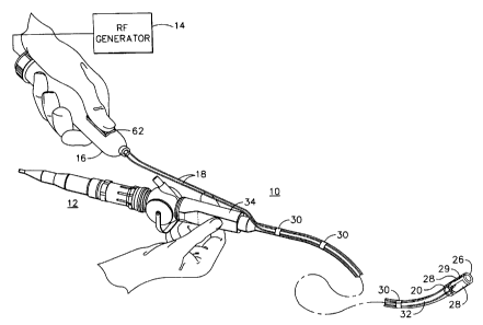
Figure 1 shows an endoscopic ablation system 10 according to the present
invention mounted on a flexible endoscope 12 (also referred to as endoscope
12),
such as the GIF-100 model available from Olympus Corporation. Flexible
endoscope 12 includes an endoscope handle 34 and a flexible shaft 32.
Endoscopic
5
CA 02440497 2003-09-11
ablation system 10 generally comprises an ablation cap 20, a plurality of
conductors 18, a handpiece 16 having a switch 62, and an RF (radio frequency)
generator 14. Ablation cap 20 fits over the distal end of flexible shaft 32
and
conductors I8 attach to flexible shaft 32 using a plurality of clips 30.
Ablation cap
20 includes a rigid support member 26, a plurality of electrodes 28, and a
viewing
window 29 positioned between electrodes 28. In this embodiment, rigid support
member 26 is made of a transparent material such as polycarbonate and viewing
window 29 is the portion of rigid support member 26 between electrodes 18.
Manual operation of switch 62 of handpiece 16 electrically connects or
disconnects
electrodes 18 to RF generator 14. Alternatively, switch 62 may be mounted on,
for example, a foot switch (not shown).
RF generator 14 is a conventional, bipolar/monopolar electrosurgical
generator such as one of many models commercially available, including Model
Number ICC 350, available from Erbe, GmbH. Either the bipolar mode or the
monopolar mode may be used for the present invention. When using the bipolar
mode with two electrodes 18 on ablation cap 20, one electrode is electrically
connected to one bipolar polarity, and the other electrode is electrically
connected
to the opposite bipolar polarity. If more than two electrodes 18 are used,
polarity
of electrodes 18 is 'alternated so that any two adjacent electrodes have
opposite
polarities.
When using the monopolar mode with two or more electrodes 18, a
grounding pad is not needed on the patient. Because a generator will typically
be
constructed to operate upon sensing connection of ground pad to the patient
when
in monopolar mode, it can be useful to provide an impedance circuit to
simulate
the connection of a ground pad to the patient. Accordingly, when the device of
the
present invention is used in monopolar mode without a grounding pad, an
impedance circuit can be assembled by one skilled in the art, and electrically
connected in series with one of conductors 18 that would otherwise be used
with a
grounding pad attached to a patient during rnonopolar electrosurgery. Use of
the
6
CA 02440497 2003-09-11
impedance circuit allows use of the generator in monopolar mode without use of
a
grounding pad attached to the patient.
The optimal power level required to operate endoscopic ablation system 10
of the present invention is approximately in the range of 10-50 watts,
although
endoscopic ablation system 10 is also functional at lower or higher power
levels.
Figure Z is an enlarged view of ablation cap 20 of endoscopic ablation
system 10 shown in Figure 1. Ablation cap 20 fits securely over the distal end
of
flexible shaft 32. Electrodes 28 are positioned on the outside surface of
rigid
support member 26, which has a circular cylinder shape in this embodiment.
Rigid support member 26 may also have alternate cylindrical shapes, including
shapes in which at least a portion of the cross sectional perimeter is non-
arcuate.
For example, rigid support member 26 may have a "D-shape" cross-section, where
electrodes 28 are positioned on the flat portion of the "D-shape." Conductors
18
are electrically insulated from each other and surrounding structures, except
for
electrical connections such as to electrodes 28. The distal end of flexible
shaft 32
of flexible endoscope 12 includes a light source 40, a viewing port 38, and a
working channel 36. Viewing port 38 transmits an image within its field of
view
to an optical device such as a CCD camera within flexible endoscope 12 so that
an
operator may view the image on a display monitor (not shown). In the
embodiment shown in Figure 2, the distal end of flexible shaft 32 is proximal
to
electrodes 28 and viewing window 29, enabling the operator to see tissue
between
electrodes 28 through viewing window 29.
Figure 3 shows the geometric relationship of a particular embodiment of
electrodes 28. In this embodiment, two rectangular electrodes 28, each having
a
width "w" and a length "L", have parallel, adjacent edges 8 that are separated
by a
distance "d". This geometric relationship may be used to calculate an ablation
index, which has particular significance to the location, size, shape, and
depth of
ablation achievable, as will be described later. Viewing window 29 (see Figure
2)
is approximately defined by the d x L rectangular area between electrodes 28.
7
CA 02440497 2003-09-11
Figure 4 is a sectional view of the lower end of an esophagus 42 and the
upper portion of a stomach S4 of a human being. Esophagus 42 has a mucosal
layer 46, a muscular layer 44, and a region of diseased tissue 48. The
boundary
between mucosal layer 46 of esophagus 42 and a gastric mucosa SO of stomach S4
S is a gastro-esophageal junction S2, which is approximately the location for
the
lower esophageal sphincter (LES). The LES allows food to enter the stomach S4
while preventing the contents of stomach S4 from refluxing into lower
esophagus
42 and damaging mucosal layer 46. Diseased tissue 48 can develop when chronic
reflux is not treated. In one form, diseased tissue 48 may be, for example,
intestinal metaplasia, which is an early stage of Barrett's esophagus. As can
be
seen in Figure 4, the esophagus is relatively flaccid and contains numerous
folds
and irregularities on the interior lining.
Figure S illustrates the use of endoscopic ablation system 10 to treat
diseased tissue 48 in lower esophagus 42. The operator positions ablation cap
20
using endoscopic visualization so that diseased tissue 48 to be treated lies
under
viewing window 29.
Figure 6 is sectional view of lower esophagus 42 showing tissue that has
been treated using endoscopic ablation system 10 according to the present
invention. In Figure 6, the size and shape of the treated tissue S6
substantially
corresponds to the size and shape of viewing window 29.
The operator may treat diseased tissue 48 using the embodiment of
endoscopic ablation system 10 of the present invention shown in Figures 1 and
5 as
follows. The operator inserts flexible shaft 32 of endoscope 12 into lower
esophagus
42 trans-orally. Rigid support member 26 holds lower esophagus 42 open as the
operator uses endoscopic visualization through ablation cap 26 to position
electrodes
28 next to the diseased tissue 48 to be treated. Rigid support member 26 opens
and
supports a portion of the flaccid, lower esophagus 42 and helps to bring the
tissue to
be treated into intimate contact with electrodes 28 and viewing window 29.
While
watching through viewing window 29, the operator actuates switch 62,
electrically
8
CA 02440497 2003-09-11
connecting electrodes 28 to RF generator 14 through conductors 18. Electric
current
then passes through the diseased tissue positioned in viewing window 29. When
the
operator observes that the tissue in viewing window 29 has been ablated
sufficiently,
the operator deactuates switch 62 to stop the ablation. The operator may
reposition
electrodes 28 for subsequent tissue treatment, or may withdraw ablation cap 26
(together with flexible endoscope 12). As illustrated in Figure 6, treated
tissue 56
has substantially the same width and length as viewing window 29.
Figure 7 shows an alternate embodiment of an endoscopic ablation system
10 and generally comprises an ablation cap 20, a sheath 63, a pair of
conductors
18, a handpiece I6 having a switch 62, and an RF generator 14. An operator may
rotate ablation cap 20 around flexible shaft 32 of flexible endoscope 12 by
manipulation of a rotation knob 58, which connects to sheath 63. Ablation cap
20
includes a rigid support member 26, at least two electrodes 28, and at least
one
viewing window 29 (between each pair of adjacent electrodes). Sheath 63
comprises a rotation tube 22 covered by an external tube 64. Ablation cap 20
attaches directly to the distal end of sheath 63. Rotation tube 22 can be made
from
a material such as, for example, corrugated polyethylene tubing, and fits
slidably
over a conventional, flexible endoscope. External tube 64 is preferably made
from
a heat-activated shrink tube material such as polyolefin. Conductors 18 are
spirally wrapped around rotation tube 22 prior to assembling and shrinking
external tube 64 onto rotation tube 22, thereby tightly retaining conductors
18 in
the wound configuration. In the embodiment shown in Figure ?, a valve 60 (also
referred to as a tapered end cover), which may be, for example, a duck bill
valve,
connects to the distal end of rigid support member 26. Valve 60 allows an
operator to extend the distal end of flexible endoscope 12 beyond the distal
end of
rigid support member 26 to improve visualization of tissue structures,
especially
during intubation. The operator may also retract the distal end of flexible
endoscope 12 within rigid support member 26 to allow visualization of viewing
window 29 and electrodes 28, while preventing bodily fluids from entering
rigid
support member 26 and impairing visualization by contact with flexible
endoscope
12.
9
CA 02440497 2003-09-11
Alternate embodiments of valve 60 may be envisioned by those skilled in
the art, each embodiment being particularly adapted to the medical procedure
and
anatomical structures involved. For example, in an alternative embodiment of
the
present invention, the distal end of valve 60 could be further tapered and
elongated
to allow for easier insertion into the esophagus. Valve 60 could further be
transparent to enable the physician to visualize through valve 60 during
intubation
into the esophagus, while preventing contact of bodily fluids against the
distal end
of flexible endoscope 12.
Figure 8 is a sectional view taken along the longitudinal axis of endoscopic
ablation system 10 of Figure 7. The distal portion of flexible shaft 32 is
inside
rotation tube 22 of endoscopic ablation system 10. A pair of conductors 18
passes
through a strain relief 66 of rotation knob 58 and between external tube 64
and
rotation tube 22. Each conductor 18 connects electrically to one of electrodes
28
on ablation cap 20. Rotation tube 22 rotatably joins rotation knob 58 to
ablation
cap 20, enabling the operator to rotatably orient electrodes 28, even after
insertion
into the esophagus, by remotely actuating rotation knob 58. The distal end of
flexible shaft 32 extends from the distal end of sheath 63 into ablation cap
20 and
proximal to electrodes I8. A viewing window 29 between electrodes 28 is within
the field of view of flexible endoscope 12, thus enabling the operator to see
on a
display monitor the tissue that is located between electrodes 18. Valve 60
extends
from the distal end of ablation cap 20 to prevent tissue or fluids from
entering
ablation cap 20.
Figure 9 is a sectional view taken along line 9-9 of ablation cap 20 of
endoscopic ablation system 10 of Figure 8. Conductors 18 connect to electrodes
28
with the portion of rigid support member 26 between electrodes 28 defining
viewing window 29. Rotation tube 22 retains flexible shaft 32. The inside
diameter of rotation tube 22 is larger than the outer diameter of flexible
endoscope
12 to allow rotation of rotation tube 22 while holding flexible endoscope 12
stationary, or vice versa. In this embodiment at least the portion of rigid
support
member 26 that forms viewing window 29 is transparent so that the operator may
CA 02440497 2003-09-11
endoscopically view the tissue between electrodes 28. Flexible endoscope 12
includes a light source 40, a viewing port 38, and a working channel 36.
Figure 10 is a sectional view taken along line 10-10 of rotation tube 22 of
endoscopic ablation system 10 of Figure 8. External tube 64 and rotation tube
22
assemble and retain conductors 18 as already described. Light source 40,
viewing
port 38, and working channel 36 of flexible endoscope 12 are shown.
Figure I 1 shows a further embodiment of an endoscopic ablation system 10
according to the present invention. A flexible ablation cap 24 includes a
flexible
support member 68 and at least two electrodes 28 mounted on an electrode sled
70,
which may be housed in or extended from a sled housing 76. Flexible ablation
cap
24 mounts over the distal end of flexible shaft 32. Conductors 18 electrically
connect to electrodes 28 as in the previous embodiments, and may be attached
to
flexible shaft 32 by a plurality of clips 30. Again, conductors 18
electrically
connect to RF generator 14 by a switch 62 of a handpiece 16.
Figure 12 is an enlarged view of flexible ablation cap 24 of the endoscopic
ablation system 10 illustrated in Figure 11 with electrode sled 70 fully
extended.
A sled housing 76 is a soft and flexible, pouch-like container, which may be
made
of a material such as PTFE in order to prevent damage to the mucosa as the
operator introduces endoscopic ablation system 10 into the esophagus. Sled
housing 76 and flexible support member 68 may be molded as a single piece.
Electrode sled 70 may be made of a clear rigid material such as, for example,
polycarbonate. As shown in Figure 12, electrode sled 70 includes two
electrodes
28, a viewing window 29, and two conductors 18. At least the portion of
electrode sled 70 that forms viewing window 29 is transparent to allow the
operator to view endoscopically the tissue between electrodes 28. Flexible
support
member 68 includes sled guides 78, which are adapted to receive electrode sled
70. Extension of sled 70 to an extended position stiffens flexible support
member
68 such as may be desired during ablation; retraction of sled 70 to a
retracted
position allows flexible support member 68 to flex such as may be desirable
during
11
CA 02440497 2003-09-11
intubation. A drive cable 74 retains conductors 18, which extends proximally
through sled housing 76 and into a sleeve 72. Sleeve 72 attaches to flexible
shaft
32 by a fixed clip 31. Thus, by extending drive cable 74, electrode sled 70
moves
distally and, by retracting drive cable 74, electrode sled 70 moves proximally
into
sled housing 76.
Figure 13 shows flexible ablation cap 24 of endoscopic ablation system 10
of Figure I1 with electrode sled 70 retracted into sled housing 76, or in a
retracted
position.
Figures 14-16 are additional views of flexible ablation cap 24 illustrated in
Figure I I. Figure 14 is a top view of flexible ablation cap 24 with electrode
sled
70 in an extended position. Figure 15 is a side view of flexible ablation cap
24
with electrode sled 70 in an extended position. In Figures 14 and 15 electrode
sled
70 includes electrodes 28, viewing window 29 and conductors 18, which are
connected to electrodes 28. Flexible support member 68 includes sled guides
78.
Drive cable 74, which houses conductors 18, is in turn housed within sled
housing
76 and extends proximally into sleeve 72. Figure 16 is an end view of the
flexible
ablation cap 24 of the endoscopic ablation system 10 illustrated in Figure I1.
Figure 16 illustrates the arrangement of sled guides 78 and the engagement of
electrode 70 by sled guides 78.
Figure 17 is an illustration of a further embodiment of an endoscopic
ablation system IO for use with an endoscope 12 having an endoscope handle 34.
Endoscopic ablation system 10 generally comprises a rotation knob S8, a sheath
63, an ablation cap 82, and a tapered end cover 84. Ablation cap 82 further
includes an ablation cap-opening 86. Conductors 18 spirally wrap around the
outside of sheath 63 in this embodiment, and at least one clip 30 attaches
conductors 18 to sheath 63. Endoscopic ablation system 10 further comprises an
actuator 90 and a timer 91. A plurality of electrodes 28 (hidden in this view)
on
ablation cap 82 electrically connect, via a pair of conductors 18, to actuator
90.
The operator actuates actuator 90 manually to enable timer 91 to electrically
I2
CA 02440497 2003-09-11
connect electrodes 28 to RF generator 14 for a predetermined period of time.
The
operator then actuates control switch 92, which may be a foot operated control
switch commonly available with RF generators, to activate RF generator 14.
When RF generator 14 is activated, timer 91 automatically connects RF
generator
14 to electrodes 28 for a predetermined length of time. For the embodiments of
an
endoscopic ablation system described herein, an appropriate predetermined
length
of time is approximately in the range of 0.1 to 10 seconds, and is preferably
about
one second. However, the length of predetermined time may vary depending on
the geometry of the electrodes, the power level used on the RF generator, the
type
of tissue being treated, and other factors. Timer 91 includes a conventional
timer
circuit that is connected in electrical series to the output of a RF generator
14
having a control switch 92. When the operator actuates control switch 92, the
electrical current from RF generator I4 induces a secondary current inside of
timer
9I. This secondary current supplies and immediately activates the timer
circuit of
timer 91, thereby connecting the output of RF generator 14 to electrodes 28
via a
relay inside of timer 91. After a predetermined period of time, the relay
disengages
automatically, therefore electrically disconnecting RF generator 14 from the
electrodes 28. Therefore, the operator controls when electrodes 28 are
energized
to begin ablation of tissue, but timer 91 controls when ablation stops, even
though
the operator may still be activating control switch 92. Timer 91 ensures
complete
ablation of diseased tissue in the viewing window and greatly reduces the
possibility of operator error associated with RF energy application.
Timer 91 and actuator 90 of Figure 17 may be provided as a handle with a
switch much like handle 16 and switch 62 of Figure 1. Alternately, timer 91
and
actuator 90 may be incorporated into a table top unit (not shown), combined
with
RF generator 14 and control switch 92, or electronically packaged in many
other
ways that are readily apparent to one skilled in the art. Actuator 90, timer
91, RF
generator 14, and control switch 92 may comprise a reusable portion of
endoscopic
ablation system 10. The remaining portion that includes conductors 18, sheath
63,
rotation knob 58, and ablation cap 82 may be provided, for example, as a
relatively low cost, sterile device that is disposable after use on one
patient.
I3
CA 02440497 2003-09-11
Figures 18, 19, and 20 are sectional views of the distal portion of
endoscopic ablation system 10 shown in Figure 17, and illustrate alternate
locations of electrodes 28. Figures 18, 19, and 20 show the distal end of
sheath 63
inserted into the proximal end of a flexible coupling 88 and attached by a
ring 94
tightly compressed around sheath 63 and the proximal end of flexible coupling
88.
The distal end of flexible coupling 88 attaches to the proximal end of a rigid
support member 26 of ablation cap 82 by the engagement of a plurality of
annular
projections 96 on the inside of the distal end of flexible coupling 88 with a
like
plurality of annular grooves 98 formed into the proximal end of rigid support
member 26. Flexible coupling 88 is made of a flexible tube material such as
silicone rubber and allows low force angulation of sheath 63 with respect to
ablation cap 82, thus facilitating passage of ablation cap 82 through the
esophagus
of the patient. The distal end of rigid support member 26 includes a plurality
of
IS annular grooves 99 for retaining a plurality of annular projections 97 on
the inside
of the proximal end of tapered end cover 84. Tapered end cover 84 is made of a
transparent, flexible material such as, for example, clear or tinted
polyurethane
that is commonly used for flexible, extruded tubing. Tapered end cover 84
further
includes an elongated, distal tip 104 that helps the operator to insert
ablation cap
82 into the esophagus.
Tapered end cover 84 is hollow in order to allow positioning of the distal
end of endoscope 12 partially into tapered end cover 84, as shown in Figure
18.
This enables the operator to view the interior of the esophagus, yet protects
the
distal end of endoscope 12 from tissue structures and bodily fluids that may
impair
visualization. Tapered end cover 84 is shaped like a bougie tube, which is
commonly used by endoscopists for dilating the esophagus prior to incubation
with
an endoscope. Distal tip 104 of tapered end cover 84 includes a channel 102 so
that the operator may pass a guide wire through ablation cap 82 and sheath 63,
in
order to facilitate positioning of ablation cap 82 inside of the esophagus.
14
CA 02440497 2003-09-11
As shown in Figures 18, 19, and 20, electrodes 28 may be mounted at
varying locations on ablation cap 82. In Figure 18, electrodes 28 are attached
to
the outside of tapered end cover 84 near distal tip 104. As indicated in
Figure 18,
electrodes 28 are positioned on a portion of tapered end cover 84 that has a
smaller
cross-sectional diameter than the diameter of the distal end of endoscope 12.
As
shown in Figure 19, electrodes 28 may also be attached to rigid support member
26, as was also described for the embodiments shown in Figures 1 and 7. In
Figure 19, a portion of one of conductors 18 is shown as it may be
electrically
connected to one of electrodes 28 by a solder and/or compression connection.
(Conductors 18 are not shown in Figures 18 and 20.) In Figure 20, electrodes
28
are positioned partially on rigid support member 26 and partially on tapered
end
cover 84. Electrodes 28 may vary in size, shape, and position on ablation cap
82,
as shown in the examples of Figures 18, 19, and 20, but importantly, still
follow
the geometric relationships described for Figure 3 in order to achieve a
desired
I S ablation quality.
Still referring to Figures 18, 19, and 20, rigid support member 26 also
includes side opening 86. In the examples shown, side opening 8b is
rectangularly
shaped and positioned between the distal end of flexible coupling 88 and the
proximal end of tapered end cover 84. In the examples shown in Figures 19 and
20, side opening 86 is on the side of rigid support member 26 opposing the
position of electrodes 26. Side opening 86 provides access to tissue
structures next
to ablation cap 82 with instrumentation passed through the working channel of
endoscope 12. In addition, side opening 86 allows fluid communication between
endoscope 12 (that normally includes suction and irrigation channels) and the
interior of the esophagus around ablation cap 86. Therefore, the operator may
position electrodes 28 adjacent to tissue to be ablated and apply the suction
provided with endoscope 12. As the lumen size of the esophagus decreases under
vacuum, the esophagus collapses around ablation cap 82, thus bringing the
tissue
to be treated in intimate contact with electrodes 28 and viewing window 29.
This
facilitates uniform electrode contact for even ablation, and improves
endoscopic
CA 02440497 2003-09-11
visualization through the viewing window of tissue being treated during the
procedure.
Figure 21 is a sectional view of the proximal portion of sheath 63, rotation
knob 58, and conductors 18 of the endoscopic ablation system 10 shown in
Figure
17. Rotation knob 58 is molded from a flexible material such as a
biocompatible
rubber. The proximal end of rotation knob 58 includes a proximal seal 110
having
a hole 111 for insertion of endoscope 12 (not shown). The interior of the
sheath
distal to proximal seal 110 and the interior of ablation cap 82 define an
enclosure
that is in fluid communication with the interior of the esophagus and the
aspiration
means of the flexible endoscope 12. Proximal seal 110 prevents fluid
communication between the air external to the patient and the interior of
sheath 63
and the interior of ablation cap 82. This allows the technique described for
Figures 18, 19, and 20 for using the suction available with endoscope 12 to
pull
the interior of the esophagus into intimate contact with electrodes 28 and
viewing
window 29. Seal 110 also wipes bodily fluids from the exterior of endoscope 12
as it is withdrawn from sheath 63. Rotation knob 58 also includes a distal
cylindrical extension 57 that fits tightly over the proximal end of a rotation
tube 22
of sheath 63. An external tube 64 fits tightly over the entire length of
sheath 63,
including the portion attached to distal cylindrical extension 57 of rotation
knob
58. Rotation tube 22 may be made of any one of a number of flexible tubing
materials, including corrugated polyethylene tubing. External tube 64 is
preferably made from polyolefin that is shrink-wrapped tightly onto rotation
tube
22 by the application of heat during assembly. In Figure 21, conductors 18 are
shown wrapped around the outside of sheath 63. Conductors 18 may also be
assembled between rotation tube 22 and external tube 64 so that the outside of
sheath 63 is relatively smooth for passage into the esophagus. Rotation knob
58
also includes a plurality of grip projections to facilitate manipulation.
Figure 22 shows the distal portion of endoscopic ablation system 10 of
Figure 17 partially inserted into the esophagus 41 of a patient. Tapered end
cover
84 dilates esophagus 41 as the operator gently inserts ablation cap 82 for
16
CA 02440497 2003-09-11
a
positioning near tissue to be ablated. Flexible couplinb 88 flexes as shown,
reducing the required insertion force and minimizing trauma (and post-
procedural
pain) to the patient.
Figure 23 is a sectional view of the distal portion of a further embodiment
of an endoscopic ablation system 10. Figure 23 shows an endoscope 12 inserted
into an ablation cap 116 that includes a sheath 63, a plurality of electrodes
28, and
a flexible coupling 88 such as was described for Figurel9. However the
embodiment in Figure 23 includes an open-end piece 114 (also referred to as a
tapered end cover) attached to the distal end of rigid support member 26. Open-
end piece 114 resembles tapered end cover 84 of Figure 17, but with all but
the
proximal portion cut off perpendicular to the longitudinal axis. The remaining
taper of open-end piece 114 facilitates passage through the esophagus and
substantially prevents body fluids on the esophageal wall from collecting
inside
ablation cap 116. Open-end piece 114 is made preferably from a flexible
material
such as silicone rubber. The operator may extend the distal end of endoscope
12
through open-end piece 114, to facilitate endoscopic visualization during
intubation
of ablation cap 116 into the esophagus. The operator may retract endoscope 12
to
a retracted position as shown in Figure 23 in order to view tissue through a
viewing window (not shown) between adjacent electrodes 28, and to watch the
progress of ablation.
Now referring again to Figure 3, the size, shape, and relative position of
electrodes 28 are shown, as they would be mounted on rigid support member 26.
The region between electrodes 28 forms the viewing window 29. In an endoscopic
ablation system according to the present invention, the size, shape and
relative
position of electrodes 28 are established by the Ablation Index, I, and:
I = P/d ( 1 )
Where:
P is the perimeter of electrodes 28 and
d is the separation between adjacent edges 8 of
electrodes 28.
17
CA 02440497 2003-09-11
In the embodiment of the invention illustrated in Figure 3:
I =2(w+L)/d (2)
Where:
w is the width of electrodes 28 and
L is the length of electrodes 28.
Although the electrodes illustrated in Figure 3 are rectangular in shape,
other
shapes having an Ablation Index I according to Equation 1 are appropriate for
use in
l0 the present invention provided that d is substantially constant, i.e. the
adjacent edges
of the electrodes are substantially parallel. In an endoscopic ablation system
according to the present invention, I can be between about 1 and about 200,
more
particularly between about 15 and 35, such as indicated by a region "A" in the
graph
of Figure 24. The graph of Figure 24 was based on data derived from
experiments
15 with many different electrode geometries for many different conditions.
Ablation
Quality is a subjective rating of between 1-10 based primarily on area, depth,
and
color of ablation achieved. Region A indicates the Ablation Index I for when
Ablation Quality is greater than or equal to 5 (an average subjective rating)
on a
scale of 1-10. In some cases, the operator may desire to maintain an ablation
index
20 where 20<I<28, as indicated by a region "B" in Figure 24. Practical
considerations
related to manufacture, type of tissue being treated, physician preferences,
and so on,
come into play when determining electrode geometry and selecting an ablation
index
range. The Ablation Index is used to define an electrode arrangement that
substantially confines the initial ablation to the tissue under the viewing
window,
25 allowing the surgeon to control the ablation process. In operation, an
endoscopic
ablation device according to the present invention includes electrodes having
an
Ablation Index within the prescribed ranges. Such an endoscopic ablation
instrument will begin to ablate tissue when an electric potential is
established
between the electrodes (i.e. the electrodes are actuated). However, during the
initial
30 ablation process little or none of the tissue directly beneath the
electrodes will be
ablated and the thermal profile within the treated tissue will have a
substantially
vertical wall at the edge of the electrodes. Further, the current density of
the
electrical current flowing between the electrodes will be very high in the
tissue under
18
CA 02440497 2003-09-11
the viewing window, accelerating the ablation of tissue within the treatment
region,
giving the surgeon precise control of the treatment region and limiting the
ablation
of healthy tissue. The operator further has precise control of the degree to
which the
treated tissue is ablated since the operator may view the entire treatment
region
through the viewing window. The operator may visually determine when the
treated
tissue is sufficiently ablated by watching to see when the ablated tissue
fills the
entire ablation window. When the ablated tissue fills the entire ablation
window, the
mucosa is consistently ablated to a predetermined depth across the treatment
region.
The actual depth of the ablation is a function of a number of variables,
including
power. Uniform ablation depths of approximately one to two millimeters are
constantly obtainable using the color of the treated tissue in the ablation
window as a
guide. Ablation depths of one to two millimeters are normally enough to ablate
the
abnormal tissue in the mucosa without significantly damaging the healthy
tissue
underneath.
Figure 25 represents an endoscopic ablation system 9 comprising an ablation
cap 152, a control unit 1 S0, and a RF generator 14. Ablation cap 152 includes
a
plurality of electrodes 156, each of which is electrically connected to
control unit
150. In this embodiment, ten electrodes labeled E1 through E10 comprise
plurality
of electrodes 156, and are printed using conventional printed circuit
manufacturing
techniques onto a transparent substrate 158 made from a material such as clear
polyacetate or Mylar film. Transparent substratE 158 is adhered to a rigid
support
member 154 using, for example, UV cured optical adhesive No. NOA 68, which is
available from Norland Products, Inc., New Brunswick, NJ. A plurality of
electrode
leads 160 are also printed onto substrate 158 and terminate at a solder pad
(not
shown) for electrical attachment to insulated wires (not shown) for electrical
connection to control unit 150. Rigid support member 154 may be identical to
rigid
support member 26 shown in Figure 2. The proximal end of ablation cap 152
attaches to flexible shaft 32 (see Figure 1). Electrode leads 160 and portions
of
electrodes 156 may be covered with a dielectric coating or shrink wrap film in
order
to be insulated from tissue. In this embodiment, a separate electrode lead is
provided for each electrode so that each electrode may be individually
actuated by
control unit 150 according to a predetermined sequence and for a predetermined
19
CA 02440497 2003-09-11
~ ! r
duration. This enables a large number of different combinations of electrode
actuation sequences and durations to obtain desired tissue ablation effects.
It is also
possible to have more than one electrode attached to a common lead. Because
rigid
housing member 154 is made of a clear material such as polycarbonate, a
plurality of
viewing windows are provided in the spaces between electrodes 156 for
endoscopically viewing tissue during the ablation procedure.
Figure 2b shows plurality of electrodes 156 of Figure 25 as they would
appear laid flat. In this embodiment, each of electrodes E1 through E10 has a
rectangular shape with length "L" and width "w", and the distance between the
parallel edges of adjacent electrodes is "d". As described for Figure 3, an
Ablation
Index, I, establishes the size, shape and relative position of electrodes 156
according to the following:
I = P/d = 2(w+L)/d (3)
Where:
P is the perimeter of electrodes 156
Although the electrodes illustrated in Figure 26 are rectangular in shape,
other shapes having an Ablation Index, I, according to Equation 3 are
appropriate for
use in the present invention provided that d is substantially constant. That
is, the
adjacent edges of the electrodes should be a constant distance apart along the
length
of the adjacent electrodes. Therefore, it is possible for electrodes 156 to
have a
curvilinear shape. As described earlier, I can be between about 1 and about
200,
more particularly between about 15 and about 35 such as indicated by a region
"A"
in the graph of Figure 24. In addition, all of electrodes 156 do not
necessarily need
to have the same width, length, or distance between electrodes 156. In other
embodiments, for example, Ablation Index may vary between pairs of adjacent
electrodes to obtain desired tissue ablation effects.
Again referring to Figure 25, control unit 25 comprises generally an internal
switching network for activating plurality of electrodes 156 according to a
predetermined sequence and pattern. When any two adjacent electrodes 156 have
opposite polarities and are in intimate contact with tissue, the tissue
between those
CA 02440497 2003-09-11
two adjacent electrodes is ablated, and tissue underneath the two adjacent
electrodes
l56 is not ablated. Control unit 25 comprises a programmable, multiplexing
system
for actuating electrodes 156 and is easily constructable by those ~ skilled in
the art.
Examples of predetermined sequences of actuation are shown in the following
tables
where E1-E10 refer to electrodes; TI-T9 refer to time periods, (+) indicates
positive
polarity, (-) indicates negative polarity, and a blank indicates electrode not
energized
during the specified time period:
Table 1
E1 E2 E3 E4 ES E6 E7 E8 E9 EIO
T1 + -
T2 + -
T3 + -
T4 +
TS + -
T6 + -
T7 + _
T8 + _
T9 + -
Table 2
E1 E2 E3 E4 ES E6 E7 E8 E9 E10
T1 - + _
T2 - + -
T3 - + -
T4 - + -
TS - + -
T6 - + -
T7 , - + _
T8 - + -
T9 ~ - - +
21
CA 02440497 2003-09-11
Table 3
E1 E2 E3 E4 ES E6 E7 E8 E9 E10
T1 - + -
T2 - + -
T3 - + -
T4 - + -
TS - +
Table 4
E1 E2 E3 E4 ES E6 E7 E8 E9 E10
T1 - + - + - + - + - +
In Table 1, electrodes E1 and E2 are energized (on) at time TI, while
electrodes E3 through E10 are not energized (off). At time T2, electrodes E2
and E3
are on, while electrodes E1 and E4 through E10 are off, and so on until all
tissue in
the viewing windows is ablated. The duration of each actuation may vary, but
can
be approximately 1-2 seconds in one embodiment. By energizing electrodes 156
sequentially in this manner, the peak power requirement for RF generator 14 is
significantly less than if all the electrodes 156 were energized
simultaneously. Also,
while all the electrodes could be energized simultaneously as in Table 4, it
may be
I S desirable to energize the electrodes in a sequential manner, as in Tables
I and 2, so
that the tissue ablation can be observed as it occurs through the appropriate
window.
A physician may use endoscopic ablation system 9 shown in Figure 25 in the
same manner as was described for endoscopic ablation system 10 of Figures I-5,
with one primary difference. That is, the physician will not need to rotate
endoscopic ablation system 9 as often within the body lumen as would be
required
for endoscopic ablation system 10, due to the larger number of electrodes 156
on the
former. If the electrodes 156 are disposed around substantially the entire
perimeter
22
CA 02440497 2003-09-11
of the ablation cap, then the device could be rotated within the body lumen
only once
in either direction, and approximately by a distance equal to the width, w, of
an
electrode 156, to provide ablation of the tissue around the circumference of
the
lumen.
In Tables 2 and 3, three electrodes and energized simultaneously. Ten
electrodes are shown in Figure 26, but it will be understood that more
electrodes or
fewer electrodes could be used, as desired.
Figure 27 is a sectional view of the distal portion of an endoscopic ablation
system 11 including a distally mounted image sensor 120. A flexible endoscope
and
a conventional video tower are not required for visualization of tissue.
Endoscopic
ablation system 11 comprises a flexible member, such as flexible shaft 138,
and an
ablation cap 146, which may be a detachable ablation cap 146. Endoscopic
ablation
system 11 can also be constructed so that ablation cap 146 is not detachable
from
flexible shaft 138. Flexible shaft 138 includes a sensor housing 140 that
contains
image sensor 120. Image sensor 120 may be a CMOS (Complementary Metallic
Oxide Sensor) camera such as Model Number OV7910, which is available from
Omnivision Technologies, Inc. (www.ovt.com). Image sensor 120 may include an
objective lens 122 as shown in this embodiment, or may be a pin-hole style
CMOS
camera that may be used with red light LED illumination, for example. A CMOS
cable 124 passing through flexible shaft 138 contains a signal wire for
connection to
a NTSC or PAL formatted display monitor and a pair of electrical leads for
connection to a SVDC-power supply.
Still referring to Figure 27, ablation cap 146 can comprise a rigid support
member 154 made of a clear plastic such as polycarbonate, and may have
approximately the same configuration as rigid support member 26 described for
Figure 18. Rigid support element 154 is hollow and has an inner surface 162
and an
outer surface 164. A plurality of illuminators 126 are surface mounted on
inner
surface 162 in order to illuminate the field of view of image sensor 120.
White light,
surface mounted LED's such as Model No. NSPWFSOBS available from Nichia
(www.nichia.co jp) are suitable as illuminators 126. Illuminator leads 128
23
CA 02440497 2003-09-11
f
electrically connect in parallel illuminators 126 to a DC power supply (not
shown).
An umbilical tube 134 has a distal end attached to rigid support member 154
and is
long enough to extend outside of the body lumen. Umbilical tube 134 removably
attaches to flexible shaft 138 with at least one clip 136. Umbilical tube 134
contains
illuminator leads 128, a plurality of bipolar electrode leads 132, and a
suction tube
130, which is connected to a vacuum source (not shown).
Figure 28 is a side view of the distal portion of endoscopic ablation system
11 shown in Figure 27. In Figure 28, ablation cap 146 and umbilical tube 134
are
shown detached from flexible shaft 138, thus allowing cleaning and reuse of a
hermetically sealed and cleanable version of flexible shaft I38 containing
image
sensor 120. Ablation cap 146 and umbilical tube 134 transport body fluids and
support components, especially electrodes I28, that may degrade with repeated
use,
and therefore may be fabricated as single patient use, disposable components.
Alternatively, the entire assembly including flexible shaft 138, ablation cap
146, and
umbilical tube 134 can be a single use, disposable unit packaged in a pre-
sterilized,
ready to use form.
Figure 28 shows one of many variations of attaching ablation cap 146 to
flexible shaft 138. Each of at least one retaining slots 144 engages with a
corresponding post 142 projecting radially from a boss 141 on the distal end
of
flexible shaft 138. (This variation of attaching two components is commonly
referred to as a "bayonet fitting.")
Electrodes having an ablation index and viewing window according to the
present invention may be used in other surgical instruments such as, for
example,
endocutters. Further, electrodes having an ablation index according to the
present
invention may be used for other treatment regimens such as tissue welding,
electrophoresis and coagulation of varicose veins and hemorrhoids.
While preferred embodiments of the present invention have been shown
and described herein, it will be obvious to those skilled in the art that such
embodiments are provided by way of example only. Numerous variations,
24
CA 02440497 2003-09-11
changes, and substitutions will now occur to those skilled in the art without
departing from the invention. Accordingly, it is intended that only the spirit
and
scope of the appended claims limit the invention.
25