Note: Descriptions are shown in the official language in which they were submitted.
' CA 02441434 2003-09-09
DESCRIPTION
TITLE OF TIi$ INVENTION
REMEDIES FOR NERVE DAMAGES
Technical Field
The present invention relates to a remedy for a nerve
dysfunctional disorder such as a central nervous system damage
including a spinal cord injury and a cerebral infarction and
the like which promotes nerve regeneration, ormore particularly,
a remedy which can be applied to gene therapies.
Background Art _
Most spinal cord injuries are traumatic, and their causes
are traffic accidents , sports , industrial accidents and the like,
whereas the causes of atraumatic injuries are inflammation,
bleeding, tumor, spinal deformation and the like. Their
pathological features are crush of a spinal cord and a compression
lesion with bleeding and edema in spinal parenchyma as a basal
plate, and a neuropathy corresponding to a in jured site occurs .
As a main clinical symptom, incompetent or competent motor palsy
and numbness occur on and under the level of injury, and for
cervical spinal cord injury, respiratory palsy and hyperpyrexia
( or severe hypothermia ) can be seen as distinctive complications .
Improvement of the aforementioned neuropathy, particularly the
improvement of dyskinesia is directly linked to the inhibition
of increment in bedridden old people and the progress of QOL
(Quality of Life) , and therefore, their importance is growing
in parallel with the extension of average life expectancy in
these years.
1
CA 02441434 2003-09-09
Therapies being conducted for the aforementioned spinal
cord injury are surgical operations for eliminating physical
compression and injuries, and steroid therapies for a spinal
cord edema at the acute stage of in jury ( N . Engl . J . Med . 322 ,
1405-1411, 1990; J. Neurosurg 93, 1-7, 2000). Among the
steroidal agents, it is reported that megadoses of
methylprednisolone are effective for the improvement of
neurological symptom associated with a spinal cord in jury ( J .
Spinal Disord. 5 ( 1 ) , 125-131, 1992 ) , however, there is a problem,
in megadoses of steroidal agents, of lowering the phylactic
function in the case of the spinal cord injuries which are
associated with infection, in addition to the strong expression
of systematic adverse reactions and the dif f iculty in controlling
them. Besides, even the efficacy of steroid-megadosed
therapies remains controversial for the present. As described
above, there has been no effective remedy for a spinal cord in jury
to date, therefore it has been aspired for the development of
a new remedy . Other therapeutic methods f or spinal cord in juries
reported in addition to the aforementioned are as follows: a
method wherein therapeutically effective amount of
glioastrocytoma which was pretreated by inflammation related
cytokine in vitro is transplanted to the injured site in the
central nervous system (CNS) (Published Japanese translation
of PCT International Publication No.2000-503983); a method
wherein regeneration of a neurological axon in the central
nervous system (CNS) of mammal animals is promoted by
administering congeneric monocular macrophages (monocytes,
macrophages, etc. ) to the injured site or disordered site, or
CNS of its vicinity (J. Mol. Med. 77, 713-717, 1999; J. Neurosci.
19(5), 1708-16, 1999; Neurosurgery 44(5), 1041-5, 1999, Trends.
2
' CA 02441434 2003-09-09
Neurosci 22(7), 295-9, 1999)(Published Japanese translation of
PCT International Publication No.Hll-13370) and the like.
Further, although the defined mechanism is uncertain, it is also
reported that restoration of motion sustainment after a spinal
cord injury was promoted by the vaccination of spinal cord
homogenate and administering a T cell specific to a myelin basic
protein which is a myelin protein (Neuron 24, 639-647, 1999;
Lancet 354, 286-287, 2000).
On the other hand, dendritic cells (DC) are the cell
population taking dendritic forms that are derived from
hematopoietic stem cells , and are widely distributed in vivo .
Immature dendritic cells undertake a role as antigen-presenting
cells that induce immunoresponse by activating antigen-specific
T cells , by way of recognizing and incorporating a foreign body
such as a virus and a bacterium which has invaded each tissue,
generating a peptide by digesting and degrading such foreign
body in the process of transfer to a lymphatic organ T cell region,
binding such peptide to a MHC molecule, and presenting such
peptide to the cell surface (Ann. Rev. Immunol. 9, 271-296, 1991;
J. Exp. Med. 185, 2133-2141, 1997).
It had been difficult to prepare a large quantity of
dendritic cells due to their low-density in each tissue despite
that they are widely distributed, however, it became possible
to easily prepare a large quantity of such cells in vitro by
adding differentiated growth factors to the culture of immature
precursor cells. Therefore, it has been started to consider
using dendritic cells as immunostimulator (J. Exp. Med. 183,
7-11, 1996). It is particularly targeted to specifically
enhance the immunoresponse by pulsing antigens to dendritic cells
against a faint tumor immunoresponse . In an animal experiment ,
3
CA 02441434 2003-09-09
it is shown that dendritic cells presenting a protein and an
antigen peptide derived from a tumor induce a specific CD8+
cytotoxic T cell. It is reported also in human that tumors
decreased or disappeared by returning a protein and an antigen
peptide derived from a tumor together with dendritic cells to
a living body. Meanwhile, it is reported that IL-12, a cytokine,
is secreted mainly from the antigen-presenting cells such as
the aforementioned dendritic cells and B cells , and acts toward
T cells and NK-cells , and has a high antitumor activation ( J
Exp. Med. 178, 1223-1230, 1993; J. Exp. Med. 189, 1121-1128,
1999) . Thus, IL-12 draws attention as a remedy for cancer, and
clinical trials have been conducted as a new immunotherapy for
cancer. However, it has not historically been applied for a
nervous system at all.
On the other hand, one of the most important elements in
the study of spiral cord in jury wherein an animal model is used
can be exemplified by the evaluation of motor function. Such
evaluation of motor function is desired to be easy and to have
high reproducibility. However, most of the historical
evaluation methods of motor function emphasize the movements
of articulations of individual posterior limbs and their
coordinated movements or the overall conditions of locomotion,
as in the BBB scoring method ( J Neurosung 93 , 266-75 , 2000 ) wherein
the locomotion of animals are evaluated by the total scores ( the
maximum score is 21 points) of various check items, and even
including the one requiring detailed measurement of the motion
which were videotaped in advance . Therefore , there was a problem
that such methods were cumbersome and might easily cause
individual variations among the experimenters.
In juries of central nervous systems including spinal cord
4
CA 02441434 2003-09-09
injuries are disorders, which are extremely difficult to be
remedied, and there has been no effective therapy to date as
described above, therefore, the development of a new therapy
is strongly expected. In addition, the number of patients
affected by nervous system disorders is on the rise in connection
with the aging of population, and it has become a ma jor social
problem. However, the central nervous system is an organ, which
is extremely difficult to be regenerated, and is a special organ
wherein immunoreaction is hard to occur. In the aforementioned
method by Schwartz et al. wherein regeneration of nervous axon
in the central nervous system (CNS) is promoted by using
macrophages , it was not clear which function of the macrophages
prompts the regeneration of an axon. When the cells such as
macrophages and the like are used, there were the problems not
only that the administration method was limited, but also that
its handling was complicated, and that it is hard to obtain
reproducible therapeutic effect since a living cell was used.
The object of the present invention is to provide a remedy for
a nerve dysfunctional disorder such as a central nervous system
injury including a spinal cord injury and a cerebral infarction,
which can be administered not only by injecting into a injured
site but also by various administration methods including
subcutaneous administration, administration to a vicinity of
lymph nodes, and intravenous administration, which can easily
be handled and stored over a long time, and can be prepared in
a large quantity at any time, and containing distinguished nerve
regeneration promotion action.
Unlike other tissues, the central nervous system is a
tissue that is isolated from the immune system. However, the
present inventors recently reported that immature T cells which
CA 02441434 2003-09-09
are not stimulated at all can not invade into the central nervous
system, however, T cells activated by an antigen in a brain can
pass through a blood brain barrier and can be reacted with a
brain tumor, as a result of experiment wherein a mouse brain
tumor model was used (Neuro-Oncologyl, S105, 1999) . Inaddition,
there is a report that restoration of central nerve damage was
promoted by administering nervous specific T cells (Lancet 354,
286-287, 2000) . It is still uncertain how the nervous specific
T cells function in the central nervous system after passing
through the blood brain barrier, for example, whether by
releasing some sort of cytokine or whether they act by directly
attaching to a nervous cell or an axon or any other, however,
the possibility of nerve regeneration by an intervention of
immune system is indicated. Meantime, it is necessary to
incorporate an antigen of a nervous system by an
antigen-presenting cell, and to present an antigen peptide
treated within the cell to T cells in order to induce a nervous
specific T cell.
The present inventors have substantiated for the first
time that exclusion of the in jured tissue at the time of spinal
cord injury is the first phase of crucial importance, and that
restoration of spinal cord function is promoted by incorporating
an antigen and directly transplanting certain dendritic cell
subsets having the highest antigen presenting ability against
T cells to the injured site of the spinal cord injured model
mouse. For the aforementioned substantiation of promoting
restoration of spinal cord function, the evaluation method for
motor function in the spinal cord in jured mouse established by
the present inventors was used. In this evaluation method for
motor function, an apparatus which was used for measuring the
6
' CA 02441434 2003-09-09
amount of motion for the purpose of analyzing sedative effects
and the like of a drug is applied to the evaluation of motor
function after a spinal cord is inured. The present inventors
targeted a substance secreted from dendritic cells generating
environmental changes including activation of T cells in the
central nervous system, or a substance inducing and proliferating,
or activating dendritic cells, and the present inventors
administered such candidate substance to the injured site of
a spinal cord injured model mouse, and screened by the
aforementioned evaluation method formotor function in the spinal
cord injured mouse, and as a result, the present inventors found
that IL-12 which are widely used as remedies for cancer but are
not used for nervous system at all, and GM-CSF promote restoration
of spinal cord function as dendritic cells do. Besides, as
described above, since significant restoration of motor function
was recognized by transplanting dendritic cell subsets into the
injured spinal cord, the present inventors analyzed a substance
promoting the nerve regeneration which are secreted from
dendritic cells , and confirmed that such dendritic cells express
a neurotrophic factor, and actually secrete the same. The
present invention has been completed as a result of these
findings.
Disvlosure of the Invention
The present invention relates to: a remedy for a nerve damage
or a nerve dysfunctional disorder wherein one or more types of
substances selected from the following, a substance secreted
from dendritic cells, a substance inducing and proliferating
dendritic cells, a substance activating dendritic cells, a
substance inducing the expression of a neurotrophic factor in
7
' CA 02441434 2003-09-09
nerve tissues, and a substance inducing and proliferating
microglias and macrophages in nerve tissues, or a dendritic cell
is used as an active ingredient ( claim 1 ) ; the remedy for a nerve
damage or a nerve dysfunctional disorder according to claim 1
wherein the substance secreted from dendritic cells, the
substance inducing and proliferating dendritic cells, the
substance activating dendritic cells, the substance inducing
the expression of a neurotrophic factor in nerve tissues, and
the substance inducing and proliferating microglias and
macrophages in nerve tissues are cytokines ( claim 2 ) ; the remedy
for a nerve damage or a nerve dysfunctional disorder according
to claim 2 wherein the cytokine secreted from dendritic cells
is an interleukin ( IL ) -12 ( claim 3 ) ; the remedy for a nerve damage
or a nerve dysfunctional disorder according to claim 2 wherein
the cytokine inducing and proliferating dendritic cells is a
granulocyte-macrophage colony-stimulating factor (GM-CSF)
( claim 4 ) ; the remedy for a nerve damage or a nerve dysfunctional
disorder according to claim 2 wherein the cytokine inducing the
expression of a neurotrophic factor in nerve tissues is a
granulocyte-macrophage colony-stimulating factor (GM-CSF)
( claim 5 ) ; the remedy for a nerve damage or a nerve dysfunctional
disorder according to claim 2 wherein the cytokine inducing and
proliferating microglias and macrophages in nerve tissues is
a granulocyte-macrophage colony-stimulating factor (GM-CSF)
( claim 6 ) ; the remedy for a nerve damage or a nerve dysfunctional
disorder according to claims 1 to 6 wherein one or more types
of the substances selected from a substance secreted from
dendritic cells, a substance inducing and proliferating
dendritic cells, and a substance activating dendritic cells are
vectors which can express such substances (claim7).
8
' CA 02441434 2003-09-09
The present invention further relates to : the remedy for
a nerve damage or a nerve dysfunctional disorder according to
claim 1 wherein the dendritic cells are dendritic cell subsets
secreting a neurotrophic factor NT-3 ( claim 8 ) ; the remedy for
a nerve damage or a nerve dysfunctional disorder according to
claim 8 wherein the dendritic cell subsets secreting a
neurotrophic factor NT-3 are immature dendritic cell subsets
expressing CNTF, TGF-,B l, IL-6 in addition to NT-3, or mature
dendritic cell subsets expressing CNTF, TGF-/31, IL-6, EGF in
addition to NT-3 (claim 9); the remedy for a nerve damage or
a nerve dysfunctional disorder according to claim 8 or 9 wherein
the dendritic cell subsets secreting a neurotrophic factor NT-3
are immature dendritic cell subsets having a surface marker of
CDllc on the cell surface, or mature dendritic cell subsets
derived from said immature dendritic cells ( claim 10 ) ; the remedy
for a nerve damage or a nerve dysfunctional disorder according
to claim 9 or 10 wherein the mature dendritic cell subsets are
mature dendritic cell subsets which can be obtained by culturing
immature dendritic cell subsets in vitro under the presence of
a stimulating agent aimed for maturing immature dendritic cells
( claim 11 ) ; the remedy for a nerve damage or a nerve dysfunctional
disorder according to any of claims 9 to 11 wherein the mature
dendritic cell subsets are mature dendritic cell subsets wherein
a protein or a peptide of a nervous system, or an expression
system of a gene that encodes them is introduced (claim 12);
a therapy method for a nerve damage or a nerve dysfunctional
disorder wherein the remedy for a nerve damage or a nerve
dysfunctional disorder according to any of claims 1 to 12 is
administered to a nerve injured site, subcutaneously, to a
vicinity of lymph nodes, or intravenously (claim 13).
9
CA 02441434 2003-09-09
Brief Desariptioa of Drawiags
Fig. 1 is a set of drawings showing the result of evaluation
of motor function of spinal cord injured model BALB/c mouse.
Fig . 2 is a set of drawings showing the result of evaluation
of motor function of spinal cord injured model C57BL/6 mouse.
Fig. 3 is a drawing showing the effect of
antigen-presenting cells including dendritic cells for a spinal
cord injury.
Fig. 4 is a drawing showing the effect of dendritic cells
of CDllc (+) for a spinal cord injury.
Fig. 5 is a drawing showing the effect of IL-12 of the
present invention for a spinal cord injury.
Fig. 6 is a drawing showing the effect of GM-CSF of the
present invention for a spinal cord injury.
Fig. 7 is a drawing showing the result of expression of
neurotrophic factor in immature dendritic cell subsets by RT-PCR .
Fig. 8 is a drawing showing the result of expression of
neurotrophic factor in mature dendritic cell subsets by RT-PCR.
Fig. 9 is a drawing showing the result of secretion of
NT-3 such as dendritic cells and the like by ELISA.
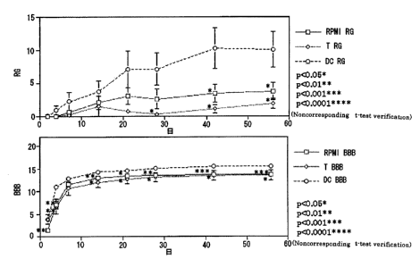
Fig. 10 is a set of drawings showing the effect of dendritic
cell subsets secreting a neurotrophic factor for a spinal cord
injury.
Fig. 11 is a set of photographs chronologically showing
a representative section particularly from marginal injured site
to cephalad direction as a result of immunostaining by using
anti-Mac-1 antibody in each of the dendritic cells (DC) and the
RPMI1640 (RPMI) transplanted group.
Fig . 12 is a drawing showing the chronological change in
' CA 02441434 2003-09-09
the number of Mac-1 positive ameboid cells by each region in
each of the dendritic cells and the RPMI1640 transplanted group.
Fig . 13 is a drawing showing the chronological change in
the number of Mac-1 positive ramified cells by each region in
each of the dendritic cells and the RPMI1640 transplanted group.
Fig . 14 is a set of photograph showing the setting of regions
for measuring the number of Musashi-1 positive cells.
Fig . 15 is a set of photographs chronologically showing
a representative section particularly from marginal inured site
to cephalad direction as a result of immunostaining by using
an anti-Musashi-1 antibody in each of the dendritic cells (DC)
and the RPMI1640 (RPMI) transplanted group.
Fig . 16 is a set of drawing showing the chronological change
in the number of Musashi-1 positive cell by each region in each
of dendritic cells and RPMI1640 transplanted group.
Fig. 17 is a drawing showing the result of expression of -
a neurotrophic factor in a spinal cord injured site after the
administration of GM-CSF by RT-PCR.
Fig. 18 is a drawing showing the setting of regions for
measuring the number of Mac-1 positive cells.
Fig. 19 is drawing showing the chronological change in
the number of endogenous microglia cells (ameboid) in each of
the GM-CSF administered group and a control ( physiological saline
administered) group.
Fig. 20 is a drawing showing the chronological change in
the endogenous microglia cells ( ramified ) in each of the GM-CSF
administered group and a control (physiological saline
administered) group.
Fig. 21 is a drawing showing the setting of regions for
measuring the number of Musashi-1 positive cells.
11
~
CA 02441434 2003-09-09
Fig. 22 is a drawing showing the chronological change in
the number of Musashi-1 positive cells in each of the GM-CSF
administered group and a control (physiological saline
administered) group.
Best Mode of Carrying out the Invention
The remedy for a nerve damage or a nerve dysfunctional
disorder of the present invention can be exemplified by the
following: asubstancesecretedfromdendriticcells; asubstance
inducing and proliferating dendritic cells; a substance
activating dendritic cells; asubstance inducing the expression
of a neurotrophic factor in nerve tissues; a substance inducing
and proliferating microglias and macrophages in nerve tissues,
wherein the substances have an effect of prevention, symptom
improvement or a therapeutic effect for a nervous in jury or a
nerve dysfunctional disorder (these substances will be
collectively referred to as a "dendritic cell related active
substance" hereinafter) , or a mixture of these substances that
are used as active ingredients . Said substance secreted from
the aforementioned dendritic cell can be eligibly exemplified
by cytokines such as IL-12 , IL-1 ~ , IL-1 ~ , IFN- r and the like ,
said substance inducing and proliferating dendritic cells can
be eligibly exemplified by cytokines such as GM-CSF, IL-4 and
the like, and said substance activating dendritic cells can be
eligibly exemplified by IL-1 ~ , CD40L and the like. Said
substance inducing the expression of a neurotrophic factor in
nerve tissues after the injury can be eligibly exemplified by
cytokines such as GM-CSF and the like , and said substance inducing
and proliferating microglias and macrophages in nerve tissues
after the injury can be eligibly exemplified by cytokines such
12
CA 02441434 2003-09-09
as GM-CSF,M-CSF and the like. The above-mentioned neurotrophic
factor can be exemplified by NT-3 inducing the effect of nerve
regeneration in vivo, the proliferation of microglias, and the
enhancement of phagocytosis; BDNF inhibiting denaturation and
omission of motor neuron of the injured spinal cord; NGF being
a neurotrophic factor of cholinergic neuron; CNTF having the
effects of denaturation and cell death protection against both
motor and sensory neurons of spinal cord, and the like.
Each known substance having the inducing andproliferating
action and the like of dendritic cells can be used as the following
substances: a substance secreted from dendritic cells; a
substance inducing and proliferating dendritic cells; a
substance activating dendritic cells; a substance inducing the
expression of a neurotrophic factor in nerve~tissues; and a
substance inducing and proliferating microglias and macrophages
in nerve tissues.-For example, said substance secreted from
dendritic cells can be obtained by culturing dendritic cells
in vitro; said substance having the inducing and proliferating
action of dendritic cells can be obtained by culturing dendritic
cells under the presence of a candidate substance in vitro, and
measuring and evaluating the extent of the induction and
proliferation of dendritic cells; said substance activating
dendritic cells can be obtained by culturing dendritic cells
under the presence of a candidate substance in vitro and measuring
and evaluating the extent of neurotrophic factor generation
ability of dendritic cells; said substance inducing the
expression of a neurotrophic factor in nerve tissues can be
obtained by measuring and evaluating the extent of the expression
and induction of a neurotrophic factor in injured neural tissues
wherein a candidate substance is administered. Said substance
13
~
CA 02441434 2003-09-09
inducing and proliferating microglias and macrophages in nerve
tissues can be obtained by measuring and evaluating the extent
of the induction and proliferation of the following cells in
injured neural tissue wherein a candidate substance is
administered: ameboid cells, in the injured neural tissues
wherein a candidate substance is administered, considered to
be the activated microglias with the strong phagocytic capacity
and macrophages derived from monocytes flown from the outside
of spinal cord; ramified cells considered to be activated
microglias secreting various neurotrophic factors and cytokines
though being lack of phagocytic capacity.
In the case where the aforementioned dendritic cell related
active substance is used as a remedy for a nerve damage or a
nerve dysfunctional disorder, various compound ingredients for
dispensing such as an ordinary carrier that is pharmaceutically
tolerated, a bonding agent , a stabilizing agent , an excipient ,
an diluent, a pH buffer agent, a disintegrant, a solubilizer,
a dissolution coadjuvant, an isotonic agent and the like can
be added . Said remedy can be administered orally or parenterally .
More specifically, it can be administered orally by ordinary
administering formulations such as formulations of powders,
granules, capsules, syrups, and liquid suspension, or it can
also be administered parenterally to the spot by injecting the
formulations of solution, emulsion, liquid suspension and the
like, or it can be further administered through the nostril by
the formulation of a spray agent.
In addition, as the aforementioned dendritic cell related
active substance, a vector which can express said substance can
be used, and when said vector is administered to the spot as
a genetic therapy, it becomes possible to provide a dendritic
14
~
CA 02441434 2003-09-09
cells related active substance to the spot stably due to the
stable expression of said substance compared to the case wherein
a remedy containing a dendritic cells related active substance
as an active ingredient is administered to the spot . In contrast
to the fact that most of the dendritic cells related active
substance of which the half-life periods are extremely short
and unstable, stable expression during the specified time can
be obtained by transferring a gene into a cell at the nerve inured
site with the use of a vector which can express a dendritic cells
related active substance. Such vectors can be eligibly
exemplified by virus vectors such as herpesvirus (HSV) vectors,
adenovirus vectors , human immunodeficiency virus ( HIV ) vectors
and the like, however, HSV vectors are preferable among these
virus vectors. HSV vectors have a high nervous affinity and
are safe since HSV is not integrated into chromosome DNA of cells ,
and it is possible to regulate the expression period of a transgene .
In addition, virus vectors that express a dendritic cells related
active substance can be prepared by ordinary protocols.
Further, a remedy for a nerve damage or a nerve
dysfunctional disorder of the present invention can be
exemplified by the one comprising dendritic cells, or
particularly preferably, dendritic cell subsets secreting a
neurotrophic factor NT-3 as an active ingredient . As for the
aforementioned dendritic cell subsets secreting the
neurotrophic factor NT-3,the following subsets are preferable:
immature dendritic cell subsets expressing the following, CNTF
showing the effects of denaturation and cell death protection
against both motor and sensory neurons of the spinal cord, TGF-
~ 1 having an inhibitory action for releasing cytotoxic substance
derived from microglias and macrophages, and IL-6 inducing the
CA 02441434 2003-09-09
protection effect for various neurons (cholin catecholamine
dopaminergic), in addition to NT-3 inducing the nerve
regeneration effect in vivo, the proliferation of microglias,
and the enhancement of phagocytosis; mature dendritic cell
subsets expressingCNTF, TGF-~ 1, IL-6andEGFwhereinthenervous
protection effect is acknowledged, in addition to NT-3. Such
subsets can be exemplified by immature dendritic cell subsets
having a surface marker of CDllc on the cell surface, and mature
dendritic cell subsets derived from said immature dendritic
cells.
As the aforementioned mature dendritic cell subsets, a
mature dendritic cell subsets , which can be obtained by culturing
immature dendritic cell subsets in vitro under the presence of
a stimulating agent for maturing immature dendritic cells such
as LPS, IL-1, TNF- CY , CD40L and the like, can be used. In this
case, there is a possibility that higher regeneration effect
can be induced due to the change in expression of neurotrophic
factor such as NT-3 and the like. Besides, mature dendritic
cell subsets wherein expression systems of myelin proteins such
as MBP (myelin basic protein), MAG (myelin-associated
glycoprotein) and the like, proteins and peptides of a nervous
system of inhibitors for the extension of nervous axon such as
Nogo and the like , or virus vectors wherein the genes encoding
them are integrated, are introduced (incorporated) can also be
used.
Dendritic cell subsets secreting a neurotrophic factor
NT-3 can be obtained by, for example, separating dendritic cell
subsets by a method wherein peripheral blood and the like are
pretreated by a density centrifugation and the like, then sorted
by FRCS with the use of a monoclonal antibody against dendritic
16
CA 02441434 2003-09-09
cell surface antigen, or by a separationmethodwherein amagnetic
beads binding monoclonal antibody against dendritic cells
surface antigen, then by selecting dendritic cell subsets
secreting NT-3 from said subsets. Said dendritic cell subsets
secreting neurotrophic factor NT-3 can be transplanted to a nerve
injured site of spinal cord and the like. Besides, mature
dendritic cell subsets wherein the expression system of a protein
or a peptide of the aforementioned nervous system, or the genes
encoding them is introduced (incorporated) can be administered
subcutaneously, or to a vicinity of lymph nodes . As described
above, a therapy method for a nerve damage or a nerve dysfunctional
disorder of the present invention can be exemplified by the method
wherein a remedy for a nerve damage or a nerve dysfunctional
disorder wherein a homogenous dendritic cell subset secreting
the aforementioned dendritic cell related active substance or
a neurotrophic factor NT-3 as an active ingredient is
administered (transplanted) to the nerve injured site,
subcutaneously or to a vicinity of lymph nodes , or intravenously.
The present invention will be further specifically
explained in the following examples, but the technical scope
of the invention will not be limited to these examples.
Example 1 (Generation of Spinal Cord Injured Model BALB/c Mouse)
BALB/c female mice ( n - 9 ) of 6 weeks old were used
respectively, the eighth thoracic vertebra was laminectomized
under ether anesthesia, and the left side of the spinal cord
was cut half by a sharp blade, and spinal cord injured model
mice ( injured group; ~ ) were generated. After the spinal cord
was injured, these mice showed palsy in the left lower limbs.
A group of BALB/c female mice of 6 weeks old (n = 9) wherein
only laminectomy was conducted were used as a control ( control
17
' CA 02441434 2003-09-09
group; 0). The amount of spontaneous motion of each of the
aforementioned mice were measured by using an action analyzing
apparatus SCANET MV-10 (Toyo Sangyo; an apparatus wherein 144
sets of near infrared radiation sensors running in all directions
are installed two-tiered in a square of 426 mm square) and motor
function was evaluated after the surgery, on day 2 and 4 as in
the acute stage, day 7 as the subacute stage, day 14, 21, 28,
and 56 as the chronic stage. In addition, measurement of
spontaneous motor quantity was set to detect and measure in forms
of two sizes of horizontal movements (Movement 1, 2; Ml, M2 for
abbreviation, it is regarded that a motion is made and the motor
quantity is measured when the motion was recognized in 12 mm
or more for Ml and in 60 mm or more for M2 ) , and vertical movements
(Rearing; RG for abbreviation, the number of uprising motion
of 6 . 75 cm or more is measured) , and it was further set to measure
for 10 minutes per mouse. The result of the case whereirrBALB/c
female mice were used is shown in Fig. 1. Besides, the p value
in the figure was calculated by using the Student's t test (*:
p < 0 . 05 , ** : p < 0 . 0l ) . As a result of comparing the evaluation
of each motor function between a control group and the in jured
group, in M1 (upper stand of Fig. 1) and M2 (middle stand of
Fig. 2) which show the evaluation of horizontal movements, a
significant difference was recognized in the acute and subacute
stage, however, significant difference was not recognized in
the chronic stage. On the other hand, in RG that shows the
evaluation of vertical movements, an obviously significant
difference was recognized between both groups until the chronic
stage (the lower stand of Fig. 1).
Example 2 (Generation of Spinal Cord Injured Model C57BL/6 Mouse)
With the exception of using C57BL/6 female mice of 6 weeks
18
CA 02441434 2003-09-09
old ( n = 16 ) instead of the aforementioned BALB/c female mice
of 6 weeks old ( n = 9 ) , evaluation of motor function was conducted
with the use of the action analyzing apparatus SCANET MV-10 in
the same manner as in Example 1. The result is shown in Fig.
2 . The p value in the f figure was calculated by us ing the Student ' s
t test ( ** : p < 0 . 01 ) . As a result of comparing the evaluation
of each motor function between a control group ( 0 ) and the injured
group ( ~ ), an obviously significant difference was not
recognized in 'M1 which shows the evaluation of horizontal
movements (upper stand in Fig. 2) and M2 (middle stand in Fig.
2) throughout the acute, subacute, and chronic stage. On the
other hand, in RG that shows the evaluation of vertical movements ,
an obviously signif scant difference was recognized in both groups
until the chronic stage ( lower stand in Fig. 2 ) . The results
of the aforementioned experiments between two different types
of mice in different strains showed that in vertical movements
( RG ) , it is possible to precisely evaluate the motor function
after the spinal injury, in contrast to the case of the amount
of horizontal movements (Ml and M2 ) wherein it was compensated
by a lower limb on the unaffected side or both upper limbs, and
it was impossible to precisely evaluate the palsy in the light
lower limb.
Example 3 (The Effect of Dendritic Cells against Spinal Cord
Injury)
Spinal cord injured model mice ( BALB/c female mice ) were
generated in the same operation as in Example 1, and immediately
thereafter, only RPMI1640 culture medium [control (0); Fig.
3 ; n = 14 , Fig . 4 ; n = 6 ] or antigen presenting cells including
dendritic cells isolated from the spleen [ 5 x 10⁵/mouse, n = 13,
( Fig . 3 ; ~ ) ] , or dendritic cells obtained by sorting a subset
19
' CA 02441434 2003-09-09
of CDllc (+) by applying the immunomagnetic beads method [1 x
10$/mouse , n = 6 , ( Fig . 4 ; C> ) ] was transplanted to the spinal
cord injured site. Besides, mice wherein only a laminectomy
was conducted were used as a control of which spinal cord is
not injured [ Fig . 3 ; 0 ( n = 6 ) ] . As in Example 1, the amount
of spontaneous vertical movement of each mouse were measured
by using the action analyzing apparatus SCANET MV-10 and the
motor function was evaluated on day 2, 4, 7, 14, 21, 28, and
56. Those results are shown in Fig. 3 and Fig. 4. In addition,
the p value in the figures was calculated by using the Student's
t test ( * : p < 0 . 05 , ** : p < 0 . O1 ) . These results showed that
a significant difference was recognized in the amount of vertical
movement compared to a control by administering CDllc (+)
dendritic cell subset to the injured site. As a result of the
aforementioned, it was revealed that restoration of the spinal
cord function is promoted by administering dendritic cells to
the nerve injured site.
Example 4 (The Effect of IL-12 against Spinal Cord Injury)
Spinal cord injured model mice were generated by conducting
the same operation as in Example 1 to the BALB/c female mice
of 6 weeks old. Besides , BALB/c female mice of 6 weeks old ( D ;
n = 6) wherein only a laminectomy was conducted were used as
a control of which spinal cord was not injured. 5 a 1 of
physiological saline only ( 0 ; n = 14 ) or IL-12 ( 100 ng/mouse;
Pharmingen ; O ; n = 14) was administered to the spinal cord
injured site immediately after the spinal cord was injured, and
then the amount of spontaneous vertical movements of each mouse
were measured by using the action analyzing apparatus SCANET
MV-10 and the motor function was evaluated on day 2 . 4 , 7 , 14 ,
21, and 28 as in Example 1. The result is shown in Fig . 5 . In
' CA 02441434 2003-09-09
addition, the p value in the figure was calculated by using the
Student's t test (*: p < 0.05, **: p < 0.01). These results
showed that an obviously significant difference was recognized
in the amount of vertical movements by administering IL-12 to
the injured site compared to the administration of physiological
saline. As a result of the aforementioned, it was revealed that
restoration of the spinal cord function is promoted by
administering IL-12 to the nerve injured $ite as in the case
of using dendritic cells mentioned above.
Example 5 (The Effect of GM-CSF for Spinal Cord Injury)
Spinal cord injured model mice were generated by conducting
the same operation as in Example 1 to the BALB/c female mice
of 6 weeks old . Besides , BALB/c female mice of 6 weeks old ( 0 ;
n = 6) wherein only a laminectomy was conducted were used as
a control of which spinal cord was not injured. 511 of
physiological saline only ( ~ ; n = 7 ) or GM-CSF ( lOng/mouse;
Genzyme ; O ; n = 6 ) was administered to the spinal cord injured
site immediately after the spinal cord was injured, and then
the amount of spontaneous vertical movements of each mouse were
measured by using the action analyzing apparatus SCANET MV-10
and the motor function was evaluated on day 2, 4, 7, 14, 21,
and 28 as in Example 1. The result is shown in Fig . 6 . In addition,
the p value in the figure was calculated by using the Student's
t test (** : p < 0 . O1 ) . These results showed that an obviously
significant difference was recognized in the amount of vertical
movement by administering GM-CSF to the injured site compared
to the administration of physiological saline. As a result of
the aforementioned, it was revealed that restoration of the
spinal cord function is promoted by administering GM-CSF to the
nerve injured site as in the case of using dendritic cells
21
' CA 02441434 2003-09-09
mentioned above.
Example 6 ( Preparation of Immature Dendritic Cell Subsets and
Mature Dendritic Cell Subsets)
Immature dendritic cells were obtained by separating CDllc
positive subsets from the spleen of BALB/c female mature mice
of 6 weeks old by applying immune magnetic beads method. More
precisely, the cell was separated as follows: the spleen was
firstly homogenated in 100 U/ml collagenase (Worthington
Biochemical Corporation ) , then coated part which is hard to be
separated was incubated in 400 U/ml collagenase at 37° C under
5% C02 for 20 minutes . The cells obtained herein were floated
in 35% BSA solution, and the RPMI1640 + 10% embryonic sera were
stratified in a centrifuge tube, centrifuged at 4° C, 3000 rpm,
for 30 minutes, then the cells at the boundary area between 35%
BSA solution and the RPMI1640 + 10% embryonic sera solution were
collected. Next, the cells obtained herein were reacted with
magnetic beads-bound monoclonal antibodies against CDllc
antigens ( 2 x 10⁸ beads , Miltenyi Biotec ) at 4° C for 15 minutes ,
and beads-bound cells were separated by magnets, and thus
fractions wherein immature dendritic cell subsets were condensed
were obtained. In addition, mature dendritic cell subsets were
obtained by culturing the immature dendritic cell subsets
obtained in the RPMI1640 + 10% embryonic sera culture solution
at 37° C, under 5% C02 for 24 hours .
Example 7 (Gene Expression of Neurotrophic Factor in Dendritic
Cells)
Total RNA was extracted from the cells of immature
dendritic cell subsets and mature dendritic cell subsets with
the use of TRIzol ( Life Technologies ) , 5 a g each of total RNA
was incubated at 42° C for 60 minutes by using AMV (Avian
22
CA 02441434 2003-09-09
Myeloblastosis Virus) reverse transcriptase and an oligo (dT)
primer, and a total amount of 200 ~l 1 cDNA was synthesized. PCR
was conducted by using a primer of ~-actin, gene expression
was confirmed, and then PCR was conducted for each neurotrophic
factor under respective conditions. PCR was conducted as
follows : gene was amplified by using 1,u 1 of cDNA as a template
and a reaction enzyme of Extaq (TAKARA) and by a thermal cycler
( Perkin-Elmer ) . The primer used and the PCR condition are shown
in Table 1. Besides, in order to show that it is not a gene
product amplified from genomic DNA which was mixed in, PCR
reaction was conducted respectively as a control by using total
RNA as a template . The result in immature dendritic cell subsets
is shown in Fig. 7 , and the result in mature dendritic cell subsets
is shown in Fig. 8, respectively.
23
' CA 02441434 2003-09-09
Table 1
__ _ Pri__m__er Sequence
. ~
.
_ _
Ident ,
SizeSense Antisense
ity
a
-acts 497 f-CATGGCATTGTTACCAACTGG-3'5'-TGTGGTGGTGAAGCTGTAGC-3'
(P1) (P2)
n
NT-3 200 f-ACTACGGCAACAGAGACGCTAC-3'f-ACAGGCTCTCACTGTCACACAC-3'
(P3) (P4)
CNTF 488 f-TGGCTAGCAAGGAAGATTCGT-3'5'-ACGGAGGTCATGGATAGACCT-3'
(P5) (P8)
IL-6 308 5'-TGCTGGTGACAACCACGGCC-3'5'-GTACTCCAGAAGACCAGAGG-3'
(P7) (P8)
TGF-
I~
462 5'-GAAGCCATCCGTGGCCAGAT-f 5'-GACGTCAAAAGACAGCCACT-3'
(P9) (P10)
1
EGF 595 5'-ACAGCCCTGAAGTGGATAGAG-f5'-GGGCTTCAGCATGCTGCCTTG-3'
(P11) (P12)
PCR Condition
94° C 1 Min. Thermal Denaturation (However, (3 -actin; 30 Secs . )
52° C 1 Min. Annealing (However, ~i -actin; 63° C, NT-3;
65° C)
72° C 2 Mins. Extension Reaction (However, a -actin, NT-3; 1 Min. )
42 cycles of the aforementioned thermal denaturation, annealing, and
extension reaction. (However, /3-actin; 30 cycles)
Expression of the following were confirmed in immature
dendritic cells: NT-3 inducing the nerve regeneration effect
in vivo, the proliferation of microglia, and the enhancement
of phagocytosis; CNTF having the protective effect for
denaturation and cell death against both motor and sensory
neurons of the spinal cord; TGF- ~ 1 having an inhibitory action
for releasing cytotoxic substance derived from microglias and
macrophages: IL-6 inducing the protection effect against various
neurons(cholin catecholamine dopaminergic)(Fig.7). Besides,
in mature dendritic cells, the expression of EGF wherein the
nervous protection effect was recognized was confirmed in
addition to NT-3, CNTF, TGF-~ 1, and IL-6 (Fig. 8). cDNA was
extracted from the gel with regard to each gene, the base sequence
was analyzed, and it was confirmed that the expression products
24
' CA 02441434 2003-09-09
were NT-3, CNTF, TGF-~1, IL-6 and BGF, respectively.
Example 8 (Secretion of Neurotrophic Factor NT-3)
With regard to NT-3, one of the neurotrophic factor
considered to be the most important for nerve regeneration, it
was further analyzed whether it was actually secreted from
dendritic cells by ELISA method using a NT-3 immunoassay system
(Promega). CDllc positive immature dendritic cells were
separated from the spleen of BALB/c female mature mice of 6 weeks
old by the immune magnetic beads method in the same manner as
in Example 1. After said 1 x 10⁵ of CDllc positive immature
dendritic cells were incubated in a culture solution of RPMI1640
+ 10% embryonic sera at 37°C under 5% C02 for 24 hours; its
conditioned media were collected. Only RPMI1640 was used as
a control, and 1 x 10⁵ of each CD4 positive T cells, CD8 positive
T cells were used. As a result of quantitative analysis of NT-3
in the media by sandwich ELISA method wherein two types of anti
NT-3 antibodies are used, it was revealed that 1 x 10⁵ of dendritic
cells secreted approximately 1. 75 ng of NT-3 for 24 hours . When
RPMI1640 only was used, and when CD4 positive T cells and CD8
positive T cells were used separately, secretion was not
recognized (Fig. 9).
Example 9 (Reconfirmation of the Effect of Dendritic Cells
against a Spinal Cord Injury)
The eighth thoracic vertebra of the BALB/c female mice
of 6 weeks old was laminectomized under ether anesthesia, and
spinal cord injured model mice of which the left side of the
spinal cord is cut in half under a microscope were generated.
After transplanting 1 x 10⁶ of dendritic cells to the spinal cord
injured site ( DC, n = 17 ) immediately, an evaluation was conducted
chronologically by applying the motorfunction evaluation method
' CA 02441434 2003-09-09
for lower limbs developed by the present inventors (RG Score
wherein the number of uprising motion is automatically analyzed
by using the action analyzing apparatus , SCANET MV-10 ) , and the
BBB scale which is among the already established motor function
evaluation method for lower limb ( it is evaluated between 0 and
21 points, 0 point means that no lower limb motor is recognized,
21 points means normal.). RPMI1640 (RPMI, n = 18) and CD8
positive T cells (T, n = 10 ) were transplanted as a control to
the spinal cord injured site in the same manner. The result
is shown in Fig . 10 . As shown in Fig . 10 , DC transplanted group
showed high scores of statistical significance in the both
evaluation methods ( RG Score and BBB scale ) compared to the cases
for T cell and RPMI of the controls. Accordingly, by
transplanting dendritic cell subsets secreting neurotrophic
factor NT-3 to the spinal cord injured site, it was reconfirmed
that restoration of spinal cord function was promoted.
Examplel0(Activation of EndogenousMicroglias by Transplanting
Dendritic Cells)
In order to examine whether any change by the transplant
of dendritic cells can be seen in the reactivity of endogenous
microglias or macrophages having invaded from the vein of the
injured part,immunohistological staining was conducted by using
Mac-lantibodies which recognize them, and chronological change
in the number of positive cells was investigated. Firstly, for
the dendritic cell transplanted mice on day 2 , 4 , 7 , and 14 of ter
the injury, transcardiac perfusion fixation was conducted with
2% paraformaldehyde, and a cryosection was generated (n = 3 ) .
The RPMI1640 transplanted group was used as a control (n = 3) .
Secondly, immunohistological staining wherein anti-mouse Mac-1
antibody (Pharmingen) was used as a primary antibody was
26
' CA 02441434 2003-09-09
conducted . Measuring region was divided into 3 parts 1 . a . , the
marginal injured site, cephalad aspect, and caudal aspect, as
a portion covering from dorsal aspect to ventral aspect at each
position of the most distal site of the gelfoam (denatured
collagen ) used for cell transplant and the site 1mm apart thereof .
Types of Mac-1 positive cells to be measured were divided into
two types, i.e. ameboid cells containing both of macrophages
derived from monocytes flown from the outside of the spinal cord
and activated macroglias wherein a phagocytic capacity is
particularly strong, and ramified cells considered to be
activated microglias lacking in phagocytic capacity.
The staining image of a representative section covering
from marginal injured site to cephalad aspect is shown in Fig.
11. In both groups, cellular infiltration is limited on day
2 after the in jury, but distinguished infiltration of ameboid
cell was recognized at the marginal in jured site on day 4 after
the injury. On and after day 4 after the injury, although
infiltration of Mac-1 positive cell was recognized at cephalad
distal part in the dendritic cell transplanted group, such change
was limited in a control group.
Subsequently, each Mac-lpositive cell was quantitatively
analyzed respectively by using an image analysis apparatus
(Flovel). Chronological change in the number of ameboid cells
by each region is shown in Fig. 12. Infiltration of ameboid
cells was mostly localized in the marginal injured site.
Although obviously different number of cells between both groups
at the marginal in jured site or the caudal aspect was not
recognized, a large number of positive cells was particularly
recognized among dendritic cell transplanted group at the
cephalad aspect on day 14 after the in jury. On the other hand,
27
' CA 02441434 2003-09-09
Fig. 13 shows the chronological' change in the number of ramified
cells by each region, a larger number of cells was recognized
in dendritic cell transplanted group in all regions , and on all
measuring days . With regard to the fact that an ameboid activated
microglia was increased in the cephalad aspect in the dendritic
cell transplanted group, it is considered that since ameboid
cells have a particularly strong phagocytic capacity, they are
eliminating denatured myelin inhibiting the extension of a
nervous axon and aprotein derived from injured tissue at a distant
place from the injured site. On the other hand, since the
increase of ramified activated microglias was seen in a wide
range, it is considered that an activated microglia itself
promoted the restoration of nervous function by secreting a
neurotrophic factor such as NT-3, CNTF, IL-6, TGF-~ l, EGF, bFGF,
NGF, BDNF, GDNF and the like.
Example 11 (Analysis of Endogenous Neural Stem Cells/Precursor -
Cells by Transplant of Dendritic Cells)_
In order to examine the reactivity of endogenous neural
stem cells/precursor cells by transplant of dendritic cells,
immunohistological staining was conducted by using Musashi-1
antibody which recognize them, and chronological change in the
number of positive cells was investigated. Firstly, for the
dendritic cell transplanted mice of day 2 , 4 , and 7 after the
in jury, transcardiac perfusion fixation was conducted with 2%
paraformaldehyde, and a cryosection was generated (n = 3) . The
RPMI1640 transplanted group was used as a control (n = 3).
Secondly, immunohistological staining wherein anti-mouse
Musashi-1 antibody is used as a primary antibody was conducted.
Musashi-1 is an RNA-bound protein of molecular weight
approximately 38 kDa which was identified by Okano et al. in
28
CA 02441434 2003-09-09
1994 (Neuron, 1994) , which was reported to strongly express in
neural stem cells/precursor cells in the analysis using a
monoclonal antibody against Musashi-1 of mouse (Dev. Biol. 1996,
J. Neurosci. 1997, Dev. Neurosci. 2000). Measuring region was
divided into 2 parts, i.e. the marginal injured site and the
distal site (cephalad aspect and caudal aspect) as a portion
covering from dorsal aspect to ventral aspect at each position
of the most distal site of the gelfoam ( denatured collagen ) used
for cell transplant and the site 1 mm apart thereof ( Fig . 14 ) .
The staining image of a representative section covering
from marginal injured site to cephalad aspect is shown in Fig.
15. In both groups, no difference can be seen on day 2 after
the in jury, however, on and after day 4 after the in jury, a large
number of Musashi-1 positive cells was recognized both in the
marginal injured site and a distal site in the dendritic cells
transplanted group, but such change was limited in a control
group.
Subsequently, Musashi-lpositive cell was quantitatively
analyzed by using an image analysis apparatus (Flovel).
Chronological change in the number of Mushashi-1 positive cells
by each region is shown in Fig. 16. More significant increase
in the number of Musashi-1 positive cells was recognized by
dendritic cells transplant both in the marginal injured site
and a distal site on and after day 4 after the injury compared
to a control. Particularly in the marginal injured site,
significant increase of Musashi-1 positive cells was recognized
in the dendritic cells transplanted group on day 2 to 4 after
the injury.
As a result of the aforementioned, it was made clear that
endogenous neural stem cells/precursor cells are induced to
29
~
CA 02441434 2003-09-09
proliferate by the transplant dendritic cells.
Example 12 (Expression Induction of Neurotrophic Factor in a
Injured Neural Tissue after Administration of GM-CSF)
Spinal cord injured model mice were generated by using
BALB/c female mice of 6 weeks old. 5 a 1 of physiological saline
only or GM-CSF (250 pg/mouse; Genzyme) was administered to the
spinal cord in jured site immediately after the in jury, and the
spinal cord was extirpated on the second day. The spinal cord
exterpated was frozen in liquid nitrogen, preserved at 80°C,
and then total RNA was extracted by using TRIzol (Life
Technologies ) . 5 ~l g each of total RNA was incubated at 42° C
for 60 minutes by using AMV (Avian Myeloblastosis Virus ) reverse
transcriptase and oligo (dT) primer, and total amount of 200
a 1 cDNA was synthesized. PCR was conducted by using a primer
of ~-actin, gene expression was confirmed, and then PCR was
conducted for each neurotrophic factor under each condition.
PCR was conducted as follows: gene was amplified by using 1
X11 of cDNA as a template and a reaction enzyme of Extaq (TAKARA)
by a thermal cycler (Perkin-Elmer). The primer used and the
PCR condition are shown in Table 2. Besides, in order to show
that it is not a gene product amplified from genomic DNA which
was mixed in , PCR reaction was conducted respectively as a control
by using total RNA as a template.
CA 02441434 2003-09-09
Table 2
Primer S equence
Ident size Sense Antisense
ity
-acts 497 f-CATGGCATTGTTACCAACTGG-3'5'-TGTGGTGGTGAi4GCTGTAGC-3'
(P1) (P2)
n
NT-3 200 f-ACTACGGCAACAGAGACGCTAC-3'5'-ACAGGCTCTCACTGTCACACAC-3'
(P3) (P4)
CNTF 468 5'-TGGCTAGCAAGGAAGATTCGT-3'f-ACGGAGGTCATGGATAGACCT-3'
(P5) (P6)
BDNF 277 f-CCAGCAGAAAGAGTAGAGGAG-3'5'-ATGAAAGAAGTAAACGTCCAC-3'
(P13) (P14)
NGF 498 5'-GTTTTGGCCTGTGGTCGTGCAG-3'5'-GCGCTTGCTCCGGTGAGTCCTG-3
(P15) '(P16)
PCR Condition
94° C 1 Min. Thermal Denaturation (However, /3 -actin; 30 Secs . )
52° C 1 Min. Annealing (However, a -actin; 63° C, NT-3;
65° C)
72° C 2 Mins. Extension Reaction (However, a -actin, NT-3; 1 Min. )
35 cycles of the aforementioned thermal denaturation, annealing, and
extension reaction. (However, a-actin; 30 cycles)
By administering GM-CSF to the inured spinal cord, it
was revealed that expressions of the following are promoted:
a neurotrophic factor, NT-3 inducing the nerve regeneration
effect in vivo, the proliferation of microglia, and the
enhancement of phagocytosis; a neurotrophic factor, BDNF
inhibiting denaturation and omission of motor neuron of the
injured spinal cord; a neurotrophic factor, NGF of cholinergic
neuron; a neurotrophic factor, CNTF having protective effect
far denaturation and cell death against both motor and sensory
neurons of the spinal cord (Fig. 17).
Examplel3(Activation of Endogenous Microglias by Administering
GM-CSF)
It is known that GM-CSF is involved in proliferation and
activation of microglias and macrophages in vitro. In order
to analyze its reactivity against microglias within a central
31
' CA 02441434 2003-09-09
nervous system tissue and macrophages having invaded from the
vein of the injured part, immunohistological staining was
conducted by using Mac-1 antibody which recognizes them, and
chronological change in the number of positive cells was
investigated. Firstly, for the GM-CSF administered mice of day
2, 4, and 7 after the injury, transcardiac perfusion fixation
was conducted with 2% paraformaldehyde, and a cryosection was
generated(n = 3). Physiological saline-administered mice were
used as control (n = 3). Secondly, immunohistological staining
wherein anti-mouse Mac-1 antibody (Pharmingen) is used as a
primary antibody was conducted. With regard to the measuring
region, the region covering 1 mm apart to dorsal direction and
ventral direction from the most distal part of the gelfoam
( denatured collagen ) used for cell transplant was analyzed ( Fig .
18) . Types of Mac-1 positive cells to be measured were divided
in two types , i . a . an ameboid cell considered to be activated
macroglias with a strong phagocytic capacity and macrophages
derived from a monocytes flown from the outside of the spinal
cord, and ramified cells considered to be activated microglias
lacking in phagocytic capacity but secreting various
neurotrophic factors and cytokines. They were quantitatively
analyzed by using an image analysis apparatus (Flovel).
Chronological change in the number of ameboid cells and
ramified cells is shown in Fig. 19 and Fig. 20, respectively.
In the GM-CSF administered group, a large number of ameboid cells
was recognized on day 2 after the in jury, and significant increase
in the number of cells compared to a control of day 7 after the
injury was confirmed. And also in ramified cells, significant
increases in the number of cells compared to a control were
recognized on day 4 and 7 after the injury. With regard to the
32
' CA 02441434 2003-09-09
fact that ameboid cells Were increased in GM-CSF administered
group, it is considered that they are eliminating denatured
myelin inhibiting the extension of a nervous axon and a protein
derivedfrom injuredtissue since ameboid cells have particularly
strong phagocytic capacity. In addition, the fact that the
increase in the number of ramified cells was also seen indicates
that activated microglia itself promoted restoration of the
nervous function by secreting a neurotrophic factor such as NT-3 ,
BDNF, NGF, CNTF and the like.
Example 14 ( Proliferation Induction of Endogenous Neural Stem
Cells/Precursor Cells by Administering GM-CSF)
In order to analyze the reactivity against neural stem
cells/precursor cells within the central nervous system by
administering GM-CSF, immunohistological system staining was
conducted by using Musashi-1 antibody that recognizes them, and
chronological change in the number of positive cells was
investigated. Firstly, for the GM-CSF administered mice of day
2, 4, and 7 after the injury, transcardiac perfusion fixation
was conducted with 2% paraformaldehyde, and a cryosection was
generated (n = 3). Physiological saline administered mice were
used as a control ( n = 3 ) . Secondly, immunohistological staining
wherein anti-Musashi-1 antibody ( Pharmingen ) is used as a primary
antibody was conducted. With regard to the measuring region;
the region covering 0 . 5 mm apart to dorsal and ventral direction
from the most distal part the gelfoam (denatured collagen) used
for cell transplanting was quantatively analyzed by using an
image analysis apparatus (Fig. 21). Chronological change in
the number of Musashi-l positive cells is shown in Fig. 22. In
GM-CSF administered group, a large number of Musashi-1 positive
cells was recognized on and after day 2 from the injury compared
33
CA 02441434 2003-09-09
to a control, and significant increase in the number of cells
was recognized on day 7 from the injury. As aforementioned,
it was revealed that endogenous neural stem cells/precursor cells
are induced to proliferate by administering GM-CSF.
As a result of the aforementioned; it is considered that
by way of transplanting dendritic cells to the injured site,
nervous function was restored through the intermediaries of the
following: direct secretion of a neurotrophic factor of its own,
secretion of a neurotrophic factor through the activation of
endogenous microglias, and the elimination action of inhibitor
for the extension of a nervous axon, and regeneration and
remyelination of a new neuron by proliferation induction of
endogenous neural stem cells/precursor cells, and the like. It
is also considered that by way of administering GM-CSF to the
injured site, nervous function is restored through the
intermediaries of the following: expression induction of a -
neurotrophic factor in a neuron , secretion of neurotrophic factor
through activation of an endogenous microglia, and elimination
action of inhibitor for extension of a nervous axon, and
regeneration and remyelination of a new neuron by proliferation
induction of endogenous neural stem cells/precursor cells, and
the like.
Industrial Applivability
The remedy for a nerve damage or a nerve dysfunctional
disorder of the present invention can be administered not only
by injecting into a injured site but also by various
administration methods including subcutaneous administration
or administration to a vicinity of lymph nodes , and intravenous
administration, which has excellent nervous function
34
CA 02441434 2003-09-09
_r
restoration action, therefore, it is useful for the disorders
of nerve dysfunctional disorders and the like such as a central
nervous system injury including a spinal cord injury and a
cerebral infarction. In addition, a dendritic cells related
active substance such as IL-12, GM-CSF and the like are useful
in that they can be easily handled and stored over a long time,
and can be prepared in a large amount at any time, and can be
applied to genetic therapies and the like.